Article

Chemical Profiles and Nitric Oxide Inhibitory Activities of the Copal Resin and Its Volatile Fraction of Bursera bipinnata

by
Silvia Marquina
1,
Mayra Antunez-Mojica
2,*,
Judith González-Christen
3,
Antonio Romero-Estrada
4,
Fidel Ocampo-Bautista
5,
Ninfa Yaret Nolasco-Quintana
1,
Araceli Guerrero-Alonso
1 and
Laura Alvarez
1,*
1
Centro de Investigaciones Químicas-IICBA, Universidad Autónoma del Estado de Morelos, Av. Universidad 1001, Col. Chamilpa, Cuernavaca 62209, Morelos, Mexico
2
SECIHTI-Centro de Investigaciones Químicas-IICBA, Universidad Autónoma del Estado de Morelos, Av. Universidad 1001, Col. Chamilpa, Cuernavaca 62209, Morelos, Mexico
3
Facultad de Farmacia, Universidad Autónoma del Estado de Morelos, Cuernavaca 62209, Morelos, Mexico
4
Instituto Tecnológico José Mario Molina Pasquel y Henriquez, Unidad Académica Tala, Tecnológico Superior de Jalisco, Av. Tecnológico S/N, Tala 45300, Jalisco, Mexico
5
Centro de Investigaciones en Biodiversidad y Conservación, Universidad Autónoma del Estado de Morelos, Av. Universidad 1001, Col. Chamilpa, Cuernavaca 62209, Morelos, Mexico
*
Authors to whom correspondence should be addressed.
Forests 2025, 16(7), 1144; https://doi.org/10.3390/f16071144
Submission received: 31 May 2025 / Revised: 7 July 2025 / Accepted: 8 July 2025 / Published: 11 July 2025
(This article belongs to the Special Issue Medicinal and Edible Uses of Non-Timber Forest Resources)

Abstract

Bursera bipinnata (DC.) Engl. (B. bipinnata), commonly known as “copal chino,” is a widely distributed Mexican tree found in transitional zones between pine-oak and deciduous forests. It is valued for its high-quality copal resin, traditionally used in ceremonies and offerings. Additionally, B. bipinnata is recognized for its significant value in traditional medicine, particularly in treating ailments associated with inflammation. In this work, the inhibition of nitric oxide (NO) production of the volatile fraction and resin of B. bipinnata in LPS-stimulated RAW 264.7 macrophage cells were demonstrated. In contrast, the volatile fraction exhibited 37.43 ± 7.13% inhibition at a concentration of 40 µg/mL. Chromatographic analyses of the total resin enabled the chemical characterization of eleven pentacyclic triterpenes belonging to the ursane, oleanane, and lupane series, as well as eight monoterpenes. Notably, the structures of compounds 15, 17, and 2935 are reported for the first time from the resin of Bursera bipinnata. The anti-inflammatory activity observed for B. bipinnata resin in this study may be attributed to its high content of the triterpenes α-amyrin (15, 29.7%) and 3-epilupeol (17, 38.1%), both known for their anti-inflammatory properties. These findings support the traditional use of this copal resin.

Graphical Abstract

1. Introduction

The Bursera genus comprises approximately 100 species throughout the American continent, of which 80 are found in México. Taxonomically, they are divided into two sections: Bullokia, or “cuajiotes,” characterized by exfoliating bark, and Bursera, or “copales,” which have non-exfoliating bark. According to Rzedowski (2005) [1], there are approximately 41 species considered within the Bursera section (Bursera copallifera (Sessé y Moc. ex DC.) Bullock, Bursera cuneata (Schltdl.) Engl., Bursera bicolor (Willd. ex Schltdl.) Engl., Bursera bipinnata, Bursera glabrifolia (Kunth) Engl., Bursera graveolens (Kunth) Triana & Planch., and Bursera penicillata (Sessé & Moc. ex DC.) Engl., among others), distributed in 26 states of México.
Species of the Copal section are very resinous, and their resins have a very characteristic aromatic smell of pine and lemon. The term copal derives from the Nahuatl word “copalli,” which describes several aromatic resins found in our territory [2]. The cultures of pre-Columbian Mesoamerica utilized copal for ceremonially burned incense and also as an adhesive. Many people still use copal in México and Central America as incense, and it plays a significant role in the economy of rural families [3,4,5]. Additionally, copal resins are used for medicinal purposes, such as creating anti-inflammatory poultices, plugging dental cavities, and treating pneumonia [6].
Pentacyclic triterpenes are common secondary metabolites in the resins of Bursera species, which contain principally triterpenes with ursane, oleanane, and lupane skeletons [7,8,9,10,11,12,13,14]. Indeed, phytochemical analysis of residues from copal resins in archaeological Aztec samples revealed the presence of triterpenes, with 3-epilupeol, 3-epi-α-amyrin, and α-amyrone being the most abundant in these samples [10].
B. bipinnata is commonly known as “copal chino, copal negro, copal santo, copalillo, jaboncillo” [1]. It is a low tree with grayish bark and many branches. It averages 6 m in height and inhabits steep areas, being part of the transitional populations between pine and oak forests, as well as lowland deciduous forests. It is found in areas between 800 and 1600 m above sea level, with a generally warm, subhumid, or dry climate. Among the Bursera species, B. bipinnata is the taxon with the widest geographic distribution in our territory. It is found mainly in Michoacán, Guerrero, Oaxaca, Puebla, and Morelos states of México, it is considered a source of high-grade copal resin, which is collected by local people in the Morelos state and used for Mexican celebrations such as “día de muertos” (Day of the Dead), and was a resin offered to the gods in Mesoamerican agricultural rituals. Additionally, B. bipinnata is regarded as having significant value in traditional medicine for treating rheumatoid arthritis, colds, cough, stroke, dental pain, and to accelerate wound healing [15,16]. Few studies have addressed the chemical constituents and pharmacological effects of B. bipinnata. Case et al. in 2003 [2] carried out the GC/MS analysis of the essential oil of a commercial copal resin identified as possible B. bipinnata; this analysis showed the presence in the oil of 14.52% of α-copaene, and 13.75% of germacrene D. Another GC/MS analysis on a derivatized commercial sample of the resin of B. bippinata identified nine triterpenes with oleanane, ursane, and lupane skeletons, these were 3-epi-β-amyrin, 3-epi-α-amyrin, 3-epi-lupeol, β-amyrone, β-amyrin, α-amyrone, α-amyrin, lupenone, and lupeol [17]. Also, the flavonoids luteolin 7-O-β-D-glucopyranoside and myricetin 3′-O-α-L-rhamnopyranoside were isolated from the methanolic extract of B. bipinnata leaves [18], and another study demonstrated the cytotoxic activity of the resin against breast carcinoma cells [19].
Despite the ethnomedicinal importance of B. bipinnata, there are no reports on its pharmacological activity or the chemical compounds responsible for this activity. Therefore, the aim of this study was to analyze the chemical composition of the resin and its volatile fraction obtained by supercritical CO2 extraction, and to evaluate the potential in vitro anti-inflammatory activity of Bursera bipinnata resin.

2. Materials and Methods

2.1. Plant Material

The resin of B. bipinnata was collected in September 2019 by Dr. Fidel Ocampo Bautista in the Sierra de Huautla Biosphere Reserve, located in the municipality of Tepalcingo, Morelos, Mexico. The resin was extracted by making incisions on the bark of thick branches (≥5 cm in diameter) using a traditional tool known as a “quichala.” After cutting, a Quercus leaf was inserted to prevent the resin flow from being obstructed. The exuding resin was directed onto a segment of agave leaf (penca) suspended below the incision site. To facilitate solidification, the resin was sealed at the end with mud or ash. Once collected, the resin was stored in plastic containers, transported in a cooler, and delivered to the Biomolecules Laboratory at the Center for Chemical Research, where it was refrigerated until chemical analysis. The plant specimen was taxonomically identified as Bursera bipinnata (DC.) Engl. (Burseraceae) by M.Sc. Gabriel Flores Franco of the HUMO herbarium of the Autonomous University of the State of Morelos, under voucher number 31840.

2.2. Extraction of Volatiles

The volatile compounds from the resin (10 g) of B. bipinnata were extracted using two different procedures. One of them consisted of using the CO2 extraction system (MelloeX CO2 Extraction System, Alegre Science, USA), with a working pressure of <900 psi (62 bar) and a release pressure of 1000 psi, utilizing 800 g of CO2 and storing it at −20 °C until analysis by GC/MS. The second method involved dissolving the resin in EtOAc for analysis by GC/MS. For that, the resin (10 g) was ground and homogenized to a uniform powder using a ceramic mortar with a pestle. The powdered resin was then totally dissolved with 30 mL of EtOAc at room temperature, filtered through Whatman No. 1 paper, and subjected to GC/MS analysis.

2.3. Gas Chromatography/Mass Spectrometry (GC/MS) Analysis

Once the volatile compounds had been extracted, 2 mg of the sample was weighed and dissolved in 0.5 mL of HPLC-grade n-hexane, and analyzed by GC/MS. The EtOAc soluble fraction obtained from the resin and the subtraction 3B (vide infra) was also analyzed by GC/MS.
For qualitative analysis of volatile compounds obtained from dried resin of B. bipinnata, an HP Agilent Technologies 6890 gas chromatograph equipped with an MSD 5973 quadrupole mass detector (HP Agilent), capillary column HP-5MS (length: 30 m; inside diameter: 0.25 mm; film thickness: 0.25 µM) was used. The GC operational conditions were as follows: injection temperature 250 °C; temperature program: 40 °C kept for 1 min, and then raised to 280 °C at 10 °C/min. The helium carrier gas was set to the column (1 mL per minute at constant flow). The mass spectrometer was operated in positive electron impact mode with an ionization energy of 70 eV. Detection was performed in selective ion monitoring (SIM) mode, and peaks were identified using target ions. Identifying organic compounds in the volatile fraction and resin was based upon comparing experimentally obtained mass spectra with those available in Mass Spectral and Retention Index Libraries, NIST version 1. 7a (The National Institute of Standards and Technology, Gaithersburg, MD, USA), with a score ≥ 80%. Relative area percentages were used to determine the relative proportion of each compound.

2.4. General Information for Chemical Characterization

Compounds were isolated employing open-column chromatography (silica gel, 70–230 mesh, ASTM). The isolation procedures and purity of compounds were checked by thin layer chromatography (silica gel 60F254 plates (Merck, Darmstadt, Germany), visualized by means of UV light (366 and 254 nm) and sprayed with Ce(SO4)2 2(NH4)2SO4 2H2O (Sigma-Aldrich, Inc., Toluca, State of México, México), followed by heating. Melting points were obtained and uncorrected on a Thermo Scientific IA9000 melting point apparatus. Optical rotation was measured using a Perkin-Elmer 341 digital polarimeter (PerkinElmer, Waltham, MA, USA). All 1D and 2D NMR experiments (1H, 13C, COSY, HSQC and HMBC) were recorded on a Varian INOVA-400 at 400 MHz, or a Bruker AVANCE III HD 500 at 500 MHz, or a Jeol ECZ600R 600 MHz (14.09 T), using acetone-d6 and CDCl3 with tetramethylsilane (TMS) as internal standard.

2.5. Phytochemical Study of the Non-Volatile Fraction of Bursera bipinnata Resin

The resin was air-dried at room temperature and used in its raw form without prior processing. A portion of powdered resin (10 g) was dissolved in acetone (30 mL) at room temperature. The resulting acetone extract was adsorbed onto silica gel (8 g) and subjected to open-column chromatography (CC) using a silica gel-packed column (430 g, 70–230 mesh; Merck, Naucalpan, State of México, México). Elution was performed with a gradient of n-heptane/acetone (95:05, 90:10, 85:15, 80:20, and 0:100, v/v). A total of 40 fractions (200 mL each) were collected and monitored by TLC using ALUGRAM® SIL G/UV254 silica gel plates (Düren, Germany). Fractions with similar profiles were combined, resulting in five subfractions: Fr-1 (1–7; 3.804 g), Fr-2 (8–16; 3.321 g), Fr-3 (17–25; 0.963 g), Fr-4 (26–33; 0.724 g), and Fr-5 (34–40; 0.372 g). Fr-2, Fr-3, and Fr-4 fractions obtained from the main column were purified separately over silica gel using a gradient system: 98:02, 96:04, 94:06, and 90:10 (n-heptane/acetone). From Fr-2 were isolated α-amyrin (15) (0.963 g; 9%), 3-epi-lupeol (17) (0.784 g; 8%), and the mixture of 15 and 17 (3.426 g; 34%). The fraction Fr-3 (963 mg) was subjected to successive chromatography on silica gel CC (35 g) using isocratic gradient n-hexane/acetone (90:10), from which two subfractions Fr-3A (647 mg) and Fr-3B (132 mg) were obtained. From Fr-3A, 3-epilupeol (17, 120 mg, 12%), α-amyrin (15, 267 mg, 27%), and 3β-Hydroxy-urs-12-en-11-one (29, 12 mg, 1%) were purified and identified. The Fr-3B subfraction showed a more complex mixture, so it was analyzed by gas chromatography coupled to mass spectrometry, identifying the compounds trans-p-mentha-2,8-dienol (19, 7%), α-terpineol (20, 4%), L-carveol (21, 49%), R-(-)-carvone (22, 4%), p-menth-2,8-dienol (23), (-)-perillyl alcohol (24), 3-oxoolean-9(11),12-diene (25), 3-oxo-ursan-9(11),12-diene (26), 3β-acetate-ursan-9 (11),12-diene (27), 3β-acetate-ursan-12-en-11-one (28), and 3β-hydroxy-ursan-12-en-11-one (29) (GC/MS are in Figures S1–S3, Supplementary Material). The Fr-4 (0.724 g) was submitted to a chromatographic open column (70 × 10 mm) previously packed with 21 g of silica gel 60 (0.040–0.063 mm). An n-hexane/acetone (90:10) isocratic system was used as the mobile phase (the volume of all samples was 5 mL). Thirty-two fractions were obtained and grouped into four final subfractions according to their chemical composition: Fr-4A (520 g), Fr-4B (76 mg), Fr-4C (54 mg), and Fr-4D (32 mg). The Fr-4A, Fr-4B, and Fr-4C subfractions were purified separately by open-column chromatography. An isocratic system of n-hexane/acetone (90:10) was used, and the fractions obtained were 3 mL each. From Fr-4A subfraction, α-amyrin (15, 264 mg, 36%) was purified. From the Fr-4B, the mixture of 3β-hydroxyolean-9(11),12-diene (30, 12 mg, 4%) and 3β-hydroxyursan-9(11),12-diene (31) was obtained. Chromatographic purification of subfraction Fr-4C afforded 8 mg (2%) of a mixture constituted by urs-12-ene-3β-11α-diol (32) and olean-12-ene-3β-11α-diol (33). The compounds p-menth-1-ene-3,6-diol (34, 8 mg) and p-menthane-2,5-diol (35, 5 mg) were purified and identified from the Fr-4D subfraction. The purification of these compounds is presented in Scheme 1, and their chemical structures (compounds 15, 17, 2935) are shown in Figure 1.
α-Amyrin (15): White amorphous solid. 1H-NMR (500 MHz, CDCl3) δH: 5.13 (1H, t, J = 3.8 and 7 Hz, H-12), 3.22 (1H, m, H-3), 1.95 (1H, t, J = 2.6 Hz, H-9), 1.88 (d, J = 3.6 Hz, H-18), 1.65 (m, H-2), 1.59 (m, H-22), 1.57 (t, H-19), 1.57 (t, H-5), 1.07 (s, H-23), 1.07 (s, H-24), 0.95 (s, H-25), 1.02 (s, H-26), 0.99 (s, H-27), 0.94 (s, H-28), 0.91 (d, J = 7.6 Hz, H-29 and H-30). 13C-NMR (125 MHz, CDCl3) δC: 139.96 (C-13), 124.65 (C-12), 79.13 (C-3), 59.33 (C-18), 55.43 (C-5), 47.96 (C-9), 41.76 (C-14), 39.89 (C-22), 39.84 (C-19), 39.03 (C-8), 39.00 (C-20, C-4), 33.98 (C-1), 33.18 (C-10), 31.49 (C-7), 28.97 (C-17), 28.36 (C-28), 28.34 (C-2), 27.51 (C-15), 26.86 (C-24), 23.60 (C-27), 23.50 (C-11), 21.61 (C-30), 18.59 (C-29), 17.69 (C-6), 17.10 (C-26), 15.90 (C-23), and 15.85 (C-25). These data match with those in the literature [20]. 1H, 13C-NMR and HSQC spectra are in Figures S4–S6 (Supplementary Material).
3-Epilupeol (17) was obtained as a white amorphous solid. 1H, 13C, and HSQC NMR data are presented in Supplementary Material Figures S7–S9 and are consistent with previously reported data [13,21].
3β-Hydroxyursan-12-en-11-one (29): Colorless needles, mp 229–231 °C [7] δH: 1H NMR (600 MHz, acetone-d6): 5.43 (1H, s, H-12), 3.28 (1H, t, J = 3.0 Hz, H-3), 2.40 (1H, s, H-9), 1.60 (1H, d, J = 4.1 Hz, H-18), 1.51–1.37 (m, 7H), 1.36 (s, 6H), 1.34–1.19 (m, 2H), 1.17 (s, 3H), 1.15 (s, 3H), 1.15–1.00 (m, 2H), 0.99 (s, 6H), 0.98–0.86 (m, 2H), 0.85 (s, 3H), 0.82 (6H, d, J = 6.2 Hz, H-29, H-30), 0.80 (s, 3H). 13C NMR (150 MHz, acetone-d6) δC: 199.22 (C-11), 164.62 (C-13), 131.12 (C-12), 78.41 (C-3), 62.22 (C-9), 54.95 C-5), 45.73, 44.38, 41.65, 40.08, 39.89, 39.78, 34.59, 33.52, 31.57, 29.21, 28.62, 28.18, 28.06, 27.92, 22.62, 21.37, 20.97, 18.99, 18.34, 17.71, 16.92, 16.33. These data match with those in the literature [22]. 1H, 13C-NMR and DEPT spectra are in Figures S10–S12 (Supplementary Material).
3β-Hydroxyolean-9(11),12-diene (30) and 3β-hydroxyursan-9(11),12-diene (31) δH: 1H NMR (600 MHz, acetone-d6) δ 5.57 (d, J = 3.7 Hz, 1H), 5.53 (d, J = 3.1 Hz, 1H), 5.23 (d, J = 3.7 Hz, 1H), 5.21 (d, J = 3.2 Hz, 1H), 3.59–3.02 (m, 2H). 13C NMR (150 MHz, acetone-d6) δC: 150.41 (C-9), 147.28 (C-9), 144.26 (C-13), 141.30 (C-13), 131.36 (C-12), 128.00 (C-12), 126.97 (C-11), 23.83 (C-11), 75.75 (C-3), 75.64 C-3), 59.02 (C-18), 50.54 (C-5), 50.35 (C-5), 46.96 (C-19) 46.82 (C-18), 44.05 (C-14), 42.82 (C-14), 42.17 (C-22), 40.44 C-20), 40.39 (C-19), 38.76 (C-1), 38.18 (C-10), 37.78 (C-22), 34.51 (C-21), 33.65 (C-29), 33.02 (C-7 and C-17), 31.61 (C-21 and C-20), 28.86 (C-28), 28.73 (C-24), 28.65 (C-15), 27.49 (C-2), 27.45 (C-16), 26.22 (C-16) (C-15), 25.65 (C-27), 25.28 (C-27), 23.97 (C-30), 22.90 (C-24), 22.22 (C- 26), 21.64 (C-30), 19.08 (C-26), 18.98 (C-23), 18.89 (C-25), 18.58 (C-6), 16.70 C-29), 15.25 (C-23). These data match with those in the literature [23,24,25,26]. 1H and 13C-NMR spectra are in Figures S13 and S14 (Supplementary Material).
Urs-12-ene-3β,11α-diol (32) and olean-12-ene-3β,11β-diol (33): 1H NMR (500 MHz, CDCl3) δH: 5.24 (d, J = 3.8 Hz, 1H), 5.19 (d, J = 3.3 Hz, 1H), 4.26 (dd, J = 8.8, 3.3 Hz, 1H), 4.20 (dd, J = 8.2, 3.8 Hz, 1H), 2.11–1.26 (m, 24H), 1.25 (d, J = 6.7 Hz, H-18), 1.23 (s, 3H), 1.11 (s, 3H), 1.09 (s, 3H), 1.07 (s, 3H), 1.05 (s, 3H), 1.01 (d, J = 7.9 Hz, 3H), 0.97 (s, 3H), 0.91 (d, J = 6.3 Hz, 3H), 0.88 (s, 3H), 0.88 (s, 3H). 13C NMR (125 MHz, CDCl3) δC: 149.62 (C-13), 146.23 (C-13), 124.90 (C-12), 121.42 (C-12), 82.04 (C-3), 81.81 (C-3), 76.24 (C-11), 76.11 (C-11), 55.94 (C-18), 49.61 (C-9), 49.54 (C-9), 47.01 (C-5), 46.91 (C-5), 46.67 (C-19), 43.65 (C-8), 43.53 (C-8), 41.50 (C-8), 41.46 (C-14), 39.50 (C-22), 39.45 C-19), 37.67 (C-20), 37.63 (C-1), 37.14 (C-4), 35.05 (C-10), 34.82 (C-22), 33.50 (C-21), 33.40 (C-20), 33.16 (C-21), 33.02 (C-7), 32.42 (C-17), 31.28 (C-7), 31.25 (C-17), 28.83 (C-2), 28.80 (C-2), 28.72 (C-16), 28.64 (C-16), 28.08C-28), 28.03 (C-30), 26.87 (C-15), 26.81 (C-27), 26.56 (C-28), 23.78 (C-23), 23.75 (C-23), 22.29 (C-30), 18.49 (C-6), 18.38 (C-6), 18.30, 18.24 (C-29), 16.88 (C-26), 16.80 (C-26), 16.76 (C-25), 16.61 (C-25). These data match with those in the literature [21,26]. 1H, 13C -NMR, and COSY spectra are in Figures S15–S17 of Supplementary Material.
p-Menth-1-ene-3,6-diol (34): crystalline solid; 1H NMR (600 MHz, acetone-d6); mp 110–112 °C; [α]20D −4.87 (c 0.6, acetone); δ 5.42 (m, H-2), 3.89 (t, J = 2.8 and 5.6 Hz, H-6), 3.82 (t, J = 7.6, 5.6 Hz, H-3), 2.15 (m, H-8), 1.72 (t, J = 2.1 and 3.5 Hz, CH3-7), 1.62 (m, H-4), 1.35 (m, H-5), 0.92 (d, J = 7 Hz, CH3-9), 0.8 (d, J = 7 Hz, CH3-10). 13C NMR (150 MHz, acetone-d6); δ 136.88 (C-2), 131.05 (C-1), 69.41 (C-6), 67.82 (C-3), 42.67 (C-4), 31.07 (C-5), 26.73 (C-8), 21.41 (C-10), 20.84 (C-7), 17.27 (C-9) [27]. 1H, 13C-NMR, DEPT, COSY, HSQC, HMBC, and MS-FAB- spectra are in Figures S18–S23 of Supplementary Material.
p-Menth-8-ene-2,5-diol (35): crystalline solid; mp 81–83 °C; [α]20D +42 (c 0.4, acetone); 1H NMR (600 MHz, acetone-d6) δH: 4.67 (d, J = 2.0 Hz, H-9a), 4.64 (d, J = 2.3 Hz, H-9b), 3.55 (sa, H-6), 3.31 (sa, H-2), 2.72 (td, J = 7.1, 3.5 Hz, H-4), 1.69 (s, CH3-10), 1.58–1.28 (m, Hs-3, H-1 and Hs-6), 0.81 (d, J = 7.8 Hz, CH3-7). 13C NMR (150 MHz, acetone-d6) δC: 151.49 (C-7), 108.56 (C-8), 75.74 (C-4), 74.06 (C-1), 47.90 (C-5), 38.37 (C-2), 35.14 (C-3), 34.40 (C-6), 21.11, 16.41 [28]. 1H, 13C-NMR, HSQC spectra and GC/MS chromatogram are in Figures S24–S27 of Supplementary Material.

2.6. In Vitro Nitric Oxide Inhibition Activity

2.6.1. Cell Culture

Murine macrophage cell line RAW 264.7 (Tib-71tm from ATCC) was grown in DMEM/F12 medium supplemented with 10% heat-inactivated FBS, without antibiotics. Cells were plated and incubated in a humidified atmosphere containing 5% CO2 at 37 °C. Cells were subcultured by scraping and seeding them in 25 cm2 flasks or 24-well plates.

2.6.2. MTS Assay to Determine Cell Viability

RAW 264.7 cells were seeded in 96-well plates at a density of 10,000 cells per well in 0.1 mL of culture medium and incubated for 24 h. The cells were then treated with various concentrations (5–40 µg/mL) of the resin or volatile fraction, vehicle control (0.21% DMSO, v/v), or etoposide (40 µg/mL) as a positive control, and incubated for an additional 22 h. Cell viability was subsequently assessed using the MTS assay. Briefly, 20 µL of MTS solution (Promega, Wisconsin, USA) was added to each well and incubated for 2 h. Absorbance was measured at 490 nm using an ELISA plate reader.

2.6.3. Treatment of Macrophages with LPS

RAW 264.7 cells were seeded in a 96-well plate (20,000 cells/well) with 0.2 mL culture medium and incubated for 24 h. Subsequently, the cells were treated with resin and volatile fraction at 0–40 μg/mL, concentration that do not affect cell viability, or vehicle (DMSO, 0.21%, v/v) or indomethacin (30 μg/mL), which served as a positive control, and were incubated for an hour. Next, the pro-inflammatory stimulus LPS was applied at 4 μg/mL to the wells that were treated with resin and volatile fraction, vehicle, and indomethacin, leaving wells with cells that were only treated with LPS (100% stimulus control) and wells with cells without any treatment (negative control) and incubated at 37 °C for 20 h. Finally, cell-free supernatants were collected and kept at −20 °C until NO quantification.

2.6.4. Determination of NO Concentration

Nitrite, the stable product of NO, was used as an indicator of NO production in the culture medium. Nitrite released into the culture medium was measured using the Griess reaction. Briefly, 50 μL of each cell culture supernatant was mixed in a 96-well plate with 100 μL of Griess reagent (50 μL of 1% sulfanilamide and 50 μL of 0.1% N-(1-naphthyl) ethylenediamine dihydrochloride in 2.5% phosphoric acid) and incubated for 10 min at room temperature. The optical density at 540 nm (OD540) was measured with a microplate reader. Fresh culture medium was used as a blank, and the nitrite concentration in the samples was calculated by comparison with the optical density (OD) at 540 nm of a standard curve of NaNO2 prepared in fresh culture medium [29].

2.6.5. Statistical Analysis

The results represent the mean ± standard deviation (SD) from at least three independent experiments. Statistical analysis was conducted using one-way analysis of variance (ANOVA), followed by Dunnett’s multiple comparisons test. All analyses were performed using GraphPad Prism® software (version 8.3.1), with p-values < 0.05 considered statistically significant. IC50 values were calculated by regression analysis.

3. Results

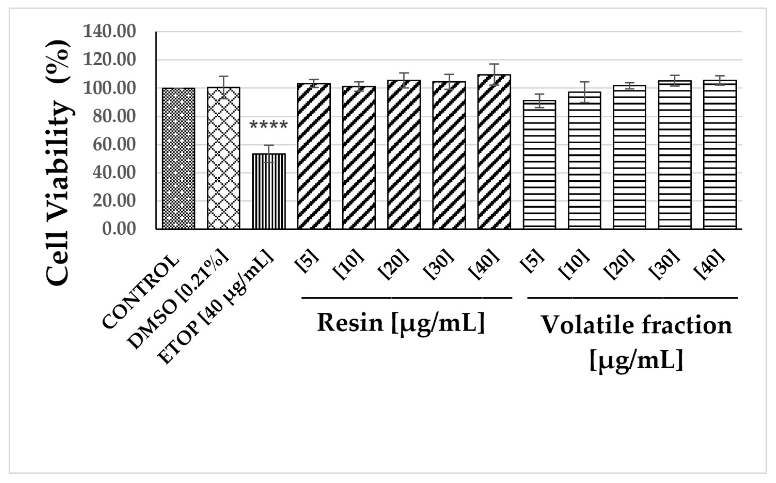
3.1. Nitric Oxide Inhibition Activity of the Total Resin and Its Volatile Fraction of Bursera bipinnata

With the aim to assess the pharmacological potential of B. bipinnata, its volatile fraction and resin were evaluated for their in vitro inhibition of NO production in LPS-stimulated RAW 264.7 cells. Firstly, to determine the effect of the samples on cell viability, RAW 264.7 cells were treated with the resin and volatile fractions at various concentrations (5–40 µg/mL) or with the vehicle (DMSO, 0.21%, v/v) or etoposide (40 µg/mL), which served as a positive control. After 22 h of incubation, cell viability was determined by the MTS assay. Results show that the resin and the volatile fraction (Figure 2) did not significantly decrease cell viability of macrophages at any of the tested concentrations compared to the etoposide treatment (positive control); therefore, the effect on the production of nitric oxide was evaluated at the same concentrations (5–40 µg/mL) (Figure 3). Cells were treated with or without the volatile fraction or resin for 1 h and then stimulated with LPS (4 μg/mL) for 20 h. Then, the amount of nitrite was measured in the medium. The results showed that NO level was increased in LPS-stimulated RAW cells, and that the positive control (indomethacin) inhibited NO production by 48.16 ± 6.38% at 30 μg/mL (p < 0.0001). This effect was significantly decreased, in a concentration-dependent manner, by treatment with the resin (p < 0.001), with a similar activity to that shown by indomethacin, with an IC50 of 30 ± 3.3 µg/mL. The inhibition percentages are shown in Figure 3 and Table 1. The inhibitory effect of the resin was not due to cytotoxicity, since it did not affect the cell viability of RAW 264.7 cells up to a concentration of 40 µg/mL (Figure 2). By the other hand, the anti-inflammatory activity of the volatile fraction, expressed as a percentage of NO inhibition, was lower than that of the total resin, reaching a 37.43 ± 7.13% of inhibition at 40 µg/mL; in contrast, the resin inhibited by 65.83 ± 8.53% at the same concentration.

3.2. Chemical Profiles

3.2.1. Volatile Compounds Present in the Resin of B. bipinnata

The chemical composition of the volatile fraction obtained by supercritical CO2 extraction from the resin of B. bipinnata is summarized in Table 2. A total of 18 volatile compounds were found, including seven monoterpenes (41%), seven sesquiterpenes (18.33%), and four triterpenes (34.48%). From these, eight were present in percentages greater than 5%: α-phellandrene (1, 24.42%), β-phellandrene (2, 8.27%), p-cymene (4, 6.72%), β-caryophyllene (9, 6.31%), cubenol (14, 8.05%), α-amyrin (15, 5.08%), β-amyrin (16, 6.28%), and lup-20(29)-en-3α-ol (17, 18.77%). It is noteworthy that α-phellandrene was the main component of the volatile fraction, and that four triterpenes represent 34.48% of the relative content (chromatogram and structures in Figures S28 and S29, respectively, in Supplementary Material).
On the other hand, for the total resin dissolved in EtOAc, 15 compounds were identified by GC/MS analysis (Table 2), those include one aromatic hydrocarbon (4.37%), seven monoterpenes (12.69%), two sesquiterpenes (2.33%), one diterpene (1.12%), and four triterpenes (79.48%). The most abundant components were the triterpenes α-amyrin (15, 29.74%) and lup-20(29)-en-3α-ol (17, 38.16%), followed by β-amyrin (16, 10.41%) and α-phellandrene (1, 5.38%). In this case, the high content of triterpenes stands out compared to that found in the volatile fraction.

3.2.2. Phytochemical Analysis of B. bipinnata Resin

Successive open-column chromatography of the total resin of B. bipinnata allowed the isolation and characterization of 19 compounds including the monoterpenes trans-p-Mentha-2,8-dienol (19), α-Terpineol (20), L-Carveol (21), R-(-)-Carvone (22), cis-p-Menth-2,8-dienol (23), (-)-Perillyl alcohol (24), p-Menth-1-ene-3,6-diol (34), and p-Menth-8-ene-2,5-diol (35), and the triterpenes α-Amyrin (15), 3-Epilupeol (17), 3-Oxoolean-9 (11),12-diene (25), 3-oxo-ursan-9 (11), 12-diene (26), 3β-acetate-ursan-9 (11),12-diene (27), 3β-acetate-ursan-12-en-11-one (28), 3β-Hydroxy-ursan-12-en-11-one (29), 3β-Hydroxyolean-9(11),12-diene (30), 3β-hydroxyursan-9 (11), 12-diene (31), urs-12-ene-3β, 11α-diol (32), and Olean-12-ene-3β, 11α-diol (33) (Figure 1). Compounds 1928 and 3031 were identified by comparing their mass spectra with those of the National Institute of Standards and Technology (NIST 1.7a) Library. The equipment used was a gas chromatograph equipped with a quadrupole mass detector in electron impact mode at 70 eV. Identification of compounds 29, 3235 was based on spectral analysis and data comparison with values described in the literature [9,23,30,31,32,33,34].

4. Discussion

Accumulating evidence has demonstrated that NO is a crucial and versatile molecule in the development of acute and chronic inflammation and host defense mechanisms. Likewise, high expression level of Nitric Oxide Synthase (iNOS) is associated with the presence of inflammation in tumors, and NO production by iNOS is a factor contributing to non-resolving tumor-promoting inflammation [35]. Therefore, identifying new agents capable of reducing the production of this pro-inflammatory agent is essential for controlling numerous inflammatory-related disorders and cancers attributed to macrophage activation [36].
The resin of B. bipinnata has been used in traditional Mexican medicine to treat various inflammatory diseases [1,5,15,16].
In this work, we assessed the effect of the volatile fraction and total resin of B. bipinnata on the nitric oxide (NO) production in LPS-stimulated RAW264.7 cells. The results showed that the positive control (indomethacin) inhibited NO production by 48.16 ± 6.38% at 30 µg/mL, while the total resin inhibited by 48.6 ± 3.57% at the same concentration; even more, the NO production was reduced in a concentration-dependent manner (Figure 3). On the other hand, the application of the volatile fraction did not inhibit the production of NO by LPS at one concentration. The inhibitory effect on NO production of total resin was not due to cytotoxicity, as it did not affect the cell viability of RAW 264.7 cells up to a concentration of 40 µg/mL (Figure 2). Importantly, the vehicle DMSO (0.21%, v/v) did not show a significant decrease in both cell viability and NO production.
Despite the importance of this plant species, to date, no reports have been found on the anti-inflammatory activity or the inhibitory activity of Bursera bipinnata on NO synthesis in LPS-activated RAW 264.7 cells. A survey in the literature showed that a few studies have been conducted on the Burseraceae family. For instance, the n-hexane leaf extract of Busera simaruba demonstrated anti-inflammatory activity in the carrageenan-induced paw edema model [37,38] and topical action on Croton oil-induced ear edema in mice [39]. A subsequent biodirected chemical study of this plant species culminated in the isolation of neophytadiene, ergost-5-en-3β-ol, 24S-stigmast-5,22E-dien-3β-ol, 24S-stigmast-5-en-3β-ol, and α-amyrin [40]. In another study performed on the volatile fraction from the bark of nine Bursera species, only Bursera lancifolia inhibited the TPA-induced edema in mice by 16.71% at 0.31 mg per ear. GC/MS analysis showed that the sesquiterpenoids elemol, agarospirol, and β-eudesmol were the most abundant components [41].
On the other hand, Acevedo et al., 2015 [42] reported the in vivo anti-inflammatory activity on the TPA-induced ear edema assay of the chloroform extracts from the bark of Bursera excelsa (IC50 = 0.26 ± 0.01 mg/mL), Bursera galeottiana (IC50 = 0.23 ± 0.02 mg/mL), and Bursera schlechtendalii (IC50 = 0.25 ± 0.02 mg/mL). This activity was statistically comparable to that of the positive control, indomethacin (IC50 = 0.19 ± 0.02 mg/mL).
In a chemical and pharmacological study performed to the dichloromethane-acetone extract of the copal resin of Bursera copallifera (B. copallifera), a topical inhibitory effect on TPA-induced auricular edema with an ID50 = 0.071 mg/ear was described. Phytochemical analysis resulted in the isolation and characterization of six pentacyclic triterpenes: 3-epilupeol formiate, lupenone, α amyrin, 3-epilupeol, and their acetates. The pharmacological study demonstrated that the anti-inflammatory activity showed by the resin can be attributed to the COX-2 inhibitory activity showed by the most abundant triterpenes α-amyrin and 3-epilupeol, together with the potent nitric oxide (NO) production inhibitory activity on RAW 264.7 macrophages, together with the moderate inhibitory activity of PLA2 enzyme displayed by all the natural triterpenes [13]. In another study, it was demonstrated that the MeOH extract of Bursera copallifera inhibited the ear edema with an IC50 = 4.4 µg/mL; this extract also inhibited cyclooxygenase-1 activity, the target enzyme for nonsteroidal anti-inflammatory drugs [43].
In the present study, phytochemical analysis of the resin of B. bipinnata allowed the identification of eleven triterpenes and two monoterpene compounds, which were characterized as α- amyrin (15), epilupeol (17), 3-oxo-olean-9(11),12-diene (25), 3-oxo-ursan-9(11),12-diene (26), 3-acetate-ursan-9 (11), 12-diene (27), 3β-acetate-ursan-12-en-11-one (28), 3β-hydroxy-ursan-12-en-11-one (29), 3β-hydroxyolean-9(11),12-diene (30), 3β-hydroxyursan-9(11), 12-diene (31), urs-12-ene-3β; 11α-diol (32), olean-12-ene-3β; 11β-diol (33), p-Menth-1-ene-3,6-diol (34), and p-Menth-8-ene-2,5-diol (35). Previous studies had already described the existence of α-amyrin (15) and 3-epilupeol (17) on a derivatized commercial sample of a resin identified as B. bipinnata, but this is the first report on the presence of triterpenes 2533 in the resin of B. bipinnata. This triterpene profile also differs from that found in the resin of Bursera copallifera, another reputed anti-inflammatory copal resin, which contains α-amyrin and 3-epilupeol, lupenone, 3-epilupeol formiate, α-amyrin acetate, and 3-epilupeol acetate [13].
Among the isolated compounds, it is worth noting the presence of high concentrations of α-amyrin (15) and 3-epilupeol (17), due to their multiple pharmacological effects. According to GC/MS, the triterpene content in the resin (79.48%) is more than double that found in the volatile fraction (34.98%). It is coincidental that the high yields of α-amyrin (15) and 3-epilupeol (17) found in the resin of B. bipinnata in this work, with the content present in the anti-inflammatory resin of B. copallifera.
Indeed, both compounds have been reported to have potential anti-inflammatory activity. In vitro studies showed that α-amyrin (15) inhibited NO production in LPS-stimulated RAW264.7 cells with IC50 = 8.98 ± 1.73 µm, while 3-epilupeol (17) did so with an IC50 = 15.50 ± 1.14 µM, as well as the in vivo inhibition of the TPA-induced inflammation [13,44,45]
Furthermore, a number of in vivo and in vitro studies have demonstrated that topical skin application of α-amyrin inhibits TPA-induced inflammation through the suppression of COX-2 expression, via inhibition of upstream protein kinases and blocking of NF-κB activation [45,46].
The chemical content of the volatile fraction obtained by supercritical CO2 extraction from the resin of B. bipinnata was identified by GC/MS (Table 2 and Figure S1). α-Phellandrene (1, TR = 7.11 min, 24.42%), β-Phellandrene (2, TR = 7.46 min, 8.27%), p-Cymene (4, TR = 7.72 min, 6.72%), and 3-epilupeol (17, TR = 38.09 min, 18.77%) were the main components. Among them, α-phellandrene is widespread in nature; in Boswellia sacra, it accounts for 42% of the essential oil. It produced cholinesterase inhibition with an IC50 of 120.2 μg/mL [47]. In rats, α-phellandrene was shown to prevent mechanical nerve injury-induced and cold hyperalgesia, and also exhibited antidepressant-like effects [48]. Phellandrene mildly enhanced macrophage proliferation and function in vivo without direct antimicrobial activity [49], suggesting the ability to suppress intracellular bacterial growth. ρ-Cymene (4), a cyclic monoterpene common in thyme (Thymus vulgaris) (27.4%), was active against Bacteroides fragilis, Candida albicans, and Clostridium perfringens [50]. It was sedative in mice at 0.04 mg in air, reducing motor activity to 47.3% of baseline [24]. Also, Siqueira et al. [51] demonstrated two mechanisms of anti-inflammatory action of α-phellandrenene: inhibition of neutrophil migration and stabilization of mast cells.
Additionally, at a dose of 50 mg/kg, it significantly reduced acetic acid-induced writhing and both the early and late phases of formalin-induced pain in mice [25]. A study showed little antioxidant or antiproliferative effects [52]. β-Caryophyllene (9) is one bicyclic sesquiterpene most common in cannabis extracts and is nearly ubiquitous in the food supply. It exhibits larvicidal activity against Anopheles subpictus, a vector of malaria, Aedes albopictus, a vector of dengue, and Culex tritaeniorhynchus, a vector of Japanese encephalitis [53]. A recent publication extends its therapeutic potential to protection from alcoholic steatohepatitis via anti-inflammatory effects and alleviation of metabolic disturbances [54]. Successive open-column chromatography of the total resin of B. bipinnata allowed the isolation and characterization of 19 compounds, including the monoterpenes trans-p-Mentha-2,8-dienol (19).

5. Conclusions

In this study, the evaluation of NO inhibition of B. bipinnata resin and its volatile fraction showed that the resin was the active part of the plant. Moreover, this study demonstrated that extraction with EtOAc facilitates the efficient extraction of anti-inflammatory triterpenes from the resin compared to supercritical CO2 extraction. Phytochemical analysis of this resin allowed the identification of eleven triterpenes and two monoterpene compounds, of which α-amyrin (15) and 3-epilupeol (17) represent more than 67.90% of the resin. The chemical profile of the resin from B. bipinnata differed from that reported for B. copallifera, another copal resin renowned for its anti-inflammatory properties. However, they coincided in the presence in high concentrations of the anti-inflammatory triterpenes alpha-amyrin (15) and epilupeol (17) whose anti-inflammatory activity has been previously demonstrated by our research team [13], and then it is probable that they were the responsible of the anti-inflammatory activity displayed by the resin of B. bipinnata. These results support the traditional use of B. bippinata for treating disorders associated with inflammation. This is the first report on the presence of triterpenes 2533 and the monoterpenes 34 and 35 in the resin of B. bipinnata. Additional studies will focus on the in vivo evaluation of the active fraction of B. bipinnata resin for future therapeutic and pharmacological research, thereby further supporting its traditional use in Mexican traditional medicine.
Future prospects. Several species of burseras are known to be sources of copal incense, but only a few are exploited extensively for commercial copal, specifically, B. bipinnata, B. copallifera, B. glabrifolia, and B. linanoe. Other important sources that are less frequently harvested commercially are B. citronella, B. excelsa, B. heteresthes, B. microphylla, B. penicillata, B. sarukhanii, B. simaruba, and B. stenophylla.
Today, copal is still used, although in smaller quantities, in celebrations, funerals, and in the traditional medicine, however, the number of communities dedicated to cultivating and obtaining these resins has decreased significantly, so to take advantage of the anti-inflammatory properties through the production of a therapeutic product, there must be a program for cultivating and preserving the trees that produce the bioactive resins.

Supplementary Materials

The following supporting information can be downloaded at https://www.mdpi.com/article/10.3390/f16071144/s1, Figure S1: GC-MS Chromatogram of Fr-3B subfraction with compounds 1922; Figure S2: GC-MS Chromatogram of Fr-3B subfraction with compounds 2324; Figure S3: GC-MS Chromatogram of Fr-3B subfraction with compounds 2528; Figure S4: 1H-NMR spectrum (400 MHz, CDCl3) of α-amyrin (15); Figure S5: 13C-NMR spectrum (100 MHz, CDCl3) of α-amyrin (15); Figure S6: HSQC spectrum (400 MHz, CDCl3) of α-amyrin (15); Figure S7: 1H-NMR spectrum (400 MHz, CDCl3) of epilupeol (17); Figure S8: 13C-NMR spectrum (100 MHz, CDCl3) of epilupeol (17); Figure S9: HSQC spectrum (400 MHz, CDCl3) of epilupeol (17); Figure S10: 1H-NMR (600 MHz, acetone-D6) of 3β-hydroxyursan-12-en-11-one (29); Figure S11: 13C-NMR (150 MHz, acetone-D6) of 3β-hydroxyursan-12-en-11-one (29); Figure S12: DEPT (150 MHz, acetone-D6) of 3β-hydroxyursan-12-en-11-one (29); Figure S13: 1H-NMR (600 MHz, acetone-D6) of 3β-Hydroxyolean-9(11),12-diene (30) and 3β-Hydroxyursan-9(11),12-diene (31); Figure S14: 13C-NMR (150 MHz, acetone-D6) of 3β-Hydroxyolean-9(11),12-diene (30) and 3β-Hydroxyursan-9(11),12-diene (31); Figure S15: 1H NMR (500 MHz, CDCl3) of urs-12-ene-3β, 11α-diol (32) and olean-12-ene-3β,11β-diol (33); Figure S16: 13C NMR (125 MHz, CDCl3) of urs-12-ene-3β, 11α-diol (32) and olean-12-ene-3β,11β-diol (33); Figure S17: COSY spectrum (500 MHz, CDCl3) of urs-12-ene-3β, 11α-diol (32) and olean-12-ene-3β,11β-diol (33); Figure S18: 1H-NMR (600 MHz, acetone-D6) of p-Menth-1-ene-3,6-diol (34); Figure S19: 13C-NMR and DEPT (600 MHz, acetone-D6) of p-Menth-1-ene-3,6-diol (34); Figure S20: HSQC (600 MHz, acetone-D6) of p-Menth-1-ene-3,6-diol (34); Figure S21: COSY (600 MHz, acetone-D6) of p-Menth-1-ene-3,6-diol (34); Figure S22: HMBC (600 MHz, acetone-D6) of p-Menth-1-ene-3,6-diol (34); Figure S23: Mass-Mass Spectrometry FAB–of p-Menth-1-ene-3,6-diol (34); Figure S24: 1H-NMR (600 MHz, acetone-D6) of p-Menth-8-ene-2,5-diol (35); Figure S25: 13C-NMR (150 MHz, acetone-D6) of p-Menth-8-ene-2,5-diol (35); Figure S26: HSQC (600 MHz, acetone-D6) of p-Menth-8-ene-2,5-diol (35); Figure S27: GC-MS of p-Menth-8-ene-2,5-diol (35); Figure S28: Chromatogram of essential oil from B. bipinnata; Figure S29: Structures of compounds identified in essential oil of B. bipinnata.

Author Contributions

Conceptualization, S.M., M.A.-M., J.G.-C., and L.A.; Data curation, J.G.-C. and F.O.-B.; Formal analysis, A.R.-E., F.O.-B., N.Y.N.-Q., and A.G.-A.; Funding acquisition, L.A.; Investigation, A.R.-E.; Methodology, A.R.-E., N.Y.N.-Q., and A.G.-A.; Project administration, J.G.-C. and L.A.; Resources, J.G.-C. and L.A.; Software, S.M., M.A.-M., N.Y.N.-Q., and A.G.-A.; Supervision, S.M. and J.G.-C.; Validation, S.M., A.R.-E., F.O.-B., N.Y.N.-Q., and A.G.-A.; Writing—original draft, S.M., M.A.-M., and L.A.; Writing—review and editing, S.M., M.A.-M., and L.A. All authors have read and agreed to the published version of the manuscript.

Funding

This research was funded by financial support from Consejo Nacional de Ciencia y Tecnología (Grant LN321131).

Data Availability Statement

Data are contained within the article and supplementary materials.

Acknowledgments

The authors thank Laboratorio Nacional de Estructura de Macromoléculas for the NMR spectra, and Consejo Nacional de Ciencia y Tecnología for financial support. We also thank Enrique Bezaleel Linares Espinoza for obtaining the volatile compounds.

Conflicts of Interest

The authors declare no competing interests.

References

  1. Rzedowski, J.; Lemos, R.M.; Rzedowski, G.C. de Inventario del conocimiento taxonómico, así como de la diversidad y del endemismo regionales de las especies mexicanas de Bursera (Burseraceae). Acta Bot. Mex. 2005, 70, 85–111. [Google Scholar] [CrossRef]
  2. Case, R.J.; Tucker, A.O.; Maciarello, M.J.; Wheeler, K.A. Chemistry and Ethnobotany of Commercial Incense Copals Copal Blanco, Copal Oro, and Copal Negro, of North America. Econ. Bot. 2003, 57, 189–202. [Google Scholar] [CrossRef]
  3. López, A.M. Copal de Bursera bipinnata. Una resina mesoamericana de uso ritual. Rev. Trace 2016, 70, 45–77. [Google Scholar] [CrossRef]
  4. Purata, V.S.E. Uso y Manejo de Copales Aromáticos: Resinas y Aceites; CONABIO/RAISES: Mexico City, Mexico, 2008. [Google Scholar]
  5. Argueta, A.; Vázquez, M.C.G. Atlas de Las Plantas de La Medicina Tradicional Mexicana; Instituto Nacional Indigenista: Mexico City, Mexico, 1994. [Google Scholar]
  6. Gigliarelli, G.; Becerra, J.X.; Curini, M.; Marcotullio, M.C. Chemical Composition and Biological Activities of Fragrant Mexican Copal (Bursera spp.). Molecules 2015, 20, 22383–22394. [Google Scholar] [CrossRef]
  7. Crowley, K.J. 818. Some Terpenic Constituents of Bursera graveolens (H. B. K.) Tr. et Pl. Var. villosula Cuatr. J. Chem. Soc. 1964, 4254–4256. [Google Scholar] [CrossRef]
  8. Syamasundar, K.V.; Mallavarapu, G.R.; Krishna, E.M. Triterpenoids of the Resin of Bursera delpechiana. Phytochemistry 1991, 30, 362–363. [Google Scholar] [CrossRef]
  9. Peraza-Sánchez, S.R.; Salazar-Aguilar, N.E.; Peña-Rodríguez, L.M. A New Triterpene from the Resin of Bursera simaruba. J. Nat. Prod. 1995, 58, 271–274. [Google Scholar] [CrossRef]
  10. Lucero-Gómez, P.; Mathe, C.; Vieillescazes, C.; Bucio, L.; Belio, I.; Vega, R. Analysis of Mexican Reference Standards for Bursera Spp. Resins by Gas Chromatography–Mass Spectrometry and Application to Archaeological Objects. J. Archaeol. Sci. 2014, 41, 679–690. [Google Scholar] [CrossRef]
  11. Álvarez, Á.L.; Habtemariam, S.; Parra, F. Inhibitory Effects of Lupene-Derived Pentacyclic Triterpenoids from Bursera simaruba on HSV-1 and HSV-2 in Vitro Replication. Nat. Prod. Res. 2015, 29, 2322–2327. [Google Scholar] [CrossRef]
  12. Messina, F.; Curini, M.; Di Sano, C.; Zadra, C.; Gigliarelli, G.; Rascón-Valenzuela, L.A.; Robles Zepeda, R.E.; Marcotullio, M.C. Diterpenoids and Triterpenoids from the Resin of Bursera microphylla and Their Cytotoxic Activity. J. Nat. Prod. 2015, 78, 1184–1188. [Google Scholar] [CrossRef]
  13. Romero-Estrada, A.; Maldonado-Magaña, A.; González-Christen, J.; Bahena, S.M.; Garduño-Ramírez, M.L.; Rodríguez-López, V.; Alvarez, L. Anti-Inflammatory and Antioxidative Effects of Six Pentacyclic Triterpenes Isolated from the Mexican Copal Resin of Bursera copallifera. BMC Complement. Altern. Med. 2016, 16, 422. [Google Scholar] [CrossRef] [PubMed]
  14. Sánchez-Monroy, M.B.; León-Rivera, I.; Llanos-Romero, R.E.; García-Bores, A.M.; Guevara-Fefer, P. Cytotoxic Activity and Triterpenes Content of Nine Mexican Species of Bursera. Nat. Prod. Res. 2021, 35, 4881–4885. [Google Scholar] [CrossRef] [PubMed]
  15. Monroy-Ortíz, C.; Castillo-España, P. Plantas Medicinales Utilizadas En El Estado de Morelos; Universidad Autónoma del Estado de Morelos, CONABIO: Cuernavaca, Morelos, México, 2007; ISBN 968-878-277-7. [Google Scholar]
  16. Dorado, O.; Maldonado, B.; Arias, D.M.; Sorani, V.; Ramirez, R.; Leyva, E.; Valenzuela, E. Programa de Conservación y Manejo Reserva de La Biosfera Sierra de Huautla; Comisión Nacional de Áreas Naturales Protegidas: Mexico City, México, 2005; ISBN 968-817-744-X. [Google Scholar]
  17. Lucero-Gómez, P.; Mathe, C.; Vieillescazes, C.; Bucio-Galindo, L.; Belio-Reyes, I.; Vega-Aviña, R. Archaeobotanic: HPLC Molecular Profiles for the Discrimination of Copals in Mesoamerica. Application to the Study of Resin Materials from Objects of Aztec Offerings. ArcheoSci. Rev. D’archéométrie 2014, 38, 119–133. [Google Scholar] [CrossRef]
  18. Hernández, J.D.; García, L.; Hernández, A.; Alvarez, R.; Román, L.U. Glicósidos de luteolina y miricetina de Burseraceae. Rev. Soc. Química México 2002, 46, 295–300. [Google Scholar]
  19. Lautié, E.; Quintero, R.; Fliniaux, M.-A.; Villarreal, M.-L. Selection Methodology with Scoring System: Application to Mexican Plants Producing Podophyllotoxin Related Lignans. J. Ethnopharmacol. 2008, 120, 402–412. [Google Scholar] [CrossRef]
  20. Chib, R.; Kumar, M.; Rizvi, M.; Sharma, S.; Pandey, A.; Bani, S.; Andotra, S.S.; Taneja, S.C.; Shah, B.A. Anti-Inflammatory Terpenoids from Boswellia Ovalifoliolata. RSC Adv. 2014, 4, 8632–8637. [Google Scholar] [CrossRef]
  21. Romero-Estrada, A.; Boto, A.; González-Christen, J.; Romero-Estudillo, I.; Garduño-Ramírez, M.L.; Razo-Hernández, R.S.; Marquina, S.; Maldonado-Magaña, A.; Columba-Palomares, M.C.; Sánchez-Carranza, J.N.; et al. Synthesis, Biological Evaluation, and Molecular Docking Study of 3-Amino and 3-Hydroxy-Seco A Derivatives of α-Amyrin and 3-Epilupeol as Inhibitors of COX-2 Activity and NF-kB Activation. J. Nat. Prod. 2022, 85, 787–803. [Google Scholar] [CrossRef]
  22. Herath, H.M.; Athukoralage, P.S.; Jamie, J.F. A New Oleanane Triterpenoid from Gordonia Ceylanica. Nat. Prod. Lett. 2001, 15, 339–344. [Google Scholar] [CrossRef]
  23. Reyes, C.P.; Jiménez, I.A.; Bazzocchi, I.L. Pentacyclic Triterpenoids from Maytenus cuzcoina. Nat. Prod. Commun. 2017, 12, 1934578X1701200508. [Google Scholar] [CrossRef]
  24. Ito, K.; Ito, M. The Sedative Effect of Inhaled Terpinolene in Mice and Its Structure–Activity Relationships. J. Nat. Med. 2013, 67, 833–837. [Google Scholar] [CrossRef]
  25. Quintans-Júnior, L.; Moreira, J.C.F.; Pasquali, M.A.B.; Rabie, S.M.S.; Pires, A.S.; Schröder, R.; Rabelo, T.K.; Santos, J.P.A.; Lima, P.S.S.; Cavalcanti, S.C.H.; et al. Antinociceptive Activity and Redox Profile of the Monoterpenes (+)-Camphene, p-Cymene, and Geranyl Acetate in Experimental Models. Int. Sch. Res. Not. 2013, 2013, 459530. [Google Scholar] [CrossRef] [PubMed]
  26. Bechkri, S.; Alabdul Magid, A.; Voutquenne-Nazabadioko, L.; Berrehal, D.; Kabouche, A.; Lehbili, M.; Lakhal, H.; Abedini, A.; Gangloff, S.C.; Morjani, H.; et al. Triterpenes from Salvia argentea Var. aurasiaca and Their Antibacterial and Cytotoxic Activities. Fitoterapia 2019, 139, 104296. [Google Scholar] [CrossRef] [PubMed]
  27. Stolow, R.D.; Sachdev, K. The p-Menth-1-Ene-3,6-Diols. Tetrahedron 1965, 21, 1889–1895. [Google Scholar] [CrossRef]
  28. Stolow, R.D.; Sachdev, K. Absolute Configurations of the P-Menthane-2,5-Diones and p-Menthane-2,5-Diols. J. Org. Chem. 1971, 36, 960–966. [Google Scholar] [CrossRef]
  29. Yang, G.; Lee, K.; Lee, M.; Ham, I.; Choi, H.-Y. Inhibition of Lipopolysaccharide-Induced Nitric Oxide and Prostaglandin E2 Production by Chloroform Fraction of Cudrania Tricuspidata in RAW 264.7 Macrophages. BMC Complement. Altern. Med. 2012, 12, 250. [Google Scholar] [CrossRef]
  30. Matsunaga, S.; Tanaka, R.; Akagi, M. Triterpenoids from Euphorbia Maculata. Phytochemistry 1988, 27, 535–537. [Google Scholar] [CrossRef]
  31. Tanaka, R.; Matsunaga, S. Triterpene Dienols and Other Constituents from the Bark of Phyllanthus Flexuosus. Phytochemistry 1988, 27, 2273–2277. [Google Scholar] [CrossRef]
  32. Yan, H.-Y.; Wang, K.-W. Triterpenoids from Microtropis fokienensis. Chem. Nat. Compd. 2017, 53, 784–786. [Google Scholar] [CrossRef]
  33. Morikawa, T.; Oominami, H.; Matsuda, H.; Yoshikawa, M. New Terpenoids, Olibanumols D–G, from Traditional Egyptian Medicine Olibanum, the Gum-Resin of Boswellia carterii. J. Nat. Med. 2011, 65, 129–134. [Google Scholar] [CrossRef]
  34. Ikuta, A.; Morikawa, A. Triterpenes from Stauntonia hexaphylla Callus Tissues. J. Nat. Prod. 1992, 55, 1230–1233. [Google Scholar] [CrossRef]
  35. Drehmer, D.; Mesquita Luiz, J.P.; Hernandez, C.A.S.; Alves-Filho, J.C.; Hussell, T.; Townsend, P.A.; Moncada, S. Nitric Oxide Favours Tumour-Promoting Inflammation through Mitochondria-Dependent and -Independent Actions on Macrophages. Redox Biol. 2022, 54, 102350. [Google Scholar] [CrossRef] [PubMed]
  36. Kobayashi, Y. The regulatory role of nitric oxide in proinflammatory cytokine expression during the induction and resolution of inflammation. J. Leukoc. Biol. 2010, 88, 1157–1162. [Google Scholar] [CrossRef] [PubMed]
  37. Noguera, B.; Díaz, E.; García, M.V.; Feliciano, A.S.; López-Perez, J.L.; Israel, A. Anti-Inflammatory Activity of Leaf Extract and Fractions of Bursera simaruba (L.) Sarg (Burseraceae). J. Ethnopharmacol. 2004, 92, 129–133. [Google Scholar] [CrossRef] [PubMed]
  38. Abad, M.J.; Bermejo, P.; Carretero, E.; Martínez-Acitores, C.; Noguera, B.; Villar, A. Anti-inflammatory Activity of Some Medicinal Plant Extracts from Venezuela. J. Ethnopharmacol. 1996, 55, 63–68. [Google Scholar] [CrossRef]
  39. Sosa, S.; Balick, M.J.; Arvigo, R.; Esposito, R.G.; Pizza, C.; Altinier, G.; Tubaro, A. Screening of the Topical Anti-Inflammatory Activity of Some Central American Plants. J. Ethnopharmacol. 2002, 81, 211–215. [Google Scholar] [CrossRef]
  40. Carretero, M.E.; López-Pérez, J.L.; Abad, M.J.; Bermejo, P.; Tillet, S.; Israel, A.; Noguera-P, B. Preliminary Study of the Anti-Inflammatory Activity of Hexane Extract and Fractions from Bursera Simaruba (Linneo) Sarg. (Burseraceae) Leaves. J. Ethnopharmacol. 2008, 116, 11–15. [Google Scholar] [CrossRef]
  41. Zúñiga, B.; Guevara-Fefer, P.; Herrera, J.; Contreras, J.L.; Velasco, L.; Pérez, F.J.; Esquivel, B. Chemical composition and anti-inflammatory activity of the volatile fractions from the bark of eight Mexican Bursera species. Planta Medica 2005, 71, 825–828. [Google Scholar] [CrossRef]
  42. Acevedo, M.; Nuñez, P.; Gónzalez-Maya, L.; CardosoTaketa, A.; Villarreal, M.L. Cytotoxic and Anti-inflammatory Activities of Bursera species from Mexico. J. Clin. Toxicol. 2015, 5, 232. [Google Scholar] [CrossRef]
  43. Columba-Palomares, M.C.; Villarreal, M.L.; Marquina, S.; Romero-Estrada, A.; Rodríguez-López, V.; Zamilpa, A.; Alvarez, L. Antiproliferative and Anti-inflammatory Acyl Glucosyl Flavones from the Leaves of Bursera copallifera. J. Mex. Chem. Soc. 2018, 62, 214–224. [Google Scholar] [CrossRef]
  44. Yasukawa, K.; Yu, S.Y.; Yamanouchi, S.; Takido, M.; Akihisa, T.; Tamura, T. Some Lupane-Type Triterpenes Inhibit Tumor Promotion by 12-O-Tetradecanoylphorbol-13-Acetate in Two-Stage Carcinogenesis in Mouse Skin. Phytomedicine 1995, 1, 309–313. [Google Scholar] [CrossRef]
  45. Medeiros, R.; Otuki, M.F.; Avellar, M.C.W.; Calixto, J.B. Mechanisms Underlying the Inhibitory Actions of the Pentacyclic Triterpene α-Amyrin in the Mouse Skin Inflammation Induced by Phorbol Ester 12-O-Tetradecanoylphorbol-13-Acetate. Eur. J. Pharmacol. 2007, 559, 227–235. [Google Scholar] [CrossRef] [PubMed]
  46. Otuki, M.F.; Vieira-Lima, F.; Malheiros, Â.; Yunes, R.A.; Calixto, J.B. Topical Anti-inflammatory Effects of the Ether Extract from Protium Kleinii and α-Amyrin Pentacyclic Triterpene. Eur. J. Pharmacol. 2005, 507, 253–259. [Google Scholar] [CrossRef] [PubMed]
  47. Bonesi, M.; Menichini, F.; Tundis, R.; Loizzo, M.R.; Conforti, F.; Passalacqua, N.G.; Statti, G.A.; Menichini, F. Acetylcholinesterase and Butyrylcholinesterase Inhibitory Activity of Pinus Species Essential Oils and Their Constituents. J. Enzym. Inhib. Med. Chem. 2010, 25, 622–628. [Google Scholar] [CrossRef] [PubMed]
  48. Piccinelli, A.C.; Santos, J.A.; Konkiewitz, E.C.; Oesterreich, S.A.; Formagio, A.S.N.; Croda, J.; Ziff, E.B.; Kassuya, C.A.L. Antihyperalgesic and Antidepressive Actions of (R)-(+)-Limonene, α-Phellandrene, and Essential Oil from Schinus Terebinthifolius Fruits in a Neuropathic Pain Model. Nutr. Neurosci. 2015, 18, 217–224. [Google Scholar] [CrossRef]
  49. Lin, J.-J.; Lin, J.-H.; Hsu, S.-C.; Weng, S.-W.; Huang, Y.-P.; Tang, N.-Y.; Lin, J.-G.; Chung, J.-G. Alpha-Phellandrene Promotes Immune Responses in Normal Mice through Enhancing Macrophage Phagocytosis and Natural Killer Cell Activities. In Vivo 2013, 27, 809–814. [Google Scholar]
  50. Carson, C.F.; Riley, T.V. Antimicrobial Activity of the Major Components of the Essential Oil of Melaleuca Alternifolia. J. Appl. Bacteriol. 1995, 78, 264–269. [Google Scholar] [CrossRef]
  51. Siqueira, H.D.S.; Neto, B.S.; Sousa, D.P.; Gomes, B.S.; da Silva, F.V.; Cunha, F.V.M.; Wanderley, C.W.S.; Pinheiro, G.; Cândido, A.G.F.; Wong, D.V.T.; et al. α-Phellandrene, a Cyclic Monoterpene, Attenuates Inflammatory Response through Neutrophil Migration Inhibition and Mast Cell Degranulation. Life Sci. 2016, 160, 27–33. [Google Scholar] [CrossRef]
  52. Fitsiou, E.; Anestopoulos, I.; Chlichlia, K.; Galanis, A.; Kourkoutas, I.; Panayiotidis, M.I.; Pappa, A. Antioxidant and antiproliferative properties of the essential oils of Satureja thymbra and Satureja parnassica and their major constituents. Anticancer Res. 2016, 36, 5757–5763. [Google Scholar] [CrossRef]
  53. Govindarajan, M.; Rajeswary, M.; Hoti, S.L.; Bhattacharyya, A.; Benelli, G. Eugenol, α-Pinene and β-Caryophyllene from Plectranthus Barbatus Essential Oil as Eco-Friendly Larvicides against Malaria, Dengue and Japanese Encephalitis Mosquito Vectors. Parasitol. Res. 2016, 115, 807–815. [Google Scholar] [CrossRef]
  54. Varga, Z.V.; Matyas, C.; Erdelyi, K.; Cinar, R.; Nieri, D.; Chicca, A.; Nemeth, B.T.; Paloczi, J.; Lajtos, T.; Corey, L.; et al. β-Caryophyllene Protects against Alcoholic Steatohepatitis by Attenuating Inflammation and Metabolic Dysregulation in Mice. Br. J. Pharmacol. 2018, 175, 320–334. [Google Scholar] [CrossRef]
Figure 1. Chemical structures of compounds isolated from the total resin of B. bipinnata.
Forests 16 01144 g001
Scheme 1. Fractionation tree diagram of the non-volatile fraction of Bursera bipinnata resin.
Forests 16 01144 sch001
Figure 2. Effect of the resin and volatile fraction of Bursera bipinnata on the viability of RAW 264.7 macrophages. Data are presented as the mean ± standard deviation (SD) from three independent experiments (n = 3). Treatments with DMSO, etoposide (ETOP), resin, and volatile fraction were compared to the control group (**** p < 0.0001). Control = untreated cells, defined as 100% viability.
Forests 16 01144 g002
Figure 3. Effect of the resin and volatile fraction from B. bipinnata on the production of nitric oxide (NO) in RAW 264.7 macrophages stimulated with LPS. Values are expressed as the mean ± SD (standard deviation) of three independent experiments (n = 3). The significant difference was determined using an ANOVA followed by Dunnett’s multiple comparison test. LPS compared to the control group (#### p ˂ 0.0001), and DMSO, INDO (indomethacin), resin, and volatile fraction compared to the LPS group (* p ˂ 0.05 or *** p ˂ 0.001 or **** p ˂ 0.0001). Control = cells without stimulus.
Forests 16 01144 g003
Table 1. Inhibitory effect of the resin and volatile fraction of B. bipinnata on nitric oxide production in RAW 264.7 macrophages stimulated with LPS.
% Inhibition
Concentration[5 µg/mL][10 µg/mL][20 µg/mL][30 µg/mL][40 µg/mL]
Resin9.86 ± 2.3715.21 ± 3.5740.42 ± 2.1148.6 ± 3.5765.83 ± 8.53
Volatile fraction12.96 ± 2.0917.64 ± 3.2324.23 ± 5.1128.56 ± 5.5037.43 ± 7.13
Indomethacin---48.16 ± 6.38-
The results were obtained from three independent experiments. Data are represented as the mean ± SD (standard deviation).
Table 2. Chemical composition of B. bipinnata resin and its volatile fraction.
Volatile Fraction (Supercritical CO2 Extraction)Resin (EtOAc)
No.CompoundsRT (min)Relative
Content (%) 1
Molecular
Formula
Mass Spectra
Match (%) 2
CompoundsRT (min)Relative
Content (%) 1
Molecular
Formula
Mass Spectra
Match (%)2
1α-Phellandrene7.1124.42C10H16136α-Phellandrene6.8735.38C10H16136
2β-Phellandrene7.468.27C10H16136m-Cymene7.1894.37C10H14134
3Carene7.230.18C10H16136ψ-Limonene7.2542.19C10H16136
4p-Cymene7.726.72C10H141344(10)-Thujen-3-ol9.9280.98C10H16O134
5Terpinolene8.350.38C10H16136exo-2-Hydroxycineole acetate11.6232.36C12H20O3126
6Thujone9.840.55C10H16O152p-Menthane11.8060.85C10H16O2135
7Carvone9.620.48C10H16O152Unnamed 12.70.54ND207
8β-Copaene12.570.54C15H24204Caryophyllene12.911.78C15H24204
9β-Caryophyllene13.216.31C15H24204p-Menthan-3-one13.0150.39C10H16O2207
10β-Caryophyllene oxide15.450.44C15H24O220Caryophyllene oxide14.9730.55C15H24O205
11Bicyclosesquiphellandrene14.131.03C15H24204α-Phellandrene, dimer17.2721.12C20H32136
121-Hydroxy-1,7-dimethyl-4-isopropyl-2,7-cyclodecadiene 14.441.33C15H26O222β-Amyrin35.910.41C30H50O426
13Calamenene14.490.63C15H222023-Epilupeol36.27538.16C30H50O426
14Cubenol17.608.05C15H26O222α-Amyrin37.10929.74C30H50O426
15α-Amyrin38.535.08C30H50O4263-Epilupeol-acetate41.7861.17C32H52O2468
16β-Amyrin36.966.28C30H50O426
173-Epilupeol38.0918.77C30H50O426
183-Epilupeol-acetate38.644.35C32H52O2468
1 Relative abundance concerning volatile fraction. 2 Score ≥ 80%, compared to mass spectra available in the NIST 1.7a library.
Disclaimer/Publisher’s Note: The statements, opinions and data contained in all publications are solely those of the individual author(s) and contributor(s) and not of MDPI and/or the editor(s). MDPI and/or the editor(s) disclaim responsibility for any injury to people or property resulting from any ideas, methods, instructions or products referred to in the content.

