Article

Phytochemical Composition, Antioxidant, Anti-Inflammatory Activity, and DNA Protective Capacity of Moss Hypnum cupressiforme Hedw. from Bulgaria

1
Department of Chemical Technology, Faculty of Chemistry, University of Plovdiv “Paisii Hilendarski”, 24 Tzar Asen Street, 4000 Plovdiv, Bulgaria
2
Department of Organic Chemistry, Faculty of Chemistry, University of Plovdiv “Paisii Hilendarski”, 4000 Plovdiv, Bulgaria
3
Department of Agrobiotechnologies, Agrobioinstitute, Agricultural Academy, 1164 Sofia, Bulgaria
4
Laboratory of Biologically Active Substances-Plovdiv, Institute of Organic Chemistry with Centre of Phyto-Chemistry, Bulgarian Academy of Sciences, 139 Ruski Blvd., 4000 Plovdiv, Bulgaria
5
Department of Molecular Biology, Faculty of Biology, University of Plovdiv “Paisii Hilendarski”, 4000 Plovdiv, Bulgaria
6
Department of Ecology and Environmental Conservation, Faculty of Biology, University of Plovdiv “Paisii Hilendarski”, 24 Tzar Asen Street, 4000 Plovdiv, Bulgaria
7
Institute of Biodiversity and Ecosystem Research, Bulgarian Academy of Sciences, 1113 Sofia, Bulgaria
*
Author to whom correspondence should be addressed.
Forests 2025, 16(6), 951; https://doi.org/10.3390/f16060951
Submission received: 15 April 2025 / Revised: 30 May 2025 / Accepted: 4 June 2025 / Published: 5 June 2025
(This article belongs to the Special Issue Medicinal and Edible Uses of Non-Timber Forest Resources)

Abstract

:
Hypnum cupressiforme Hedw. is a widely distributed moss species with significant bioactive potential, but the phytochemical composition and biological activity of this species are not well documented yet, particularly in Bulgaria. The current study aimed to investigate the amino acid composition, free sugars, sterol profile, total polyphenol content, antioxidant activity, DNA-protective effect, and the anti-inflammatory activity of ethanolic extracts of H. cupressiforme. Amino acid analysis revealed that proline (2.282 g/100 g), isoleucine (2.047 g/100 g), and glutamic acid (1.746 g/100 g) were the dominant constituents. The moss contained mannose (1.76 g/100 g) and fructose (1.43 g/100 g) as major free sugars and a diverse sterol profile containing stigmasterol (4.37 mg/g), β-sitosterol (4.29 mg/g), and campesterol (3.34 mg/g) as major phytosterols, which are known for their potential health benefits, such as cholesterol regulation and anti-inflammatory effects. The antioxidant activity of the extracts was determined by DPPH and ABTS assays and expressed moderate free radical scavenging ability (2.56 and 4.15 mM TE/g DW). Furthermore, the extracts also exhibited a potent DNA-protective effect against oxidative damage and anti-inflammatory activity. These findings contribute to the phytochemical knowledge of H. cupressiforme and suggest that Bulgarian specimens may be worth further investigation for pharmaceutical, nutraceutical, and cosmetic applications.

1. Introduction

The natural sources of bioactive compounds play an important role in the pharmaceutical industry. The efforts of researchers worldwide are focused mainly on the isolation of a myriad of health-promoting components from vascular plants, while the utilization of bryophytes is often neglected. However, mosses have been used in traditional medicine worldwide since ancient times, especially in China [1,2]. Bryophytes are a large group of plants distributed globally in almost every geographic region. They are small in size, lacking vascular tissue, which limits water transportation. Bryophytes are represented by more than 20,000 species divided into three phylums: liverworts (appr. 6000 species), mosses (about 14,000 species), and hornworts (about 300 species) [3]. More recent studies have continuously revealed the presence of secondary metabolites in different bryophytes, such as phenolic compounds, polyphenols, flavonoids, bibenzyls, and terpenoids, as well as lipids, essential amino acids, quinones, and many others, which can have biological activity [1,4,5]. For example, several authors reported that some bryophytes exhibit antioxidant, antibacterial, antifungal, antiviral, insecticidal, and neuroprotective activities [2,6,7,8,9].
Mosses are the biggest taxonomical group of the bryophytes and the second most diverse phylum of land plants. They play key ecosystem roles: retain soil moisture, aid nutrient cycling and soil stabilization, and provide microhabitats. They are easily adapted to various substrates and can grow on different soils, stones, rocks, trees, etc. [10,11]. It is established that one of the main components of the mosses is carbohydrates (including polysaccharides, which are related to the resistance of the plants to changes in environmental factors), as well as some secondary metabolites [2,12].
Hypnum cupressiforme Hedw. is a variable pleurocarpous moss with a cosmopolitan distribution [13]. It forms dense mats on wood and rock surfaces, as well as on anthropogenic structures, and exhibits broad ecological amplitude. It can thrive on substrates ranging from moderately acidic to calcareous and is tolerant of both shaded forest understories and more exposed, sunlit environments.
H. cupressiforme is a key biomonitoring species in southeastern Europe, used in moss surveys under the ICP Vegetation programme. In recent years, it has attracted growing scientific interest due to its diverse bioactive properties, particularly its high polyphenol content and associated antioxidant potential. Polyphenols are secondary metabolites in plants known for their strong antioxidant properties, which can play a vital role in protecting cells from oxidative damage. Some studies have shown that H. cupressiforme exhibits high antioxidant activity, and its extracts possess antibacterial, antifungal, and antiproliferative properties [13,14,15,16]. Lunić et al. [13] established the impact of seasonality on the chemical composition and biological activity of this moss species. They reported that the total phenolic content, total phenolic acid content, total flavonoids, total flavonols, and triterpenoid contents in the ethyl acetate extracts from H. cupressiforme had the highest levels in the summertime compared to the other seasons. The same authors also found the highest antioxidant activity (using linoleic acid/β-carotene bleaching assay) for the extracts during the summer. These findings have direct relevance for Bulgarian populations, which inhabit similar climates in the Balkans. Petkova et al. [17] reported H. cupressiforme from Bulgaria to be rich in lipid-soluble bioactives (sterols, tocopherols, unsaturated fatty acids) with favorable health-related indices. The current research is expected to complement the findings of Lunić et al. [13] by further highlighting the ecological ubiquity and pharmacological potential of H. cupressiforme across the region.
Badridze et al. [18] examined the content of active metabolites in some species of mosses in Georgia and established that H. cupressiforme contained 43.42% ascorbic acid, 11.3 mg/g soluble phenols, 211.96 μg/g proline, 40.6 mg/g soluble carbohydrates, 90.54 mg/g total proteins, and its antioxidant activity was 7.2% of inhibition. Smolińska-Kondla et al. [19] investigated the antioxidant capacity and the phenolic compounds of various extracts from five common European mosses, including H. cupressiforme. It was revealed that the aqueous and 50% ethanol extracts of this species expressed the weakest antioxidant activity using the DPPH assay, and naringenin (0.57 mg/L) was the main phenolic compound. On the other hand, other researchers established that kaempferol, p-hydroxybenzoic acid, protocatechuic acid, and p-coumaric acid were the main compounds in different H. cupressiforme extracts [1]. The water extract (18.21 mg GAE/g extract) had the highest total phenolic content, while the ethyl acetate extract possessed the highest amount of total flavonoids (58.86 mg QE/g extract) [1].
The characterization of both primary (e.g., amino acids, sugars) and secondary metabolites (e.g., phenolic compounds, flavonoids, sterols) is important for understanding the biological potential of bryophytes like H. cupressiforme. Primary metabolites are essential for growth and survival, but many—such as essential amino acids, proline, and simple sugars—also serve as precursors for secondary metabolite biosynthesis and play roles in stress adaptation. Secondary metabolites, particularly phenolic compounds, are well-known for their antioxidant, anti-inflammatory, antimicrobial, and DNA-protective properties. In vascular plants, these compounds have been extensively studied and linked to applications in pharmaceuticals, nutraceuticals, and cosmetics. However, in mosses, and especially in H. cupressiforme, such chemical profiling remains scarce and fragmented. By analyzing the phytochemical composition of Bulgarian populations of this species, this study aims to both bridge a critical knowledge gap and assess its value as a novel source of multifunctional bioactive compounds.
Therefore, the primary aim of this study was to examine the phytochemical profile—including amino acids, water-soluble sugars, sterols, carotenoids, and total polyphenol content—and to assess the antioxidant, DNA-protective, and anti-inflammatory activities of ethanolic extracts from the species sampled in Bulgaria, while also supplementing existing data from other regions. Additionally, this comprehensive study can also underscore the potential of the moss H. cupressiforme as a valuable resource for various industries.

2. Materials and Methods

2.1. Materials

The moss samples (H. cupressiforme) were collected in April 2024 from a non-urban area in Southern Bulgaria (42.03353 N, 25.22516 E) during the growing season. The specimens’ affiliation was confirmed under a microscope at the Faculty of Biology, Plovdiv University.

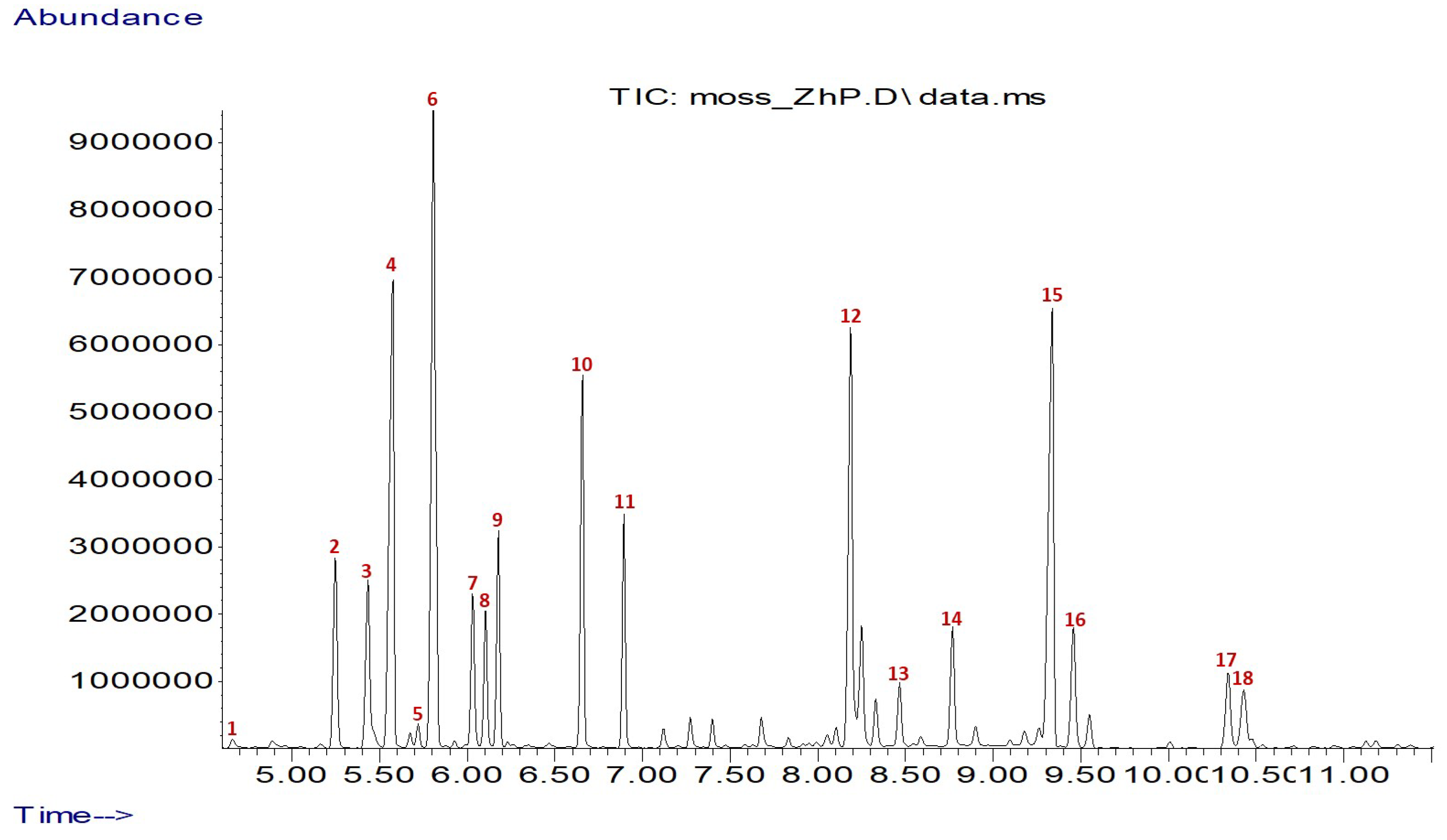
2.2. GC-MS Determination of Amino Acid Composition

Using the following protocol, the moss samples were subjected to acid hydrolysis to determine their amino acid composition. First, 50 mg of the sample was mixed with 2 mL of hydrolysis solution (6N HCl) in a glass bottle and incubated in a thermostat at 110 °C for 24 h. The resulting extract was centrifuged at 3000 rpm, and the supernatant was collected. Then, 100 µL was transferred into a GC vial and mixed with 30 µL of internal standard nor-Valine (0.2 mg/mL in water). The sample was dried completely using a vacuum concentrator at 60 °C for 1 h. Once dried, 50 µL of methoxyamine solution (20 mg/mL methoxyamine in pyridine) was added. The mixture was briefly vortexed and incubated at 80 °C for 1 h. Next, 50 µL of BSTFA was added, followed by an additional 1 h incubation at 80 °C. Finally, the sample was transferred into another glass vial with a microvolume insert, and 50 µL of pyridine was added prior to analysis using a GC/MS instrument.
The analysis was conducted using an Agilent Technologies gas chromatograph (model 7890A) coupled with a mass spectrometric detector (model 5975C). A column DB-5 ms (30 m × 0.32 mm × 0.25 µm) was utilized. The carrier gas was helium (1.0 mL/min). The temperature program was 100 °C for 2 min, increasing to 180 °C (15 °C/min) with a 1 min hold, then a rise (5 °C/min) to 300 °C, maintained for 10 min. The injector and detector temperatures were 250 °C. The mass spectrometric detector operated within a scan range of m/z = 50–550, while the sample injection volume was 1.0 µL (split ratio was 30:1). The retention times and Kovats retention indices (RI) were compared with those of reference standards, and the mass spectral data were matched with entries from the NIST’08 library (National Institute of Standards and Technology (NIST), Gaithersburg, MD, USA) to achieve compound identification.
The following formula was used for the calculation of the amino acid score [20]:
Amino   acid   score = mg   of   amino   acid   in   1   g   of   test   protein mg   of   amino   acid   in   1   g   of   reference   protein

2.3. HPLC Determination of Free Sugars

One gram of the sample was extracted with 30 mL of a 3% aqueous meta-phosphoric acid solution at 30 °C for 1 h under continuous agitation. The extract was centrifuged at 6000× g for 20 min and filtered through a 0.45 µm PTFE membrane. Free sugars were quantified using a UHPLC system (Nexera-i LC2040C Plus, Shimadzu, Kyoto, Japan) equipped with a refractive index detector. Separation was performed on an Agilent ZORBAX Carbohydrate column (5 µm, 4.6 × 150 mm) with a guard column, using acetonitrile/water (80:20, v/v) at 25 °C and a flow rate of 1.0 mL/min. Quantification was based on a calibration curve from five standard concentrations, and the compounds were confirmed by comparing retention times with D-fructose (D-Fru) and D-mannose (D-Man) standards.

2.4. Sterol Composition

The glyceride oil was obtained through the extraction of the moss sample with n-hexane, and the oil content was 1.2%. After that, 2 g of the oil was mixed with 2 N KOH, subjected to saponification, and the unsaponifiable matter was extracted with hexane [21]. Sterols were separated from this fraction through thin-layer chromatography (TLC), followed by the determination of individual sterol profiles using a gas chromatograph (HP 5890) with a capillary column DB-5 (25 m × 0.25 mm). The temperature program included an initial hold at 90 °C for 3 min, then an increase to 290 °C (rate of 15 °C/min), followed by a final rise to 310 °C at 4 °C/min, maintained for 10 min. The injector and detector temperatures were set at 300 °C and 320 °C, with hydrogen serving as the carrier gas. Identification of sterols was accomplished by comparing retention times with a reference mixture containing β-sitosterol (approximately 75% β-sitosterol and 10% campesterol, Acros Organics, NJ, USA), cholesterol (purity 95%, Acros Organics, NJ, USA), and stigmasterol (purity 95%, Sigma-Aldrich, St. Louis, MO, USA) [22].

2.5. Total Chlorophyll and Carotenoids Content

For the analysis of chlorophyll a (Ca), chlorophyll b (Cb), chlorophyll a + b (Ca + b), and total carotenoids (Cx + c), the sample was extracted with 100% acetone (Merck KgaA, Darmstadt, Germany) employing a sample to solvent ratio of 1:50 (w/v). Extraction was carried out in duplicate in an ultrasonic bath at room temperature for 20 min. Then, the extracts were passed through filter paper, and their absorbance (A) was determined at 470 nm, 645 nm, and 662 nm. The pigment concentration was determined using Equations (2)–(5), as described by Lichtenthaler et al. [23].
Chl a (µg/mL) = 11.24A662 − 2.02A645
Chl b (µg/mL) = 20.13A645 − 4.19A662
Chl a + b (µg/mL) = 7.05A662 − 18.09A645
C x + c (µg/mL) = (1000A470 − 1.82Chl a − 63.14Chl b)/214

2.6. Preparation of Ethanolic Extracts with Ultrasound-Assisted Extraction

For the extraction procedure, the sample was mixed with 80% ethanol in a ratio of 1:10 (w/v). The extraction was carried out at 40 °C for 20 min in an ultrasonic bath (Asonic, Ljubljana, Slovenia) at a frequency of 40 kHz. The extraction process was carried out in duplicate, and the resulting extracts were combined and filtered through filter paper. Then, the collected filtrate was utilized for further analysis of antioxidant activity and total polyphenolic content.

2.7. Total Phenolic Content

The total phenolic content was determined using a Folin–Ciocalteu reagent (Sigma-Aldrich, Merck, Munich, Germany), according to the procedure described by Ivanov et al. [24] with some modifications. The results were presented as mg equivalent of gallic acid/g of dry weight moss sample (mg GAE/g DW) [24].

2.8. HPLC Determination of Phenolic Compounds

Prior to analysis, 0.5 g of ground sample was stirred for 1 h with 40 mL 60% ethanol in 0.5% formic acid, then the mixture was centrifuged at 6000× g for 20 min. The resulting supernatant was used for HPLC analysis. The UHPLC system Nexera-i LC2040C Plus (Shimadzu Corporation, Kyoto, Japan) was utilized, equipped with a UV-VIS detector and a binary pump. A Poroshell 120 EC-C18 column (3 mm × 100 mm, 2.7 µm) was maintained at 26 °C. The utilized mobile phase was 0.5% acetic acid (A) and 100% acetonitrile (B). The flow rate was 0.3 mL/min, and the injection volume was 5 µL. Detection of derivatives was performed at 280 nm. The gradient conditions commenced with 14% (B), gradually increasing to 25% (B) between 6 and 30 min, and reaching 50% (B) at 40 min. The identity of each compound was verified through retention time comparisons against standard solutions and calibration curves for various phenolics. The limit of detection (LOD) and the limit of quantification (LOQ) of the identified quercetin were 0.83 μg/mL and 2.0 μg/mL. The results are given in mg per 100 g DW.

2.9. Antioxidant Activity

2.9.1. DPPH Radical Scavenging Assay

A mixture consisting of 2.85 mL of DPPH reagent (2,2-diphenyl-1-picrylhydrazyl) (Sigma-Aldrich, Merck, Munich, Germany) and 0.15 mL of 80% ethanol extract was incubated at 37 °C for 15 min. The absorbance was then recorded at 517 nm. Antioxidant activity was expressed in mM Trolox equivalents per gram of dry weight (mM TE/g DW) of the moss sample [24].

2.9.2. ABTS Radical Scavenging Assay

The ABTS reagent (2,2′-azino-bis(3-ethylbenzothiazoline-6-sulfonic acid) diammonium salt) was prepared following the method outlined by Ivanov et al. [24]. After incubation (at 37 °C for 15 min in a dark place) of 2.85 mL of ABTS•+ solution and 0.15 mL of the prepared extract, the absorbance was measured at 734 nm. Antioxidant activity was quantified using a calibration curve and expressed in mM TE/g DW of the moss sample [24].

2.10. In Vitro Albumin Denaturation Inhibition Assay

H. cupressiforme extract was prepared in 80% ethanol; the hydromodulus was 1:10. A single extraction was performed in an ultrasonic bath at 40 °C for 40 min. The extract was passed through filter paper for purification, and the solvent was subsequently removed using a rotary vacuum evaporator under reduced pressure. The dry H. cupressiforme extract was dissolved in DMSO (10 mg/mL concentration). The plant extract was subjected to an in vitro albumin denaturation inhibition assay.
The anti-denaturation assay was conducted following the method of Milusheva et al. [25], and Equation (6) was used for the calculation of the percentage of inhibition of protein denaturation (% IPD):
%   Inhibition   of   denaturation = ( Abs   control Abs   sample ) Abs   control ×100
The control represents 100% protein denaturation. For comparative analysis, readily available anti-inflammatory drugs are assessed utilizing the same methodology applied to the H. cupressiforme extract.

2.11. DNA Nicking Protection Assay

Supercoiled plasmid DNA (pUC19) was used for determining the DNA protective effect of the H. cupressiforme extracts prepared in 80% ethanol [26,27]. In brief, the moss extract (1 μL) was combined with pUC19 (250 ng) and incubated at 37 °C with Fenton’s reagent (30 min). pUC 19 plasmid was purified from recombinant E. coli Neb10 strain using QIAprep Spin Miniprep Kit (QIAGEN). The experiments were performed in a total volume of 20 µL. Various concentrations (25, 50, and 100 mg/mL) of 6-hydroxy-2,5,7,8-tetramethylchromane-2-carboxylic acid (Trolox, Merck KGaA, Darmstadt, Germany) were prepared in 10 µL aliquots, with water serving as both the positive and negative control. The resulting reactions were analyzed via 1.0% agarose gel electrophoresis in 0.5× TBE buffer at 120 V for 1 h [28]. DNA nicking was evaluated through the Gel Doc™ EZ Imaging system (Bio-Rad, Hercules, CA, USA).

2.12. Statistics

All analyses were performed in triplicate (n = 3), and the results are reported as the mean value along with the corresponding standard deviation (SD).

3. Results and Discussion

Numerous nutrients and secondary metabolites contribute to plants’ biological activity and antioxidant capacity. Therefore, the identification of these components in Hypnum cupressiforme Hedw. plays a crucial role in understanding the potential applications of this moss species.

3.1. Amino Acid Composition

Amino acids play vital roles in plant metabolism, serving as building blocks for proteins and participating in various metabolic pathways. They are essential for the structure and function of the plant cells. Proteins are involved in nearly all aspects of plant life, including enzyme activity, structural support, and transport mechanisms [29,30]. Some amino acids are also established to take part in the development of plants and their response to environmental stresses [29]. Therefore, revealing the amino acid composition of H. cupressiforme is important to evaluate its biological functions.
The results of the bound amino acids in the studied moss sample using gas chromatography–mass spectrometry (GC-MS) are presented in Table 1 and Figure 1, respectively.
Eighteen amino acids were identified, and the main ones were proline (2.282 g/100 g) and isoleucine (2.047 g/100 g). Several other amino acids were detected in the sample in comparatively high amounts: glutamic acid (1.746 g/100 g), leucine (1.463 g/100 g), methionine (1.358 g/100 g), valine (1.201 g/100 g), lysine (1.175 g/100 g), and aspartic acid (1.132 g/100 g). The rest of the identified amino acids were found to range from 0.095 g/100 g (alanine) to 1.015 g/100 g (threonine). Nine essential amino acids were identified in the samples of H. cupressiforme (valine, leucine, isoleucine, threonine, methionine, phenylalanine, lysine, and tryptophan), including the amino acid that is essential for children’s nutrition (histidine), and there was one semi-essential amino acid (arginine). These essential amino acids cannot be synthesized in the human body, and they have to be taken from the diet, mainly acquired via plant sources [29,30]. The amino acids present in H. cupressiforme in the lowest quantities were alanine (0.095 g/100 g), histidine (0.149 g/100 g), and tyrosine (0.194 g/100 g). The amount of the essential amino acids in the moss sample was higher (9.051 g/100 g) than the quantity of the non-essential ones (7.700 g/100 g) and the ratio of the essential to non-essential amino acids was calculated to be 1.18: 1. It was also obvious that lysine, which was present in very small level in most of the plants limiting their nutritional value [29], was actually in higher concentration in H. cupressiforme.
The obtained results differed significantly from the amino acid composition of other moss species. Nordin and Gunnarsson [31] determined the amino acid composition of Sphagnum mosses (Sphagnum fuscum (Schimp.) H.Klinggr., Sphagnum magellanicum Brid., and Sphagnum rubellum Wilson) and found that arginine was the main one. Different results for the total amino acids were reported by Semerjyan and Semerjyan [32] for some mosses grown in Armenia (Mnium spinosum (Voit) Schwaegr, Plagiomnium cuspidatum (Hedw), Brachythecium salebrosum (Web. et Mohr) B.S.G., Thuidium recognitum (Hedw) Lindb, Dicranum scoparium (Hedw), Anomodon viticulosus (Hedw), Drepanocladus aduncus (Hedw)). The amount ranged from 57.62 mg/100 g to 113.47 mg/100 g. The main prevailing amino acids in the investigated mosses were glutamic acid, alanine, and threonine, but methionine was not found in all samples. Another study revealed that the amino acid content was from 590 to 3266 mg/100 g dry moss in Aulacomnium palustre (Hedw.) Schwägr., Hylocomium splendens (Hedw.) Schimp., Polytrichum commune Hedw., Polytrichum juniperum Hedw., Pleurozium schreberi (Brid.) Mitt., Dicranum polysetum Sw., Climacium dendroides (Hedw.) F. Weber & D. Mohr, Ptilium crista-castrensis (Hedw.) De Not., Rhytidiadelphus triquetrus (Hedw.) Warnst., Sphagnum fallax (H.Klinggr.) H.Klinggr., S. magellanicum, S. rubellum, and Sphagnum tenellum (Brid.) Brid. The major amino acids were glutamic acid, valine, aspartic acid, and phenylalanine [2].
Generally, the amino acids serve as precursors for secondary metabolites, which play roles in plant defense, pigmentation, and interaction with other organisms. They are indispensable for plant metabolism, influencing everything from growth and development to stress responses and nutrient assimilation. Their multifaceted roles make them key players in maintaining plant health and productivity. Certain amino acids help plants cope with stress. Proline accumulates in response to drought and salinity stress, helping to stabilize proteins and membranes [29]. Glutamine and asparagine are involved in nitrogen storage and transport, which is crucial during nutrient deficiency [30]. The amino acid profile of the examined moss H. cupressiforme shows a balanced composition of essential and non-essential amino acids, indicating its potential as a nutritious source. The high levels of proline, isoleucine, and glutamic acid are noteworthy, suggesting significant roles in structural integrity, muscle metabolism, and neurotransmission.
The amino acid score (AAS) is a widely accepted method for assessing the quality of dietary proteins based on the essential amino acid composition. Table 2 presents the AAS of H. cupressiforme, calculated against the reference amino acid profile of cow’s milk protein—considered a complete, high-quality protein.
The amino acid score indicates the extent to which the moss meets human amino acid requirements, with a score of 1.0 representing adequacy and values below 1.0 indicating limiting amino acids [20,33]. The obtained results reveal that H. cupressiforme has a relatively low amino acid score profile, and none of the essential amino acids reach or surpass 0.50, which indicates that the moss cannot serve as a sole source of high-quality dietary protein. Several of the essential amino acids are significantly below the threshold requirement when compared to cow’s milk protein, a known complete protein source [34]. In the moss protein, histidine is particularly deficient (only 6% of the required level), which is a critical limitation because it is semi-essential for infants and children, potentially limiting growth, hematopoiesis, and immune function [35]. Similarly, phenylalanine (essential) and tyrosine (conditionally essential) are critically low; as precursors for neurotransmitters and thyroid hormones, their deficiency may impair neurological and endocrine functions [36,37]. Lysine (often the first limiting amino acid in cereal-based diets) and leucine (a key branched-chain amino acid critical for muscle protein synthesis) are present at only 15% of reference levels, suggesting limited support for protein synthesis and muscle repair [34,38,39]. Tryptophan, available at 18% of the requirement and necessary for serotonin and niacin production, further indicates that the moss protein alone may be insufficient for optimal metabolic and neurotransmitter functions [40,41]. Additionally, while valine and threonine are only moderately provided—supporting muscle metabolism and gut integrity, respectively [35]—methionine and isoleucine, the highest scoring amino acids (0.41 and 0.44), still fall short of adequacy, limiting their roles in cell metabolism and muscle function [42].
From a nutritional standpoint, the amino acid profile of H. cupressiforme clearly indicates multiple limiting essential amino acids, with histidine and phenylalanine + tyrosine being the most critically deficient. Such a profile places the moss below high-quality protein standards as defined by digestibility and amino acid composition [43], making it unsuitable as a primary protein source. However, it could be viewed as part of a complementary dietary approach, especially when combined with other protein sources that are rich in the limiting amino acids. Its relatively higher content of methionine and isoleucine could make it a supportive ingredient in plant-based protein blends or for food fortification strategies [44]. Additionally, from a sustainability and food security perspective, interest in unconventional or underutilized protein sources like mosses is growing in order to establish resilient food systems, especially in regions where conventional agriculture is extremely limited [45]. However, significant protein isolation, nutrient enhancement, or biofortification would be required for H. cupressiforme in order to be an acceptable, food-grade protein for broader nutritional use.

3.2. Sugars

In general, amino acid metabolism is connected to energy and carbohydrate metabolism, and some amino acids are precursors for synthesizing several secondary metabolites [46].
The results for the free sugar composition of H. cupressiforme are arranged in Table 3 and Supplementary Material S1.
The results indicated the presence of mannose and fructose. The amount of mannose (1.76 g/100 g) was slightly higher than that of fructose. Mannose is an aldohexose sugar commonly found in plant-derived materials, particularly in hemicelluloses and glycoproteins [47]. It should be mentioned that mannose is a precursor for glycosylation pathways and contributes to cell wall integrity; thus, its presence might be associated with the physiological adaptation of H. cupressiforme to environmental conditions. Fructose was identified as the second most abundant free sugar, with a concentration of 1.43 g/100 g. It is a key monosaccharide involved in energy metabolism and is often found in plants as a product of photosynthesis or carbohydrate breakdown [47]. The detection of fructose in H. cupressiforme suggests its role as an immediate energy source, contributing to the osmotic regulation and stress tolerance of the moss, particularly under fluctuating environmental conditions. The total content of free sugars in H. cupressiforme was relatively low (3.19 g/100 g), which suggests that this moss species might not store high levels of free sugars like some vascular plants. Instead, the moss might regulate its sugar levels dynamically to adapt to environmental stressors such as desiccation or nutrient limitation.
Comparing the sugar composition of H. cupressiforme to other bryophytes revealed both similarities and differences in their carbohydrate profiles. Studies on other bryophytes identified the presence of different sugars. For instance, in Rhodobryum ontariense Kindb., sucrose was the main soluble sugar, and a novel fructooligosaccharide, 1-kestose, was also detected [48]. Additionally, research on the moss Plagiomnium affine (Blandow ex Funck) T.J.Kop. indicated that sucrose played a significant role in frost tolerance, with increased cell sucrose concentrations correlating with enhanced frost hardiness [49]. These variations suggest that different bryophyte species may prioritize different sugars, such as sucrose, glucose, or fructose, depending on their specific physiological needs and environmental adaptations. The relatively low total free sugar content in H. cupressiforme compared to the higher sucrose concentrations in other species may indicate alternative strategies in carbon storage and stress response mechanisms.

3.3. Sterols, Chlorophyll, and Carotenoids Content

The analysis of the chemical composition of H. cupressiforme was further extended by investigating its phytosterol profile, as presented in Table 4.
Phytosterols, also known as plant sterols, have several important functions. They can reduce the amount of low-density lipid (LDL) cholesterol, have anti-inflammatory, anti-cancer, and antioxidant activity, and participate in immune regulation [50].
The determination of the sterol composition is preceded by the isolation of the glyceride oil contained in the moss, with its content being 1.2%. Overall, H. cupressiforme showed a diverse sterol profile with a notable presence of stigmasterol (4.37 mg/g fresh weight), β-sitosterol (4.29 mg/g fresh weight), and campesterol (3.34 mg/g fresh weight), which are known for their potential health benefits. Minor amounts of ∆7-stigmasterol (0.71 mg/g fresh weight), ∆5-avenasterol (0.16 mg/g fresh weight), and brassicasterol (0.05 mg/g fresh weight) were also identified. Cholesterol was a common sterol in animals but could also be found in small amounts in plants—which accounted for 0.48 mg/g fresh weight in H. cupressiforme. The total sterol content sums up to 13.40 mg/g, indicating a notable level of these compounds in the moss sample. β-Sitosterol is one of the most abundant plant sterols, found in nuts, seeds, and vegetable oils; stigmasterol is present in many legumes and vegetables; and campesterol is also found in many plants, including soybeans and corn [47]. The sterol concentrations in H. cupressiforme (stigmasterol, β-sitosterol, and campesterol) are relatively high for a non-vascular plant and comparable to certain seeds. A study analyzing sterol profiles in 19 nuts and seeds found that their total sterols ranged from 10 mg/100 g sample (in chestnuts) to 331 mg/100 g sample (in sesame seeds) [51]. These values indicate that the sterol content in the examined moss sample (13.40 mg/g FW or 1340 mg/100 g FW) is relatively high compared to some seeds and nuts commonly used in commercial applications, suggesting its potential as a significant natural source of phytosterols.
The composition of the examined moss H. cupressiforme suggests that it could have beneficial properties, particularly in terms of cholesterol management and anti-inflammatory effects. Plant sterols, including those found in H. cupressiforme, are known for their ability to lower cholesterol levels and reduce inflammation. These effects are beneficial for cardiovascular health and may also have other health-promoting properties [52].
Chlorophyll and carotenoids are important components of plants. They play vital roles in processes like photosynthesis and protection against oxidative damage, contributing significantly to plant health and growth. The results for chlorophyll and carotenoid content in H. cupressiforme are presented in Table 5.
Chlorophyll a had the highest concentration among the measured components, indicating its dominance in the photosynthetic pigments of the moss. Chlorophyll b was present in a significant amount but was less abundant than chlorophyll a. The content of the carotenoids in the moss H. cupressiforme was found to be lower than that of the chlorophyll and consisted of 91.53 μg/g DW. The established carotenoid content in the present study was slightly lower than the reported results of samples from Poland, which were about 160 μg/g DW [19].
Chlorophyll is an essential biomolecule in plants and plays an important role in enabling them to synthesize carbohydrates using sunlight. Beyond its function as a color pigment, chlorophyll is integral to the plant’s physiological processes and offers numerous health benefits [53]. It is known to alleviate the pain from pancreatitis, has a significant role in preventing various diseases, including cancer, cardiovascular conditions, and other chronic illnesses, and takes part in traditional medicine because of its therapeutic effect [53,54]. Generally, the chlorophyll content of the plants varies from 0.6 to 1.6% on dry weight, and usually, the ratio between the chlorophyll a and b is 3:1 [53]. Carotenoids are vital for photosynthesis and photoprotection, and they also can have various health benefits. They are useful for the immune system and help with better eyesight [55].

3.4. Phenolic Compounds and Antioxidant Activities

Table 6 presents the results for total phenolic content and antioxidant activity in H. cupressiforme. These bioactive compounds are of significant interest due to their potential therapeutic effects, such as antioxidant, antimicrobial, and anti-inflammatory properties [56,57].
The total phenolic content of the samples (TPC) was 0.98 mg GAE/g DW. The modest presence of phenols in H. cupressiforme suggests the potential for free radical scavenging, as phenols are known to donate hydrogen atoms or electrons to neutralize reactive oxygen species (ROS) [56]. These results were lower than the data presented for the 96% ethanolic and water/ethanol (1:1, v/v) extracts from the same species grown in Serbia, which were 6.25 and 7.38 mg GAE/g extract, respectively. Additionally, they were significantly lower than the total phenolic content (TPC) of the ethyl acetate and water extracts, which were 15.33 and 18.21 mg GAE/g extract, respectively [1]. The only flavonoid detected in the moss sample of H. cupressiforme was quercetin, with a content of 0.12 mg/g (Supplementary Material S2). This compound is a well-known flavonol and has significant antioxidant and anti-inflammatory properties [57]. Its presence in H. cupressiforme reinforces its potential as a bioactive agent for various pharmacological applications. Lower concentrations of quercetin were also observed for water extracts from Phoenix dactylifera L. obtained by Soxhlet extraction (3.271–4.259 mg/g), but they were much higher in their ethanol and chloroform extracts (37.32–54.469 mg/g) [58]. The current results significantly differed from a previous study in which researchers established that kaempferol, protocatechuic acid, p-coumaric acid, and p-hydroxybenzoic acid were the main compounds in different extracts of H. cupressiforme collected from Serbia [1]. On the other hand, Smolińska-Kondla et al. [19] reported that naringenin (0.57 mg/L) was the main phenolic compound in H. cupressiforme collected from southern Poland. These variations can be explained by the fact that environmental aspects such as light exposure, soil composition, pollutants, and climate can significantly affect the levels of flavonoids [13]. If certain stressors stimulate quercetin production while others are suppressed, this could clarify the absence of additional polyphenols. Furthermore, the levels of secondary metabolites may fluctuate based on the moss’s growth stage, nutrient availability, or seasonal changes [13]. If quercetin synthesis is particularly heightened during the sampling period while the synthesis of others is downregulated, it could emerge as the predominant polyphenol.
Antioxidant activity was assessed using two widely accepted assays: DPPH (2,2-diphenyl-1-picrylhydrazyl) and ABTS (2,2′-azino-bis(3-ethylbenzothiazoline-6-sulfonic acid)). These assays measure the ability of compounds to scavenge free radicals and indicate overall antioxidant potential. For the DPPH assay, the inhibition percentage (I, %) was 14.80%. This relatively moderate inhibition suggests that H. cupressiforme possesses free radical-scavenging properties but may not be as potent as some other plant sources known for high antioxidant capacities, such as Cotinus coggygria Scop (7.05 mM TEAC) and Camellia sinensis Kuntze (5.91 mM TEAC) [59]. The antioxidant capacity in terms of Trolox equivalents was 2.56 mM TE/g DW. For the ABTS assay, the inhibition percentage (I, %) was significantly higher at 33.28%, which suggested that H. cupressiforme might be more effective in neutralizing ABTS radicals than DPPH radicals, which could be due to differences in the chemical nature of the antioxidants present. The antioxidant capacity in terms of Trolox equivalents was 4.15 mM TE/g DW. Smolińska-Kondla et al. [19] also established that the antioxidant activity of the aqueous and 50% ethanol extracts of H. cupressiforme grown in Poland was higher when measured by ABTS assay rather than DPPH assay—79.00 and 15.02 µmolTE/L, and 101.21 and 60.10 µmolTE/L.
Other than that, some amino acids may contribute to the antioxidant activity of H. cupressiforme as well. Xu et al. [60] reported that seven amino acids exhibited particularly high antioxidant activity, including arginine, cysteine, histidine, lysine, methionine, tryptophan, and tyrosine. The content of the aromatic amino acids phenylalanine (0.390 g/100 g), tyrosine (0.194 g/100 g), and tryptophan (0.253 g/100 g) collectively surpasses 0.8 g/100 g in the examined moss samples. These amino acids, possessing aromatic rings capable of donating hydrogen atoms or electrons, contribute to radical scavenging in a manner analogous to classical polyphenols. Since the Folin–Ciocalteu reagent primarily interacts with free phenolic hydroxyl groups, the reactivity of peptide-bound phenolic amino acids may be diminished, resulting in their underrepresentation. Nonetheless, the combined presence of these amino acids, together with minor amounts of quercetin and other phenolics, likely leads to an increase in the antioxidant activity observed in the DPPH and ABTS assays.
The differences in antioxidant effectiveness between the DPPH and ABTS assays indicate the specificity of different radical scavenging mechanisms in H. cupressiforme. ABTS inhibition being more than twice that of DPPH suggests that the compounds present in the moss may interact more favorably with hydrophilic radicals. Since ABTS can measure both hydrophilic and lipophilic antioxidant activity, this higher value may imply a broader spectrum of antioxidant compounds in H. cupressiforme. Furthermore, the combined results of phenols, quercetin content, and antioxidant assays suggest that H. cupressiforme could be a valuable natural source of bioactive compounds with potential applications in nutraceuticals, pharmaceuticals, and cosmetics. The high quercetin content supports its use in mitigating oxidative stress-related diseases. However, studies on H. cupressiforme from different countries suggest variations in secondary metabolites, probably due to environmental factors such as pollution levels, soil composition, climate, and seasonality, which highly impact the components present in the plants [13]. Lunić et al. (2022) [13] reported substantial seasonal variation in the total secondary metabolite content of H. cupressiforme extracts, with the highest concentrations observed during the summer. This seasonal trend was reflected as well as in the antioxidant activity, which also peaked in the summer months, suggesting a correlation between metabolite accumulation and enhanced bioactivity.
Overall, the distinct chemical profile of H. cupressiforme may be influenced by the environmental conditions at the collection site in Southern Bulgaria, characterized by a transitional Continental–Mediterranean climate. Hot, dry summers and cold winters can stimulate the production of stress-responsive metabolites, such as polyphenols, proline, and carotenoids [61]. Additionally, elevated UV radiation during summer months likely enhances flavonoid synthesis [62]. The sampling site’s probable mid-altitude and non-urban forested habitat with acidic or organic-rich substrates further contributes to increased secondary metabolite biosynthesis, as nutrient-poor soils often trigger defensive responses in plants [61]. These environmental factors may collectively explain the observed accumulation of phenolic amino acids, sterols, and antioxidant compounds in the moss.

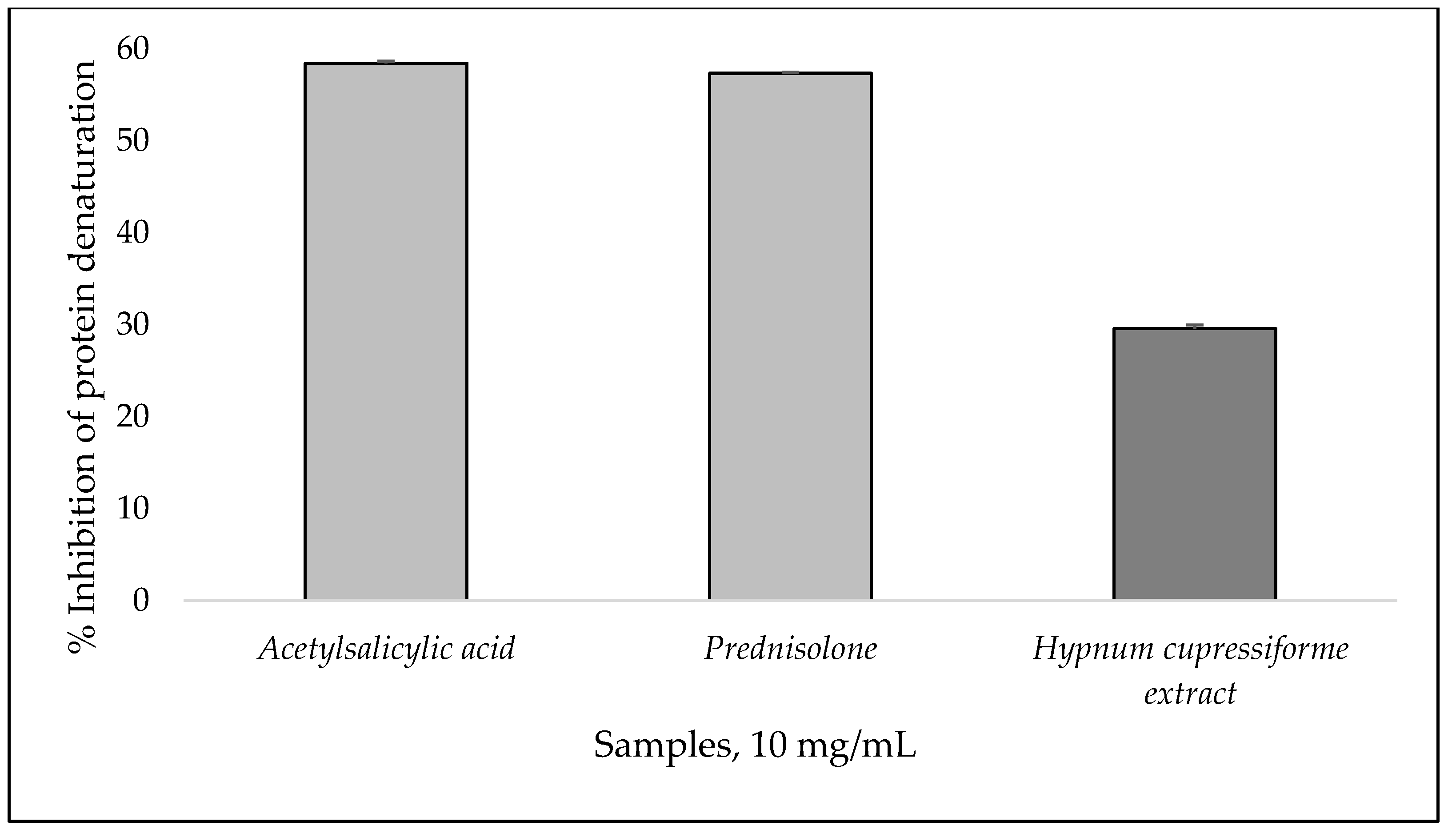
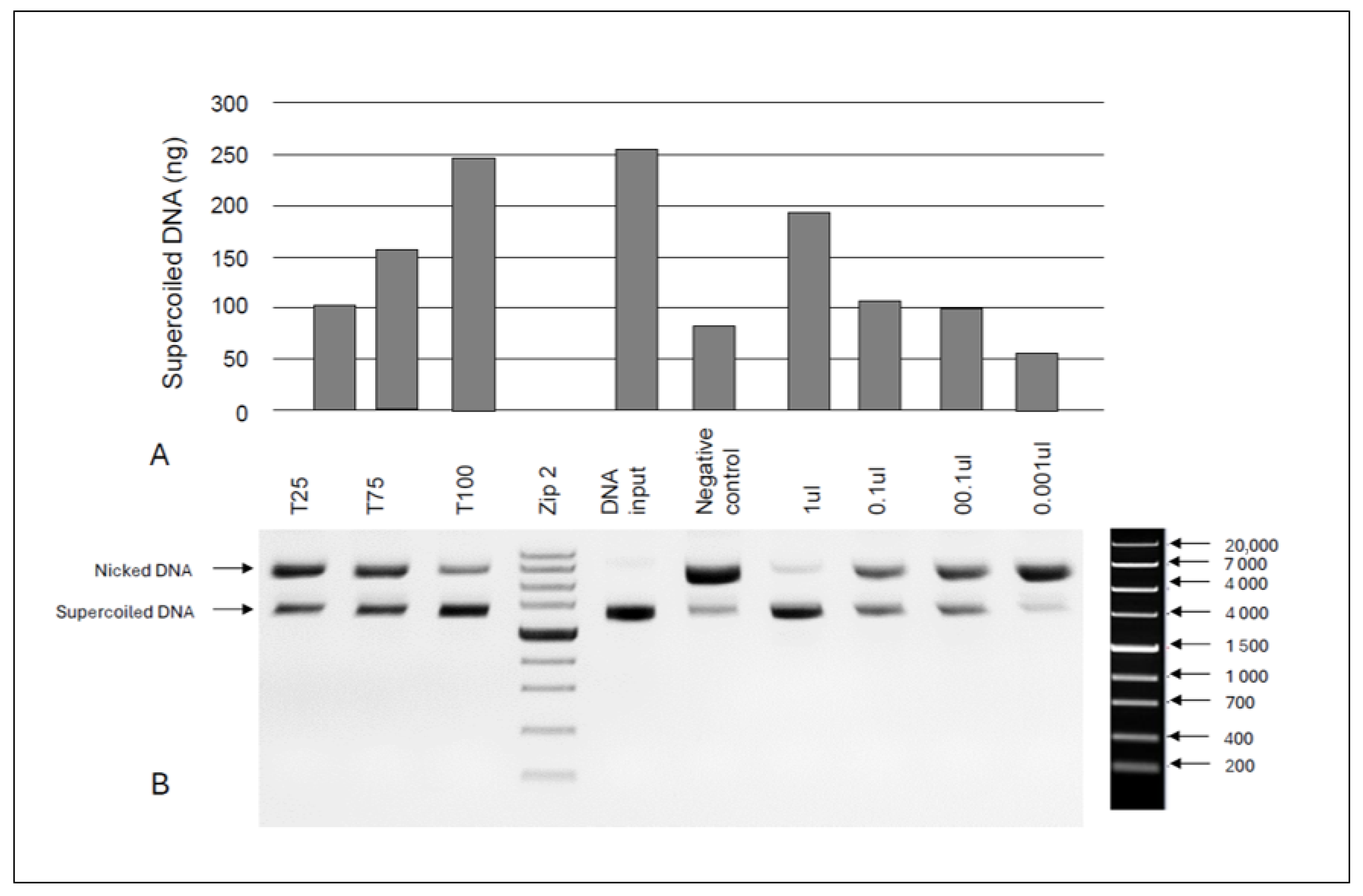
3.5. In Vitro Anti-Inflammatory Activity and DNA Protective Capacity

Protein denaturation is a critical event observed during inflammatory conditions [63]. Inflammation, which can be categorized as either acute or chronic, induces the disruption of secondary and tertiary protein structures, thereby impairing their biological functionality [64,65]. Evidence suggests that the inhibition of protein denaturation correlates with a reduction in inflammatory activity. The anti-inflammatory properties of substances or therapeutic interventions are characterized by their ability to mitigate the inflammatory response [63]. Management of inflammation is typically achieved through the administration of nonsteroidal anti-inflammatory drugs (NSAIDs) and steroidal anti-inflammatory drugs (SAIDs), although their usage is associated with adverse side effects [66,67].
An alternative or concomitant therapy to conventional anti-inflammatory drugs could be various plant extracts based on the biologically active substances they contain and their potential therapeutic effect [68]. In recent years, there has been a significant number of studies on the anti-inflammatory activity of extracts from various parts of plants, using the inhibition of albumin denaturation assay [68,69].
A rapid and inexpensive method was used to evaluate the anti-inflammatory activity of H. cupressiforme extract, and it was compared with two anti-inflammatory drugs. Figure 2 presents the results as percentage inhibition of albumin denaturation.
The tested extract demonstrated 29.58 ± 0.39% protection of human albumin against thermal denaturation at 10 mg/mL concentration. The two standards, acetylsalicylic acid and prednisolone, protected human albumin against heat-induced denaturation, demonstrating a percentage inhibition of 58.44 ± 0.44% and 57.34 ± 0.22%, respectively [70].
The tested H. cupressiforme extract showed inhibitory potential on protein denaturation, most likely due to the content of biologically active substances that contribute to the stabilization of the protein structure [69].
Additionally, the DNA protective capacity of the extracts was tested with an in vitro nicking assay (Figure 3). The amounts used for assays were titrated to 0.001 μL per assay in order to demonstrate the moss extracts’ DNA nicking protection activity. No significant protective effect was found at lower moss extract concentrations (0.001 μL and 0.01 μL). However, when 1 μL of moss extract was tested, DNA nicking protection was found to be commensurable to the protective effect of 75 mg/mL Trolox solution. This finding supports our hypothesis for the putative antioxidant and DNA protective capacity of H. cupressiforme extracts. Yayintas and Demir [71] examined the seasonal changes of DNA damage protection activity of H. cupressiforme extracts using the agarose gel electrophoresis method. They found that the chloroform extract of the samples in the spring season led to complete DNA disintegration at a concentration of 1 μM, which was similar to the findings in the present study.

4. Conclusions

This study provides an analysis of the phytochemical composition and assessment of the in vitro biological activity of Hypnum cupressiforme Hedw. The predominant amino acids were proline, isoleucine, and glutamic acid. The essential amino acids were found in higher concentrations than the non-essential ones, highlighting the potential nutritional value of this moss species. Mannose and fructose were identified in the samples, which suggests their key role in energy metabolism and stress tolerance in H. cupressiforme. Stigmasterol, β-sitosterol, and campesterol were the major phytosterol constituents of this species, which can contribute to their potential health benefits, such as cholesterol-lowering and anti-inflammatory effects. H. cupressiforme contained substantial amounts of polyphenols and quercetin, contributing to its moderate-to-high antioxidant activity. While its DPPH radical scavenging ability was moderate, its ABTS radical scavenging activity was more pronounced, suggesting the presence of both hydrophilic and lipophilic antioxidants. The ethanolic extract of H. cupressiforme demonstrated moderate anti-inflammatory activity. In addition, the DNA nicking protection assay confirmed the potential of this moss extract to protect DNA from oxidative damage, which further increases its value as a source of natural antioxidants with pharmaceutical potential.
Despite this, further research must be conducted to reveal the therapeutic potential of H. cupressiforme. Future studies can be focused on the isolation and characterization of the individual bioactive compounds from this moss species and leading in vivo tests to validate their pharmacological application, as well as comprehensive toxicity evaluations through in vitro cytotoxicity assays and in vivo animal models to establish safe dosage ranges. Seasonal variations in chemical composition and biological activity must also be considered to optimize the timing of harvesting for optimal bioactivity.
Overall, H. cupressiforme is a promising natural source possessing diverse phytochemicals with biological activity, which may be utilized as a potential alternative plant species in various industries.

Supplementary Materials

The following supporting information can be downloaded at: https://www.mdpi.com/article/10.3390/f16060951/s1, Supplementary Material S1. Calibration curve of fructose and mannose; Supplementary Material S2. Chromatogram of the sugars in Hypnum cupressiforme Hedw.; Supplementary Material S3.1. Chromatogram of the phenolic compounds in H. cupressiforme; Supplementary Material S3.2. Chromatogram of the quercetin standard; Supplementary Material S3.3. Calibration curve of quercetin.

Author Contributions

Conceptualization, Z.P. and G.G.; methodology, Z.P., G.A., M.T., I.D., M.O. and S.N.; validation, I.D., M.O. and G.A.; formal analysis, Z.P., M.T., I.D., M.O., S.N., E.A., G.A. and O.T.; investigation, Z.P., O.T., G.G. and G.A.; resources, O.T. and G.G.; data curation, Z.P., M.T., I.D., M.O., S.N., E.A. and G.A.; writing—original draft preparation, Z.P., G.A. and S.N.; writing—review and editing, M.T., I.D., M.O., E.A., O.T. and G.G.; visualization, Z.P. and O.T.; supervision, Z.P.; project administration, Z.P.; funding acquisition, Z.P. All authors have read and agreed to the published version of the manuscript.

Funding

This study is financed by the European Union-NextGenerationEU, through the National Recovery and Resilience Plan of the Republic of Bulgaria, project No. BG-RRP-2.004-0001-C01.

Data Availability Statement

The data presented in this study are available on request from the corresponding author.

Conflicts of Interest

The authors declare no conflicts of interest.

References

  1. Lunić, T.M.; Oalđe, M.M.; Mandić, M.R.; Sabovljević, A.D.; Sabovljević, M.S.; Gašić, U.M.; Duletić-Laušević, S.N.; Božić, B.D.J.; Božić Nedeljković, B.D. Extracts Characterization and In Vitro Evaluation of Potential Immunomodulatory Activities of the Moss Hypnum Cupressiforme Hedw. Molecules 2020, 25, 3343. [Google Scholar] [CrossRef] [PubMed]
  2. Klavina, L.; Springe, G.; Nikolajeva, V.; Martsinkevich, I.; Nakurte, I.; Dzabijeva, D.; Steinberga, I. Chemical Composition Analysis, Antimicrobial Activity and Cytotoxicity Screening of Moss Extracts (Moss Phytochemistry). Molecules 2015, 20, 17221–17243. [Google Scholar] [CrossRef]
  3. Vollár, M.; Gyovai, A.; Szűcs, P.; Zupkó, I.; Marschall, M.; Csupor-Löffler, B.; Bérdi, P.; Vecsernyés, A.; Csorba, A.; Liktor-Busa, E.; et al. Antiproliferative and Antimicrobial Activities of Selected Bryophytes. Molecules 2018, 23, 1520. [Google Scholar] [CrossRef]
  4. Cianciullo, P.; Maresca, V.; Sorbo, S.; Basile, A. Antioxidant and Antibacterial Properties of Extracts and Bioactive Compounds in Bryophytes. Appl. Sci. 2021, 12, 160. [Google Scholar] [CrossRef]
  5. Kirisanth, A.; Nafas, M.N.M.; Dissanayake, R.K.; Wijayabandara, J. Antimicrobial and Alpha-Amylase Inhibitory Activities of Organic Extracts of Selected Sri Lankan Bryophytes. Evid.-Based Complement. Altern. Med. 2020, 2020, 3479851. [Google Scholar] [CrossRef]
  6. Cheng, X.; Xiao, Y.; Wang, X.; Wang, P.; Li, H.; Yan, H.; Liu, Q. Anti-Tumor and Pro-Apoptotic Activity of Ethanolic Extract and Its Various Fractions from Polytrichum commune L. ex Hedw in L1210 Cells. J. Ethnopharmacol. 2012, 143, 49–56. [Google Scholar] [CrossRef] [PubMed]
  7. Krzaczkowski, L.; Wright, M.; Reberioux, D.; Massiot, G.; Etievant, C.; Gairin, J.E. Pharmacological Screening of Bryophyte Extracts That Inhibit Growth and Induce Abnormal Phenotypes in Human HeLa Cancer Cells. Fundam. Clin. Pharmacol. 2009, 23, 473–482. [Google Scholar] [CrossRef]
  8. Wang, X.N.; Yu, W.T.; Lou, H.X. Antifungal Constituents from the Chinese Moss Homalia trichomanoides. Chem. Biodivers. 2005, 2, 139–145. [Google Scholar] [CrossRef]
  9. Singh, M.; Govindarajan, R.; Nath, V.; Rawat, A.K.S.; Mehrotra, S. Antimicrobial, Wound Healing and Antioxidant Activity of Plagiochasma appendiculatum Lehm. et Lind. J. Ethnopharmacol. 2006, 107, 67–72. [Google Scholar] [CrossRef]
  10. Mandić, M.R.; Oalđe, M.M.; Lunić, T.M.; Sabovljević, A.D.; Sabovljević, M.S.; Gašić, U.M.; Duletić-Laušević, S.N.; Božić, B.D.; Nedeljković, B.D.B. Chemical Characterization and In Vitro Immunomodulatory Effects of Different Extracts of Moss Hedwigia ciliata (Hedw.) P. Beauv. from the Vršačke Planine Mts., Serbia. PLoS ONE 2021, 16, e0246810. [Google Scholar] [CrossRef]
  11. Asakawa, Y.; Ludwiczuk, A. Chemical Constituents of Bryophytes: Structures and Biological Activity. J. Nat. Prod. 2017, 81, 641–660. [Google Scholar] [CrossRef]
  12. Klavina, L.; Ramawat, K.; Mérillon, J.M. Polysaccharides from Lower Plants: Bryophytes. In Polysaccharides; Springer: Cham, Switzerland, 2015; pp. 145–160. [Google Scholar]
  13. Lunić, T.M.; Mandić, M.R.; Oalđe Pavlović, M.M.; Sabovljević, A.D.; Sabovljević, M.S.; Božić Nedeljković, B.D.; Božić, B.D. The Influence of Seasonality on Secondary Metabolite Profiles and Neuroprotective Activities of Moss Hypnum cupressiforme Extracts: In Vitro and In Silico Study. Plants 2022, 11, 123. [Google Scholar] [CrossRef]
  14. Glime, J.M. Medical Uses: Medical Conditions. Chapter 2-1. In Bryophyte Ecology; Glime, J.M., Ed.; Michigan Technological University and the International Association of Bryologists: Houghton, MI, USA, 2017; Available online: http://digitalcommons.mtu.edu/bryophyte-ecology/ (accessed on 14 May 2025).
  15. Karataşa, H.; Yayintasb, O. Green Synthesis, Characterization and Biological Activity of Silver Nanoparticles of Hypnum cupressiforme Hedw Extract. J. Optoelectron. Biomed. Mater. 2024, 16, 63–72. [Google Scholar] [CrossRef]
  16. Lunić, T.; Božić, B.; Nedeljković, B.B. Immunomodulatory Potential of Hedwigia ciliata and Hypnum cupressiforme. In Bioactive Compounds in Bryophytes and Pteridophytes; Murthy, H.N., Ed.; Springer: Cham, Switzerland, 2023. [Google Scholar] [CrossRef]
  17. Petkova, Z.; Teneva, O.; Antova, G.; Angelova-Romova, M.; Gecheva, G.; Dimitrova-Dyulgerova, I. Chemical Composition, Lipid-Soluble Bioactive Compounds and Potential Health Benefits of the Moss Hypnum cupressiforme Hedw. Plants 2023, 12, 4190. [Google Scholar] [CrossRef]
  18. Badridze, G.; Chkhubianishvili, E.; Rapava, L.; Kikvidze, M.; Chigladze, L.; Tsiklauri, N.; Tsilosani, K.; Chanishvili, S. Content of Active Metabolites in Some Species of Mosses of Georgia. Bull. Georg. Natl. Acad. Sci. 2020, 14, 127–131. [Google Scholar]
  19. Smolińska-Kondla, D.; Zych, M.; Ramos, P.; Wacławek, S.; Stebel, A. Antioxidant Potential of Various Extracts from 5 Common European Mosses and Its Correlation with Phenolic Compounds. Herba Pol. 2022, 68, 54–68. [Google Scholar] [CrossRef]
  20. World Health Organization; Food and Agriculture Organization. Protein Quality Evaluation. In Proceedings of the Report of the Joint FAO/WHO Expert Consultation, Bethesda, MD, USA, 4–8 December 1989; FAO Food and Nutrition Paper No. 51. Food and Agriculture Organization: Rome, Italy, 1991. [Google Scholar]
  21. ISO 18609:2000; Animal and Vegetable Fats and Oils. Determination of Unsaponifiable Matter. Method Using Hexane Extraction. ISO: Geneva, Switzerland, 2000.
  22. ISO 12228-1:2014; Animal and Vegetable Fats and Oils. Determination of Individual and Total Sterols Contents. Gas Chromatographic Method. ISO: Geneva, Switzerland, 2014.
  23. Lichtenthaler, H.K.; Wellburn, A.R. Determination of Total Carotenoids and Chlorophylls a and b of Leaf in Different Solvents. Biochem. Soc. Trans. 1983, 11, 591–592. [Google Scholar] [CrossRef]
  24. Ivanov, I.; Vrancheva, R.; Marchev, A.; Petkova, N.; Aneva, I.; Denev, P.; Georgiev, V.; Pavlov, A. Antioxidant Activities and Phenolic Compounds in Bulgarian Fumaria Species. Int. J. Curr. Microbiol. Appl. Sci. 2014, 3, 296–306. [Google Scholar]
  25. Milusheva, M.; Gledacheva, V.; Stefanova, I.; Feizi-Dehnayebi, M.; Mihaylova, R.; Nedialkov, P.; Cherneva, E.; Tumbarski, Y.; Tsoneva, S.; Todorova, M.; et al. Synthesis, molecular docking, and biological evaluation of novel anthranilic acid hybrid and its diamides as antispasmodics. Int. J. Mol. Sci. 2023, 24, 13855. [Google Scholar] [CrossRef]
  26. Andonova, T.; Petkova, Z.; Teneva, O.; Antova, G.; Apostolova, E.; Naimov, S.; Mladenova, T.; Slavov, I.; Dimitrova-Dyulgerova, I. Ailanthus altissima seed oil–A valuable source of lipid-soluble components with DNA protective and antiproliferative activities. Foods 2024, 13, 1268. [Google Scholar] [CrossRef]
  27. Rajiv, C.; Roy, S.S.; Tamreihao, K.; Kshetri, P.; Singh, T.S.; Sanjita Devi, H.; Sharma, S.K.; Ansari, M.A.; Devi, E.D.; Devi, A.K.; et al. Anticarcinogenic and Antioxidant Action of an Edible Aquatic Flora Jussiaea repens L. Using in vitro Bioassays and in vivo Zebrafish Model. Molecules 2021, 26, 2291. [Google Scholar] [CrossRef]
  28. Sambrook, J.; Fritsch, E.R.; Maniatis, T. Molecular Cloning: A Laboratory Manual, 2nd ed.; Cold Spring Harbor Laboratory Press: Cold Spring Harbor, NY, USA, 1989. [Google Scholar]
  29. Trovato, M.; Funck, D.; Forlani, G.; Okumoto, S.; Amir, R. Editorial: Amino Acids in Plants: Regulation and Functions in Development and Stress Defense. Front. Plant Sci. 2021, 12, 772810. [Google Scholar] [CrossRef]
  30. Yang, Q.; Zhao, D.; Liu, Q. Connections Between Amino Acid Metabolisms in Plants: Lysine as an Example. Front. Plant Sci. 2020, 11, 928. [Google Scholar] [CrossRef]
  31. Nordin, A.; Gunnarsson, U. Amino acid accumulation and growth of Sphagnum under different levels of N deposition. Écoscience 2000, 7, 474–480. [Google Scholar] [CrossRef]
  32. Semerjyan, G.H.; Semerjyan, I.H. Investigation of amino acid composition of some mosses gathered from Armenia. Proc. YSU B Chem. Biol. Sci. 2021, 55, 290–295. [Google Scholar] [CrossRef]
  33. Hoffman, J.R.; Falvo, M.J. Protein—Which is Best? J. Sports Sci. Med. 2004, 3, 118–130. [Google Scholar]
  34. Joint WHO/FAO/UNU Expert Consultation. Protein and Amino Acid Requirements in Human Nutrition; WHO Technical Report Series 935; WHO: Geneva, Switzerland, 2007. [Google Scholar]
  35. Wu, G. Amino Acids: Biochemistry and Nutrition; CRC Press: Boca Raton, FL, USA, 2013. [Google Scholar]
  36. Fernstrom, J.D.; Fernstrom, M.H. Tyrosine, phenylalanine, and catecholamine synthesis and function in the brain. J. Nutr. 2007, 137, 1539S–1547S. [Google Scholar] [CrossRef]
  37. Elango, R.; Ball, R.O.; Pencharz, P.B. Amino acid requirements in humans: An update. Curr. Opin. Clin. Nutr. Metab. Care 2010, 13, 72–77. [Google Scholar]
  38. Pencharz, P.B. Protein and amino acids. In Present Knowledge in Nutrition, 10th ed.; Erdman, J.W., Jr., Macdonald, I.A., Zeisel, S.H., Eds.; Wiley-Blackwell: Hoboken, NJ, USA, 2012. [Google Scholar]
  39. Norton, L.E.; Layman, D.K. Leucine regulates translation initiation of protein synthesis in skeletal muscle after exercise. J. Nutr. 2006, 136, 533S–537S. [Google Scholar] [CrossRef]
  40. Richard, D.M.; Dawes, M.A.; Mathias, C.W.; Acheson, A.; Hill-Kapturczak, N.; Dougherty, D.M. L-Tryptophan: Basic metabolic functions, behavioral research and therapeutic indications. Int. J. Tryptophan Res. 2009, 2, 45–60. [Google Scholar] [CrossRef]
  41. Young, S.N.; Gauthier, S. Tryptophan Availability and the Control of 5-Hydroxytryptamine and Tryptamine Synthesis in Human CNS. In Serotonin; Haber, B., Gabay, S., Issidorides, M.R., Alivisatos, S.G.A., Eds.; Advances in Experimental Medicine and Biology; Springer: Boston, MA, USA, 1981; Volume 133, pp. 221–230. [Google Scholar] [CrossRef]
  42. Brosnan, J.T.; Brosnan, M.E. The sulfur-containing amino acids: An overview. J. Nutr. 2006, 136, 1636S–1640S. [Google Scholar] [CrossRef] [PubMed]
  43. FAO. Dietary Protein Quality Evaluation in Human Nutrition; Report of an FAO Expert Consultation; FAO: Rome, Italy, 2013. [Google Scholar]
  44. Schaafsma, G. The protein digestibility–corrected amino acid score. J. Nutr. 2000, 130, 1865S–1867S. [Google Scholar] [CrossRef]
  45. de Boer, J.; Aiking, H. On the merits of plant-based proteins for global food security: Marrying macro and micro perspectives. Ecol. Econ. 2011, 70, 1259–1265. [Google Scholar] [CrossRef]
  46. Pratelli, R.; Pilot, G. Regulation of amino acid metabolic enzymes and transporters in plants. J. Exp. Bot. 2014, 65, 5535–5556. [Google Scholar] [CrossRef] [PubMed]
  47. Belitz, H.D.; Grosch, W.; Schieberle, P. Food Chemistry, 4th ed.; Springer: Berlin/Heidelberg, Germany, 2009. [Google Scholar]
  48. Pejin, B.; Iodice, C.; Tommonaro, G.; Sabovljevic, M.; Bianco, A.; Tesevic, V.; Vajs, V.; De Rosa, S. Sugar composition of the moss Rhodobryum ontariense (Kindb.) Kindb. Nat. Prod. Res. 2012, 26, 209–215. [Google Scholar] [CrossRef]
  49. Rütten, D.; Santarius, K.A. Relationship between frost tolerance and sugar concentration of various bryophytes in summer and winter. Oecologia 1992, 91, 260–265. [Google Scholar] [CrossRef]
  50. Xinmei, X.; Ning, X.; Jianbin, H. Physiological function of phytosterol and its application. Anim. Husb. Feed Sci. 2015, 7, 67–69. [Google Scholar]
  51. Vecka, M.; Staňková, B.; Kutová, S.; Tomášová, P.; Tvrzická, E.; Žák, A. Comprehensive Sterol and Fatty Acid Analysis in Nineteen Nuts, Seeds, and Kernel. SN Appl. Sci. 2019, 1, 1531. [Google Scholar] [CrossRef]
  52. Othman, R.A.; Moghadasian, M.H. Beyond cholesterol-lowering effects of plant sterols: Clinical and experimental evidence of anti-inflammatory properties. Nutr. Rev. 2011, 69, 371–382. [Google Scholar] [CrossRef]
  53. Pareek, S.; Sagar, N.A.; Sharma, S.; Kumar, V.; Agarwal, T.; González-Aguilar, G.A.; Yahia, E.M. Chlorophylls: Chemistry and biological functions. In Fruit and Vegetable Phytochemicals: Chemistry and Human Health, 2nd ed.; Yahia, E.M., Ed.; Wiley-Blackwell: Hoboken, NJ, USA, 2017; pp. 269–284. [Google Scholar]
  54. Sangeetha, R.K.; Baskaran, V. Carotenoid composition and retinol equivalent in plants of nutritional and medicinal importance: Efficacy of β-carotene from Chenopodium album in retinol-deficient rats. Food Chem. 2010, 119, 1584–1590. [Google Scholar] [CrossRef]
  55. Sun, T.; Rao, S.; Zhou, X.; Li, L. Plant carotenoids: Recent advances and future perspectives. Mol. Hortic. 2022, 2, 3. [Google Scholar] [CrossRef] [PubMed]
  56. Platzer, M.; Kiese, S.; Tybussek, T.; Herfellner, T.; Schneider, F.; Schweiggert-Weisz, U.; Eisner, P. Radical Scavenging Mechanisms of Phenolic Compounds: A Quantitative Structure-Property Relationship (QSPR) Study. Front. Nutr. 2022, 9, 882458. [Google Scholar] [CrossRef]
  57. Ginwala, R.; Bhavsar, R.; Chigbu, D.G.I.; Jain, P.; Khan, Z.K. Potential Role of Flavonoids in Treating Chronic Inflammatory Diseases with a Special Focus on the Anti-Inflammatory Activity of Apigenin. Antioxidants 2019, 8, 35. [Google Scholar] [CrossRef]
  58. Baqer, S.H.; Al-Younis, Z.K.; Al-Shawi, S.G. Extracting quercetin from different plant sources, purifying it using different extraction methods (chemical, physical, and enzymatic), and measuring its antioxidant activity. Front. Biosci. 2024, 16, 35. [Google Scholar] [CrossRef] [PubMed]
  59. Ivanova, D.; Gerova, D.; Chervenkov, T.; Yankova, T. Polyphenols and antioxidant capacity of Bulgarian medicinal plants. J. Ethnopharmacol. 2005, 96, 145–150. [Google Scholar] [CrossRef]
  60. Xu, N.; Chen, G.; Liu, H. Antioxidative Categorization of Twenty Amino Acids Based on Experimental Evaluation. Molecules 2017, 22, 2066. [Google Scholar] [CrossRef] [PubMed]
  61. Rao, M.J.; Zheng, B. The Role of Polyphenols in Abiotic Stress Tolerance and Their Antioxidant Properties to Scavenge Reactive Oxygen Species and Free Radicals. Antioxidants 2025, 14, 74. [Google Scholar] [CrossRef]
  62. Chaves, N.; Escudero, J.C. Variation of Flavonoid Synthesis Induced by Ecological Factors. In Principles and Practices in Plant Ecology; CRC Press: Boca Raton, FL, USA, 2023; pp. 267–285. [Google Scholar]
  63. Sangeetha, G.; Vidhya, R. In vitro anti-inflammatory activity of different parts of Pedalium murex (L.). Int. J. Herb. Med. 2016, 4, 31–36. [Google Scholar]
  64. Okoli, C.O.; Akah, P.A.; Nwafor, S.V.; Anisiobi, A.I.; Ibegbunam, I.N.; Erojikwe, O. Anti-inflammatory activity of hexane leaf extract of Aspilia africana C.D. Adams. J. Ethnopharmacol. 2007, 109, 219–225. [Google Scholar] [CrossRef]
  65. Eming, S.A.; Krieg, T.; Davidson, J.M. Inflammation in wound repair: Molecular and cellular mechanisms. J. Investig. Dermatol. 2007, 127, 514–525. [Google Scholar] [CrossRef]
  66. Bindu, S.; Mazumder, S.; Bandyopadhyay, U. Non-steroidal anti-inflammatory drugs (NSAIDs) and organ damage: A current perspective. Biochem. Pharmacol. 2020, 180, 114147. [Google Scholar] [CrossRef] [PubMed]
  67. Del Grossi Moura, M.; Cruz Lopes, L.; Silva, M.T.; Barberato-Filho, S.; Motta, R.H.L.; Bergamaschi, C.C. Use of steroid and nonsteroidal anti-inflammatories in the treatment of rheumatoid arthritis: Systematic review protocol. Medicine 2018, 97, e12658. [Google Scholar] [CrossRef] [PubMed]
  68. Madhuranga, H.D.T.; Samarakoon, D.N.A.W. In vitro anti-inflammatory egg albumin denaturation assay: An enhanced approach. Nat. Ayurvedic Med. 2023, 7, 1–6. [Google Scholar]
  69. Dharmadeva, S.; Galgamuwa, L.S.; Prasadinie, C.; Kumarasinghe, N. In vitro anti-inflammatory activity of Ficus racemosa L. bark using albumin denaturation method. AYU 2018, 39, 239–242. [Google Scholar] [CrossRef]
  70. Bailey-Shaw, Y.A.; Williams, L.A.; Green, C.E.; Rodney, S.; Smith, A.M. In-vitro evaluation of the anti-inflammatory potential of selected Jamaican plant extracts using the bovine serum albumin protein denaturation assay. Int. J. Pharm. Sci. Rev. Res. 2017, 47, 145–153. [Google Scholar]
  71. Yayintas, O.T.; Demir, N. Seasonal Changes of Antioxidant Activity and DNA Damage Protection Potential of Fontinalis antipyretica Hedw. and Hypnum cupressiforme Hedw. Fresenius Environ. Bull. 2019, 28, 6978–6987. [Google Scholar]
Figure 1. GC-MS chromatogram of the amino acids in Hypnum cupressiforme Hedw. Legend: The number of the peaks represents the number of the specific amino acid shown in Table 1.
Forests 16 00951 g001
Figure 2. In vitro anti-inflammatory activity of the H. cupressiforme extract and the controls (10 mg/mL) expressed as inhibition of albumin denaturation (%). Legend: All analyses were conducted in triplicate (n = 3), and the results are presented as mean ± standard deviation (SD).
Forests 16 00951 g002
Figure 3. DNA protective capacity of H. cupressiforme extracts: (A) relative levels of nicked plasmid DNA—lanes 1–3 represent varying Trolox concentrations (25, 75, and 100 mg/mL); lane 4—ZipRuler 2 Express DNA Ladder (Thermo Scientific, SM1373); lane 5—plasmid DNA input; lane 6—negative control; lanes 7–10 correspond to tenfold serial dilutions of the tested extract (1, 0.1, 0.01, and 0.001 μL/reaction); and (B) agarose gel electrophoresis results.
Forests 16 00951 g003
Table 1. Amino acid composition of Hypnum cupressiforme Hedw. (g/100 g FW).
No.RT * (min)Amino AcidsMean ± SD
14.66Alanine0.095 ± 0.03
25.25Valine 11.201 ± 0.31
35.43Leucine 11.463 ± 0.42
45.55Isoleucine 12.047 ± 0.73
55.71Glycine0.220 ± 0.06
65.82Proline2.282 ± 0.21
76.02Serine0.939 ± 0.24
86.09Threonine 11.015 ± 0.30
96.15Methionine 11.358 ± 0.41
106.66Aspartic acid1.132 ± 0.18
116.89Pyroglutamic acid0.738 ± 0.15
128.19Glutamic acid1.746 ± 0.12
138.47Phenylalanine 10.390 ± 0.09
148.78Arginine 30.354 ± 0.14
159.11Histidine 20.149 ± 0.03
169.38Lysine 11.175 ± 0.30
1710.33Tryptophan 10.253 ± 0.07
1810.46Tyrosine0.194 ± 0.04
Total content of amino acids16.75 ± 3.83
* RT—Retention time; all analyses were conducted in triplicate (n = 3), and the results are presented as mean ± standard deviation (SD); 1 Essential amino acids, 2 Essential amino acids in child nutrition, 3 Semi-essential amino acids.
Table 2. Amino acid score of H. cupressiforme.
Amino AcidsScoring Pattern, g/100 g *Amino Acid Score
Histidine2.70.06
Isoleucine4.70.44
Leucine9.50.15
Lysine7.80.15
Methionine +cystine3.30.41
Phenylalanine + Tyrosine10.20.06
Threonine4.40.23
Tryptophan1.40.18
Valine6.40.19
* Amino acid scoring pattern (g/100 g of protein) of cow’s milk [18].
Table 3. Content and composition of sugars in H. cupressiforme (g/100 g DW) *.
No.RT (min) **Free SugarsMean ± SD
1.7.550Fructose1.43 ± 0.1
2.8.145Mannose1.76 ± 0.1
Total3.19 ± 0.2
* All analyses were conducted in triplicate (n = 3), and the results are presented as mean ± standard deviation (SD); ** RT—Retention time.
Table 4. Individual sterol composition of H. cupressiforme (mg/g FW) *.
No.RT (min) **SterolsMean ± SD
113.702Cholesterol0.48 ± 0.02
214.508Brassicasterol0.05 ± 0.00
314.937Campesterol3.34 ± 0.08
415.352Stigmasterol4.37 ± 0.12
516.153β-Sitosterol4.29 ± 0.10
617.3815-Avenasterol0.16 ± 0.03
717.4827-Stigmasterol0.71 ± 0.04
Total13.40 ± 0.39
* All analyses were conducted in triplicate (n = 3), and the results are presented as mean ± standard deviation (SD); ** RT—Retention time.
Table 5. Chlorophyll and carotenoid content of H. cupressiforme (μg/g DW) *.
ComponentsMean ± SD
Chl a237.37 ± 0.13
Chl b117.39 ± 0.51
Chl a + b354.77 ± 0.64
Carotenoids91.53 ± 1.36
* All analyses were conducted in triplicate (n = 3), and the results are presented as mean ± standard deviation (SD).
Table 6. Total phenols and antioxidant activity of H. cupressiforme *.
IndicatorsMean ± SD
Total phenols (mg GAE/g DW)0.98 ± 0.04
Content of quercetin, mg/g0.12 ± 0.01
Antioxidant activity
DPPHI, % **14.80 ± 0.08
(mM TE/g DW)2.56 ± 0.02
ABTSI, %33.28 ± 0.12
(mM TE/g DW)4.15 ± 0.12
* All analyses were conducted in triplicate (n = 3), and the results are presented as mean ± standard deviation (SD); ** I—Inhibition percentage.
Disclaimer/Publisher’s Note: The statements, opinions and data contained in all publications are solely those of the individual author(s) and contributor(s) and not of MDPI and/or the editor(s). MDPI and/or the editor(s) disclaim responsibility for any injury to people or property resulting from any ideas, methods, instructions or products referred to in the content.

