Article

The First Report of Hypogeous Geastrum in Hungary in Mattirolomyces terfezioides-Producing Forests in the Carpathian Basin

1
Department of Plant Physiology and Molecular Plant Biology, Eötvös Loránd University (ELTE), 1/C Pázmány Péter Sétány, 1117 Budapest, Hungary
2
Department of Biology, Mekdela Amba University, South Wollo P.O. Box 32, Ethiopia
*
Author to whom correspondence should be addressed.
Forests 2025, 16(7), 1136; https://doi.org/10.3390/f16071136
Submission received: 31 May 2025 / Revised: 28 June 2025 / Accepted: 1 July 2025 / Published: 10 July 2025
(This article belongs to the Section Forest Biodiversity)

Abstract

Hungary has some of the best-researched hypogeous fungal flora in Europe, with a large number of genera and species already having been discovered in this country. In this study, we performed morphological and molecular analysis of unidentified hypogeous fungi samples collected from Hungary. We confirmed that they belong to the hypogeous earthstar species Geastrum nadalii (Paz et al., 2024), marking the first report of this species in this country and in a continental climate. We also assume that the habitat preference of this mushroom species is similar to the habitat preference of Mattirolomyces terfezioides, with both occurring in planted non-native Robinia pseudoacacia forests, suggesting that these are secondary habitats for these species. We also conclude that this Mediterranean species has appeared only recently in the Hungarian mycota.

1. Introduction

Earthstars are a morphological and taxonomically diverse group of macrofungi. Despite their lack of commercial use, they are well-studied fungi, because of their unique morphological appearance [1]. Earthstars belong to the family Geastraceae. Most species in this family are considered saprotrophic, but there is evidence that some species can form ectomycorrhiza [2]. The basidiome of earthstars develops underground; then, throughout the maturation process, most earthstars grow above ground, and their exoperidium rips open and spores are released through a hole in the endoperidium [1]. However, there are species in which the fruitbody remains hypogeous, the exoperidium does not rip, and the endoperidium is underdeveloped or absent [3]. These taxa demonstrate the typical evolutionary line of hypogeous fungi [4]. Because they lose the ability to release their spores into the air, they usually develop an aromatic odor to attract vector animals [4].
The genus Radiigera was described by Sanford Myron Zeller in 1944 [5], with the type species as Radiigera fuscogleba. Later on, it was placed in the Geastraceae family and considered a close relative of the genus Geastrum [6]. But, in their 2014 article [7], Zamora et al. revised the systematics of Geastrum and Radiigera and merged the two genera into one. Since then, four former Radiigera species—R. bushnellii, R. flexuosa, R. fuscogleba, and R. taylorii—have been considered to belong to the genus Geastrum. Another former species, R. atrogleba, is now considered a synonym of Schenella simplex [8].
There is a very limited number of records of hypogeous Geastraceae samples in Europe. G. flexuosum has been identified in Sweden [3]. Another species, known as Radiigera romana, was described in Italy [9], and later renamed as Schenella romana [8]. Another specimen that has been found in Italy is Radiigera atrogleba, discovered in Montecchi, and Sarassini’s book [10] notes that it is extremely rare.
In this study, we confirmed the occurrence of a hypogeous Geastrum species in Hungary, based on morphological and molecular analysis, and examined its habitat preferences. In our herbarium, we had samples from the time span of 2013 to 2024. They were labeled as “Radiigera sp.”, although there are no previous reports of this genus and other hypogeous Geastraceae from Hungary, and according to our analysis, the samples were morphologically and phylogenetically different from the hypogeous Geastrum species known at the time. The samples were collected in a period lasting from September to December, from habitats in sandy soil deposited by the Danube, in planted man-made Robinia pseudoacacia forests (Robinietum cultum, [11]). We observed that all fungal nests were located near honey locust (Gleditsia triacanthos) trees. Other common woody plants that can be found nearby the basidiomata are Celtis occidentalis and Crataegus monogyna.

2. Materials and Methods
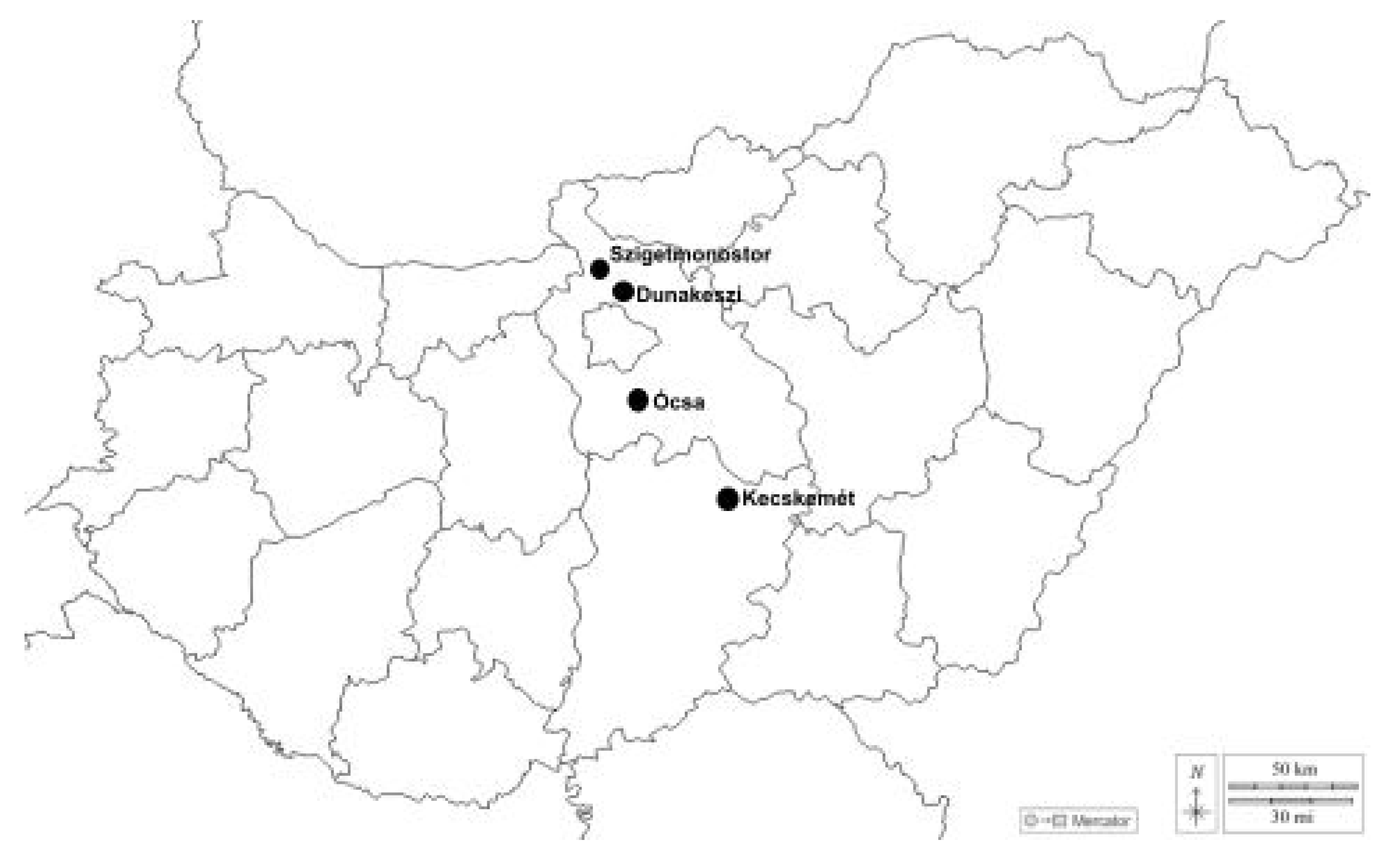
2.1. Study Area

All our collections were made in areas with sandy soil deposited by the river Danube (see Table 1 and Figure 1). These habitats are highly similar to those of the sand truffle (Mattirolomyces terfezioides), which is a well-known and valuable commercial truffle in Hungary, but there are many unanswered questions about its origin and habitat preferences [12], and it forms a mycorrhizal connection with the black locust tree (Robinia pseudoacacia) [13]. According to our collection data, this Geastrum species has only been found in the same habitats as the sand truffle in Hungary, in the Pest Plain and Kiskunság microregions.

2.2. Fungi Collection, Sampling

Fresh basidiomata were collected from natural habitats (see Figure 2), with the help of truffle hunters, members of the First Hungarian Truffle Society (EMSzE) with certificates, and trained truffling dogs, and were deposited in the society’s mycotheca (ZB) [14]. The fruitbodies were transferred to our lab, where some were examined when fresh, then dried and stored in our herbarium.

2.3. Soil Sampling

Soil samples were taken in the vicinity of the fruitbodies (less than 1 m from them) with the help of shovels. Four soil samples were taken from each mushroom habitat, approximately till 15 cm depth. Samples were packed in plastic bags, and after drying and sieving, they were sent to an accredited lab for soil analysis (Nemzeti Élelmiszerlánc-biztonsági Hivatal Élelmiszerlánc-biztonsági Laboratórium Igazgatóság Növény-és Talajvédelmi Nemzeti Referencia Laboratórium, Velence, Hungary). The examined soil parameters and results can be found in Supplement S2.

2.4. Morphological and Molecular Study

The samples’ macromorphological characteristics, including their basidiome shape, surface, color, and gleba color and shape, were examined with a Nikon SMZ-2T stereo microscope. PZO Biolar, Zeiss Ergaval, Nikon Optiphot-2, and Nikon SMZ-2T microscopes were used for measuring the micromorphological structures of the fungi, such as their peridium, columella, gleba, and spores. In addition to this, the Piximétre program was used to measure the spore sizes. The ultrastructure of the peridium, spores, and capillitia were examined under a scanning electron microscope. For this examination, the gleba of dried ascomata was scraped, and pieces of it were fixed onto a taped disk. The tissue fragments were gold-coated, and adhered spores were inspected under a Hitachi 2360N scanning electron microscope (SEM). Gleditsia roots were stained to investigate possible mycorrhizal connections, according to Vierheilig’s method [15].
Genomic DNA was isolated from dried basidiomata with the DNeasy Plant Mini Kit (Qiagen, Courtaboeuf, France), according to the manufacturer’s instructions, with a few modifications based on our experience. The amplified loci were Internal Transcribed Spacer (ITS) and Large Subunit (LSU) regions of the fungal nuclear ribosomal DNA locus. For the ITS, we used ITS1F/ITS4 primers, and for the LSU, we used LROR/LR5 primers for amplification of the genomic region of interest [16,17]. PCR was conducted under the following conditions: initial denaturation at 94 °C for 4.5 min; 33 cycles of denaturation at 94 °C for 30 s; annealing at 51 °C for 30 s; elongation at 72 °C for 45 s; and, after the last cycle, a final synthesis stage at 72 °C for 7 min. For gel electrophoresis of the amplification products, 1% agarose gel was used. Staining was performed with ethidium bromide. For the purification of the PCR products, we used the QIAquick® PCR Purification Kit. Sanger sequencing was carried out by BIOMI Ltd. (Gödöllő, Hungary).
The electropherograms were checked and edited using FinchTV 1.4.0 (https://digitalworldbiology.com/finchtv, first access date: 29 July 2022). The resulting sequences were compared to other sequences in GenBank through a BLASTn (version: 2.16.0+) search [18], and then were submitted to the GenBank database. The sequences generated in this study, together with additional sequences from GenBank, were aligned using MAFFT [19] on XSEDE (7.402), on the CIPRES Science Gateway [20] (http://www.phylo.org/ first access date: 29 July 2022). The aligned DNA sequences were manually corrected using MEGA 6.06 [21] in cases of misalignment. The file formats were converted with the ALTER online application [22]. All the sequences were deposited in GenBank with accession numbers.
After this, the most taxonomically relevant sequences were gathered for further evaluation regarding the available ecological and distributional data. The final dataset contains our newly generated sequences and the selected reference sequences that were selected based on the current taxonomic results. RAxML 8.2.12 [23], as implemented on CIPRES [20], was used to perform Maximum Likelihood (ML) analysis using fast bootstrap (1000×). We used jModelTest [24] to find the optimal substitution model, which was the General Time Reversible model, for nucleotide substitutions, combined with a gamma distribution to account for rate heterogeneity across sites, and incorporating a proportion of invariable sites in the evolutionary model. The likelihood of the final tree was optimized under the GAMMA substitution model, and FigTree V. 1.4.4 [25] was used to visualize the phylogenetic tree (Figure 3). As an outgroup, Myriostoma coliforme (GenBank accession numbers: KF988337 and KF988348) was chosen.
The DNA extraction, PCR, sequence processing, and phylogenetic tree generation were conducted according to our lab protocol, in a similar way to work on other hypogeous fungi [26].

3. Results

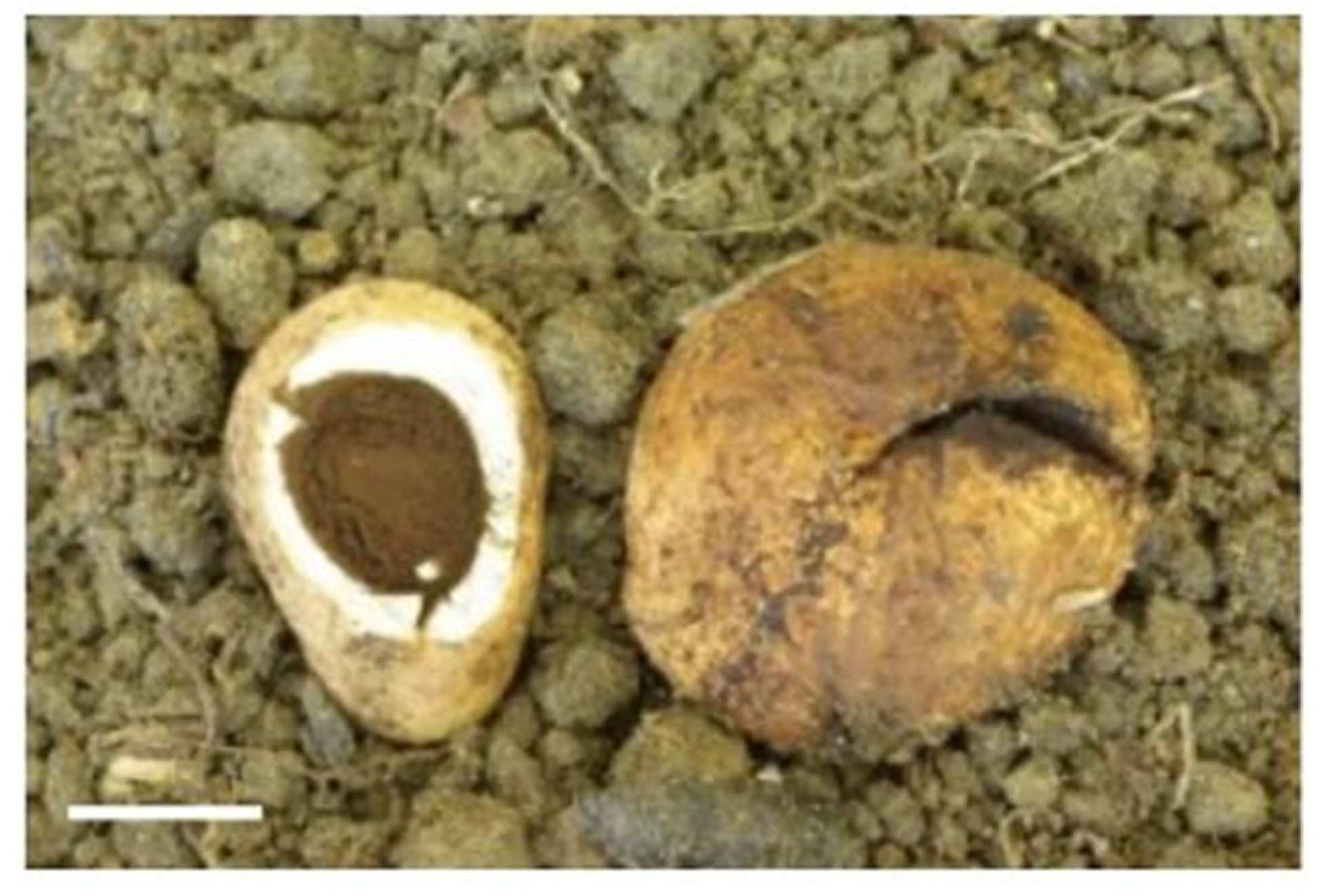
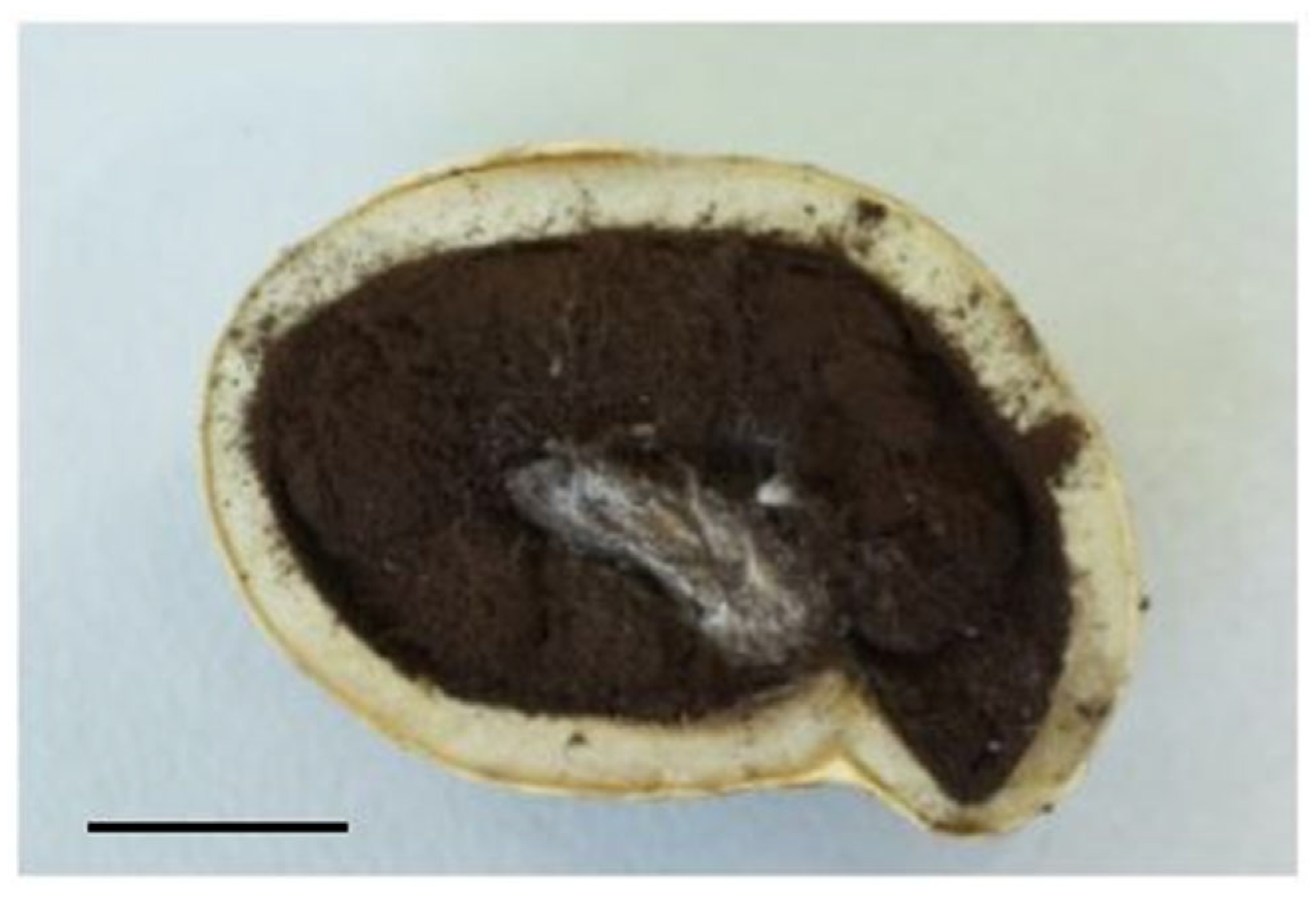
3.1. Macromorphological Characteristics

The microscopy results showed that the basidiome forms below the soil surface, and does not rise visibly to the soil surface even when mature; it is 10–36 mm in diameter, spherical or slightly flattened spherical, sometimes slightly wrinkled, and smooth or slightly rough, and the surface is covered with an easily detachable mycelium and some rhizomorph-like structures.
Color of fresh basidiome: ocher cream—pale beige—rose ocher—pink—bright pink—purplish pink.
Dried material colors: clay buff (32), fawn (29), fulvous (12) (color key of the Royal Botanical Garden, Edinburgh (RBGE) 1969).
The smell is metallic in fresh material; this is not observed in the exsiccata.
The exoperidium is thin and brownish at the cut surface of the fruiting body.
The exoperidial layer adheres to the 1–2 mm thick mesoperidial layer or detaches from it after drying.
The outer part of the mesoperidium, a thinner band, is yellowish-brown, and the thicker inner part is whitish. The inner side of the mesoperidial layer directly borders the gleba.
The endoperidium is absent.
The gleba is dark gray-black, and cotton-like when mature, with a disintegrating spore mass.
The columella is well-developed, with a width of 4–7 mm and a length of 10 mm, and it extends halfway into the gleba (see Figure 3).

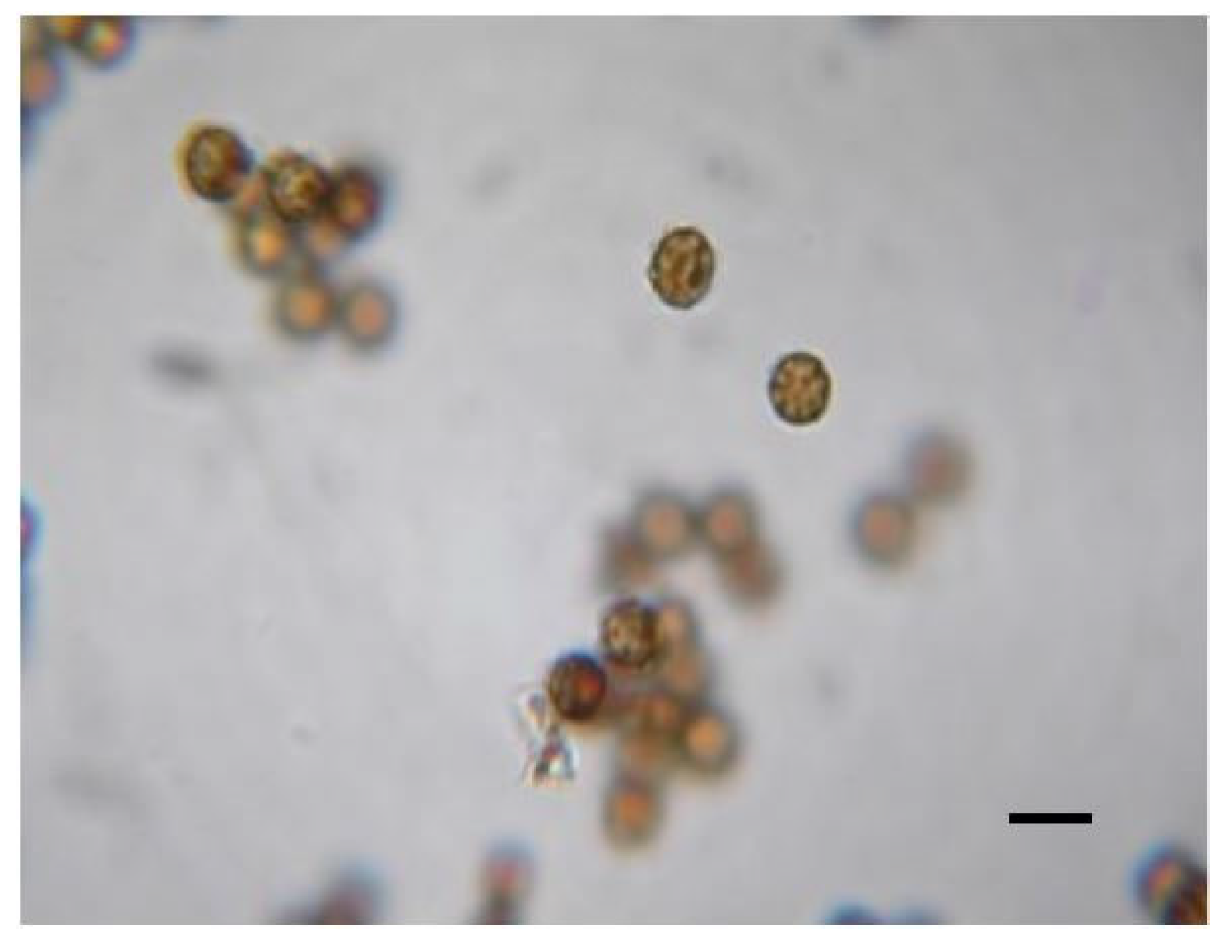
3.2. Micromorphological Characteristics

The total width of the peridium is 1632–2224 µm.
The exoperidium consists of two layers. The cortical external layer is 32–112 µm, with dense interwoven yellowish 2–5 µm wide hyphae and 0.5–0.7 µm thick cell walls; the hyphae are rarely branched. Internal layer: 144–320 µm, has an incompact structure, strongly coiled interwoven hyphae, pale yellow, rarely branched, septum not detected, cells 1.7–4 µm thick, cell walls 0.5–1.5 µm.
The width of the mesoperidium is 1504–1968 µm; the hyaline pseudoparenchymatous cells are of mixed type and of various shapes, with rounded corners, or completely spherical, with an irregular ovoid shape, sometimes with angular or pointed corners wedged between neighboring cells.
External mesoperidial layer textura globosa, with smaller subglobose cells of 2.5–10.9 × 3.3–18.3 µm, are arranged in a few cell rows at the border of the exoperidium and the mesoperidium. Deeper towards to the gleba, the internal layer is textura globosa to textura prismatica, with larger and larger shapeless angular cells of 5.9–30 × 8.3–43.3 µm located mixed in with the small subglobose cells.
Columella hyphae are hyaline or yellowish, rarely branched, (1.5–)2–3(–4) µm wide, 0.5–0.7(–1.0) µm wall thickness, septa rarely visible.
Capillitium is 1–2.5(–3.5) µm wide hyphal hyaline, clamp and septa not detected, sometimes undulating, sometimes widening, their wall thickness varies, 0.5–0.7 µm.
Spores are globose–subglobose, ornamented, brown, diameter: 3.2–5.1 µm (average 3.7–4.1 µm), Q: 1–1.1(–1.2) (see Figure 4). Ornamentation: slightly rounded, coarse warts, knotted (sometimes almost irregularly areolate-reticulate) 0.1–0.6 µm (average = 0.27–0.37 µm) (see Figure 5).
Basidium is not detected.
Crystals are not detected, not on hyphae nor on spores.
After staining of the Gleditsia roots, only arbuscular mycorrhizal structures were detected, which cannot be connected to the Geastrum genus.

3.3. Phlyogenetic Analysis

3.4. Soil Analysis

The results of the soil analysis can be found in Supplement S2.

4. Discussion

Based on the morphological and phylogenetic data, we can conclude that these hypogeous Geastrum samples belong to the recently described Geastrum nadalii species [27]. All previous records come from regions with a Mediterranean climate; the samples studied here constitute the first report from an area with a continental climate. Based on the phylogenetic data, we found that this species is most closely related to Geastrum violaceum, which has been identified in Brazil [28]. According to the molecular data, this species belongs to the Corollina section and the Plicostomata subsection [7]; however, this could not be confirmed morphologically due to a lack of crystals and a peristome, the shapes of which are the main morphological characteristics of this section. Among other European species, Geastrum morganii is the most closely related species. Because all the samples were collected from known Mattirolomyces habitats, we suggest that in Hungary, this species shares the same habitat with the sand truffle. The soil data was highly similar to that reported for sand truffle nests. We also noticed that this species always grows under or nearby honey locust trees (Gleditsia triacanthos), which is a plant introduced from North America, like the black locust (Robinia pseudoacacia). We did not find any special fungal structures at the actual time of sampling, besides arbuscular mycorrhizae in the honey locust roots, suggesting that this connection might not be mycorrhizal. Both fungal species have been identified in the Mediterranean, so we can conclude that planted Robinietum forests might be secondary habitats of both the sand truffle and Geastrum nadalii. These species are additional good examples of Mediterranean mushroom species spreading into northern Europe, as is well documented for some other truffle species, like Tuber magnatum [29,30,31].

5. Conclusions

Hungary has a rich and well-researched hypogeous mycota, and in this study, we confirmed the presence of a previously unknown species, Geastrum nadalii, the first hypogeous Geastrum to be identified in this country. Analysis of its growing sites showed that it shares habitat preferences with Mattirolomyces terfezioides, occurring in planted Robinia forests. We conclude that this species has appeared recently in Hungary, as most samples have been collected in the Mediterranean, and this species is currently spreading north in Europe. Our findings show that there are still many unidentified species of the hypogeous mycota, even in well-screened territories like the Carpathian Basin.

Supplementary Materials

The following supporting information can be downloaded at https://www.mdpi.com/article/10.3390/f16071136/s1: Supplement S1: Geastrum collections used in phylogenetic analyses. Sequences in bold were newly generated for this study. Supplement S2: Soil data for the different Geastrum nadalii mushroom nests.

Author Contributions

Conceptualization and methodology: Á.H., P.C., B.P., A.A.H., and Z.B.; field collections: Á.H., A.A.H., B.P., and Z.B.; morphological analysis: Á.H., P.C., and I.N.; phylogenetic analysis: B.P., A.A.H., P.C., and Á.H.; writing—manuscript preparation: Á.H., A.A.H., I.N., B.P., and Z.B.; supervision: Z.B.; project administration: Z.B. All authors have read and agreed to the published version of the manuscript.

Funding

This research received no external funding.

Data Availability Statement

The molecular data is publicly available in GenBank, and all other data is available in the manuscript.

Acknowledgments

The authors would like to express their gratitude to the truffle hunters, who helped in the collection of the samples. Furthermore, we express our thanks to Biomi Ltd. for the sequencing of our samples. We are very grateful to Károly Bóka for his help in the scanning electron microscopy work.

Conflicts of Interest

The authors have no conflicts of interest.

References

  1. Jeppson, M.; Nilsson, R.H.; Larsson, E. European earthstars in Geastraceae (Geastrales, Phallomycetidae)—A systematic approach using morphology and molecular sequence data. Syst. Biodivers. 2013, 11, 437–465. [Google Scholar] [CrossRef]
  2. Agerer, R. Geastrum fimbriatum Fr. + Fagus sylvatica L. Descr. Ectomyc. 1998, 3, 13–18. [Google Scholar]
  3. Dominguez de Toledo, L.S.; Castellano, M.A. A revision of the genera Radiigera and Pyrenogaster. Mycologia 1996, 88, 863–884. [Google Scholar] [CrossRef]
  4. Moreno, G.; Alvarado, P.; Manjón, J.L. Hypogeous Desert Fungi. In Desert Truffles; Springer: Berlin/Heidelberg, Germany, 2014; pp. 3–20. [Google Scholar]
  5. Zeller, S.M. Representatives of the Mesophelliaceae in North America. Mycologia 1944, 36, 627–637. [Google Scholar] [CrossRef]
  6. Askew, B.; Miller, O.K., Jr. New evidence of close relationships between Radiigera and Geastrum (Lycoperdales). Can. J. Bot. 1977, 55, 2693–2700. [Google Scholar] [CrossRef]
  7. Zamora, J.C.; de Diego Calonge, F.; Hosaka, K.; Martín, M.P. Systematics of the genus Geastrum (Fungi: Basidiomycota) revisited. Taxon 2014, 63, 477–497. [Google Scholar] [CrossRef]
  8. Estrada-Torres, A.; Gaither, T.W.; Miller, D.L.; Lado, C.; Keller, H.W. The myxomycete genus Schenella: Morphological and DNA sequence evidence for synonymy with the gasteromycete genus Pyrenogaster. Mycologia 2005, 97, 139–149. [Google Scholar] [CrossRef]
  9. Quadraccia, L. Studies on Italian gasteromycetes. I. Two new species of Arachnion and Radiigera (Basidiomycotina, Lycoperdales) from Rome and its environs. Mycotaxon 1996, 58, 331–341. [Google Scholar]
  10. Montecchi, A.; Sarasini, M. Funghi Ipogei D’Europa; A.M.B. Fondazione Centro Studi Micologici: Vicenza, Italy, 2000. [Google Scholar]
  11. Borhidi, A. Magyarország Növénytársulásai; Akadémiai Kiadó: Budapest, Hungary, 2003. [Google Scholar]
  12. Csorbainé, A.G.; Bratek, Z.; Merényi, Z.; Illyès, Z.; Dimény, J. Choiromyces meandriformis and Mattirolomyces terfezioides: Peculiar truffles with new perspectives. In Proceedings of the XVII Convegno Nazionale di Micologia, Pavia, Italy, 10–12 November 2008; pp. 10–12. [Google Scholar]
  13. Bratek, Z.; Jakucs, E.; Bóka, K.; Szedlay, G. Mycorrhizae between black locust (Robinia pseudoacacia) and Terfezia terfezioides. Mycorrhiza 1996, 6, 271–274. [Google Scholar] [CrossRef]
  14. Merényi, Z.; Illyés, Z.; Völcz, G.; Bratek, Z. A database and its application for the development of truffle cultivation methods. Osterr. Z. Für Pilzkd. 2010, 19, 239–244. [Google Scholar]
  15. Vierheilig, H.; Coughlan, A.P.; Wyss, U.R.S.; Piché, Y. Ink and vinegar, a simple staining technique for arbuscular-mycorrhizal fungi. Appl. Environ. Microbiol. 1998, 64, 5004–5007. [Google Scholar] [CrossRef] [PubMed]
  16. Vilgalys, R.; Hester, M. Rapid genetic identification and mapping of enzymatically amplified ribosomal DNA from several Cryptococcus species. J. Bacteriol. 1990, 172, 4238–4246. [Google Scholar] [CrossRef] [PubMed]
  17. Gardes, M.; Bruns, T.D. ITS primers with enhanced specificity for basidiomycetes-application to the identification of mycorrhizae and rusts. Mol. Ecol. 1993, 2, 113–118. [Google Scholar] [CrossRef] [PubMed]
  18. Altschul, S.F.; Madden, T.L.; Schäffer, A.A.; Zhang, J.; Zhang, Z.; Miller, W.; Lipman, D.J. Gapped BLAST and PSI-BLAST: A new generation of protein database search programs. Nucleic Acids Res. 1997, 25, 3389–3402. [Google Scholar] [CrossRef]
  19. Katoh, K.; Toh, H. Recent developments in the MAFFT multiple sequence alignment program. Brief. Bioinform. 2008, 9, 286–298. [Google Scholar] [CrossRef]
  20. Miller, M.A.; Pfeiffer, W.; Schwartz, T. Creating the CIPRES Science Gateway for inference of large phylogenetic trees. In Proceedings of the 2010 Gateway Computing Environments Workshop (GCE), New Orleans, LA, USA, 14 November 2010; pp. 1–8. [Google Scholar]
  21. Tamura, K.; Stecher, G.; Peterson, D.; Filipski, A.; Kumar, S. MEGA6: Molecular evolutionary genetics analysis version 6.0. Mol. Biol. Evol. 2013, 30, 2725–2729. [Google Scholar] [CrossRef]
  22. Glez-Peña, D.; Gómez-Blanco, D.; Reboiro-Jato, M.; Fdez-Riverola, F.; Posada, D. ALTER: Program-oriented format conversion of DNA and protein alignments. Nucleic Acids Res. 2010, 38, W14–W18. [Google Scholar] [CrossRef]
  23. Stamatakis, A. RAxML version 8: A tool for phylogenetic analysis and post-analysis of large phylogenies. Bioinformatics 2014, 30, 1312–1313. [Google Scholar] [CrossRef]
  24. Posada, D. jModelTest: Phylogenetic model averaging. Mol. Biol. Evol. 2008, 25, 1253–1256. [Google Scholar] [CrossRef]
  25. Rambaut, A. FigTree. Tree Figure Drawing Tool. 2009. Available online: http://tree.bio.ed.ac.uk/software/figtree/ (accessed on 25 May 2025).
  26. Cseh, P.; Merényi, Z.; Bóna, L.; Varga, T.; Bóka, K.; Nagy, I.; Kaounas, V.; Vidal, J.M.; Paz, A.; Bratek, Z. Taxonomic characterisation of the Regianum clade (genus Tuber) and the trait evolution of spore size among true truffles. Mycol. Progress. 2024, 23, 11. [Google Scholar] [CrossRef]
  27. Paz, A.; Lavoise, C.; Chautrand, P.; Slavova, M.; Daniëls, P.P.; Rojo, C. Geastrum nadalii sp. nov. In: Crous, P.W.; et al. Fungal Planet description sheets: 1614–1696. Fungal Syst. Evol. 2024, 13, 241–242. [Google Scholar]
  28. Sousa, J.O.; Morais, L.A.; Nascimento, Y.M.; Baseia, I.G. Updates on the geographic distribution of three Geastrum species from Brazilian semi-arid region. Mycosphere 2014, 5, 467–474. [Google Scholar] [CrossRef]
  29. Čejka, T.; Trnka, M.; Büntgen, U. Sustainable cultivation of the white truffle (Tuber magnatum) requires ecological understanding. Mycorrhiza 2023, 33, 291–302. [Google Scholar] [CrossRef] [PubMed]
  30. Belfiori, B.; D’Angelo, V.; Riccioni, C.; Leonardi, M.; Paolocci, F.; Pacioni, G.; Rubini, A. Genetic structure and phylogeography of Tuber magnatum populations. Diversity 2020, 12, 44. [Google Scholar] [CrossRef]
  31. Bratek, Z.; Gogan, A.; Halasz, K.; Bagi, I.; Erdei, V.; Bujaku, G. The northernmost habitats of Tuber magnatum known from Hungary. In Proceedings of the First Hypogean Mushroom Conference, Rabat, Morocco, 6–8 April 2004; Kabar, L., Ed.; Rabat Net: Rabat, Morocco, 2004; pp. 28–30. [Google Scholar]
Figure 1. Hypogeous Geastrum sampling sites in Hungary.
Forests 16 01136 g001
Figure 2. Basidiomata of examined samples, photographed in situ at the sampling site (photo: Zoltán Bratek) (scale 1 cm).
Forests 16 01136 g002
Figure 3. Section of fresh basidiome (scale 1 cm).
Forests 16 01136 g003
Figure 4. Spores of samples examined under a light microscope (magnification 300×, scale = 10 µm).
Forests 16 01136 g004
Figure 5. A spore of a sample examined under a scanning electron microscope (scale marked).
Forests 16 01136 g005
Figure 6. The phylogenetic tree of the Geastrum genus, based on Internal Transcribed Spacer (ITS) and 28S rRNA (LSU), with GenBank herbarium voucher codes in brackets (see in Supplement S1). The new samples examined in this study are framed in red. Respective Maximum Likelihood (ML > 60%) bootstrap values are indicated at the nodes of branches. The scale bar represents 0.05 expected nucleotide changes per site.
Forests 16 01136 g006
Table 1. Data of the collected basidiomata from Hungary identified as hypogeous Geastrum.
Lab Herbarium CodeGeographical MicroregionLocalityDate of CollectionNumber of Collected BasidiomataGenBank VoucherGenBank Accession Number
ZB4964KiskunságNear Kecskemét14 October 20131nono
ZB5788Pest PlainÓcsa16 October 20195nono
ZB5906Pest PlainDunakeszi21 October 20212Rad1PQ784779
ZB5972Szentendre IslandSzigetmonostor2 October 20229Rad2PQ784780
ZB5978Pest PlainÓcsa9 November 202212Rad3PQ784781
Disclaimer/Publisher’s Note: The statements, opinions and data contained in all publications are solely those of the individual author(s) and contributor(s) and not of MDPI and/or the editor(s). MDPI and/or the editor(s) disclaim responsibility for any injury to people or property resulting from any ideas, methods, instructions or products referred to in the content.

