Article

Morphology and Molecular Phylogenetic Characterization of Novel Tar Spot Disease-Causing Fungi on Fabaceae Trees in Thailand

by
Sukanya Haituk
1,2,
Anuruddha Karunarathna
1,2,
Dulanjalee Lakmali Harishchandra
1,2,
Saruta Arayapichart
1,
Chiharu Nakashima
3,
Rampai Kodsueb
4,
Sararat Monkhung
5 and
Ratchadawan Cheewangkoon
1,*
1
Department of Entomology and Plant Pathology, Faculty of Agriculture, Chiang Mai University, Chiang Mai 50200, Thailand
2
Office of the Research Administration, Chiang Mai University, Chiang Mai 50200, Thailand
3
Graduate School of Bioresources, Mie University, Kurima-machiya 1577, Tsu, Mie 514-8507, Japan
4
Microbiology Program, Faculty of Science and Technology, Pibulsongkram Rajabhat University, Phitsanulok 65000, Thailand
5
Crop Production Technology Program, Faculty of Animal Science and Agricultural Technology, Silpakorn University, Phetchaburi Information Technology Campus, Phetchaburi 76120, Thailand
*
Author to whom correspondence should be addressed.
Forests 2025, 16(4), 556; https://doi.org/10.3390/f16040556
Submission received: 11 February 2025 / Revised: 18 March 2025 / Accepted: 20 March 2025 / Published: 21 March 2025
(This article belongs to the Special Issue Pathogenic Fungi in Forest)

Abstract

:
Tar spot diseases have a huge impact on various plants by reducing the photosynthesis surface and allowing secondary severe infections on the host. Phyllachoraceae species causing tar spots infect both monocot and dicot plants and are known as obligate parasites. In the current study, two different tar spot disease symptoms were observed on Pterocarpus macrocarpus and Dalbergia sp. in northern Thailand. The phylogeny of a combined matrix of LSU, SSU, and ITS and morphology revealed that both causal species of tar spot diseases belong to the genus Neophyllachora. Furthermore, these results showed that these are novel species of the genus Neophyllachora, N. pterocarpi-macrocarpae on P. macrocarpus and N. dalbergiae on Dalbergia sp. with high bootstrap supports.

1. Introduction

Tar spot diseases are caused by species belonging to Phyllachoraceae and Rhytismataceae, including Phyllachora spp., Neophyllachora spp., and Rhytisma spp. Of them, Rhytisma spp. (Rhytismataceae) causes tar spots mainly on perennial plants in the Northern Hemisphere while species from Phyllachora and Neophyllachora cause tar spot disease on a variety of monocot and dicot plant hosts. Tar spot symptoms caused by these two families show slight differences in disease morphology. Rhytisma-like species form large black stromata on living leaves with one or several immersed apothecial ascomata, erumpent through the outer layers and exposing the hymenium [1]. In contrast, species in Phyllachoraceae form immersed clypeate pseudostroma in leaf tissues, varying from a subcuticular or intra-epidermal to a generalized infection of the entire section of the mesophyll. This forms characteristic slightly raised, semi-circular, dark brown to black lustrous spots. These raised melanized structures named stromata/pseudostroma are commonly referred to as tar spots [2,3].
The members of Phyllachoraceae cause tar spots on the leaves, stems, and fruits [4] and are well-known as obligate parasites. Phyllachoraceae was established by Theissen and Sydow [5] with Phyllachora as the type genus. It accommodates more than 1000 species worldwide and consists of 54 genera [4,5]. Many species in Phyllachoraceae are considered important plant pathogens, causing diseases that severely affect agricultural food crops [6], forest plants and ornamental plants [7,8], and grasses [9]. However, species in Neophyllachora are usually recognized as minor pathogens but can often facilitate secondary infections by severe pathogens [10].
Initially, the taxonomic classification of Phyllachoraceae was performed mainly based on the morphological characteristics and differences of host plants. Based on this hypothesis, most species are assumed to be host-specific. However, with the development of a polyphasic approach through molecular phylogeny of multiple loci, morphology, and considering the hosts and their ecological niches, the taxonomic position of Phyllachoraceae started to be resolved [10]. Proper taxonomic clarification supports accurate diagnosis and the understanding of disease dynamics [11]. For example, although Phyllachora maydis causes devastating tar spot diseases on corn [12], it is recognized as a species complex composed of several closely related species, having different host ranges [13]. Hence, it is crucial to apply a polyphasic approach in diagnosing diseases and identifying the Phyllachoraceae species.
The genus Neophyllachora Dayar. & K.D. Hyd was introduced by Dayarathne et al. [10], designating N. myrciae as the type. Neophyllachora myrciae, N. myrciariae, N. cerradensis, N. subcircinans, and N. truncatispora were introduced as novel combinations through the reexamination of morphology and molecular phylogeny. Neophyllachora differs from Phyllachora with subepidermal to intra-epidermal stromata with less deep mesophyll invasion and clavate asci, whereas Phyllachora consists mostly of epidermal clypeus. Neophyllachora species have been previously reported on the plant family Moraceae from the Far East and Southeast Asia [14,15], and Myrtaceae from South America [10].
During a random collection visit in the forest areas of Chiang Mai and Lampun provinces in Northern Thailand, tar-spot-like symptoms were observed on plants in Fabaceae (Pterocarpus macrocarpus and Dalbergia sp.). Pterocarpus macrocarpus is widely grown for timber and Dalbergia species are commonly found in deciduous forests in northern Thailand. Dalbergia species are capable of withstanding frequent fires and becoming increasingly common in degraded areas where their ability to fix atmospheric nitrogen gives them a competitive advantage [16]. Therefore, Dalbergia species play a crucial role in Southeast Asian forests. In this study, two novel species of Neophyllachora and the role of Fabaceae hosts in the speciation are discussed.

2. Materials and Methods

2.1. Sample Collection, Morphological Study, and Herbarium Deposit

Symptomatic leaves of tar spot diseases on Pterocarpus macrocarpus (Fabaceae) and Dalbergia sp. (Fabaceae) were collected during random collection visits conducted in 2024 in Chiang Mai and Lamphun Provinces, Thailand. The specimens were transported to the laboratory in plastic bags and examined under 20×–40× magnification using a Zeiss Stemi 305 stereo microscope (Zeiss, Jena, Germany). Ascomata were studied by preparing hand-cut sections, which were mounted in lactic acid and ddH2O water. Microscopic features, such as ascomata, peridium, paraphyses, asci, and ascospores, were observed using a Zeiss Axiovision Scope-A1 microscope (Zeiss, Jena, Germany) equipped with a Canon EOS 6D digital camera (Canon, Tokyo, Japan). Measurements were taken using the Tarosoft Image Frame Work software version 0.9.7 (Tarosoft, Bangkok, Thailand), and photographic plates were prepared with Adobe Photoshop CC 2014 version 15 (Adobe Systems, San Jose, CA, USA). The specimens were deposited in the collection of the Department of Entomology and Plant Pathology (CDEP), Faculty of Agriculture, Chiang Mai University, Chiang Mai, Thailand. Index Fungorum numbers were registered as outlined in Index Fungorum [17].

2.2. DNA Extraction, PCR Amplification, and Sequencing

Total genomic DNA was extracted directly from the stromata using the DNA Extraction Mini Kit (FAVORGEN, Ping-Tung, Taiwan) according to the manufacturer’s protocol. The extracted DNA was then amplified using polymerase chain reaction (PCR). Three loci, large subunit rRNA (LSU), internal transcribed spacer region (ITS), and small subunit rRNA (SSU) were targeted for amplification [7,10]. The LSU gene was amplified using the primers LR0R and LR5 [18], the ITS region with the primers ITS1F and ITS4 [19,20], and the SSU region with the primers NS1 and NS4 [19]. Each amplification reaction was conducted in a 25 μL volume, consisting of 12.5 μL of master mix (Quick Taq HS DyeMix, TOYOBO, Osaka, Japan), 1 µL of each forward and reverse primer (Macrogen Inc., Seoul, Republic of Korea), 9.5 µL of PCR-grade water, and 1 µL of DNA template. The PCR thermal cycling conditions for the amplification of LSU, ITS, and SSU were as follows: 30 cycles of denaturation at 98 °C for 10 s, annealing at 56 °C for 10 s, and extension at 72 °C for 10 s (ITS and SSU) or 20 s (LSU), with a final extension at 72 °C for 1 min. The PCR products were visualized on a 1% agarose gel and sequenced using Sanger sequencing by First Base Company (Kembangan, Malaysia). The resulting nucleotide sequences were deposited in GenBank (accession numbers listed in Table 1).

2.3. Sequence Alignment and Phylogenetic Analyses

The DNA sequences from Sanger sequencing were analyzed and contig sequences were prepared using SeqMan software (SeqMan NGen®. Version 5.0). The sequences of other Neophyllachora and Phyllachoraceae species were retrieved from NCBI GenBank. A data matrix composed of 63 taxa belonging to Phyllachoraceae, six strains obtained in the current study, and two outgroup species, Telimena bicincta MM 108 and Telimena bicincta MM 133, was analyzed. The datasets were prepared for LSU, SSU, and ITS. Each dataset was aligned with MAFFT V.7.036 (http://mafft.cbrc.jp/alignment/server/, accessed on 5 November 2024) [21] and edited manually where necessary using BioEdit v. 7.2 [22]. Data in FASTA format were converted to PHYLIP format using the Alignment Transformation Environment online (https://sing.ei.uvigo.es/ALTER/, accessed on 5 November 2024) [23]. The RAxML maximum likelihood phylogeny was performed using the CIPRES Science Gateway portal (https://www.phylo.org, accessed on 12 January 2025) [24], using RAxML-HPC2 on ACCESS (8.2.12)—Phylogenetic tree inference, using maximum likelihood/rapid bootstrapping run on XSEDE, and employed a GTR+I+G model of nucleotide substitution with 1000 bootstrap replications for each gene to check any incongruences in the datasets. The datasets without incongruences were concatenated in the order of LSU, SSU, and ITS. Multilocus phylogenetic analyses were performed for maximum likelihood (ML). The best-scoring ML tree was used to represent the phylogenetic tree and the resulting tree was visualized in FigTree v.1.4.3 [25] and edited using Adobe Illustrator CC 2019.

3. Results

3.1. Phylogenetic Analysis
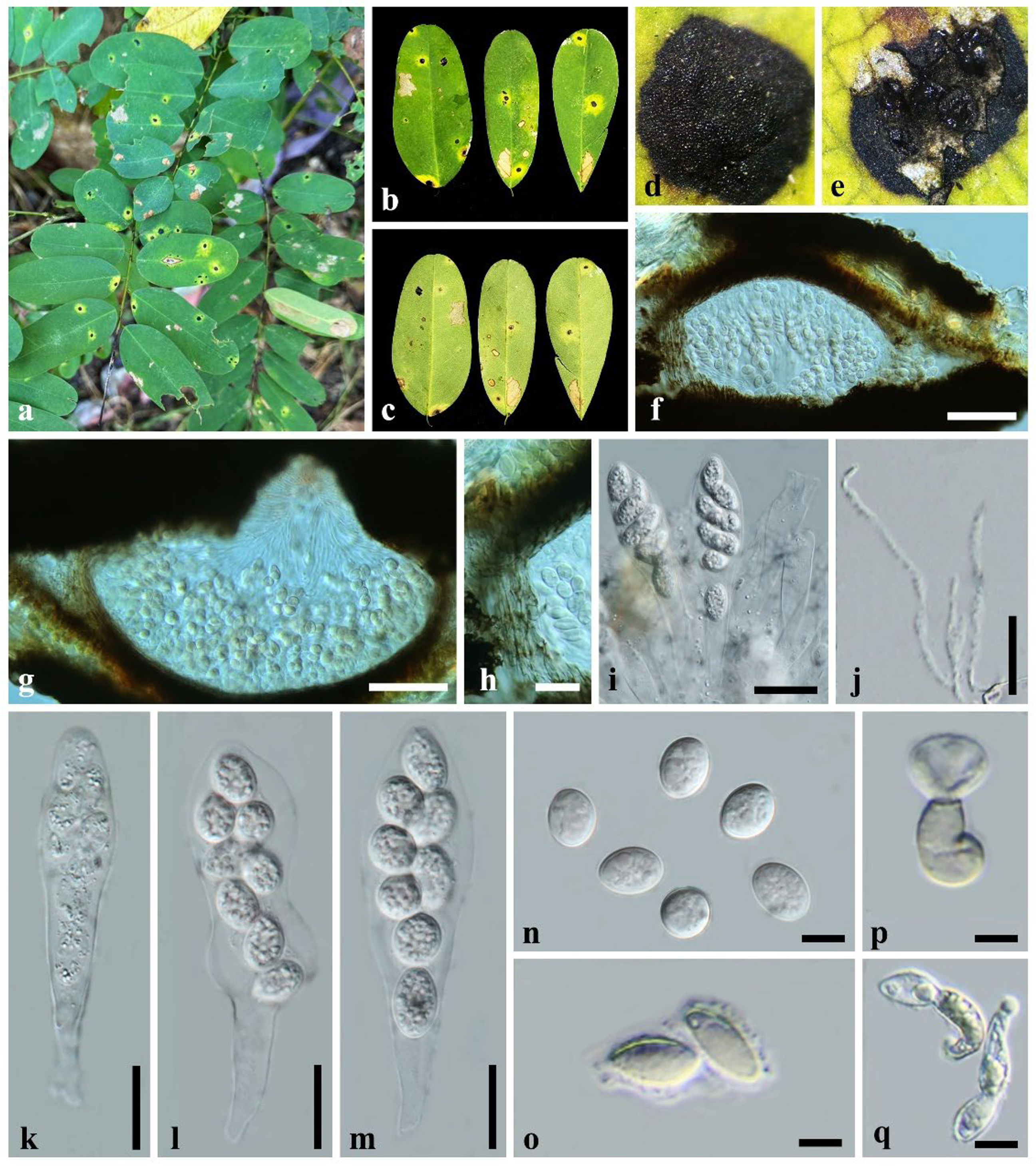
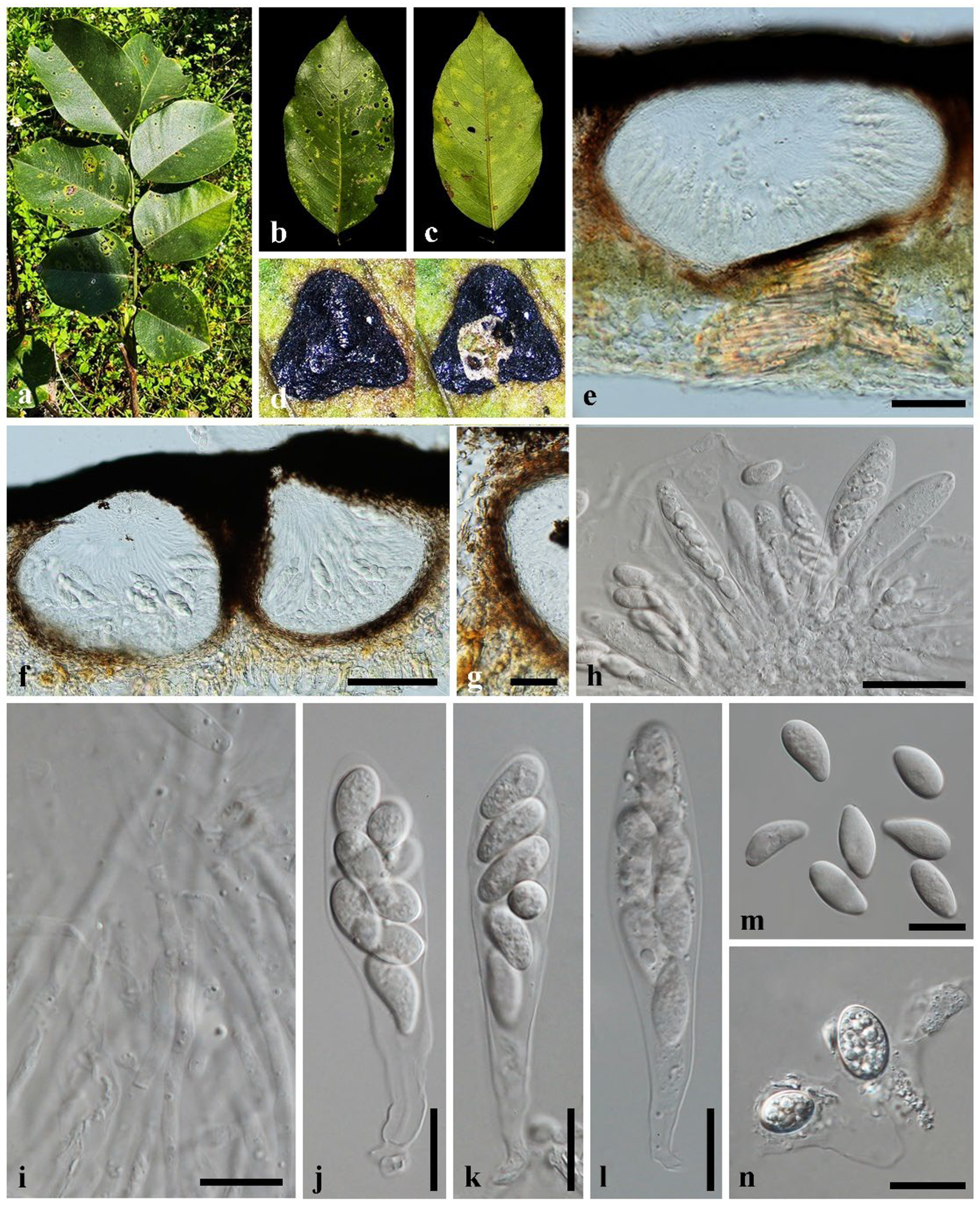
A concatenated LSU, SSU, and ITS dataset comprising 2879 characters (LSU: 889, SSU: 1119, ITS: 819) was analyzed. Maximum likelihood (ML) phylogenetic analysis using the dataset generated the best-scoring tree with a final ML optimization likelihood value of -26897.804397. The estimated base frequencies for the GTR+I+G model of the combined dataset were A–0.252985, C–0.233243, G–0.278171, and T–0.235601. The substitution rates were AC–1.200579, AG–2.174439, AT–1.437903, CG–0.713782, CT–4.521910, and GT–1.000000. Further, the proportion of invariable sites was I–0.140638 and the gamma distribution shape parameter was 0.537932. The matrix had 1578 distinct alignment patterns, with 44.23% of undetermined characters or gaps. The best-scoring ML tree is given in Figure 1.

3.2. Taxonomy

Neophyllachora dalbergiae Haituk, Karunarathna & Cheewangkoon sp. nov. Figure 2.
Etymology: Derived from the host plant genus.
Index Fungorum number: IF903460.
Holotype: Thailand, Lumphun Province (18°38′17″ N 99°2′20″ E), on living leaves of Dalbergia sp. (Fabaceae), 28 March 2024, Sukanya Haituk, CDEP-50.
Parasitic on Dalbergia sp. Leaf symptoms visible as tar spots, amphigenous, scattered, small, black with yellow halo. Pseudostromata 1–3 mm diam., circular, black, intra-epidermal, scattered, prominent, multilocular, epiphyllous, glabrous, shiny. Sexual morph: Ascomata perithecial, globose to sub-globose, solitary or aggregated, immersed in pseudostromata occupying parenchymatous mycelial tissues, 509–620 × 272–329 µm ( x ¯ = 573 × 340 µm, n = 20), ostiolate; ostiole conspicuous, periphysate; peridium 25–35 µm thick, dark brown to black, wider at the lateral than the basal part, compactly arranged strongly melanized cells; paraphyses 1.1–2.3 µm wide, numerous, persistent, filiform, aseptate, branched. Asci 8-spored, unitunicate, persistent, cylindrical to clavate, short to medium pedicellate, with walls uniform in thickness, not specially thickened at apex, 90–100 × 19–25 µm ( x ¯ = 96 × 21 µm, n = 20). Ascospores overlapping, uni- or biseriate, hyaline, globose to elliptical, guttulate, smooth to rough, often with mucilage sheath, 13–17 × 11–13 µm ( x ¯ = 15.8 × 11.9 µm, n = 30). Asexual morph: Not observed.
Additional material examined: Thailand, Lumphun Province (18°38′17″ N 99°2′20″ E), on living leaves of Dalbergia sp. (Fabaceae), 28 March 2024, Sukanya Haituk, CDEP-51.
Notes: The disease symptoms and the typical tar spot appearance of Neophyllachora dalbergiae indicate Neophyllachora within Phyllachoraceae. The present species demonstrates closer morphological similarity towards N. fici. Both species show globose ascospores (Table 2). Contrastingly, N. dalbergiae shows more globose to subglobose spores without central concave depression and a gelatinous sheath, which is absent in N. fici. Moreover, the asci of N. fici are fusoid with a narrow apex, while N. dalbergiae is fusoid and club-shaped with a broader apex. Further, N. dalbergiae differs from N. subcircinans and N. cerradensis, having globose to subglobose spores, due to its unique globose shape. Multi-locus phylogeny using a matrix of concatenated LSU, SSU, and ITS shows N. dalbergiae is located in a basal clade with N. pterocarpi-macrocarpae within Neophyllachora with 100% RAxML statistical support (Figure 1).
Neophyllachora pterocarpi-macrocarpae Haituk, Karunarathna & Cheewangkoon sp. nov. Figure 3.
Etymology: Derived from the host plant species.
Index Fungorum number: IF903461.
Holotype: Thailand, Chiang Mai Province (18°52′11.6″ N 98°56′25.0″ E), on living leaves of Pterocarpus macrocarpus Kurz (Fabaceae), 20 December 2023, Sukanya Haituk, CDEP-52.
Parasitic on Pterocarpus macrocarpus. Leaf symptoms visible as tar spots, small, circular to irregular, distinct, black with a narrow halo at the upper leaf surface, indistinct, pale greenish-yellow at the lower leaf surface. Pseudostromata 2–3 mm diam., black, various in shape, elongated, irregular, discrete, sparse, rarely, coalescent, glabrous, shiny, intraepidermal to subepidermal, mainly epiphyllous, multilocular, occasionally amphigenous, rarely scattered whole leaf surface. Sexual morph: Ascomata perithecial, globose to ampulliform, solitary or aggregated, immersed in pseudostromata occupying parenchymatous mycelial tissues, 456–497 × 213–337 µm ( x ¯ = 477 × 271 µm, n = 20), ostiolate; ostiole conspicuous, periphysate; peridium 22–40 µm thick, dark brown to black, wider at the lateral than the basal part, compactly arranged strongly melanized cells; paraphyses 2.8–5.0 µm wide, numerous, persistent, filiform, septate, branched. Asci 8-spored, unitunicate, persistent, cylindrical to clavate, short to medium pedicellate, with walls uniform in thickness, not specially thickened at apex, 81–106 × 19–29 µm ( x ¯ = 92 × 23 µm, n = 20). Ascospores overlapping, irregularly biseriate, hyaline, various in shape, globose, ovoid, ellipsoid, pyriform, fusiform, amygdaliform, guttulate, sometimes concave at the center, smooth to rough, surrounded by a gelatinous sheath, 18–21 × 9–12 µm ( x ¯ = 20.6 × 10.3 µm, n = 30). Asexual morph: Not observed.
Additional materials examined: Thailand, Chiang Mai Province (18°52′11.6″ N 98°56′25.0″ E), on living leaves of Pterocarpus macrocarpus (Fabaceae), 20 December 2023, Sukanya Haituk, CDEP-53.
Notes: Neophyllachora pterocarpi-macrocarpae was collected from Pterocarpus macrocarpus. On the plant genus Pterocarpus, Phyllachora pterocarpi Syd. & P. Syd. is previously known [26]. N. pterocarpi-macrocarpae is easily distinguishable from P. pterocarpi in the larger ascomata, asci, and ascospores (Table 2). Further, N. pterocarpi-macrocarpae differs from other Neophyllachora species in having various shapes of ascospores (Table 2). Additionally, phylogenetic analysis indicates that N. pterocarpi-macrocarpae is phylogenetically a sister to N. dalbergiae (Figure 1).

4. Discussion

Species in Phyllachoraceae are non-culturable obligate parasites [6]. The majority of species in Phyllachoraceae were introduced mainly based only on morphology. In the study by Dayarathne et al. [10] introducing Neophyllachora, they used taxonomy, molecular phylogeny of SSU, LSU, and ITS, and the host specificity towards Myrtaceae as the distinctive characteristics of this genus. However, later studies by Tennakoon et al. [14] and Literatus et al. [15] introduced Neophyllachora species from Moraceae, and our study introduces two novel Neophyllachora species from Fabaceae. Therefore, unlike Phyllachora, where most species are only reported from hosts of the Poaceae family, Neophyllachora can infect various plant hosts across different plant families.
In this study, two species of Neophyllachora, N. dalbergiae and N. pterocarpi-macrocarpae, are described based on morphology, phylogeny, and host associations. Interestingly, Neophyllachora shows a relatively higher morphological diversity of ascospore and conidia than that of other Phyllacholaceae fungi. For example, Neophyllachora myrciae forms unique lunate ascospores and both ellipsoidal and falcate conidia [3], and contrastingly, N. fici and N. dalbergiae have globose ascospores. Furthermore, ascospores of N. pterocarpi-macrocarpae are oblong to ellipsoid, N. truncatispora is sublunate to fusoid, and N. myrciariae is elliptical. These substantial morphological differences and their respective host associations help in separating the species within Neophyllachora. The updated key to Neophyllachora is provided for further clarification.
Key to species of Neophyllachora updated from Literatus et al. [15]
1.
Parasitic on Dalbergia Ficus, Myrcia, Myrciaria, and Pterocarpus species..................................................................................................................................................2
1′.
Parasitic on Psidium species, ascospores thin-walled, short-ellipsoidal covered with thin-walled gelatinous sheath..............................................................................N. subcircinans
2.
Parasitic on Dalbergia, Ficus, Myrcia, and Pterocarpus species..............................................3
2′.
Parasitic on Myrciaria species, clavate-fusoid asci...........................................N. myrciariae
3.
Parasitic on Dalbergia, Ficus, Myrcia and Pterocarpus, species..............................................4
3′.
Parasitic on Myrcia species, lunate-reniform to half-moon shape ascospores with thick walled ....................................................................................................................N. truncatispora
4.
Parasitic on Myrcia species…………………………………………….……………………..5
4′.
Parasitic on Dalbergia, Ficus and Pterocarpus, species…………….…………..……………6
5.
Elliptic-Oblong microguttulate ascospores...................................................... N. cerradensis
5′.
Thin walledLunate ascospores …… ..................................................................... N. myrciae
6.
Parasitic on Ficus species………………………………………………………….………….7
6′.
Parasitic on Dalbergia and Pterocarpus………………………………………………………8
7.
Parasitic on Ficus species, globose to ellipsoidal ascospores with gelatinous ........N. religiosa
7′.
Parasitic on Ficus species, globose to subglobose ascospores without gelatinous sheath..................................................................................................................................... N. fici
8.
Parasitic on Dalbergia species………………………….………………………. N. dalbergiae
8′.
Parasitic on Pterocarpus species…………………….…………….N. pterocarpi-macrocarpae
Species belonging to Phyllachoraceae infect a wide range of host families such as Arecaceae, Fabaceae, Juncaceae, Marantaceae, Melastomataceae, Myrtaceae, Poaceae, Vochysiaceae, and other monocot and dicot plants in tropic and temperate regions. As they are unculturable in artificial media, the host range is difficult to confirm by inoculation tests. Furthermore, overlapping morphologies within Phyllachoraceae cause taxonomic controversies. Therefore, the diagnosis of the causal species and identification faces a number of difficulties, impeding progress. Hence, it is important to identify Phyllachoraceae species by the polyphasic approach based on the morphology, host plant, and phylogeny, as mentioned above.
The seven species of Neophyllachora described in previous studies were associated with only two plant families. These are Myrtaceae, which originated in Gondwana and spread to the southern hemisphere [27], and Moraceae, which originated in Eurasia and spread to the southern hemisphere [28]. Regarding the associated plant hosts of the newly described two species of Neophyllachora from this study, Fabaceae originated around the Tethys Seaway in the early Tertiary period [29,30].
Referring to the phylogenetic tree of Neophyllachora, we can observe that a common ancestral strain acquired parasitism to one of these plant families and jumped to other plant families. The member in this genus might be speciated with the divergence of host plants on each continent. These are Myrtaceae plants for N. cerradensis, N. myrciae, N. myrciariae, N. subcircinans, and N. truncatispora in South America; Moraceae plants for N. fici and N. religiosa; and Fabaceae plants for N. dalbergiae and N. pterocarpi-macrocarpae in Asia. The divergence of obligate parasites on those herbaceous and/or broadleaf tree host plants is known in the case of powdery mildew and their relative plant hosts [31,32]. On the other hand, the co-speciation of the fungal species occurring on Gondwanan plants was denied [33]. Neophyllachora might be a good example for revealing the coevolution of fungi and their host plants. However, further in-depth studies with a larger number of specimens are required to prove this assumption.

5. Conclusions

In this study, two new species, N. pterocarpi-macrocarpae and N. dalbergiae in Neophyllachora, are introduced with a polyphasic approach from Pterocarpus macrocarpus and Dalbergia sp., respectively. Our study expands the host range of Neophyllachora to include an additional plant family other than Myrtaceae and Moraceae. This is the first time Neophyllachora has been reported from Fabaceae. The current reports on the collection of Neophyllachora species are limited geographically to South America and East Asia. The results from our study together with Literatus et al. [15] broaden the geographic distribution of Neophyllachora within Southeast Asia (Thailand).

Author Contributions

Conceptualization, R.C., S.H. and A.K.; methodology, S.H. and S.A.; validation, R.C., C.N., R.K. and S.M.; formal analysis, A.K. and S.H.; writing—original draft preparation, A.K., D.L.H. and S.H.; writing—review and editing, R.C., S.H., C.N., D.L.H. and A.K.; visualization, S.H.; supervision, R.C. and C.N.; project administration, R.C.; funding acquisition, R.C. and S.H. All authors have read and agreed to the published version of the manuscript.

Funding

This project is funded by the National Research Council of Thailand (NRCT) grant number: 4737753 and was also partially funded by the Big Bang International Projects 2024—M67IN00041, Chiang Mai University, Thailand.

Data Availability Statement

Accession numbers for the DNA sequence data are contained within the article. These can be found at: https://www.ncbi.nlm.nih.gov/.

Acknowledgments

Sukanya Haituk (EX010024), Anuruddha Karunarathna (EX010177), and Dulanjalee Lakmali Harishchandra (EX010075) would like to thank the CMU Proactive Researcher Postdoctoral Program, Chiang Mai University, Thailand, for the support. We would like to thank Angkhana Inta for supporting the plant identification.

Conflicts of Interest

The authors declare no conflicts of interest.

References

  1. Wang, Q.-T.; Guo, M.-J.; Lv, T.; Zhou, H.; Wang, S.; Wang, S.-J.; Lin, Y.-R.; Gronefeld, S.; Kirschner, R.; Piepenbring, M.; et al. Phylogeny and Taxonomy of Rhytisma-like Species Worldwide. Fungal Divers. 2023, 120, 77–119. [Google Scholar] [CrossRef]
  2. Cannon, P.F. A Revision of Phyllachora and Some Similar Genera on the Host Family Leguminosae; Mycological Papers; CAB International: Wallingford, UK, 1991; pp. 1–302. [Google Scholar]
  3. Dos Santos, M.D.M.; de Noronha Fonseca, M.E.; Silva Boiteux, L.; Câmara, P.E.A.S.; Dianese, J.C. ITS Phylogeny and Taxonomy of Phyllachora Species on Native Myrtaceae from the Brazilian Cerrado. Mycologia 2016, 108, 1141–1164. [Google Scholar] [CrossRef] [PubMed]
  4. Cannon, P.F. Diversity of the Phyllachoraceae with Special Reference to the Tropics. Biodivers. Trop. Microfungi 1997, 8, 255–278. [Google Scholar]
  5. Theissen, F.; Sydow, H. Die Dothideales. Kritisch-Systematische Original Untersuchungen. Ann. Mycol. 1915, 13, 147–746. [Google Scholar]
  6. MacCready, J.S.; Roggenkamp, E.M.; Gdanetz, K.; Chilvers, M.I. Elucidating the Obligate Nature and Biological Capacity of an Invasive Fungal Corn Pathogen. MPMI 2023, 36, 411–424. [Google Scholar] [CrossRef]
  7. Yang, C.-L.; Xu, X.-L.; Liu, Y.-G.; Hyde, K.D.; Mckenzie, E.H.C. A New Species of Phyllachora (Phyllachoraceae, Phyllachorales) on Phyllostachys heteroclada from Sichuan, China. Phytotaxa 2019, 392, 186. [Google Scholar] [CrossRef]
  8. Sutton, B.C.; Hodges, C.S. Hawaiian Forest Fungi. III. A New Species, Gloeocoryneum Hawaiiense, on Acacia Koa. Mycologia 1983, 75, 280–284. [Google Scholar] [CrossRef]
  9. Gabel, A.W. Host-Parasite Relations of Phyllachora Species on Native Grasses. Mycologia 1989, 81, 702–708. [Google Scholar] [CrossRef]
  10. Dayarathne, M. Neophyllachora Gen Nov. (Phyllachorales), Three New Species of Phyllachora from Poaceae and Resurrection of Polystigmataceae (Xylariales). Mycosphere 2017, 8, 1598–1625. [Google Scholar] [CrossRef]
  11. Manawasinghe, I.S.; Phillips, A.J.L.; Xu, J.; Balasuriya, A.; Hyde, K.D.; Stępień, Ł.; Harischandra, D.L.; Karunarathna, A.; Yan, J.; Weerasinghe, J.; et al. Defining a Species in Fungal Plant Pathology: Beyond the Species Level. Fungal Divers. 2021, 109, 267–282. [Google Scholar] [CrossRef]
  12. Mueller, D.S.; Wise, K.A.; Sisson, A.J.; Allen, T.W.; Bergstrom, G.C.; Bissonnette, K.M.; Bradley, C.A.; Byamukama, E.; Chilvers, M.I.; Collins, A.A.; et al. Corn Yield Loss Estimates Due to Diseases in the United States and Ontario, Canada, from 2016 to 2019. Plant Health Prog. 2020, 21, 238–247. [Google Scholar] [CrossRef]
  13. Broders, K.; Iriarte-Broders, G.; Bergstrom, G.C.; Byamukama, E.; Chilvers, M.; Cruz, C.; Dalla-Lana, F.; Duray, Z.; Malvick, D.; Mueller, D.; et al. Phyllachora Species Infecting Maize and Other Grass Species in the Americas Represents a Complex of Closely Related Species. Ecol. Evol. 2022, 12, e8832. [Google Scholar] [CrossRef] [PubMed]
  14. Tennakoon, D.S.; Kuo, C.-H.; Maharachchikumbura, S.S.N.; Thambugala, K.M.; Gentekaki, E.; Phillips, A.J.L.; Bhat, D.J.; Wanasinghe, D.N.; De Silva, N.I.; Promputtha, I.; et al. Taxonomic and Phylogenetic Contributions to Celtis formosana, Ficus ampelas, F. septica, Macaranga tanarius and Morus australis Leaf Litter Inhabiting Microfungi. Fungal Divers. 2021, 108, 1–215. [Google Scholar] [CrossRef]
  15. Literatus, I.C.E.; Maharachchikumbura, S.S.N.; Withee, P.; Haituk, S.; Tamakaew, N.; Nguanhom, J.; Monkhung, S.; Thiyagaraja, V.; Cheewangkoon, R. Taxonomic and Phylogenetic Appraisal of a New Holomorphic Neophyllachora Species from Chiang Mai, Thailand. Phytotaxa 2023, 600, 259–271. [Google Scholar] [CrossRef]
  16. Gardner, S.; Sidisunthorn, P.; Anusarnsunthorn, V. (Eds.) A Field Guide to Forest Trees of Northern Thailand; Kobfai Publ. Project: Bankok, Thailand, 2000; ISBN 978-974-7798-29-6. [Google Scholar]
  17. Index Fungorum. Available online: https://www.indexfungorum.org/names/Names.asp (accessed on 20 August 2024).
  18. Vilgalys, R.; Hester, M. Rapid Genetic Identification and Mapping of Enzymatically Amplified Ribosomal DNA from Several Cryptococcus Species. J. Bacteriol. 1990, 172, 4238–4246. [Google Scholar] [CrossRef]
  19. White, T.J.; Bruns, T.; Lee, S.; Taylor, J. Amplification and direct sequencing of fungal ribosomal RNA genes for phylogenetics. In PCR Protocols; A Guide to Methods and Applications; Innis, M.A., Gelfand, D.H., Sninsky, J.J., White, T.J., Eds.; Academic Press: San Diego, CA, USA, 1990; pp. 315–322. [Google Scholar]
  20. Gardes, M.; Bruns, T.D. ITS Primers with Enhanced Specificity for Basidiomycetes—Application to the Identification of Mycorrhizae and Rusts. Mol. Ecol. 1993, 2, 113–118. [Google Scholar] [CrossRef]
  21. Katoh, K.; Rozewicki, J.; Yamada, K.D. MAFFT Online Service: Multiple Sequence Alignment, Interactive Sequence Choice and Visualization. Brief. Bioinform. 2019, 20, 1160–1166. [Google Scholar] [CrossRef]
  22. Hall, T.A. BioEdit: A User-Friendly Biological Sequence Alignment Editor and Analysis Program for Windows 95/98/NT. Nucleic Acids Symp. Ser. 1999, 41, 95–98. [Google Scholar]
  23. Glez-Peña, D.; Gómez-Blanco, D.; Reboiro-Jato, M.; Fdez-Riverola, F.; Posada, D. ALTER: Program-Oriented Conversion of DNA and Protein Alignments. Nucleic Acids Res. 2010, 38, W14–W18. [Google Scholar] [CrossRef]
  24. Miller, M.A.; Pfeiffer, W.; Schwartz, T. Creating the CIPRES Science Gateway for Inference of Large Phylogenetic Trees. In Proceedings of the 2010 Gateway Computing Environments Workshop (GCE), New Orleans, LA, USA, 14 November 2010; pp. 1–8. [Google Scholar]
  25. Rambaut, A. FigTree 2012. FigTree v1.3.1. Institute of Evolutionary Biology, University of Edinburgh, Edinburgh. Available online: http://tree.bio.ed.ac.uk/software/figtree/ (accessed on 5 November 2024).
  26. Sydow, H.; Sydow, P. Beschreibungen Neuer Südafrikanischer Pilze. Ann. Mycol. 1912, 10, 33–45. [Google Scholar]
  27. De Souza Neto, J.D.; Dos Santos, E.K.; Lucas, E.; Vetö, N.M.; Barrientos-Diaz, O.; Staggemeier, V.G.; Vasconcelos, T.; Turchetto-Zolet, A.C. Advances and Perspectives on the Evolutionary History and Diversification of Neotropical Myrteae (Myrtaceae). Bot. J. Linn. Soc. 2022, 199, 173–195. [Google Scholar] [CrossRef]
  28. Zerega, N.J.C.; Clement, W.L.; Datwyler, S.L.; Weiblen, G.D. Biogeography and Divergence Times in the Mulberry Family (Moraceae). Mol. Phylogenetics Evol. 2005, 37, 402–416. [Google Scholar] [CrossRef]
  29. Doyle, J.J.; Luckow, M.A. The Rest of the Iceberg. Legume Diversity and Evolution in a Phylogenetic Context. Plant Physiol. 2003, 131, 900–910. [Google Scholar] [CrossRef] [PubMed]
  30. Eric Schranz, M.; Mohammadin, S.; Edger, P.P. Ancient Whole Genome Duplications, Novelty and Diversification: The WGD Radiation Lag-Time Model. Curr. Opin. Plant Biol. 2012, 15, 147–153. [Google Scholar] [CrossRef]
  31. Braun, U.; Cook, R.T.A. Taxonomic Manual of the Erysiphales (Powdery Mildews); CBS Biodiversity Series; CBS-KNAW Fungal Biodiversity Centre: Utrecht, The Netherlands, 2012; ISBN 978-90-70351-89-2. [Google Scholar]
  32. Takamatsu, S. Studies on the Evolution and Systematics of Powdery Mildew Fungi. J. Gen. Plant Pathol. 2018, 84, 422–426. [Google Scholar] [CrossRef]
  33. Niinomi, S.; Takamatsu, S.; Havrylenko, M. Molecular Data Do Not Support a Southern Hemisphere Base of Nothofagus Powdery Mildews. Mycologia 2008, 100, 716–726. [Google Scholar] [CrossRef]
Figure 1. Phylogenetic tree generated by maximum likelihood analysis using a concatenated dataset of ITS, LSU, and SSU loci of the genera in Phyllachoraceae. Nodes are marked with maximum likelihood bootstrap proportions ≥60% (ML). The tree is rooted to Telimena bicincta MM 108 and T. bicincta MM 133. Newly obtained strains are in red. The type strains are bold.
Forests 16 00556 g001
Figure 2. Neophyllachora dalbergiae (CDEP-50, holotype). (a) Tar spot on living leaves. (b) Upper leaf surface with symptoms. (c) Lower leaf surface with symptoms. (d) Pseudo-stroma. (e) Horizontal section through pseudostroma. (f,g) Vertical section through ascomata. (h) Vertical section through peridium. (i,km) Asci. (j) Paraphyses. (n) Ascospores. (o) Ascospore with mucilaginous sheath. (p,q) Germinated ascospores. Scale bars: 20 µm (fg,km), 10 µm (h,j,nq), 30 µm (i).
Forests 16 00556 g002
Figure 3. Neophyllachora pterocarpi-macrocarpae (CDEP-52, holotype). (a) Tar spot on living leaves. (b) Upper leaf surface with symptoms. (c) Lower leaf surface with symptoms. (d) Pseudostroma and horizontal section through pseudostroma. (e,f) Vertical section through ascomata. (g) Vertical section through peridium. (h) Asci and paraphyses. (i) Paraphyses. (jl) Asci. (m) Ascospores. (n) Ascospore with mucilaginous sheath. Scale bars: 100 µm (e), 200 µm (f), 10 µm (g), 50 µm (h), 20 µm (in).
Forests 16 00556 g003
Table 1. Taxa table for phylogenetic analysis. Culture collection numbers with “T” designate the type.
SpeciesSample CodeLSU SSUITS
Camarotella costaricensisMM-21KX430490KX451851KX451900
Coccodiella calatheaeMP5133TMF460370MF460376MF460366
Coccodiella melastomatumCMU78543 U78543
Coccodiella miconiaeppMP1342KX430506KX451871MF460365
Coccodiella miconiicolaSO-15 MF460374MF460380MF460369
Coccodiella toledoiMM-165KX430488KX451865KX451917
Neophyllachora cerradensisUB21823KC683470
Neophyllachora cerradensisUB21908TKC683471
Neophyllachora dalbergiaeCDEP-50TPV241510PV241504
Neophyllachora dalbergiaeCDEP-51PV241511PV241505
Neophyllachora ficiMFLU 19-2702MW114384
Neophyllachora ficiNCYU 19-0061MW114385
Neophyllachora ficiNCYU 19-0326MW114386
Neophyllachora myrciaeUB21292KC683463
Neophyllachora myrciaeUB22192KC683476
Neophyllachora myrciariaeUB21781TKC683469
Neophyllachora pterocarpi-macrocarpaeCDEP-52TPV241512PV241165PV241506
Neophyllachora pterocarpi-macrocarpaeCDEP-53PV241513PV241166PV241507
Neophyllachora pterocarpi-macrocarpaeCDEP-54PV241514PV241167PV241508
Neophyllachora pterocarpi-macrocarpaeCDEP-55PV241515PV241168PV241509
Neophyllachora subcircinansUB09748KC683441
Neophyllachora subcircinansUB21347KC683466
Neophyllachora subcircinansUB21747KC902622KC683467
Neophyllachora truncatisporaUB14083KC902614KC683448
Neophyllachora religiosaMFLU 23-0258OQ821004
Phyllachora arthraxonisMHYAU:072MG269803MG269749
Phyllachora arundinellaeMHYAU:108MG269815MG269761
Phyllachora capillipediicolaMHYAU 20089MG356698KY498084
Phyllachora chloridisMFLU 15-0173TMF197499MF197505KY594026
Phyllachora chloridis-virgataeMHYAU 20136MG356685KY498122
Phyllachora chongzhouensisSICAU 24-0044PP785312PP785323PP785301
Phyllachora chrysopogonicolaMFLU 16-2096TMF372146MF372145
Phyllachora cynodonticolaMFLU 16-2977TMF197501MF197507KY594024
Phyllachora cynodontisMHYAU:20043KY498081KY471329
Phyllachora dendrocalami-hamiltoniicolaMHYAU 221MK614118
Phyllachora dendrocalami-membranaceiMHYAU 220MK614117MK614102
Phyllachora flaccidudisIFRD9445TON072101ON072097ON075524
Phyllachora graminisSICAU 24-0051 PP785306PP785317PP785295
Phyllachora heterocladaeMFLU 18-1221TMK296472MK296468MK305902
Phyllachora huiliensisSICAU 24-0048PP785308PP785319PP785297
Phyllachora imperataeMHYAU:014MG269800MG269746
Phyllachora indosasaeMHYAU 125MG195662MG195637
Phyllachora isachnicolaMHYAU:179TMH018563MH018561
Phyllachora jiaensisIFRD9448TON075440ON072100ON075527
Phyllachora keralensisMHYAU:20082MG269792KY498106
Phyllachora maydisBPI 893231KU184459
Phyllachora maydisBPI 910560MG881846
Phyllachora miscanthiSICAU 24-0050PP785305PP785316PP785294
Phyllachora neidongensisSICAU 24-0046PP785314PP785325PP785303
Phyllachora panicicolaMFLU 16-2979TMF197503MF197504KY594028
Phyllachora pogonatheriMHYAU:071MG269802MG269748
Phyllachora pomigenaCBS 193.33MH866860MH855409
Phyllachora qualeaeUB 21159KU682781
Phyllachora sandiensisIFRD9446TON075528ON072098ON075525
Phyllachora sinobambusaeMHYAU 085MG195655MG195630
Phyllachora sphaerocaryiMHYAU 178TMK614114MK614100
Phyllachora thysanolaenaeMFLU 16-2071MF372147
Phyllachora virgataesIFRD9447TON075439ON072099ON075526
Phyllachora yushaniae-falcatiauritaeMHYAU 123MG195656MG195631
Phyllachora yushaniae-polytrichaeMHYAU 122MG195657MH992455MG195632
Polystigma pusillumMM-19KX430489KX451850KX451899
Telimena bicinctaMM-108 KX430473KX451857KX451906
Telimena bicinctaMM 133KX430478KX451861KX451910
Abbreviations: MFLUCC = Mae Fah Luang University Culture Collection; CBS = Westerdijk Institute, Utrecht, The Netherlands; CDEP = Culture Collection of Department of Entomology and Plant Pathology; IFRD = International Fungal Research & Development Centre; MHYAU = Mycological Herbarium of the Yunnan Agricultural University; SICAU = Herbarium of Sichuan Agricultural University, Chengdu, China; NCYU = National Chiayi University Herbarium; UB = Department of Botany at the University of Brasilia Herbarium; BPI = U.S. National Fungus Collections.
Table 2. Synopsis of Neophyllachora species.
Neophyllachora SpeciesPhyllachora s.l.
Morphological CharacteristicsN. cerradensisN. ficiN. myrciaeN. myrciariaeN. subcircinansN.
truncatispora
N. religiosaN. dalbergiaeN. pterocarpi-macrocarpaeP. pterocarpi
Pseudostromata (mm in diam)2–42–33–6 0.5–1.51–4 2–5 2–31–32–30.75–2
Shape of AscomataAmpulliform to GloboseGlobose to SubgloboseGlobose to AmpulliformAmpulliformAmpulliformGlobose to OvoidGlobose to SubgloboseGlobose to sub globoseGlobose to AmpulliformRotundate to illegular
Position of AscomataOccasionally coalescingEpiphyllousOccasionally coalescentImmersed in the pseudostromataImmersed in the pseudostromatic tissueImmersed in pseudostromaEpiphyllousEpiphyllousEpiphyllousEpiphyllous
Peridium thickness (µm)17–2120–25 -19–2415–21-15–40 25–3522–40-
Size of Ascomata (µm)260–362 × 168–264150–300 × 200–400 205–485 × 148–207161–432 × 126–267329–417 × 193–275160–200 × 100–350 150–300 × 200–400 272–329 × 509–620213–337 × 456–497150–200
Size of Asci (µm)69–92 × 17–2890–100 × 15–1989–117 × 13–1963–90 × 11–1573–100 × 12–1969–117 × 14–2555–185 × 11–2690–100 × 19–2581–106 × 19–2945–80 × 15–25
Shape of AsciFusoidCylindrical to FusiformFusoidClavate–FusoidCylindrical-Cylindrical to FusiformCylindrical to clavateCylindrical to clavateCylindrical clavate to clavate
Width of Paraphyses (µm)1.6–31.5–2.52.5–4.52–32–3.5 µm2.5–51.5–2.51.1–2.3 2.8–5.0-
Septate of ParaphysesSeptateAseptateSeptateSeptateSeptateSeptateSeptateAseptateSeptate-
Size of Ascospores (µm)15–22 × 6–912–13 × 10–1114–18 × 5–714–19 × 5–811–16 × 7–918–26 × 7–8 8–15 × 5–1213–17 × 11–1318–21 × 9–1214–18 × 8–11
Shape of AscosporesElliptic–OblongGlobose to SubgloboseLunateEllipticalOblong to EllipsoidSublunate to FusoidGlobose to ellipticalGlobose to elliptical with a central concave depressionGlobose, ovoid, ellipsoid, pyriform, fusiform, amygdaliformEllipsoid
Color of AscosporesHyalineHyalineHyalineHyalineHyaline to Light olivaceousHyalineHyaline to Light olivaceousHyalineHyalineHyaline
Ascospores arrangementBiseriate1–2 seriateBiseriate to multi-seriateObliquely biseriateMostly uniseriate, sometimes with biseriate-Mostly uniseriate, sometimes with biseriateObliquely uni- or biseriateIrregularly biseriate1–2 seriate
Ascospores wallCovered by a thin gelatinous sheathAbsentThin-walledCovered by a thin gelatinous sheathThin wall surrounded by a gelatinous sheathWall thickenings at both acute endsCovered by a thick gelatinous sheathCovered by a gelatinous sheath, smooth to roughCovered by a gelatinous sheath, smooth to rough-
GuttulesMicroguttulate cytoplasm--Irregularly guttulateCentrally guttulate-Irregularly guttulateGuttulateGuttulate-
Asexual morphCoelomycete UnknownCoelomycete UnknownUnknownCoelomycete UnknownUnknownUnknown-
HostLeaves of Myrcia tortaLeaves of Ficus septicaMyrcia sp.Leaves of Myrciaria delicatulaPsidium sp.Leaves of Myrcia camapuanensisLeaves of Ficus religiosaDalbergia sp.Pterocarpus macrocarpusPterocarpus angolensis
DistributionBrazilTaiwanBrazilBrazilBrazilBrazilThailandThailandThailandSouth Africa
References[3][14][3][3][3][3][15]Present studyPresent study[26]
Disclaimer/Publisher’s Note: The statements, opinions and data contained in all publications are solely those of the individual author(s) and contributor(s) and not of MDPI and/or the editor(s). MDPI and/or the editor(s) disclaim responsibility for any injury to people or property resulting from any ideas, methods, instructions or products referred to in the content.

