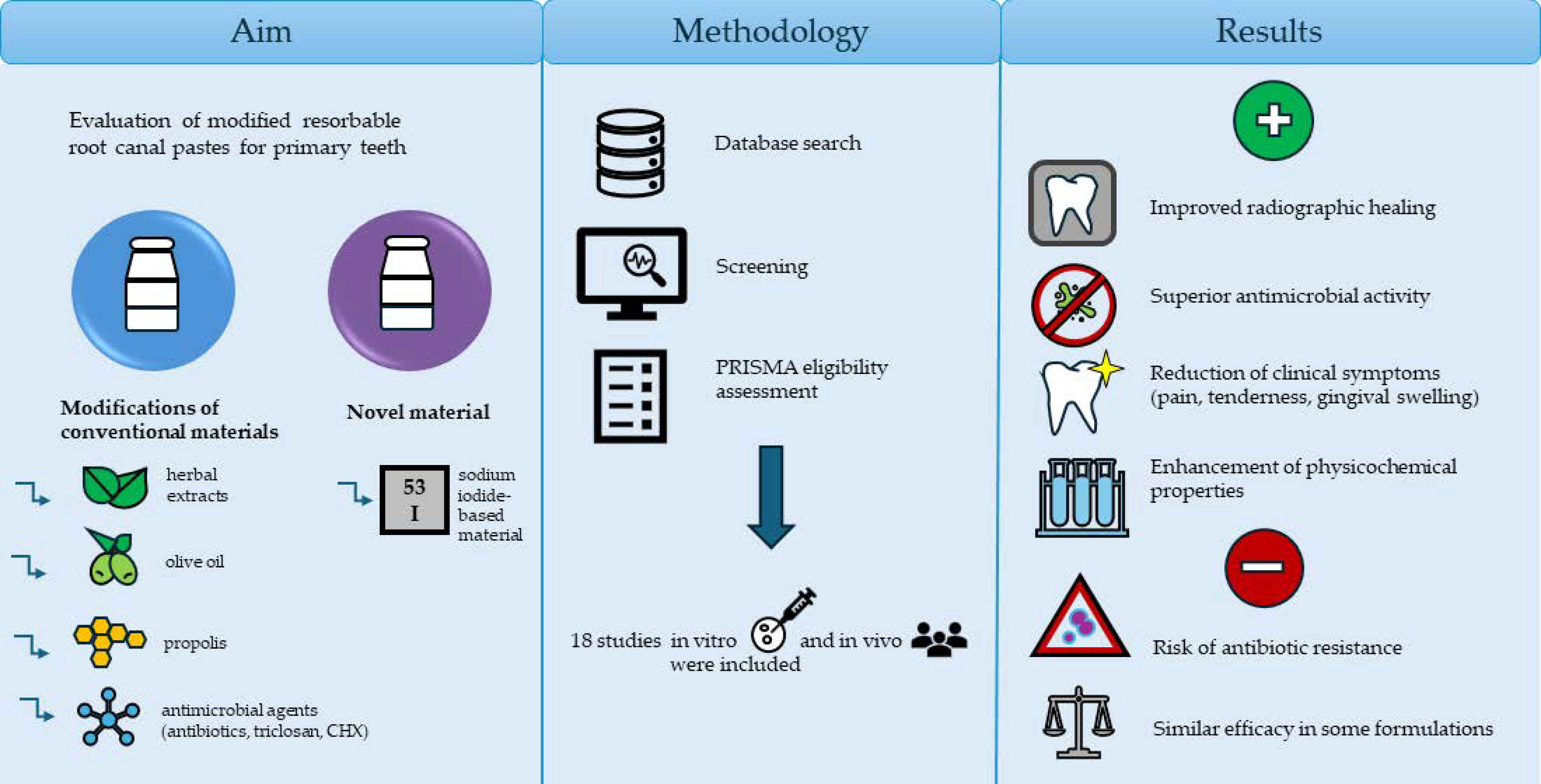
Modifications of Resorbable Root Canal Filling Materials for Primary Teeth: A Systematic Review

 , , , , , ,
  and
and 
Abstract
1. Introduction
2. Materials and Methods
2.1. Focused Question
2.2. Protocol
2.3. Eligibility Criteria
- Studies evaluating resorbable root canal filling materials in primary teeth;
- Studies investigating modifications to root canal filling materials (e.g., addition of antibacterial agents, bioactive materials, nanoparticles, or other additives);
- In vitro experimental studies;
- In vivo including non-randomized controlled clinical trials (NRSs) and Randomized controlled clinical trials (RCTs);
- Studies including a control group, such as conventional unmodified resorbable root canal filling materials.
- Studies published in English.
- Studies focusing on endodontic treatment of permanent teeth;
- Studies evaluating non-resorbable root canal filling materials (e.g., gutta-percha, resin-based sealers intended for permanent teeth);
- Case reports, clinical reports, editorials, letters or opinion papers;
- Narrative reviews, systematic reviews or meta-analyses;
- Studies without accessible full-text;
- Duplicated publications
2.4. Information Sources, Search Strategy, and Study Selection
2.5. Data Collection Process and Data Items
2.6. Quality Assessment and Risk of Bias
2.7. Data Synthesis and Statistical Analysis
3. Results
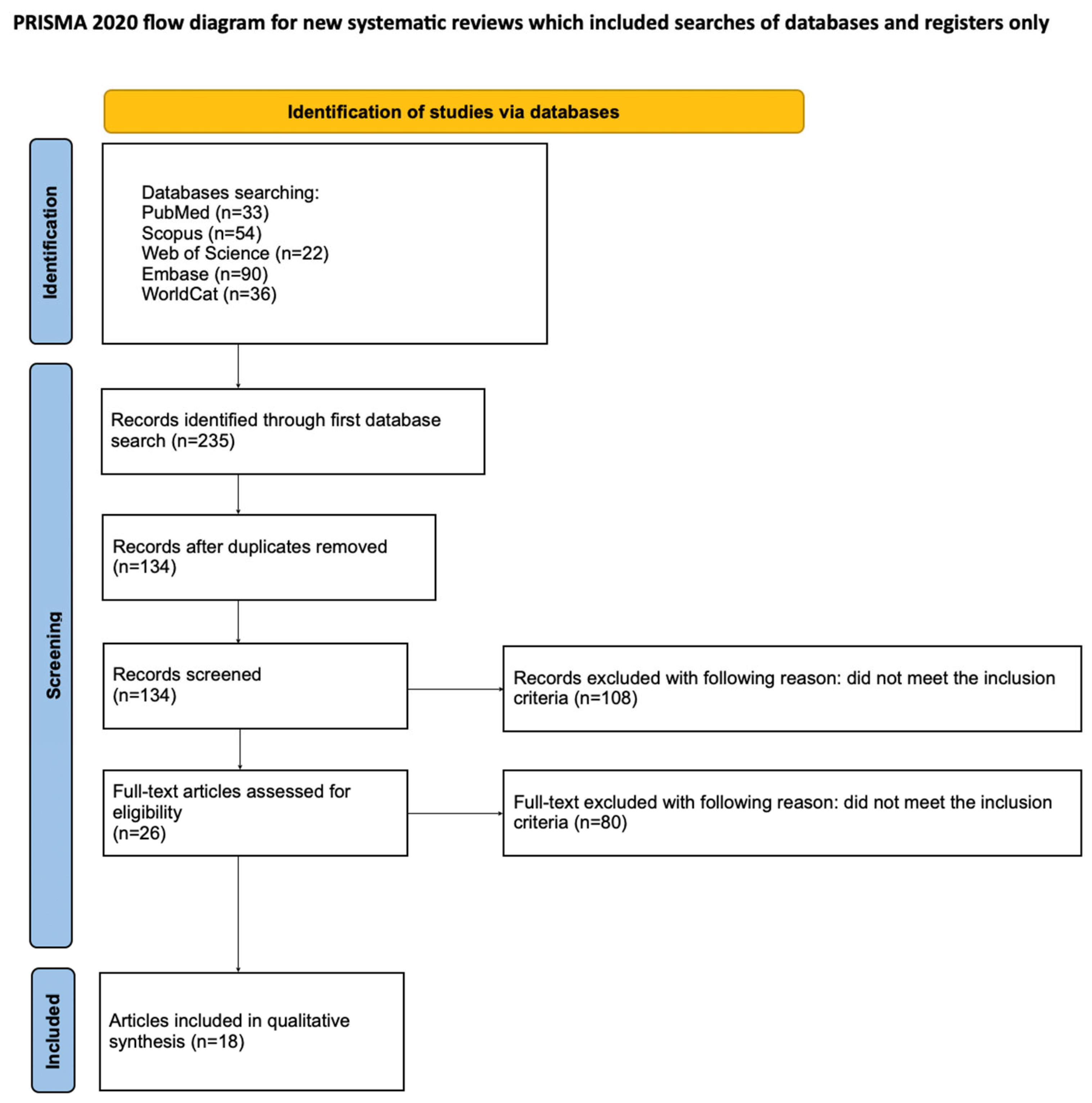
3.1. Study Selection
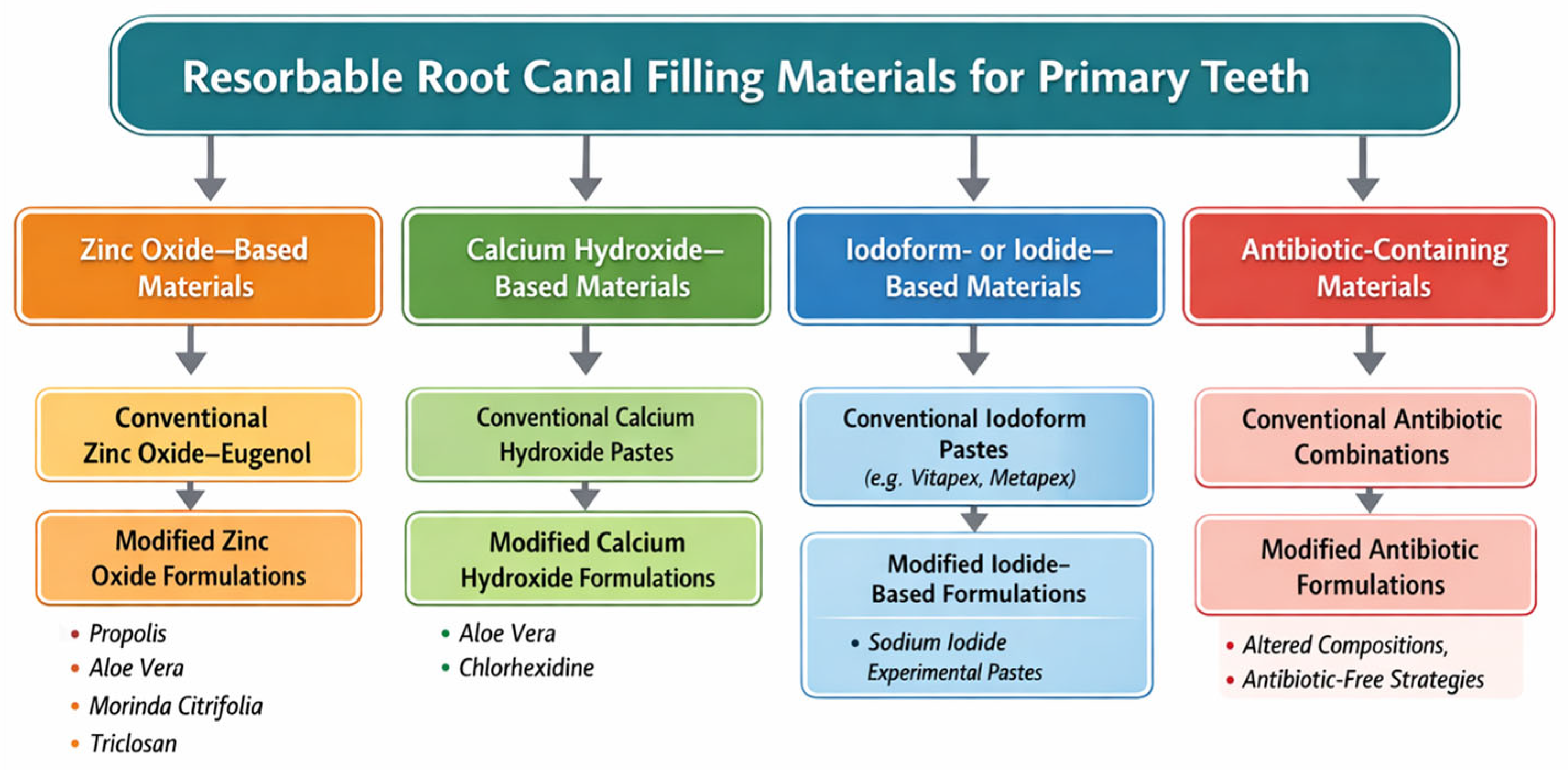
3.2. General Characteristics of the Included Studies
3.3. Main Study Outcomes
3.3.1. In Vitro Studies—Detailed Outcomes

| Study | Samples/Specimens | Filling Materials | Antibacterial/Antifungal Findings | Radiographic Outcomes | Other Findings |
|---|---|---|---|---|---|
| Alashbal et al. [49] | -In vitro, -Primary teeth, -Samples taken from 21 children aged 3–8. | -Zinc oxide + eucalyptus -Zinc oxide eugenol -Metapex | Inhibiting zone in millimeters: Streptococcus spp. Zinc oxide eucalyptus 16.4 ± 2.366 Zinc oxide eugenol 17.300 ± 3.743 Metapex 8.000 ± 4.967 Candida albicans Zinc oxide eucalyptus 31.200 ± 3.490 Zinc oxide eugenol 30.200 ± 3.259 Metapex 7.400 ± 6.899 | No data | No data |
| Park et al. [48] | -In vitro -Samples of sodium iodide-based pastes after modifications | L0: sodium iodide, calcium hydroxide, silicone oil L5: sodium iodide, calcium hydroxide, silicone oil, lanolin Control-Vitapex | Log CFU/mL L0: ~6.6 L5: ~6.75 Vitapex: ~6.9 Vitapex > L5 > L0 Antimicrobial reduction (%) L0: ~35% L5: ~20% Vitapex: ~5% L0 > L5 > Vitapex | No data | Physicochemical tests: Solubility: L0 > L5 > Vitapex Radiopacity: Vitapex > L5 > L0 Injectability: Vitapex > L5 > L0 Complex Viscosity Vitapex > L5 > L0 pH: -all pH values were approximately 12 Evaluation of root canal obturation: -all materials exhibited comparable ability to fill the root canal up to the apex. |
| EL-Desouky et al. [27] | -In vivo -90 secondary primary second molars from 108 children aged 4–8. -Teeth are divided into 3 groups of 30 teeth each. | Group I—zinc oxide ozonated olive oil Group II—zinc oxide with olive oil Group III (control)—zinc oxide with eugenol. | No data | Group I: -the most favorable radiographic outcomes, with the highest bone density at 12 months (114.78 ± 29.05) -the greatest reduction in periodontal ligament width (final value 0.17 ± 0.02). Group II: -faster early bone density improvement, with values at 3 months (62.32 ± 11.09) and 6 months (79.43 ± 18.05), -comparable periodontal ligament width at 12 months (0.17 ± 0.02), though the final bone density was lower (106.64 ± 26.20). Group III: -the least favorable outcomes, lower bone density at all time points, reaching 88.86 ± 31.33 at 12 months, -a smaller reduction in periodontal ligament width (0.18 ± 0.03). | Group I: -comparable results with Group II, -a stable success rate of 92.6% and low symptom rates at 12 months. Group II: -the best clinical outcomes, -the highest success at 6 and 12 months (96.4%) and the lowest incidence of symptoms at 12 months. Group III: -the least favorable outcomes, -higher symptom prevalence at 12 months and lower success rates at 6 and 12 months (85.7%). |
| Pinky et al. [1] | -In vivo, -40 primary teeth from 28 children aged 4–10. -Divided into 2 groups of 20 teeth each. | Group A—antibacterial paste: ciprofloxacin, metronidazole, minocycline, propylene glycol; Group B—antibacterial paste: ciprofloxacin, ornidazole, minocycline, propylene glycol; | No data | Interradicular radiolucency: -progressive improvement in both groups, with greater reduction in Group B at 6 months (30% vs. 35%) and 12 months (60% vs. 55%). Increased/no increased interradicular radiolucency: -at 6 months, no increase was observed in either group. -at 12 months, increased radiolucency occurred only in Group A (10%), while Group B showed no increase (0%). | Pain: -at 3 and 6 months, no pain was reported. -at 12 months, Group A: 10%, Group B: 0%. Intraoral abscess: -initially Group A: 75%, Group B: 70%; -resolved completely by 3 months and remained absent at 6 and 12 months in both groups. Extraoral abscess: -initially Group A: 30%, Group B: 40%; -absent at 3, 6, and 12 months in both groups. Mobility: -initially Group A: 60%, Group B: 65%; -resolved in both groups at 3, 6, and 12 months. Tenderness: -no tenderness at 3 and 6 months -minimal at 12 months in Group A: 10% |
| Singh et al. [24] | -In vivo, -60 primary molars from 50 children aged 4–9. -Divided into 3 groups of 20 teeth each. | Group 1: Zinc oxide + eugenol (ZOE), Group 2: Zinc oxide + propolis Group 3: Endoflas | No data | Furcation radiolucency: -gradual decrease in unchanged cases, with higher rates of reduction in Groups 2 and 3 at 6 months (45–50% vs. 35% in Group 1) and 9 months (45–55% vs. 35% in Group 1). Furcation radiolucency: -elevated radiolucency observed in Group 1 at 6 and 9 months (20%); Groups 2 and 3 showed minimal elevation (0–5%). -reduction in radiolucency was highest in Group 2 at 9 months (55%), with Groups 1 and 3 showing 45–50%. | Pain: -high pain incidence in 1st week after treatment in all groups -resolved completely by 3 months. Mobility: -minimal mobility was observed 1 week after treatment in Group 1: 5% and Group 3: 5% -at 6 months Group 1: 5% -at 9 and 12 months no mobility in all groups. Sinus manifestation: -initially present in all groups -mostly resolved by 3 months (Group 1: 5%, Group 2: 10%, Group 3: 0%), -absent in all groups at 6 and 9 months, except Group 1: 15% at 6 months. Tenderness on percussion: -1 week after treatment (Group 1: 45%, Group 2: 70%, Group 3: 55%), -resolved completely by 3, 6, and 9 months. Resorption of material: -no resorption at 1 and 6 months in any group. -at 9 months, Group 1 and Group 2 showed 1/10 cases, while Group 3 showed none. |
| Goel et al. [25] | -In vivo -120 primary teeth from children aged 3–9 | Group A: ZOE 4:1 ratio Group B: ZnO-Aloe vera 1:2 ratio Group C: ZnO-10%NaF Group D: Endoflas | No data | All four materials showed a general progressive radiographic healing, though differences between the groups were not statistically significant. | Root and filling material resorption were well matched in all groups, but zinc oxide–eugenol showed slower resorption and retained excess material longer than the others. Comparison of filling materials vs. root resorption 1. slower than root ZOE 96.70% ZnO-Aloe vera 11.10% ZnO-NaF 0%, Endoflas 0% 2. same as root ZOE 66.70%, ZnO-Aloe vera 77.80% ZnO-NaF 50%, Endoflas 100% 3. faster than the root ZOE 0%, ZnO-Aloe vera 11.1% ZnO-NaF 50% Endoflas 0% |
| Al-Ostwani et al. [26] | -In vivo -64 primary teeth from 39 children aged 3–9 -Divided into 4 group of 16 teeth each | Group 1: ZOE Group 2: Endoflas-chlorophenol-free Group 3: zinc oxide and propolis (ZOP pastes) Group 4: Metapex (calcium hydroxide with iodoform) | No data | Radiological success rate: After 6 months: ZOE -success 56.3% -suspicion 25% -failure 18.8% Endoflas-CF -success 81.3% -failure 18.8% ZOP -success 75% -suspicion 6.3% -failure 18.8% Metapex -success 75% -failure 25% After 12 months: ZOE -success 56.3% -suspicion 25% -failure 6.3% -extracted 12.5% Endoflas-CF -success 81.3% -failure 6.3% -extracted 12.5% ZOP -success 62.5% -suspicion 6.3% -failure 25% -extracted 6.3% Metapex -success 75% -failure 12.5% -extracted 12.5% | Clinical success rate after 6 months ZOE -success 93.8% -failure 6.3% Endoflas-CF -success 100% ZOP -success 100% Metapex -success 93.8% -failure 6.3% -extracted 0% After 6 months ZOE -success 87.5% -extracted 12.5% Endoflas-CF -success 87.5% -extracted 12.5% ZOP -success 93.8% -extracted 6.3% Metapex -success 87.5% -extracted 12.5% Comparison of filling materials vs. root resorption 1. slower than root ZOE 31.3% Endoflas CF 0% ZOP O% Metapex 0% 2. same as root ZOE 62.5% Endoflas CF 43.8% ZOP 37.5 Metapex 56.3% 3. faster than the root ZOE 6.3% Endoflas-CF 56.3% ZOP 37.5% Metapex 56.3% |
| Hegde et al. [52] | -In vitro -Research conducted on bacterial strains | Group 1: ZOE Group 2: ZnO + Ca(OH)2 + 10%NaF Group 3: ApexCal Group 4: Metapex Group 5: Endoflas Group 6: Vaseline (control group) | Zones of inhibition (mm): ZOE: Strong inhibition of S. aureus (19 ± 2.82), S. epidermis (19 ± 2.82), B. subtilis (20.5 ± 0.70), P. aeruginosa (18.5 ± 2.12), and C. albicans (26 ± 1.41); medium inhibition of other microorganisms. ZO + Ca(OH)2: No inhibition of S. aureus, E. faecalis, P. aeruginosa, C. albicans; weak inhibition of S. epidermis (6 ± 1.41); medium inhibition of others ZO + Ca(OH)2 + NaF: Medium inhibition of S. epidermis (12 ± 5.65), S. mutans (13.5 ± 0.70), S. aureus (7 ± 0), B. subtilis (7 ± 2.82); weak or no inhibition of others. ApexCal: Medium inhibition of C. albicans (12 ± 1.41), S. epidermis (9 ± 2.82), B. subtilis (9.5 ± 2.12); weak inhibition of S. aureus (3 ± 0), S. mutans 5 ± 2.82), P. aeruginosa (6 ± 1.41); no inhibition of E. coli, E. faecalis. Metapex: Mostly non-inhibitory; medium inhibition only against B. subtilis (10 ± 4.24). Endoflas: Strong inhibition of C. albicans (26 ± 1.41); medium inhibition of other microorganisms. Vaseline (control): No inhibition of any microorganisms. | No data | No data |
| Rojaramya et al. [34] | -In vivo -40 primary teeth from children aged 4–8 | Group 1: zinc oxide-propolis mixture (ZOP) Group 2: ZOE (control) | No data | Radiological success rate: -After 6 months -ZOP 100% -ZOE 80% -After 12 months -ZOP 95% -ZOE 80% -After 24 months -ZOP 95% -ZnOE 70% | No data |
| Wasnik et al. [4] | -In vitro, -Study performed on bacterial strains | Tested materials: -Zinc Oxide Eugenol (ZOE) -Zinc oxide + Morinda citrifolia -Zinc oxide + Aloe vera -Zinc oxide + Neem -Control: Petroleum jelly (Vaseline) | Inhibition zone (mm): Against S. aureus (24 h): ZOE 20.66 mm (strong), ZnO + M. citrifolia 16.33 mm (medium), ZnO + A. vera 16.50 mm (medium), ZnO + Neem 16.33 mm (medium), Control 0 mm. Against P. aeruginosa (24 h): ZnO + M. citrifolia 28.50 mm (strong—most effective), ZnO + A. vera 25.83 mm (strong), ZnO + Neem 24.66 mm (strong), ZOE 24.33 mm (strong), Control 0 mm. Against C. albicans (24 h/48 h): ZOE 32.16/32.00 mm (strong), ZnO + A. vera 12.66/13.50 mm (medium), ZnO + Neem 11.66/12.50 mm (medium), ZnO + M. citrifolia 10.00/10.16 mm (weak), Control 0 mm. | No data | No data |
| Deepak et al. [37] | -In vitro -Research conducted on bacterial strain Enterococcus faecalis | Tested materials: -Group I: Zinc Oxide Eugenol paste (ZOE) -Group Ia: ZOE + 2.5% Triclosan -Group II: Endoflas -Group IIa: Endoflas FS + 2.5% Triclosan | Inhibition zone (mm): At 24 h: Group I 16.13 ± 1.60 mm, Group Ia 21.47 ± 1.77 mm, Group II 24.67 ± 2.44 mm, Group IIa 31.67 ± 3.16 mm (highest). At 6th Day (Day 7): Group I 14.4 ± 1.64 mm, Group Ia 16.53 ± 1.36 mm, Group II 20.93 ± 2.28 mm, Group IIa 27.60 ± 3.44 mm (highest). At 29th Day (Day 30): Group I 8.93 ± 2.60 mm (lowest) Group Ia 10.60 ± 3.23 mm, Group II 16.67 ± 2.02 mm, Group IIa 18.33 ± 2.02 mm (highest). Highest activity: 24 h post-mixing. | No data | No data |
| Kriplani et al. [36] | -In vitro -Microbiological samples collected from 20 primary molars with abscesses or sinus tracts in children aged 4–8 years, comprising facultative/aerobic Gram-positive (14 strains) and Gram-negative (4 strains) bacteria. | Tested materials: -Aloe vera + Sterile Water -Zinc Oxide Eugenol (ZOE) -ZOE + Aloe vera -Calcium Hydroxide + Sterile Water -Calcium Hydroxide + Sterile Water + Aloe vera -Metapex -Vaseline (control) | Inhibition Zone (mm): against all 18 Strains: Aloe vera + Water: range 10.83–27.66 mm (highest overall activity). ZOE + Aloe vera: range 11.5–25.66 mm (second highest). ZOE: range 9–20.66 mm. Ca(OH)2 + Aloe vera: range 0.66–21.16 mm. Ca(OH)2 + Water: range 0–12.16 mm (weakest among active materials). Metapex: range 0–10.33 mm (mostly ineffective). Vaseline: 0 mm (no inhibition—control). Ranking: Weak: 0.1–11.5 mm Medium: 11.5–19.7 mm Strong: >19.7 mm | No data | No data |
| Freire et al. [38] | -In vitro -5 samples of pastes based on zinc oxide | -Terpineol, -Cinnamaldehyde, -Terpineol + cinnamaldehyde -Chlorhexidine, -Chloramphenicol + tetracycline + zinc oxide + eugenol (control) | After 24 h, only the terpineol paste failed to inhibit E. faecalis, however after 72 h, all samples were effective. Both the MIC (Minimum Inhibitory Concentration) and MBC (Minimum Bactericidal Concentration) were 2000 μg/mL for terpineol and 500 μg/mL for cinnamaldehyde. | No data | After 24 h the terpineol paste turned out to be highly soluble, becoming completely dissolved within 48 h. After 144 h, the terpineol paste solubility was followed by the control paste. Other pastes did not show a significant difference in solubility. |
| Velasco-Loera et al. [50] | -In vitro -Microbial specimens collected from 21 deciduous teeth with at least one necrotic canal or sinus tract | -Group 1-Ultrapex -Group 2-metronidazole, ciprofloxacin, and minocycline | The modified paste showed a stronger antimicrobial effect, while Ultrapex exhibited minimal or no inhibition effect. However, Clostridium ramosum and Candida albicans turned out to be resistant to both examined materials. | No data | No data |
| Silva et al. [51] | -In vivo -40 teeth from 40 children aged 3–7. | -Calen + 1% CHX -Control—Calen | CFU Control 100% reduction Study group: Anaerobic bacteria—93.5% reduction Black-pigmented bacilli (BPB)—100% reduction Aerobic bacteria—96.5% reduction Streptococci (total)—92.4% reduction Streptococcus mutans—100% reduction | No data | CHX loses activity at high pH Ca(OH)2; possible reduced biocompatibility when combined |
| Antoniazzi et al. [2] | -In vitro -Research-tested, reference strains | G1—Control—Jodoform + Rifocort G2—Jodoform + Nebacetin G3—Jodoform + 2% CHX G4—Jodoform + Maxitrol | All tested pastes showed bacteriostatic activity against all microorganisms. G2, G3, and G4 performed similarly against E. faecalis, E. coli, B. subtilis, S. oralis, and S. mutans, while differences were observed for S. aureus, with G3 showing the strongest (10 ± 2.0 mm of inhibition zone) and G2 the weakest effect (5 ± 0.0 mm). All pastes were bactericidal against E. coli, S. aureus, S. oralis, and S. mutans. Only G3 and G4 were bactericidal against E. faecalis, and none showed bactericidal activity against B. subtilis. | No data | No data |
| Rivera-Albarrán et al. [5] | -In vitro -Streptococcus mutans (strains A, B, C, D) and Enterococcus faecalis (strains A, B, C, D, E) isolated from 34 primary teeth from children aged 6–10 | -Guedes-Pinto modified (GPM) past -CTZ (chloramphenicol, tetracycline, zinc oxide eugenol) paste -Chlorhexidine-positive control -Distilled water-negative control | Inhibition zones varied among S. mutans and E. faecalis strains according to their antibiotic profiles. CTZ paste produced larger inhibition zones than GPM in most cases, though similar effects were observed for S. mutans C and E. faecalis D and E, and GPM was more effective against S. mutans D. Chlorhexidine showed consistently large inhibition zones, while distilled water produced minimal inhibition. | No data | No data |
| Jahan et al. [53] | -In vivo -90 primary teeth from 90 children aged 4–9. | -Group A Control—Zinc Oxide Eugenol (ZOE) -Group B ZOE + Calcium Hydroxide + Iodoform | No data | Radiographic outcomes: After 3 months: A: 39 teeth (43.3%)—reduction in radiolucency; 6 teeth (6.7%)—no improvement B: 45 teeth (100%)—reduction in radiolucency; 0 teeth (0%)—no improvement After 6 months: A: 39 teeth (43.3%)—reduction in radiolucency; 6 teeth (6.7%)—no improvement B: 45 teeth (100%)—reduction in radiolucency; 0 teeth (0%)—no improvement | Pain status Before 100% After 3 months: A: 88.9% B: 0% After 6 months: no pain in both groups Tenderness on percussion Before: 100% After 3 months: A: 77.8% B: 0% After 6 months: none Gingival swelling: Before: A: 23.3% B: 22.2% After 3 months: A: 1% B: 0% After 6 months no swelling in both groups |
3.3.2. In Vivo Studies—Detailed Outcomes
3.4. Quality Assessment of Included Studies
4. Discussion
5. Conclusions
Supplementary Materials
Author Contributions
Funding
Institutional Review Board Statement
Data Availability Statement
Conflicts of Interest
Abbreviations
| WoS | Web of Science |
| NiTi | Nickel-Titanum |
| NaOCl | sodium hypochlorite |
| CHX | Chlorheksidine |
| EDTA | ethylenediaminetetraacetic |
| NRSs | Non-randomized controlled clinical trials |
| RCTs | Randomized controlled clinical trials |
| CTZ | Chloramphenicol, tetracycline, zinc oxide |
| ZOE | Zinc oxide-eugenol |
| MIC | Minimal Inhibitory Concentration |
| MBC | Minimum Bactericidal Concentration |
References
- Pinky, C.; Shashibhushan, K.K.; Subbareddy, V.V. Endodontic Treatment of Necrosed Primary Teeth Using Two Different Combinations of Antibacterial Drugs: An in Vivo Study. J. Indian Soc. Pedod. Prev. Dent. 2011, 29, 121–127. [Google Scholar] [CrossRef] [PubMed]
- Antoniazzi, B.F.; Pires, C.W.; Bresolin, C.R.; Weiss, R.N.; Praetzel, J.R. Antimicrobial Activity of Different Filling Pastes for Deciduous Tooth Treatment. Braz. Oral Res. 2015, 29, 1–6. [Google Scholar] [CrossRef] [PubMed][Green Version]
- Zawiślak, I.; Kiryk, S.; Kiryk, J.; Kotela, A.; Kensy, J.; Michalak, M.; Matys, J.; Dobrzyński, M. Toxic Metal Content in Deciduous Teeth: A Systematic Review. Toxics 2025, 13, 556. [Google Scholar] [CrossRef] [PubMed]
- Wasnik, M.B.; Mittal, R.; Sajjanar, A.; Gahlod, N.; Khekade, S.; Shukla, H. Comparative Evaluation of Antimicrobial Efficacy of Zinc Oxide Eugenol with Zinc Oxide Mixed with Three Herbal Products to Be Used as Root Canal Filling Material: An In Vitro Study. Int. J. Clin. Pediatr. Dent. 2022, 15, S40–S46. [Google Scholar] [CrossRef]
- Rivera-Albarrán, C.A.; Morales-Dorantes, V.; Ayala-Herrera, J.L.; Castillo-Aguillón, M.; Soto-Barreras, U.; Cabeza-Cabrera, C.V.; Domínguez-Pérez, R.A. Antibiotic Resistance Decreases the Efficacy of Endodontic Filling Pastes for Root Canal Treatment in Children’s Teeth. Children 2021, 8, 692. [Google Scholar] [CrossRef]
- Chawla, H.; Setia, S.; Gupta, N.; Gauba, K.; Goyal, A. Evaluation of a Mixture of Zinc Oxide, Calcium Hydroxide, and Sodium Fluoride as a New Root Canal Filling Material for Primary Teeth. J. Indian Soc. Pedod. Prev. Dent. 2008, 26, 53–58. [Google Scholar] [CrossRef]
- Mahesh, R.; Nivedhitha, M. Root Canal Morphology of Primary Mandibular Second Molar: A Systematic Review. Saudi Endod. J. 2020, 10, 1–6. [Google Scholar] [CrossRef]
- Iranparvar, P.; Ghasemi, A.; Iranparvar, P. Adhesion of Glass Ionomer Cements to Primary Dentin Using a Universal Adhesive. Dent. Med. Probl. 2024, 61, 93–98. [Google Scholar] [CrossRef]
- Chen, X.; Liu, X.; Zhong, J. Clinical and Radiographic Evaluation of Pulpectomy in Primary Teeth: A 18-Months Clinical Randomized Controlled Trial. Head Face Med. 2017, 13, 12. [Google Scholar] [CrossRef]
- Boutsiouki, C.; Frankenberger, R.; Krämer, N. Clinical and Radiographic Success of (Partial) Pulpotomy and Pulpectomy in Primary Teeth: A Systematic Review. Eur. J. Paediatr. Dent. 2021, 22, 273–285. [Google Scholar] [CrossRef]
- Lin, G.S.S.; Chin, Y.J.; Choong, R.S.; Wafa, S.W.W.S.S.T.; Dziaruddin, N.; Baharin, F.; Ismail, A.F. Treatment Outcomes of Pulpotomy in Primary Teeth with Irreversible Pulpitis: A Systematic Review and Meta-Analysis. Children 2024, 11, 574. [Google Scholar] [CrossRef] [PubMed]
- Coll, J.A. Pulpectomy for Primary Teeth. In Handbook of Clinical Techniques in Pediatric Dentistry; Wiley: Hoboken, NJ, USA, 2021; pp. 79–91. [Google Scholar] [CrossRef]
- Cameron, A.; Widmer, R. Handbook of Pediatric Dentistry E-Book; Elsevier: Amsterdam, The Netherlands, 2021. [Google Scholar]
- Jena, A. Pulpectomy: A Comprehensive Review. Indian J. Forensic Med. Toxicol. 2020, 14, 9321–9324. [Google Scholar] [CrossRef]
- Ahmed, H.M.A.; Abbott, P.V. Discolouration Potential of Endodontic Procedures and Materials: A Review. Int. Endod. J. 2012, 45, 883–897. [Google Scholar] [CrossRef]
- Dantham, P.; Nuvvula, S.; Ismail, A.F.; Akkilagunta, S.; Mallineni, S.K. Association between Passive Smoking and Dental Caries Status in Children: A Cross-Sectional Analytical Study. Dent. Med. Probl. 2024, 61, 209–216. [Google Scholar] [CrossRef]
- Botton, G.; Pires, C.W.; Cadoná, F.C.; Machado, A.K.; Azzolin, V.F.; Cruz, I.B.M.; Sagrillo, M.R.; Praetzel, J.R. Toxicity of Irrigating Solutions and Pharmacological Associations Used in Pulpectomy of Primary Teeth. Int. Endod. J. 2016, 49, 746–754. [Google Scholar] [CrossRef]
- Valizadeh, M.; Gheidari, A.; Daghestani, N.; Mohammadzadeh, Z.; Khorakian, F. Evaluation of Various Root Canal Irrigation Methods in Primary Teeth: A Systematic Review. BMC Oral Health 2024, 24, 1535. [Google Scholar] [CrossRef]
- Rayad, S.; Klimas, S.; Janeczek, M.; Małyszek, A.; Bort, M.; Małysa, A.; Dominiak, M.; Dobrzyński, M. Studies on the Content of Toxic Metals in Teeth: A Narrative Review of Literature. Dent. Med. Probl. 2024, 61, 943–961. [Google Scholar] [CrossRef]
- Álvarez-Vásquez, J.L.; Erazo-Guijarro, M.J.; Domínguez-Ordoñez, G.S.; Ortiz-Garay, É.M. Epoxy Resin-Based Root Canal Sealers: An Integrative Literature Review. Dent. Med. Probl. 2024, 61, 279–291. [Google Scholar] [CrossRef] [PubMed]
- Mittal, N.; Thangamuthu, T.; Gupta, S.; Gupta, S.; Aggarwal, H.; Kharat, S. Comparative Evaluation of Resin-Based Sealers and Bioceramic Sealers for Postoperative Pain after Endodontic Treatment: A Systematic Review. Dent. Med. Probl. 2024, 61, 293–300. [Google Scholar] [CrossRef]
- Shindova, M. Root Canal Filling Materials in Primary Teeth—Review. Folia Med. 2021, 63, 657–662. [Google Scholar] [CrossRef]
- El Hachem, C.; Chedid, J.C.A.; Nehme, W.; Kaloustian, M.K.; Ghosn, N.; Sahnouni, H.; Mancino, D.; Haikel, Y.; Kharouf, N. Physicochemical and Antibacterial Properties of Conventional and Two Premixed Root Canal Filling Materials in Primary Teeth. J. Funct. Biomater. 2022, 13, 177. [Google Scholar] [CrossRef]
- Singh, S.; Saxena, A.; Khandelwal, V.; Verma, N.; Jain, A. Evaluation of Zinc Oxide-Eugenol, Zinc Oxide-Propolis and Endoflas as an Obturating Materials in Primary Molar Pulpectomies—A Comparative Study. Indian J. Dent. Res. 2025, 36, 54–59. [Google Scholar] [CrossRef]
- Goel, H.; Mathur, S.; Sachdev, V. Clinical and Radiographic Evaluation of Four Different Zinc-Oxide Integrated Root Canal Obturating Materials Used in Primary Teeth. Pediatr. Dent. J. 2018, 28, 73–86. [Google Scholar] [CrossRef]
- Al-Ostwani, A.O.; Al-Monaqel, B.M.; Al-Tinawi, M.K. A Clinical and Radiographic Study of Four Different Root Canal Fillings in Primary Molars. J. Indian Soc. Pedod. Prev. Dent. 2016, 34, 55–59. [Google Scholar] [CrossRef]
- EL-Desouky, S.S.; Omer, S.M.M.; Ghouraba, R.F.; Latif, R.M.A.A.; Kabbash, I.A.; Hadwa, S.M. Zinc Oxide-Ozonated Olive Oil as a New Root Canal Filling Material in Primary Molars: A Clinical Randomized Controlled Trial. Clin. Oral Investig. 2023, 27, 7395–7405. [Google Scholar] [CrossRef] [PubMed]
- Petel, R.; Moskovitz, M.; Tickotsky, N.; Halabi, A.; Goldstein, J.; Houri-Haddad, Y. Cytotoxicity and Proliferative Effects of Iodoform-Containing Root Canal-Filling Material on RAW 264.7 Macrophage and RKO Epithelial Cell Lines. Arch. Oral Biol. 2013, 58, 75–81. [Google Scholar] [CrossRef] [PubMed]
- Al-Hiyasat, A.S.; El-Farraj, H.S.; Alebrahim, M.A. The Effect of Calcium Hydroxide on Dentine Composition and Root Fracture Resistance of Human Teeth: An in Vitro Study. Eur. J. Oral Sci. 2021, 129, e12798. [Google Scholar] [CrossRef] [PubMed]
- Naseri, M.; Eftekhar, L.; Gholami, F.; Atai, M.; Dianat, O. The Effect of Calcium Hydroxide and Nano–Calcium Hydroxide on Microhardness and Superficial Chemical Structure of Root Canal Dentin: An Ex Vivo Study. J. Endod. 2019, 45, 1148–1154. [Google Scholar] [CrossRef]
- Sahebi, S.; Moazami, F.; Abbott, P. The Effects of Short-term Calcium Hydroxide Application on the Strength of Dentine. Dent. Traumatol. 2010, 26, 43–46. [Google Scholar] [CrossRef]
- Prahasti, A.E.; Yuanita, T.; Rahayu, R.P. Novel Combination of Nanostructured Calcium Hydroxide and Natural Materials: Formulation and Characterization. Dent. Med. Probl. 2024, 61, 721–728. [Google Scholar] [CrossRef]
- Zakrzewski, W.; Dobrzynski, M.; Nowicka, J.; Pajaczkowska, M.; Szymonowicz, M.; Targonska, S.; Sobierajska, P.; Wiglusz, K.; Dobrzynski, W.; Lubojanski, A.; et al. The Influence of Ozonated Olive Oil-Loaded and Copper-Doped Nanohydroxyapatites on Planktonic Forms of Microorganisms. Nanomaterials 2020, 10, 1997. [Google Scholar] [CrossRef] [PubMed]
- Rojaramya, K.; Vinay, C.; Uloopi, K.; Chandrasekhar, R. In Vivo Evaluation of Zinc Oxide-Propolis Mixture as Root Canal Filling Material in the Primary Molars: A 24-Month Follow-up Randomized Controlled Trial. J. Indian Soc. Pedod. Prev. Dent. 2020, 38, 171–176. [Google Scholar] [CrossRef] [PubMed]
- Boyapati, R.; Peeta, J.; Dhulipalla, R.; Kolaparthy, L.; Adurty, C.; Cheruvu, R.N.S. Comparative Evaluation of the Efficacy of Probiotic, Aloe Vera, Povidine-Iodine, and Chlorhexidine Mouthwashes in the Treatment of Gingival Inflammation: A Randomized Controlled Trial. Dent. Med. Probl. 2024, 61, 181–190. [Google Scholar] [CrossRef] [PubMed]
- Kriplani, R.; Thosar, N.; Baliga, M.S.; Kulkarni, P.; Shah, N.; Yeluri, R. Comparative Evaluation of Antimicrobial Efficacy of Various Root Canal Filling Materials along with Aloevera Used in Primary Teeth: A Microbiological Study. J. Clin. Pediatr. Dent. 2013, 37, 257–262. [Google Scholar] [CrossRef]
- Deepak, B.M.; Prabhakar, A.R.; Karuna, Y.M.; Sugandhan, S.; Zahoor, N.; Shagale, A.M. Evaluation of the Antibacterial Activity of Triclosan-Incorporated Root Canal Filling Materials for Primary Teeth against Enterococcus Faecalis. Int. J. Clin. Pediatr. Dent. 2021, 14, 393–397. [Google Scholar] [CrossRef]
- Freire, A.R.; Freire, D.E.W.G.; de Sousa, S.A.; Serpa, E.M.; de Almeida, L.d.F.D.; Cavalcanti, Y.W. Antibacterial and Solubility Analysis of Experimental Phytotherapeutic Paste for Endodontic Treatment of Primary Teeth. Pesqui. Bras. Odontopediatria Clin. Integr. 2020, 21, e0007. [Google Scholar] [CrossRef]
- Govindaraju, L.; Jeevanandan, G.; Vishwanathaiah, S.; Maganur, P.C.G. Clinical and Radiographic Success Rate of the Root Canal Filling Materials Used in Primary Teeth: A Systematic Review. Dent. Med. Probl. 2024, 61, 447–455. [Google Scholar] [CrossRef]
- Rhaiem, M.; Elelmi, Y.; Baaziz, A.; Chatti, M.; Maatouk, F.; Ghedira, H. Alternative Root Canal Filling Materials to Zinc Oxide Eugenol in Primary Teeth: A Systematic Review of the Literature. Eur. Arch. Paediatr. Dent. 2023, 24, 533–547. [Google Scholar] [CrossRef]
- Huang, X.; Lin, J.; Demner-Fushman, D. Evaluation of PICO as a Knowledge Representation for Clinical Questions. AMIA Annu. Symp. Proc. 2006, 2006, 359–363. [Google Scholar]
- Page, M.J.; Mckenzie, J.E.; Bossuyt, P.M.; Boutron, I.; Hoffmann, T.C.; Mulrow, C.D.; Shamseer, L.; Tetzlaff, J.M.; Akl, E.A.; Brennan, S.E.; et al. The PRISMA 2020 Statement: An Updated Guideline for Reporting Systematic Reviews. BMJ 2021, 372, n71. [Google Scholar] [CrossRef]
- Dobrzyński, W.; Piszko, P.J.; Kiryk, J.; Kiryk, S.; Michalak, M.; Kotela, A.; Kensy, J.; Świenc, W.; Grychowska, N.; Matys, J.; et al. Dental Resin Composites Modified with Chitosan: A Systematic Review. Mar. Drugs 2025, 23, 199. [Google Scholar] [CrossRef] [PubMed]
- Piszko, P.J.; Piszko, A.; Kiryk, J.; Lubojański, A.; Dobrzyński, W.; Wiglusz, R.J.; Matys, J.; Dobrzyński, M. The Influence of Fluoride Gels on the Physicochemical Properties of Tooth Tissues and Dental Materials—A Systematic Review. Gels 2024, 10, 98. [Google Scholar] [CrossRef] [PubMed]
- Małyszek, A.; Kiryk, S.; Kensy, J.; Kotela, A.; Michalak, M.; Kiryk, J.; Janeczek, M.; Matys, J.; Dobrzyński, M. Identification of Factors Influencing Fluoride Content in Tea Infusions: A Systematic Review. Appl. Sci. 2025, 15, 5974. [Google Scholar] [CrossRef]
- Kiryk, J.; Kiryk, S.; Kensy, J.; Świenc, W.; Palka, B.; Zimoląg-Dydak, M.; Dobrzyński, W.; Matys, J.; Dobrzyński, M. Effectiveness of Laser-Assisted Teeth Bleaching: A Systematic Review. Appl. Sci. 2024, 14, 9219. [Google Scholar] [CrossRef]
- Oleniacz-Trawińska, M.; Kotela, A.; Kensy, J.; Kiryk, S.; Dobrzyński, W.; Kiryk, J.; Gerber, H.; Fast, M.; Matys, J.; Dobrzyński, M. Evaluation of Factors Affecting Fluoride Release from Compomer Restorative Materials: A Systematic Review. Materials 2025, 18, 1627. [Google Scholar] [CrossRef]
- Park, H.; Kim, Y.; Chang, S.; Lee, H.; Han, M.; Lee, J.; Kim, J.; Kim, J.; Shin, J.; Lee, J. Optimization of Sodium Iodide-Based Root Filling Material for Clinical Applications: Enhancing Physicochemical Properties. Pharmaceutics 2024, 16, 1031. [Google Scholar] [CrossRef]
- Alashbal, L.A.; Jafar, Z.J.; Aldhaher, Z.A. Antimicrobial Effect of Eucalyptus Oil as a Root Canal Filling Material for Primary Teeth in Comparison with Other Filling Materials against C. albicans and Streptococcus spp. J. Pure Appl. Microbiol. 2019, 13, 1537–1542. [Google Scholar] [CrossRef]
- Velasco-Loera, N.; De Alba-Vazquez, Y.; Garrocho-Rangel, A.; Gonzalez-Amaro, A.M.; Flores-Reyes, H.; Pozos-Guillen, A.J. Comparison of the Antibacterial Effect of Modified 3-Mix Paste versus Ultrapex over Anaerobic Microorganisms from Infected Root Canals of Primary Teeth: An in Vitro. J. Clin. Pediatr. Dent. 2012, 36, 239–244. [Google Scholar] [CrossRef]
- Silva, L.; Romualdo, P.; Silva, R.; Souza-Gugelmin, M.; Pazelli, L.; De Freitas, A.; Faria, G.; Nelson-Filho, P. Antibacterial Effect of Calcium Hydroxide With or Without Chlorhexidine as Intracanal Dressing in Primary Teeth With Apical Periodontitis. Pediatr. Dent. 2017, 39, 28–33. [Google Scholar] [PubMed]
- Sapna, H.; Priti Kamlesh, L.; Dinesh Rao, B.; Shubha, A.B. An in Vitro Evaluation of Antimicrobial Efficacy of Primary Root Canal Filling Materials. J. Clin. Pediatr. Dent. 2012, 37, 59–64. [Google Scholar] [CrossRef]
- Jahan, I.; Siddiqua, F.; Akter, S.; Hossain, M.Z.; Islam, M.A.; Nessa, J. Efficacy of Zinc Oxide Eugenol Mixed with Calcium Hydroxide and Iodoform as Obturation Material in Pulpectomy of Primary Teeth. Bangladesh Med. Res. Counc. Bull. 2024, 50, 119–126. [Google Scholar] [CrossRef]
- Nochahrly, N.; Hachem, C.; Geitani, R.; Chedid, J.-C.A. Comparison of Antibacterial Activity of Five Root Canal Filling Materials in Primary Dentition: An in Vitro Study. J. Stomatol. 2025, 78, 194–202. [Google Scholar] [CrossRef]
- Parga, A.; Mattu, J.; Belibasakis, G.N.; Kline, K.A.; Leprince, J.G.; Manoil, D. A Polymicrobial Perspective into the Ecological Role of Enterococcus Faecalis in Dental Root Canal Infections. NPJ Biofilms Microbiomes 2025, 11, 83. [Google Scholar] [CrossRef]
- Dobrzynski, M.; Pajaczkowska, M.; Nowicka, J.; Jaworski, A.; Kosior, P.; Szymonowicz, M.; Kuropka, P.; Rybak, Z.; Bogucki, Z.A.; Filipiak, J.; et al. Study of Surface Structure Changes for Selected Ceramics Used in the CAD/CAM System on the Degree of Microbial Colonization, In Vitro Tests. BioMed Res. Int. 2019, 2019, 9130806. [Google Scholar] [CrossRef]
- Priya, M.H.; Bhat, S.S.; Hegde, K.S. Comparative Evaluation of Bactericidal Potential of Four Root Canal Filling Materials against Microflora of Infected Non-Vital Primary Teeth. J. Clin. Pediatr. Dent. 2010, 35, 23–29. [Google Scholar]
- Govindaraju, L.; Jeevanandan, G. Evaluation of the Antimicrobial Efficacy of Different Concentrations of a Novel Root Canal Filling Material for Primary Teeth-An in Vitro Study. Dent. Res. J. 2023, 20, 20. [Google Scholar] [CrossRef]
- Gautam, S.; Rajkumar, B.; Landge, S.P.; Dubey, S.; Nehete, P.; Boruah, L.C. Antimicrobial Efficacy of Metapex (Calcium Hydroxide with Iodoform Formulation) at Different Concentrations against Selected Microorganisms-An in Vitro Study. Nepal Med. Coll. J. 2011, 13, 297–300. [Google Scholar] [PubMed]
- Takushige, T.; Cruz, E.V.; Asgor Moral, A.; Hoshino, E. Endodontic Treatment of Primary Teeth Using a Combination of Antibacterial Drugs. Int. Endod. J. 2004, 37, 132–138. [Google Scholar] [CrossRef] [PubMed]
- Moraes, R.D.R.; Perez, R.; Da Silva, A.S.S.; Machado, A.S.; Lopes, R.T.; Pintor, A.V.B.; Primo, L.G.; Neves, A.D.A. Micro-CT Evaluation of Root Canal Preparation with Rotary Instrumentation on Prototyped Primary Incisors. Braz. Oral Res. 2021, 35, e132. [Google Scholar] [CrossRef]
- Srivastava, S. Root Canal Instrumentation: Current Trends and Future Perspectives. Cureus 2024, 16, e58045. [Google Scholar] [CrossRef]
- Manchanda, S.; Sardana, D.; Yiu, C.K.Y. Review and Meta-analysis of Randomized Clinical Trials Comparing Rotary Canal Instrumentation Techniques with Manual Instrumentation Techniques in Primary Teeth. Int. Endod. J. 2020, 53, 333–353. [Google Scholar] [CrossRef]
- Thakur, S.; Deep, A.; Singhal, P.; Chauhan, D. A Randomized Control Trial Comparing the Efficacy of 3Mixtatin and Modified 3Mix-MP Paste Using Lesion Sterilization and Tissue Repair Technique to Conventional Root. Dent. Res. J. 2021, 18, 93. [Google Scholar] [CrossRef] [PubMed]
- Duggal, M.S.; Philip, N.; Nazzal, H.; Robertson, A.J. Pulp Therapy for Primary Teeth. In Restorative Techniques in Paediatric Dentistry; CRC Press: Boca Raton, FL, USA, 2024; pp. 40–71. [Google Scholar] [CrossRef]
- Błaszczyk-Pośpiech, A.; Struzik, N.; Szymonowicz, M.; Sareło, P.; Wiśniewska-Wrona, M.; Wiśniewska, K.; Dobrzyński, M.; Wawrzyńska, M. Endodontic Sealers and Innovations to Enhance Their Properties: A Current Review. Materials 2025, 18, 4259. [Google Scholar] [CrossRef] [PubMed]
- Nieto Marín, V.; Buccini, D.F.; Gomes da Silva, V.; Fernandez Soliz, I.A.; Franco, O.L. Nanoformulations of Bioactive Compounds Derived from Essential Oils with Antimicrobial Activity. Nano Trends 2025, 9, 100070. [Google Scholar] [CrossRef]
- Karnwal, A.; Jassim, A.Y.; Mohammed, A.A.; Sharma, V.; Al-Tawaha, A.R.M.S.; Sivanesan, I. Nanotechnology for Healthcare: Plant-Derived Nanoparticles in Disease Treatment and Regenerative Medicine. Pharmaceuticals 2024, 17, 1711. [Google Scholar] [CrossRef]
- Wang, L.; Hu, C.; Shao, L. The Antimicrobial Activity of Nanoparticles: Present Situation and Prospects for the Future. Int. J. Nanomed. 2017, 12, 1227. [Google Scholar] [CrossRef]


| Study | Aim of the Study | Material and Methods | Conclusions |
|---|---|---|---|
| Alashbal et al. [49] | In vitro evaluation of the antimicrobial potential of a zinc oxide–eucalyptus oil paste compared with conventional zinc oxide–eugenol paste and Metapex for root canal obturation in primary teeth. | The in vitro study was conducted on Streptococcus spp. and Candida albicans strains isolated from 21 necrotic primary teeth of children aged 3–8 years, with or without periapical lesions. The tested materials were placed on agar plates inoculated with the microorganisms, and the diameter of the inhibition zones was measured after overnight incubation at 37 °C. | The experimental paste zinc oxide–eucalyptus oil paste, exhibited an antimicrobial effect comparable to that of zinc oxide–eugenol. Metapex showed the lowest antimicrobial activity against the tested microorganisms. |
| Park et al. [48] | Evaluation of the physicochemical properties of an experimental root canal pastes containing sodium iodide (instead of iodoform) with and without lanolin, in comparison with Vitapex. | Based on evaluations of pastes with different compositions and ingredients, two formulations were selected for study: L0 group, which exhibited the lowest solubility and contained calcium hydroxide, sodium iodide, and lanolin, and L5 group, which had the same composition without lanolin, compared to Vitapex as the control. Physicochemical properties were evaluated according to ISO 6876:2012, including solubility, flow, film thickness, radiopacity, injectability, viscosity, pH changes, ion release, root canal filling ability, removability of the paste from the canal, and antibacterial activity against Enterococcus faecalis. | The experimental sodium iodide–based pastes may serve as an alternative for filling the root canals of primary teeth compared to conventional materials such as Vitapex. However, further in vivo biocompatibility studies are required before clinical application. |
| EL-Desouky et al. [27] | In vivo evaluation of the clinical and radiographic efficacy of two experimental zinc oxide pastes: with ozonated olive oil and with non-ozonated olive oil for root canal obturation in primary teeth. | In a randomized study, 90 s primary molars of children aged 4–8 years were divided into three groups and obturated with the following materials: zinc oxide–ozonated olive oil, zinc oxide–olive oil, and zinc oxide–eugenol (control). Clinical and radiographic assessments were conducted at 3, 6, and 12 months. | The mixtures of zinc oxide with ozonated and non-ozonated olive oil are more effective as root canal filling materials in primary teeth than conventional zinc oxide with eugenol. |
| Pinky et al. [1] | In vivo evaluation of the clinical and radiographic efficacy of two antibiotic mixtures in the infected primary teeth treatment. | In the study, 40 primary molars of children aged 4–10 years were obturated with a paste containing ciprofloxacin, metronidazole, and minocycline, or with a paste containing ciprofloxacin, ornidazole, and minocycline. Clinical evaluations were performed at 15 and 30 days, followed by both clinical and radiographic assessments at 3, 6, and 12 months. | The paste containing ornidazole instead of metronidazole demonstrated better clinical efficacy but requires further studies with longer follow-up periods until tooth exfoliation. |
| Singh et al. [24] | In vivo assessment of the clinical and radiographic efficacy of zinc oxide–propolis paste compared with zinc oxide–eugenol and Endoflas in deciduous teeth pulpectomy. | In the study, 60 primary molars from children aged 4–9 years after pulpectomy were obturated with zinc oxide–eugenol, zinc oxide–propolis and Endoflas. Clinical and radiographic results were evaluated at 3, 6, and 9 months. | The combination of zinc oxide with and propolis and Endoflas are more effective materials than traditional zinc oxide with eugenol. |
| Goel et al. [25] | To compare four different zinc oxide integrated root canal obturating materials. | The effects of treatment of 120 primary molars with irreversible pulpitis filled with zinc oxide eugenol, zinc oxide powder with 10% sodium fluoride, zinc oxide powder with Aloe vera and Endoflas. Single sitting pulpectomy was carried out and restored with a preformed crown in next visit. | Sodium fluoride and aloe vera can serve as effective, low-cost alternatives to root canal obturating materials for primary teeth. |
| Al-Ostwani et al. [26] | To assess the effectiveness of pulpectomy in nonvital primary molars using 4 different root canal filling materials: zinc oxide and propolis, Endoflas-chlorophenol-free, Metapex paste, and zinc oxide and eugenol | The study involved 64 nonvital primary molars randomly assigned to four groups based on the root canal filling material. Pulpectomy was completed in a single session using 5.25% sodium hypochlorite for irrigation and restored with stainless-steel crowns | Zinc oxide-propolis paste showed strong potential due to its natural antibacterial properties, while zinc oxide-eugenol performed comparably to the other material |
| Hegde et al. [52] | To compare the antimicrobial effectiveness of 6 commonly used root canal filling materials for primary teeth against typical infection-causing microorganisms. | The in vitro experiment tested the antimicrobial effects of 6 root canal filling materials (zinc oxide and eugenol paste, a mixture of zinc oxide powder and calcium hydroxide paste in distilled water, a mixture of zinc oxide powder and calcium hydroxide paste in 10% sodium fluoride, calcium hydroxide paste, calcium hydroxide with iodoform, and a mixture of iod-oform, calcium hydroxide and zinc oxide) against 8 microbial strains using agar diffusion. | The tested filling materials showed different levels of antimicrobial activity, with zinc oxide-based materials working better against the microorganisms than those without zinc oxide. |
| Rojaramya et al. [34] | To evaluate and compare how well a zinc oxide-propolis mixture works as a root canal filling material in non-vital primary molars, compared to traditional zinc oxide eugenol (ZOE). | Forty primary molars needing pulpectomies were split into two groups: one received a zinc oxide-propolis mixture, and the other received ZOE. All teeth were restored with stainless steel crowns, and children were checked at 6, 12, and 24 months. A Chi-square test was used to analyze the results. | Zinc oxide-propolis mixture showed good clinical and radiographic success after 24 months, suggesting it could be a suitable alternative root canal filling material for primary teeth. |
| Wasnik et al. [4] | The antimicrobial efficacy of zinc oxide eugenol (ZOE) and zinc oxide mixed with Morinda citrifolia, Aloe vera, or neem extracts. | MIC determined via broth dilution. Test materials: zinc oxide mixed with MIC of extracts (M. citrifolia 6.25%, A. vera 6.25–25%, neem 6.25–12.5%) compared to ZOE and vaseline (control). Agar diffusion was performed using 3 mm wells. Inhibition zones measured after incubation at 37 °C: 24 h for S. aureus and P. aeruginosa; 24/48 h for C. albicans. | ZOE proved most effective against S. aureus and C. albicans, while zinc oxide combined with M. citrifolia outperformed ZOE against P. aeruginosa. The herbal extract combinations mixed with zinc oxide show potential as alternative root canal filling materials for primary teeth. |
| Deepak et al. [37] | To compare the antibacterial activity of zinc oxide eugenol (ZOE) and Endoflas FS with/without 2.5% triclosan incorporation against Enterococcus faecalis. | Four groups (n = 15): ZOE, ZOE + 2.5% triclosan, Endoflas FS, Endoflas FS + 2.5% triclosan. Double-layer agar well diffusion tested against E. faecalis using five 10 × 4 mm wells per plate, incubated at 37 °C for 24 h. Testing at 24 h, day 6, and day 29 with fresh plates; specimens rinsed and stored in sterile water between tests. | Incorporating 2.5% triclosan enhanced the antimicrobial activity of ZOE and Endoflas FS against E. faecalis. Endoflas FS with triclosan demonstrated superior antibacterial efficacy. All materials maintained sustained antimicrobial activity through day 29, effectiveness gradually decreased. Triclosan addition to root canal filling materials improves the elimination of residual microflora. |
| Kriplani et al. [36] | To calculate the MIC of Aloe vera and evaluate the antimicrobial efficacy of six root canal filling materials (Aloe vera + water, ZOE, ZOE + Aloe vera, calcium hydroxide + water, calcium hydroxide + water + Aloe vera, Metapex) and vaseline control against bacteria isolated from infected primary teeth. | Aloe vera was processed and sterilized; MIC = 400 mg/mL, MBC = 500 mg/mL. Eighteen bacterial strains (14 Gram-positive, 4 Gram-negative) were isolated from 20 infected deciduous molars in children aged 4–8 years. Inhibition zones were measured at 16–24 h and categorized as: None (0 mm), Weak (0.1–11.5 mm), Medium (11.5–19.7 mm), or Strong (>19.7 mm). | Aloe vera + water showed the strongest antimicrobial activity, followed by ZOE + Aloe vera, calcium hydroxide + Aloe vera, ZOE, calcium hydroxide, and Metapex; vaseline was inactive. Aloe vera is a potential root canal filling material for primary teeth and significantly enhances ZOE and calcium hydroxide’s antimicrobial effectiveness against bacteria from infected primary teeth. |
| Freire et al. [38] | Evaluation of the solubility and antibacterial effect of experimental root canal pastes with terpineol and cinnamaldehyde compared to conventional paste in primary teeth. | Five zinc oxide-based pastes were prepared (terpineol, cinnamaldehyde, terpineol + cinnamaldehyde, chlorhexidine, chloramphenicol+ tetracycline + zinc oxide + eugenol). Solubility was measured with a spectrophotometer after 48 h and 144 h immersion of the specimens in distilled water at 37 °C. Antibacterial activity was tested by the direct contact method using E. faecalis after 24 h and 72 h. | Experimental pastes containing cinnamaldehyde or terpineol + cinnamaldehyde showed CTZ-comparable antibacterial activity and lower solubility. |
| Velasco-Loera et al. [50] | Evaluation of antibacterial effect of a modified paste (zinc oxide with (metronidazole, ciprofloxacin and minocycline)) compared to iodoform paste. | Control and experimental pastes were sandwiched between two filter paper disks. The disk-diffusion tests were performed in an anaerobic chamber using 21 microbial samples from necrotic pulp canals. Disks were incubated at 35 °C for 48 h. | The experimental paste (zinc oxide with (metronidazole, ciprofloxacin and minocycline)) exhibited a superior antibacterial effect compared to the iodoform paste. |
| Silva et al. [51] | In vivo evaluation of the antibacterial effect of calcium hydroxide (CH) with or without chlorhexidine (CHX) in primary teeth with periapical inflammation. | In a randomized study, 40 root canals in children aged 3–7 years were filled with CH or CH + CHX paste for 30 days, and the number and elimination of microorganisms in microbiological samples taken before and after treatment were compared. | The addition of chlorhexidine to calcium hydroxide does not provide additional antibacterial benefits in root canal treatment of primary teeth with periapical inflammation. |
| Antoniazzi et al. [2] | Assessment of the antibacterial potential of filling pastes for primary teeth in which the withdrawn ingredient Rifocort was replaced with other preparations with known antimicrobial activity. | In vitro tests were performed using the diffusion method, comparing four paste combinations (with Rifocort, Nebacetin, 2% chlorhexidine and Maxitrol) against six strains of aerobic and facultatively anaerobic bacteria. | All tested pastes have antibacterial potential suitable for use in endodontic treatment of primary teeth, but require further evaluation for biocompatibility before clinical use. |
| Rivera-Albarrán et al. [5] | Assessment of the influence of bacterial resistance on the effectiveness of antibiotic filling pastes used in root canal treatment of primary teeth. | An in vitro study was conducted with Streptococcus mutans and Enterococcus faecalis isolated from the root canals of necrotic teeth of 34 children, identifying their resistance profile to tetracycline, chloramphenicol and rifampicin, and then testing the activity of CTZ and Guedes-Pinto modified (GPM) toothpastes against sensitive and resistant strains. | Bacterial resistance to antibiotics reduces the effectiveness of antibiotic endodontic pastes, which undermines the validity of simplified root canal treatment techniques based on such preparations and indicates the need to develop alternative methods without the use of antibiotics. |
| Jahan et al. [53] | Evaluation of the therapeutic efficacy of a mixture of zinc oxide with eugenol, calcium hydroxide and iodoform in comparison with traditional zinc oxide with eugenol in root canal treatment of primary teeth. | In an experimental study, 90 children aged 4–9 years underwent pulpectomy using ZOE (control) and ZOE with Ca(OH)2 and iodoform in two comparative groups, respectively; clinical and radiological results were assessed after 3 and 6 months. | The combination of zinc oxide with eugenol, calcium hydroxide and iodoform is a more effective and safe filling material in pulpectomy of primary teeth than the classic zinc oxide with eugenol. |
| Quantitative Randomized Controlled Trials | Methodological Quality Criteria | ||||
|---|---|---|---|---|---|
| Authors | 1. Is randomization appropriately performed? | 2. Are the groups compared at the baseline? | 3. Are there complete outcome data? | 4. Are outcome assessors blinded to the intervention provided? | 5. Did the participants adhere to the assigned intervention? |
| Al-Ostwani et al. [26] | Yes | Yes | Yes | Yes | Yes |
| Pinky et al. [1] | Yes | Yes | Yes | No | Yes |
| Alshabal et al. [49] | Yes | Not applicable | Yes | Yes | Not applicable |
| El-Desouky et al. [27] | Yes | Yes | No | Yes | Yes |
| Goel et al. [25] | Yes | Yes | No | No | Yes |
| Jahan et al. [53] | Yes | Yes | Yes | No | Yes |
| Rojaramya et al. [34] | Yes | Yes | Yes | Yes | Yes |
| Silva et al. [51] | Yes | Yes | No | Yes | Yes |
| Singh et al. [24] | Yes | Yes | Yes | Yes | Yes |
| Quantitative descriptive | Methodological quality criteria | ||||
| Authors | 1. Is the sampling strategy relevant to address the research question? | 2. Is the sample representative of the target population? | 3. Are the measurements appropriate? | 4. Is the risk of nonresponse bias low? | 5. Is the statistical analysis appropriate to answer the research question? |
| Park et al. [48] | Yes | Not applicable | Yes | Not applicable | Yes |
| Hegde et al. [52] | Yes | Not applicable | Yes | Not applicable | Yes |
| Wasnik et al. [4] | Yes | Not applicable | Yes | Not applicable | Yes |
| Deepak et al. [37] | Yes | Not applicable | Yes | Not applicable | Yes |
| Kriplani et al. [36] | Yes | Not applicable | Yes | Not applicable | Yes |
| Freire et al. [38] | Yes | Not applicable | Yes | Not applicable | Yes |
| Velasco-Loera et al. [50] | Yes | Not applicable | Yes | Not applicable | Yes |
| Antoniazzi et al. [2] | Yes | Not applicable | Yes | Not applicable | Yes |
| Rivera-Albarrán et al. [5] | Yes | Not applicable | Yes | Not applicable | Yes |
Disclaimer/Publisher’s Note: The statements, opinions and data contained in all publications are solely those of the individual author(s) and contributor(s) and not of MDPI and/or the editor(s). MDPI and/or the editor(s) disclaim responsibility for any injury to people or property resulting from any ideas, methods, instructions or products referred to in the content. |
© 2026 by the authors. Licensee MDPI, Basel, Switzerland. This article is an open access article distributed under the terms and conditions of the Creative Commons Attribution (CC BY) license.
Share and Cite
Błaszczyk-Pośpiech, A.; Kiryk, S.; Nawrot, N.; Kensy, J.; Kiryk, J.; Kotela, A.; Wawrzyńska, M.; Szymonowicz, M.; Matys, J.; Dobrzyński, M. Modifications of Resorbable Root Canal Filling Materials for Primary Teeth: A Systematic Review. Materials 2026, 19, 950. https://doi.org/10.3390/ma19050950
Błaszczyk-Pośpiech A, Kiryk S, Nawrot N, Kensy J, Kiryk J, Kotela A, Wawrzyńska M, Szymonowicz M, Matys J, Dobrzyński M. Modifications of Resorbable Root Canal Filling Materials for Primary Teeth: A Systematic Review. Materials. 2026; 19(5):950. https://doi.org/10.3390/ma19050950
Chicago/Turabian StyleBłaszczyk-Pośpiech, Anna, Sylwia Kiryk, Natalia Nawrot, Julia Kensy, Jan Kiryk, Agnieszka Kotela, Magdalena Wawrzyńska, Maria Szymonowicz, Jacek Matys, and Maciej Dobrzyński. 2026. "Modifications of Resorbable Root Canal Filling Materials for Primary Teeth: A Systematic Review" Materials 19, no. 5: 950. https://doi.org/10.3390/ma19050950
APA StyleBłaszczyk-Pośpiech, A., Kiryk, S., Nawrot, N., Kensy, J., Kiryk, J., Kotela, A., Wawrzyńska, M., Szymonowicz, M., Matys, J., & Dobrzyński, M. (2026). Modifications of Resorbable Root Canal Filling Materials for Primary Teeth: A Systematic Review. Materials, 19(5), 950. https://doi.org/10.3390/ma19050950