1. Introduction
Acute myeloid leukemia (AML) is a fatal hematologic cancer characterized by clonal expansion and differentiation arrest of hematopoietic stem cells and progenitor cells [
1,
2]. To date, the main clinical treatment options include chemotherapy, targeted therapy and hematopoietic stem cells transplantation. Although the remission rate among treated patients has improved, the prognosis of elderly patients remains unsatisfactory. With the continuous advancements of anti-tumor technology, numerous novel treatment approaches, such as chemodynamic therapy (CDT), starvation therapy and immunotherapy, have been widely used to enhance tumor treatment outcomes [
3,
4]. Monotherapy is initially highly effective; however, resistance to monotherapy inevitably develops over time [
5]. In contrast, therapies that integrate different mechanisms of action can inhibit and eliminate tumor cells through multiple pathways, holding promise for avoiding drug resistance and enhancing AML therapeutic efficacy. Furthermore, CXCR4/CXCL12 axis contributes to AML resistance. Most types of AML cells highly express CXCR4, while bone marrow stromal cells secrete its ligand, CXCL12 [
6]. Owing to the interactions between CXCR4 and CXCL12, leukemia cells migrate and adhere to the bone marrow microenvironment, where leukemia cells subsequently obtain survival, proliferation and drug resistance signals [
7,
8]. Therefore, it is urgent to design a combination treatment strategy to combat drug resistance and enhance AML therapeutic efficacy.
In recent years, various blocking strategies against CXCR4/CXCL12 axis have been explored to inhibit tumor cell metastasis. For example, Gu et al. constructed breast cancer cell membrane-modified quantum dots to block CXCR4/CXCL12 axis for evaluating lung metastasis of breast cancer [
9]. They found that the quantum dots inherited the CXCR4 expression of breast cancer cells, enabling them to effectively bind to CXCL12 protein and inhibit CXCL12-induced tumor cells metastasis. Motivated by this research, targeting and inhibiting CXCR4 by bone marrow stromal cell membrane holds promise for disturbing CXCR4/CXCL12 axis, which could block leukemia cells from returning to the protective bone marrow environment.
Immunotherapy is a powerful anti-tumor strategy that has achieved breakthrough in the treatment of hematologic malignancies. Among various cancer immunotherapies, bispecific antibodies represent a promising therapeutic approach. Bispecific antibodies are engineered antibodies that recognize two distinct targets. For example, bispecific T cell-redirecting antibodies, a subclass of bispecific antibodies, simultaneously target CD3 on T cells and tumor-specific antigen on tumor cells [
10]. By bridging T cell and tumor cell, bispecific T cell-redirecting antibodies trigger T cell activation to dissociate tumor cell [
11]. T cell-redirecting antibodies, such as Her2/CD3, CD123/CD3, and CD20/CD3, have been explored for tumor treatment [
12,
13,
14]. However, their applications are faced with major challenges including complex production processes, high fabrication cost and rapid blood clearance. Chemodynamic therapy (CDT) is an emerging tumor therapy technology that converts hydrogen peroxide (H
2O
2) into poisonous hydroxyl radicals (·OH) via a Fenton or Fenton-like reaction, thereby expanding the oxidative stress reaction in tumor cells and specifically inducing tumor cell death [
15,
16]. Nanozymes, a class of nanomaterials with enzyme-like activity, have shown great therapeutic potential for CDT [
17]. For example, peroxidase (POD)-like nanozyme can decompose H
2O
2 into highly toxic ·OH to induce tumor cell apoptosis, thus achieving CDT [
18]. To date, POD-like nanozymes have made significant progress for tumor therapy because of their advantages including mild catalytic conditions, good stability and low fabrication cost. Nevertheless, both the limited H
2O
2 and acidity of the tumor microenvironment restrict the generation of adequate ·OH, leading to poor therapeutic effect [
19]. It has been reported that glucose oxidase (GOX) catalyzes intracellular glucose to produce gluconic acid and H
2O
2, thereby providing sufficient substrate and acidity for the following POD-like catalytic reaction [
20,
21]. Hence, GOX can serve as a reinforced strategy for CDT.
Nanotechnology offers advantages in improving the pharmacokinetic behavior and stability of loaded therapeutic cargos [
22,
23]. Moreover, it provides a platform for combination therapy with multiple mechanisms of action [
24]. On one hand, nanocarriers exert intrinsic POD-like activity for CDT. On the other hand, their structural characteristics facilitate loading of therapeutic cargos with different mechanisms. Porphyrins are ideal materials to construct high-efficiency nanozymes due to their advantages of porous structural features and outstanding catalytic performance and good biocompatibility. Porphyrins are conjugated macrocycles formed by four pyrrole rings through methylene bridges in which the four nitrogen atoms can form efficient and stable coordination with metal ions. Among these metal ions, the coordination compatibility of porphyrins with iron ions is particularly outstanding, which enables rapid interconversion between Fe
2+ and Fe
3+ and highly mimics the active center of natural enzymes. It has been demonstrated that iron porphyrins possess excellent redox catalytic properties and mimic multiple enzymes such as POD, catalase and monooxygenase [
25]. Benefiting from these advantages of unique structure, outstanding catalytic ability and good biosafety, iron porphyrins show great potential for biomedical applications including antioxidant, antibacterial therapy and biosensing. Covalent organic frameworks are a new type of crystalline porous material assembled from organic units linked by strong covalent bonds, which were regarded as promising platforms due to their large surface areas, good structural stability, adjustable pore size and structural designability [
26,
27]. Encouraged by the unique features of porphyrins, porphyrins and their metallic derivatives can be integrated into covalent organic frameworks as organic ligands to endow them with good catalytic ability. So far, it has been confirmed that a covalent organic framework chelating iron with porphyrin exhibits enhanced catalytic activity [
28,
29].
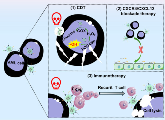
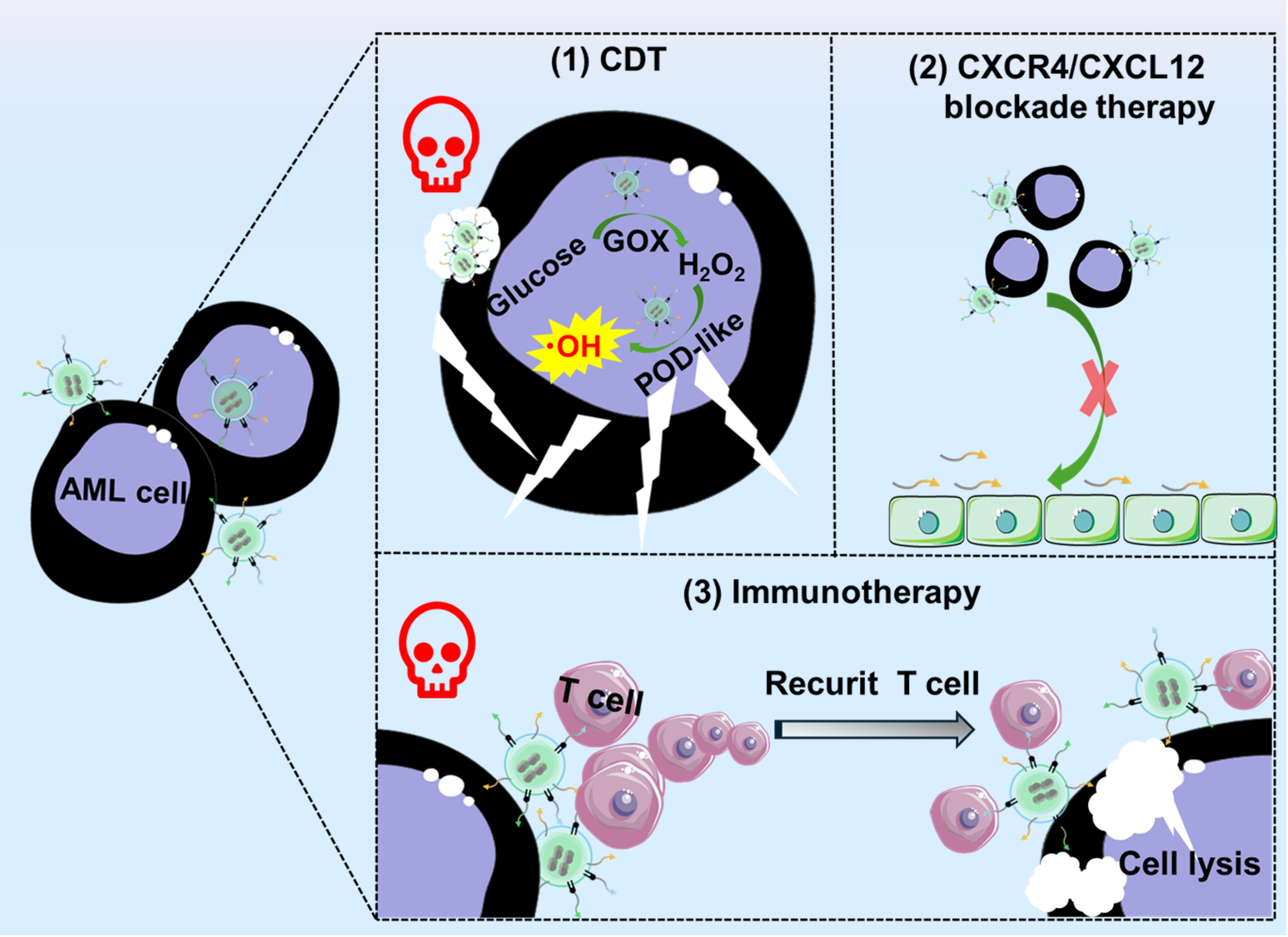
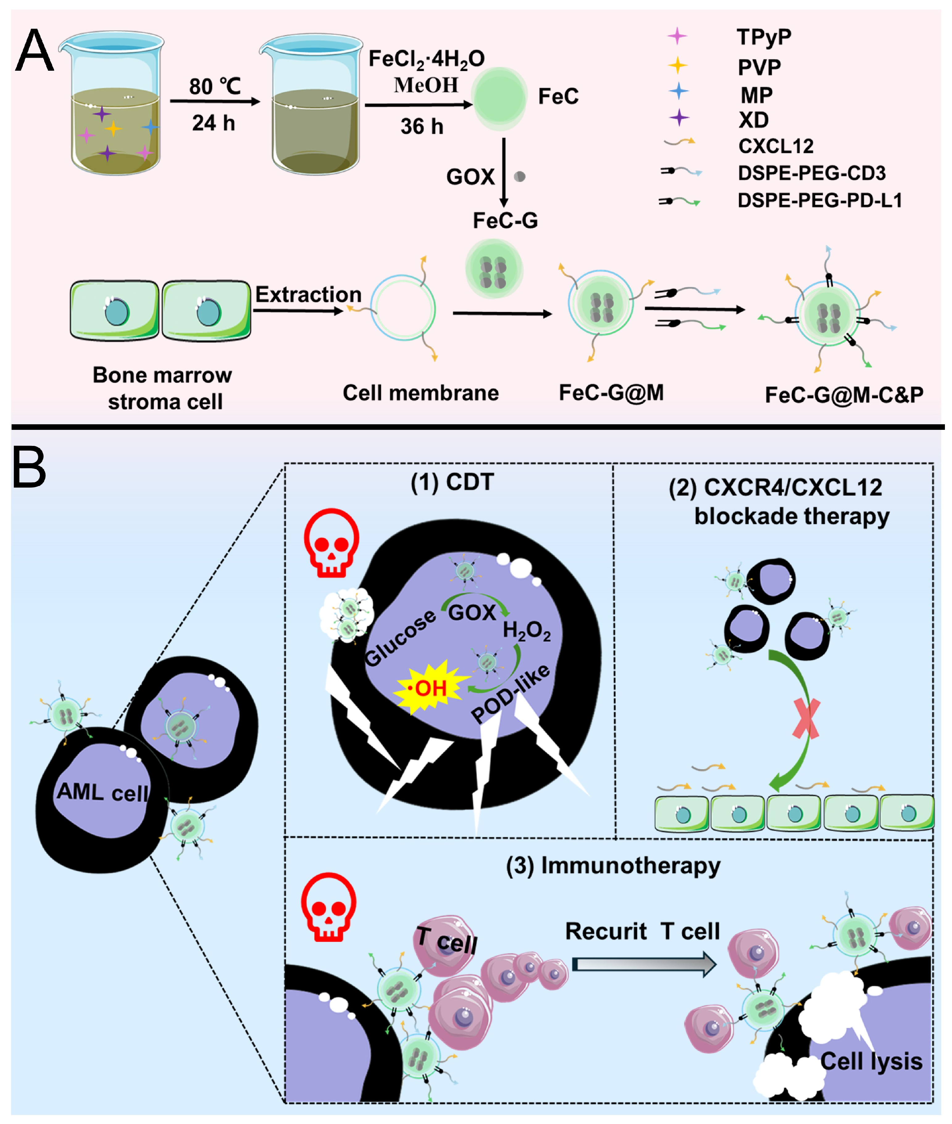
Herein, we designed a covalent organic framework bispecific nanosystem for the combined treatment of AML (
Scheme 1). The synthetic procedures were described as follows. First, iron porphyrin-based covalent organic framework (abbreviated FeC) was prepared by a facile method. Then GOX was encapsulated to prepare GOX-loaded FeC (abbreviated FeC-G). Subsequently, bone marrow stromal cell membrane was employed to prepare membrane coated FeC-G (abbreviated FeC-G@M). Finally, anti-CD3 and anti-PD-L1 antibodies pre-modified with 1,2-distearoyl-sn-glycero-3-phosphoethanolamine-(polyethylene glycol)-NHS ester (DSPE-PEG-NHS) were anchored into the phospholipid bilayer via physical interactions to prepare FeC-G@M modified with anti-CD3 and anti-PD-L1 antibodies (abbreviated FeC-G@M-C&P). The FeC core triggered the Fenton-like reaction in the acidic tumor microenvironment to produce ROS. Meanwhile, GOX depleted glucose and generated abundant H
2O
2 to amplify CDT induced by FeC. Bone marrow stromal cell membrane endowed the nanosystem with strong targeting ability, which aimed at enhancing CDT efficacy and realizing CXCR4/CXCL12 blockade therapy. Anti-CD3 and anti-PD-L1 antibody moieties simultaneously recognized CD3 on the surface of T cells and PD-L1 on the surface of leukemia cells to form intercellular crosslink between T cells and leukemia cells, narrowing cell–cell distance and activating anti-leukemia immune responses. The nanosystem achieved combined therapeutics effects of enhanced CDT, CXCR4/CXCL12 blockade therapy and immunotherapy in pursuit of solving poor efficiency of single therapy and drug resistance.
2. Materials and Methods
2.1. Chemical Reagents
5,10,15,20-tetra(4-pyridyl)-21H,23H-porphine (TPyP), 3,3′,5,5′-tetramethylbenzidine (TMB), 2,2′-azino-bis-(3-ethylbenzthiazoline-6-sulphonate) (ABTS), DSPE-PEG-NHS and methanol (MeOH) were provided by Shanghai Aladdin Biochemical Technology Co., Ltd. (Shanghai, China). 1-methyl-2-pyrrolidinone (MP) was provided by Beijing J&K Scientific Co., Ltd. (Beijing, China). p-xylylene dibromide (XD) was provided by Tianjin Heowns Biochemical Technology Co., Ltd. (Tianjin, China). Polyvinylpyrrolidone (PVP), ferrous chloride tetrahydrate (FeCl2·4H2O), and glucose were brought from Sinopharm Chemical Reagent Co., Ltd. (Shanghai, China). GOX and methyl red were brought from Shanghai Macklin Biochemical Co., Ltd. (Shanghai, China). Anti-CD3 and anti-PD-L1 antibody were brought from Bio X Cell (NH, USA). FITC-NHS was brought from Shanghai Toyang Biotechnology Co., Ltd. (Shanghai, China). Cy5-NHS was brought from Dalian Meilun Biotechnology Co., Ltd. (Dalian, China). Anti-CXCL12 antibody was brought from Beijing Biosynthesis Biotechnology Co., Ltd. (Beijing, China). Mouse leukemia cell (C1498) and human umbilical vein endothelial cells (HUVECs) were purchased from Shanghai Zhong Qiao Xin Zhou Biotechnology Co., Ltd. (Shanghai, China). Mouse Stromal-5 (MS-5) were provided by Shanghai Qingqi Biotechnology Development Co., Ltd. (Shanghai, China). Dulbecco’s modified eagle medium (DMEM) and Annexin V-FITC/PI apoptosis detection kit were purchased from Nanjing Keygen Biotech Co., Ltd. (Nanjing, China). Fetal bovine serum (FBS) was purchased from Hangzhou Si Ji Qing Biological Engineering Materials Co., Ltd. (Hangzhou, China). Penicillin-streptomycin solution was purchased from Wuhan Servicebio Technology Co., Ltd. (Wuhan, China). Iron assay kit was supplied by Nanjing Jiancheng Bioengineering Institute. Cell counting kit-8 (CCK-8), ROS assay kit and BCA protein assay kit were supplied by Shanghai Beyotime Biotechnology Co., Ltd. (Shanghai, China). CXCL12 was supplied by BD Bioscience (San Jose, CA, USA). Enzyme-linked immunosorbent assay (ELISA) kits for tumor necrosis factor-alpha (TNF-α), interleukin-2 (IL-2), and interferon-gamma (IFN-γ) were purchased from Abbkine Scientific Co., Ltd. (Wuhan, China). ELISA kits for perforin and granzyme B were purchased from Wuhan Huamei Biotech Co., Ltd. (Wuhan, China).
2.2. Isolation of Bone Marrow Stromal Cell Membrane
Cell membranes were isolated by hypotonic treatment and liquid nitrogen freeze–thaw method. MS-5 cells were collected by centrifugation, and thoroughly suspended in sterile 0.1 × PBS solution. Then cell suspensions were placed in a liquid nitrogen canister to freeze for 8 s, followed by fully thawing at room temperature. The process was repeated 6 times to destroy cell membrane. The obtained cell suspension was centrifuged at 12,000 rpm/min for 20 min at 4 °C. After discarding the supernatant, 1 mL of sterile water was added to suspend the cell membranes. BCA protein assay kit was used to measure protein concentration. The resulting cell membrane samples were kept at −80 °C for further use.
2.3. Preparation of FeC, FeC-G and FeC-G@M
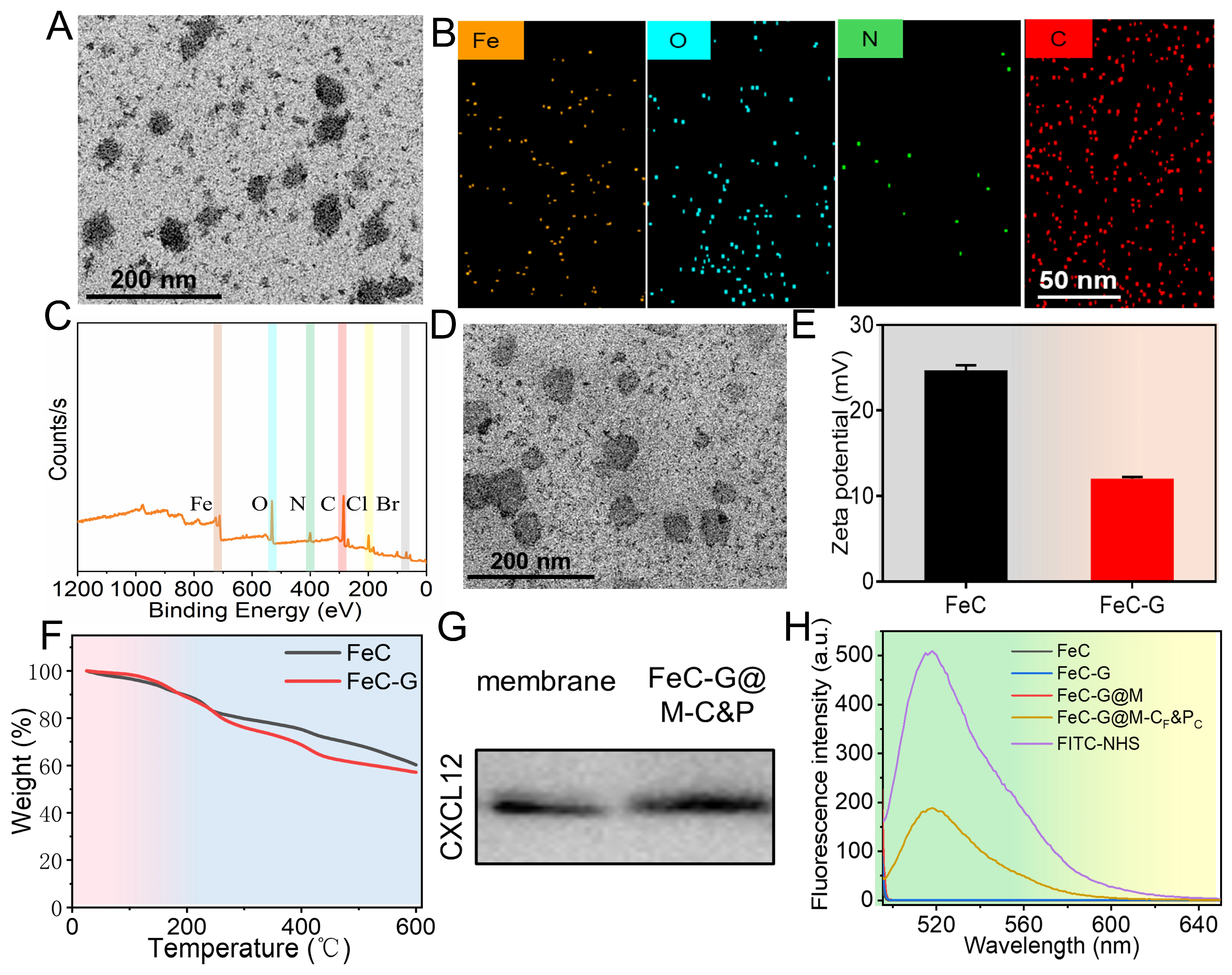
30 mg of TPyP and 365 mg of PVP were dissolved in a flask containing 15 mL of MP. Then, 40 mg of XD was added into the flask, and stirred for 20 min. After purging the mixture with nitrogen gas for 15 min, the flask was placed in the water bath at 80 °C for 24 h. At the end of the reaction, the mixture was centrifuged, and the supernatant was discarded. Next, 40 mg of FeCl2·4H2O and 5 mL of MeOH were added into the obtained composite, and subsequently stirred at room temperature for 36 h. Upon completion of the reaction, the mixture was centrifuged, and the supernatant was discarded. The product FeC was collected after washing with MeOH. To prepare FeC-G, GOX and FeC were mixed at the mass ratio of 1:1, and stirred for 2.5 h at room temperature. FeC-G@M was prepared by incubating FeC-G with 0.5 mg of cell membranes at the mass ratio of 1:1 in an ultrasonic bath for 5 min.
For the preparation of FITC-labeled FeC, FITC and FeC were mixed at a mass ratio of 1:10 and stirred at room temperature in the dark overnight. Afterwards, the obtained product was centrifuged and thoroughly washed to remove free FITC. The preparation of FITC-labeled FeC@M was almost the same as that of FITC-labeled FeC, except that the product was further incubated with 0.5 mg of isolated cell membranes at a mass ratio of 1:1 in an ultrasonic bath for 5 min.
2.4. Preparation of FeC-G@M-P and FeC-G@M-C&P
First, antibodies were modified with DSPE-PEG-NHS via amide reaction. Specifically, DSPE-PEG-NHS was mixed with 100 μg of anti-CD3 antibody and 100 μg of anti-PD-L1 antibody with a molar ratio of 5:1 respectively, and the pH was adjusted to 8 by adding sodium bicarbonate solution. After incubation on a shaker at room temperature for 2 h, DSPE-PEG-NHS modified anti-CD3 antibody (DSPE-PEG-CD3) and anti-PD-L1 antibody (DSPE-PEG-PD-L1) were obtained via ultrafiltration. Then, the obtained antibodies were incubated with the resulting FeC-G@M at 37 °C for 1.5 h. The FeC-G@M-C&P was finally collected after centrifugation. At the same time, FeC-G@M modified with anti-PD-L1 antibody (abbreviated FeC-G@M-P) was prepared for comparison. The preparation procedure of FeC-G@M-P was almost the same as that of FeC-G@M-C&P except that DSPE-PEG-CD3 was replaced with DSPE-PEG-NHS, which anchored onto the phospholipid bilayer via physical interaction.
The preparation procedure of FeC@M and FeC@M-P were almost the same as that of FeC-G@M and FeC-G@M-P except the removing of GOX. The preparation of FeC-G@M modified with FITC-labeled anti-CD3 and Cy5-labeled anti-PD-L1 antibodies (abbreviated FeC-G@M-CF&PC) was almost the same as that of FeC-G@M-C&P except that FITC-NHS and DSPE-PEG-NHS co-modified anti-CD3 antibody as well as Cy5-NHS and DSPE-PEG-NHS co-modified anti-PD-L1 antibody were used.
2.5. Characterization
Transmission electron microscopy (TEM) images were captured by a JEM-1200EX electron microscope (Tokyo, Japan). X-ray photoelectron spectroscopy (XPS) measurements were detected by a Thermo Escalab 250Xi (, DE, USA). The polydispersity index (PDI) and zeta potential were measured by a Zetasizer Nano ZS90 (Malvern, UK). Thermal gravimetric analysis (TGA) was analyzed by a STA 449 F3 thermal analyzer (Selb, Germany). Fluorescence spectra were detected by an RF-5301PC luminescence spectrometer (Kyoto, Japan). Absorption spectra were obtained by a Nicolet evolution 300 spectrophotometer (Madison, WI, USA). CCK-8 experiment was conducted using an EnSpire 2300 microplate detector (Waltham, MA, USA). The expression of CXCL12 was detected by Western blotting using a ChemiDoc™ MP imaging system, Hercules, CA, USA).
2.6. Evaluation of POD-like Activity and Cascade Catalytic Activity
The following POD-like activity was evaluated by catalyzing ABTS/TMB oxidation method.
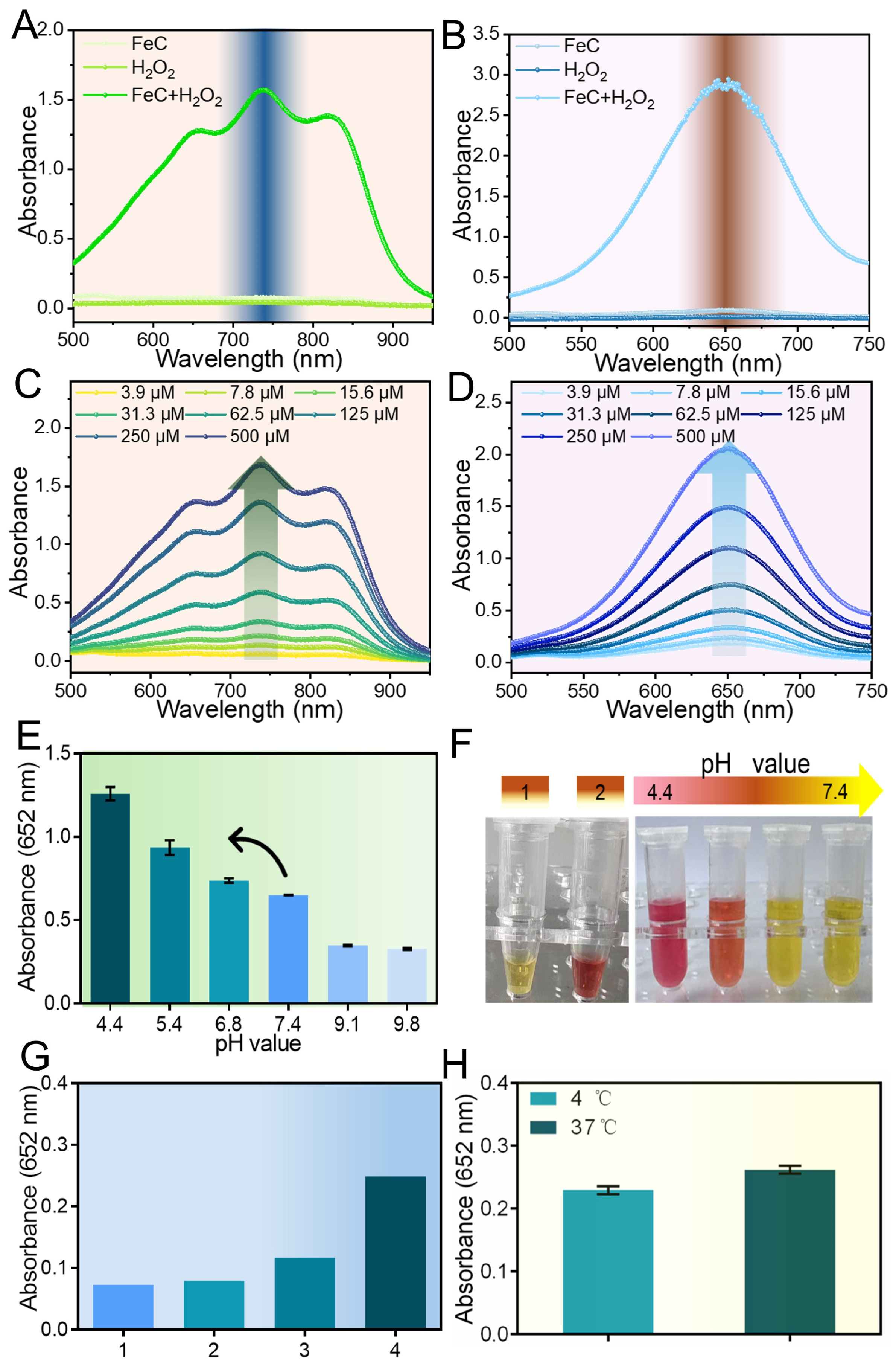
Detection of POD-like activity of FeC: For ABTS oxidation experiment, the catalytic system was configured as follows: the mixture of H2O2 (20 mM), FeC (500 μg/mL), ABTS (5 mg/mL), and buffer at a volume ratio of 1:1:1:7 was prepared, followed by incubating at room temperature for 10 min. Set groups without the addition of H2O2 or FeC as control. Ending incubation, absorption spectra of the solution were recorded by ultraviolet–visible spectrophotometer. For the TMB oxidation experiment, the operation was the same as that of ABTS excepting the replacement of ABTS by TMB.
Effect of H2O2 concentrations on POD-like activity of FeC: For ABTS oxidation experiment, the catalytic system was configured as follows: the mixture of H2O2 with various concentrations, FeC (250 μg/mL), ABTS (5 mg/mL), and buffer at the volume ratio of 1:1:1:7 was prepared, followed by incubating at room temperature for 25 min. Ending incubation, absorption spectra of the solution were recorded by ultraviolet–visible spectrophotometer. For the TMB oxidation experiment, the operation was the same as that of ABTS excepting the replacement of ABTS by TMB.
Effect of pH on POD-like activity: The mixture of H2O2 (20 mM), FeC (250 μg/mL), TMB (5 mg/mL), and buffers of different pH at a volume ratio of 1:1:1:7 was prepared, followed by incubating at room temperature for 10 min. Ending incubation, absorbance at 652 nm of the solution was recorded by a microplate reader.
Acidity changes in the reaction system catalyzed by FeC-G@M-C&P: The catalytic system was configured as follows: the mixture of glucose (300 mM), FeC-G@M-C&P (500 μg/mL), and buffer at a volume ratio of 1:2:7 was prepared, followed by incubating at room temperature for 24 h. Set the group without the addition of glucose as control. Ending incubation, methyl red indicator was added to the reaction solution. The color was photographed and recorded.
Cascade catalytic activity of FeC-G@M-C&P: The experimental group was prepared by mixing glucose (300 mM), FeC-G@M-C&P (500 μg/mL), TMB (5 mg/mL), and buffer at a volume ratio of 1:1:1:7. Glucose + TMB, FeC-G@M-C&P + TMB, and Glucose + GOX + TMB groups were set as controls. The mixture was incubated at room temperature for 10 min, and the absorbance at 652 nm of the solution was recorded by a microplate reader.
Effect of temperature on POD-like activity: Preparation of the catalytic system was the same as that in the upper part. After incubation at 4 °C or 37 °C for 10 min, the absorbance at 652 nm of the solution was recorded by a microplate reader.
2.7. Cell Culture
C1498 cells were incubated in DMEM supplemented with 1% streptomycin–penicillin solution and 10% FBS. MS-5 cells and HUVECs were incubated in RPMI 1640 medium supplemented with 1% streptomycin–penicillin solution and 10% FBS. Cells were incubated in a humidified atmosphere containing 5% CO2 at 37 °C.
2.8. Evaluation of Targeting, ROS Generation Ability and Cell Apoptosis Rate
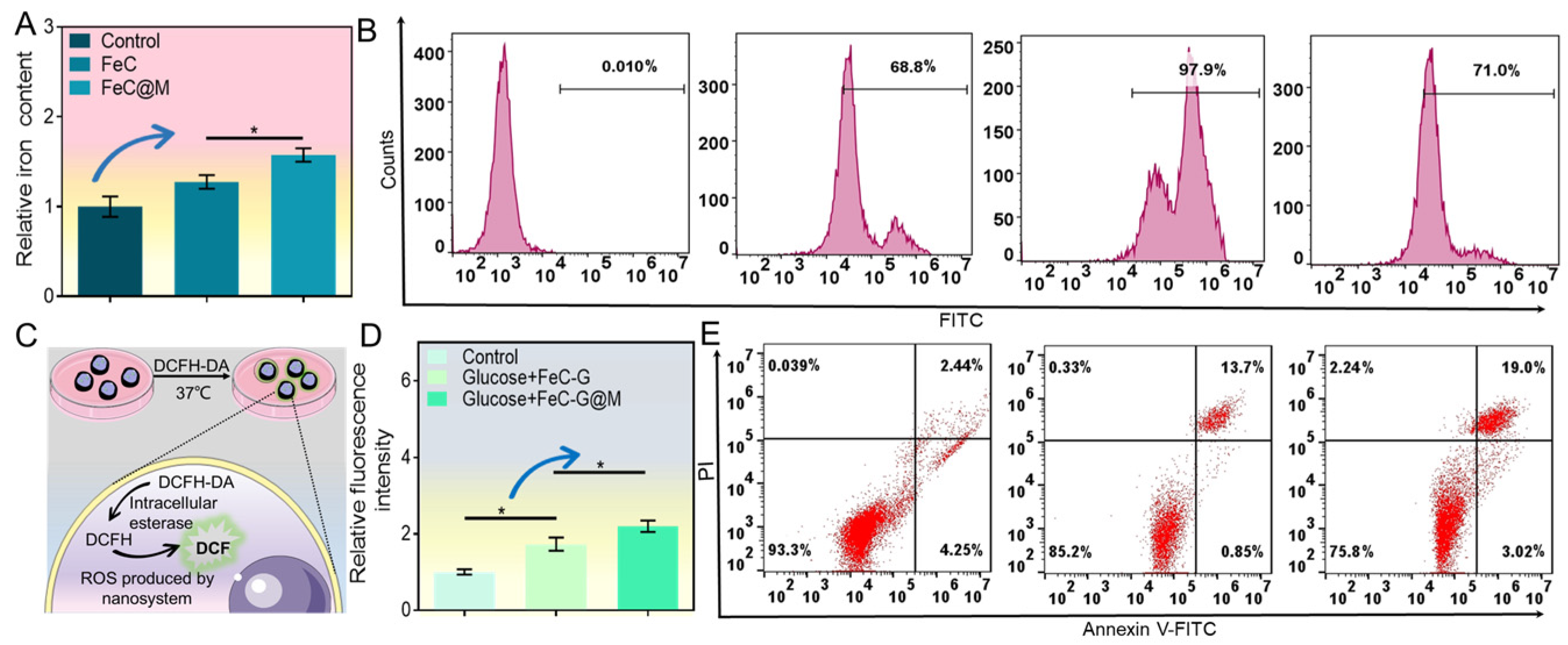
C1498 cells were seeded into 6-well plates at a density of 1 × 10⁶ cells/well. The cells of experimental groups were separately treated with FeC and FeC@M at a concentration of 100 μg/mL. PBS treated cells were served as control. After 4 h of treatment, cell suspensions were centrifuged at 1000 rpm/min for 5 min. The cells were collected and washed three times with sterile PBS. The content of iron was measured according to the instruction of the test kit, and the relative iron content was calculated.
C1498 cells were seeded into 6-well plates at a density of 1 × 10⁶ cells/well. The cells of experimental groups were separately treated with FITC-labeled FeC, FITC-labeled FeC@M and CXCL12 + FITC-labeled FeC@M at a concentration of 100 μg/mL. The concentration of CXCL12 was 5 μg/mL, with a 0.5 h pre-incubation prior to treatment. After 4 h of treatment, the cell suspensions were centrifuged at 1000 rpm/min for 5 min. The collected cells were washed three times with sterile PBS and detected by a Beckman Coulter CytoFLEX flow cytometer.
C1498 cells were seeded into 6-well plates at a density of 1 × 10⁶ cells/well. The cells of experimental groups were separately treated with Glucose + FeC-G and Glucose + FeC-G@M. The concentration of glucose and nanomaterial was 30 mM and 100 μg/mL, respectively. PBS treated cells were served as control. Twelve hours later, old culture media were replaced by that containing 10 μM DCFH-DA, and further incubated in the cell culture incubator. After 20 min of incubation, the cell suspensions were centrifuged at 1000 rpm/min for 5 min. The cells were washed three times. Fluorescence intensity was measured by a microplate (excitation: 488 nm; emission: 525 nm). The relative fluorescence intensity of cells was calculated.
C1498 cells were seeded in 6-well plates at a density of 1 × 10⁶ cells/well. The cells of experimental groups were separately treated with Glucose + FeC-G and Glucose + FeC-G@M for 24 h at a concentration of 100 μg/mL. The concentration of glucose was 30 mM. After treatment, cell suspensions were centrifuged at 1000 rpm/min for 5 min. The cells were collected and washed, and then incubated with 5 μL Annexin V-FITC and 5 μL PI at room temperature in the dark for 10 min. Cell apoptosis was determined using a Beckman Coulter CytoFLEX flow cytometer.
2.9. Evaluation of Migration and Adhesion Ability
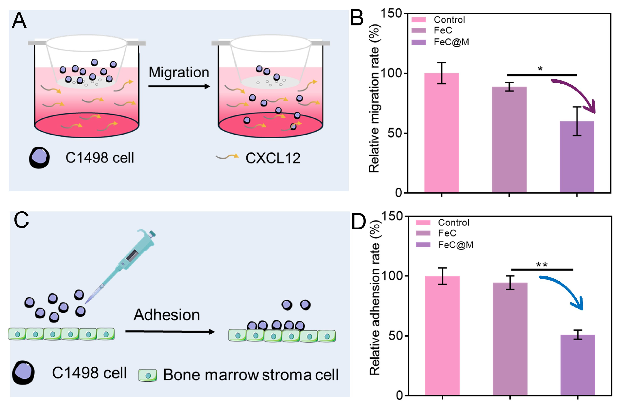
For the migration assay, C1498 cells were seeded in 6-well plates. The cells of the experimental group were separately treated with FeC and FeC@M at a concentration of 100 μg/mL, while that of the control and blank group received no treatment. After 1 h of treatment, 3 × 10⁵ cells were inoculated into the upper chambers of the Transwell. The lower chambers of the experimental and control group were added with cell culture medium containing CXCL12 (0.2 μg/mL), while that of the blank group was added with an equal volume of blank cell culture medium. After 24 h of incubation, cells in the lower chambers were collected. CCK-8 reagent was added and placed the plate in the cell culture incubator in the dark for 3 h. The absorbance at 450 nm was measured by a microplate reader. Relative migration rate of cells was calculated.
For adhesion assay, MS-5 cells were seeded in 6-well plates. After formed stromal cell layers, 1 × 10⁵ cells were further seeded into cell culture plates. C1498 cells in the experimental group were pre-treated with FeC and FeC@M (100 μg/mL) for 1 h, respectively, while that in the control group received no treatment. After 24 h of incubation, non-adherent cells were gently removed. CCK-8 reagent was added and placed the plate in the cell culture incubator in the dark for 3 h. The absorbance at 450 nm was measured using a microplate reader. After deducting the absorbance of the group only containing MS-5 cells, relative adhesion rate of cells was calculated.
2.10. Evaluation of the Enhanced T Cell Cytotoxicity and Combined Anti-Leukemia Effect of FeC-G@M-C&P
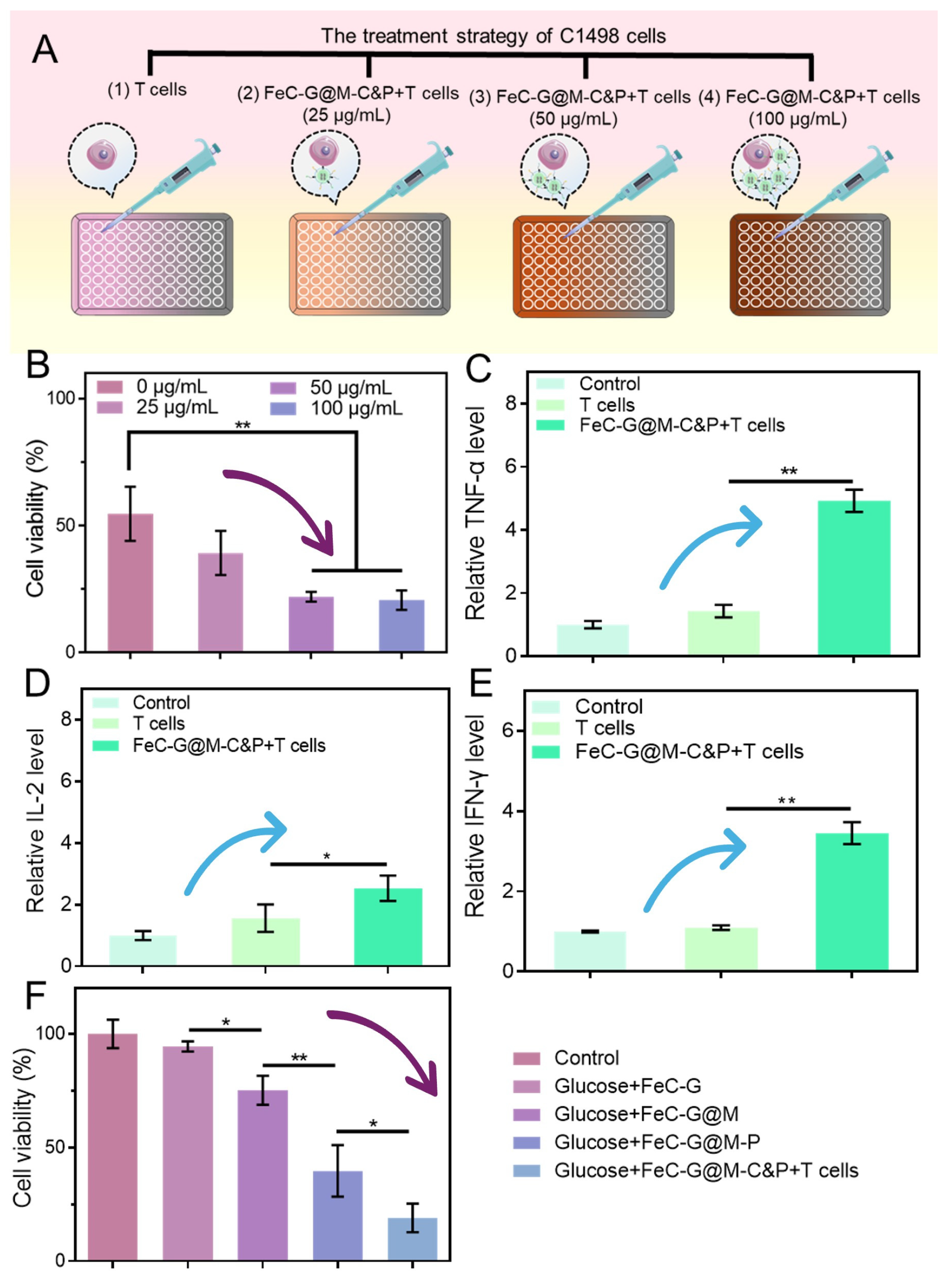
C1498 cells (target cells) were seeded in 96-well culture plates at a density of 5 × 10⁴ cells/well. C1498 cells were co-incubated with different concentrations of FeC-G@M-C&P and T cells (effector cells). T cells were obtained from the spleens of healthy mice and expanded for two weeks prior to use. The effector/target ratio was set as 5:1. After 48 h of incubation, CCK-8 reagent was added into each well and placed the plate in the cell culture incubator for 3 h. Absorbance at 450 nm was measured using a microplate reader, and the cell viability was calculated.
C1498 cells were seeded in 96-well culture plates at a density of 5 × 10⁴ cells/well. To demonstrate the benefits of dual antibody loading, C1498 cells were randomly divided into the T cells group, FeC-G@M-P + T cells group and FeC-G@M-C&P + T cells group. The effector/target ratio was set as 5:1, and the concentration of the FeC-G@M-C&P was 100 μg/mL. After 24 h incubation, CCK-8 reagent was added into each well and placed the plate in the cell culture incubator for 3 h. Absorbance at 450 nm was measured using a microplate reader, and the cell viability was calculated.
C1498 cells were seeded in 96-well plates at a density of 5 × 10⁴ cells/well. The cells were randomly divided into the control, T cells, and FeC-G@M-C&P + T cells groups, and received corresponding treatments. The effector/target ratio was set as 5:1, and the concentration of the FeC-G@M-C&P was 100 μg/mL. After 48 h treatment, cell suspensions were centrifuged for 5 min at 4 °C. The supernatant was carefully collected, and the contents of TNF-α, IL-2, and IFN-γ in samples were determined following the manufacturer’s instructions of the ELISA kits. The levels of perforin and granzyme B in cell culture supernatants were measured by the identical procedure. The relative level of cytokines was calculated.
C1498 cells were seeded in 96-well plates at a density of 5 × 10⁴ cells/well. The cells were divided into the control group, Glucose + FeC-G, Glucose + FeC-G@M, Glucose + FeC-G@M-P, and the Glucose + FeC-G@M-C&P + T cells group. The effector/target ratio was set as 5:1. The concentration of glucose and nanomaterial is 30 mM and 25 μg/mL, respectively. After 24 h treatment, CCK-8 reagent was added and placed the plate in the culture incubator for 3 h. Absorbance at 450 nm was measured using a microplate reader, and the cell viability was calculated.
2.11. Evaluation of Biosafety and Biocompatibility
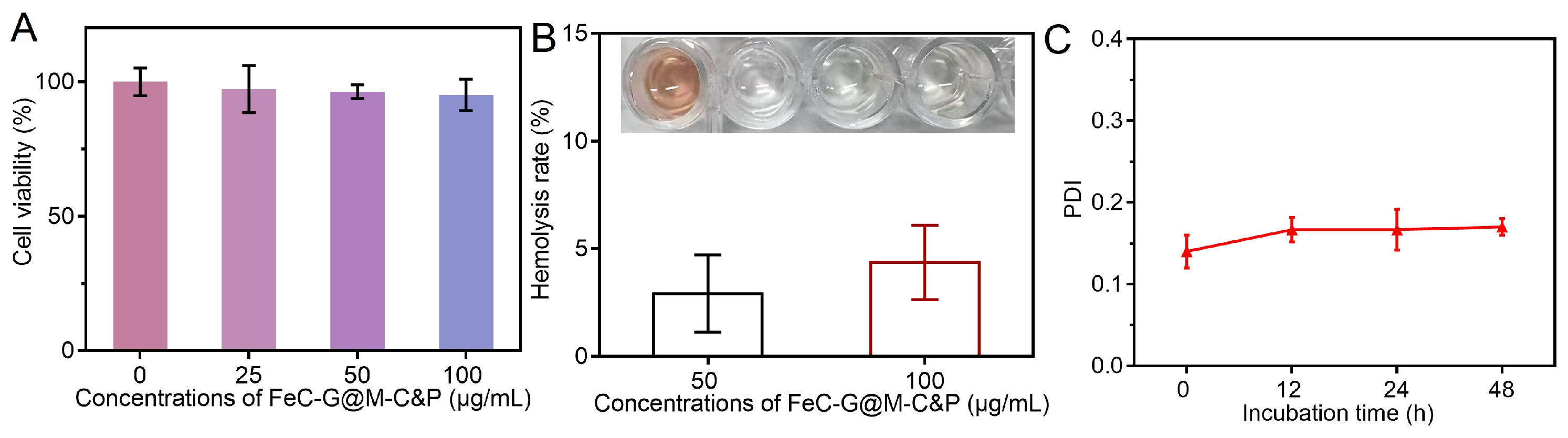
HUVECs were seeded in 96-well plates at a density of 5 × 10⁴ cells/well and cultured overnight. Subsequently, the old culture medium was replaced with a fresh medium containing different concentrations of FeC-G@M-C&P ranging from 0 to 100 μg/mL, while PBS-treated cells were set as the control group. After 48 h incubation, CCK-8 reagent was added to each well, and the plates were further incubated at 37 °C for 3 h in the dark. Finally, the absorbances at 450 nm were detected by a microplate reader to calculate the cell viability.
Fresh mouse blood was collected and centrifuged to harvest the red blood cells (RBCs), which were repeatedly washed with PBS to remove impurities. Then 2% RBCs was prepared for subsequent experiments. In detail, 500 μL RBCs was separately mixed with an equal volume of pure water, PBS or FeC-G@M-C&P dispersions with different concentrations. The pure water group was served as the positive control, and the PBS was served as the negative control. After incubation at 37 °C for 30 min, all mixtures were centrifuged at 3500 rpm for 5 min. The supernatants were transferred to a 96-well plate for visual observation, followed by measuring the absorbances at 545 nm by a microplate reader to calculate the hemolysis rate.
FeC-G@M-C&P was dispersed in PBS solution, followed by incubation at 37 °C. At predetermined time points (0, 12, 24, 48 h), 1 mL of sample dispersion was gently taken off and loaded into a disposable dynamic light scattering cuvette. The PDI was determined by Zetasizer Nano ZS90 (Malvern, UK).
2.12. Statistical Analysis
Data fitting and analysis were performed by GraphPad Prism 6 and SPSS Statistics 27 software. Data were presented as mean ± standard deviation. Briefly, one-way analysis of variance coupled with Tukey’s post hoc test was applied for statistical evaluation. * p < 0.05 and ** p < 0.01 represent that the difference was statistically significant.