Influence of Final Irrigation on Calcium Silicate-Based Sealer Dentinal Tubular Penetration: A Systematic Review

 , , and
Abstract
1. Introduction
2. Materials and Methods
2.1. Protocol and Registration
2.2. Search Strategy and Eligibility Criteria
2.3. Study Screening
2.4. Data Extraction
2.5. Quality Assessment
3. Results
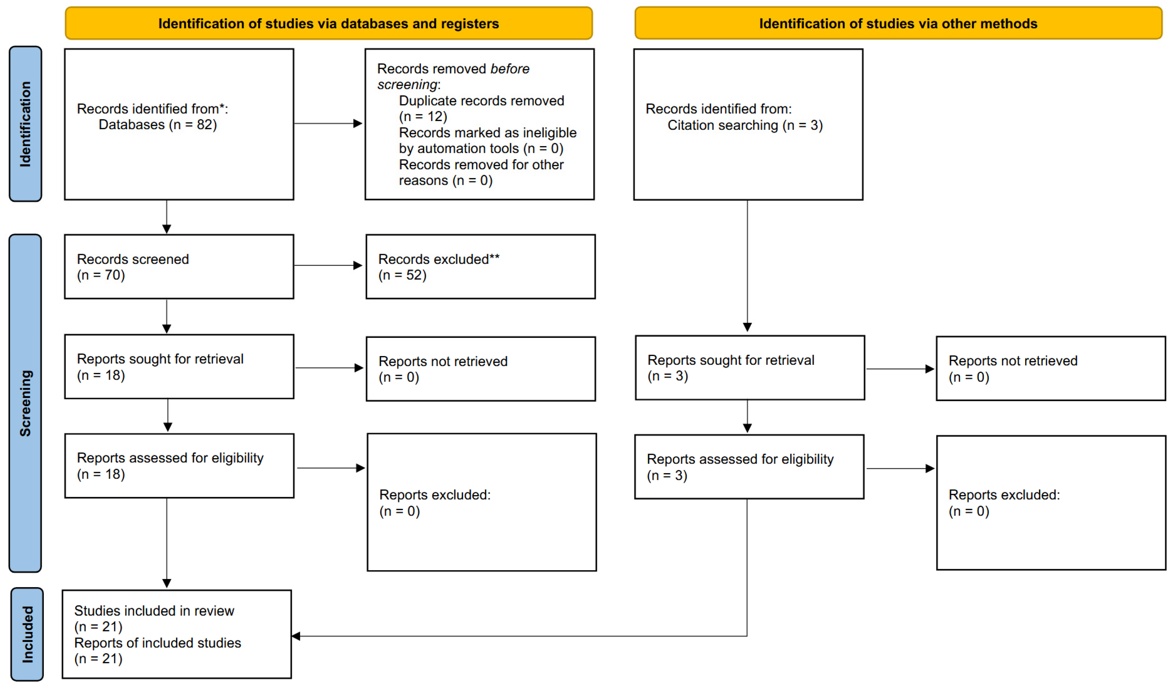
3.1. Study Selection and Flow Diagram
3.2. Quality Assessment
3.3. Study Methodology and Results
4. Discussion
4.1. Interpretation of the Main Findings
4.2. Methodological Considerations
4.3. Risk of Bias and Overall Confidence of the Evidence
4.4. Clinical Implications and Limitations
4.5. Future Perspectives
5. Conclusions
Supplementary Materials
Author Contributions
Funding
Institutional Review Board Statement
Informed Consent Statement
Data Availability Statement
Conflicts of Interest
References
- Haapasalo, M.; Shen, Y.; Qian, W.; Gao, Y. Irrigation in endodontics. Dent. Clin. N. Am. 2010, 54, 291–312. [Google Scholar] [CrossRef] [PubMed]
- Real Academia Española. Irrigar. In Diccionario de la Lengua Española, 23rd ed.; Real Academia Española: Madrid, Spain, 2024; Available online: https://dle.rae.es/irrigar?m=form (accessed on 11 November 2025).
- Zehnder, M. Root canal irrigants. J. Endod. 2006, 32, 389–398. [Google Scholar] [CrossRef] [PubMed]
- Plotino, G.; Grande, N.M.; Mercadé, M. New technologies to improve root canal disinfection. Aust. Dent. J. 2016, 61, 14–23. [Google Scholar]
- Ballal, N.V.; Kandian, S.; Mala, K.; Bhat, K.S. Comparison of the efficacy of maleic acid and ethylenediaminetetraacetic acid in smear layer removal at the apical third of root canals. J. Endod. 2009, 35, 402–405. [Google Scholar]
- Uloopi, K.S.; Madhuri, G.; Vinay, C.; Chandrasekhar, R. Comparative evaluation of smear layer removal by maleic acid, EDTA, and citric acid—An in vitro study. Int. J. Clin. Pediatr. Dent. 2011, 4, 176–179. [Google Scholar]
- Reddy, P.; Bhavana, V.; Srinivasulu, G. Maleic acid as a final irrigant: A review. J. Int. Soc. Prev. Community Dent. 2014, 4, 5–10. [Google Scholar]
- Dal Bello, Y.; Fracaro Porsch, H.; Farina, A.P.; Souza, M.A.; Nogueira Leal Silva, E.J.; Bedran-Russo, A.; Cecchin, D. Glycolic acid as the final irrigant in endodontics: Mechanical and cytotoxic effects. Mater. Sci. Eng. C Mater. Biol. Appl. 2019, 100, 323–329. [Google Scholar] [CrossRef] [PubMed]
- Sarı, M.; Ayhan, A.A.; Tüfenkçi, P.; Çelikten, B. Efficacy of glycolic acid at different concentrations for smear layer removal with various activation systems. BMC Oral Health 2025, 25, 1695. [Google Scholar] [CrossRef] [PubMed]
- Mullick, T.; Ballal, N.V. Effect of glycolic acid, maleic acid, and EDTA in the removal of smear layer from root canal dentin. Pesqui. Bras. Odontopediatria Clin. Integr. 2023, 23, e220058. [Google Scholar] [CrossRef]
- Ali, A.M.M.; Raab, W.H.M. Smear layer removal efficiency using apple vinegar: An in vitro scanning electron microscope study. Am. J. Dent. 2019, 32, 21–27. [Google Scholar]
- Candeiro, G.T.M.; Matos, I.B.; Costa, C.F.E.; Fonteles, C.S.R.; do Vale, M.S. A comparative scanning electron microscopy evaluation of smear layer removal with apple vinegar and sodium hypochlorite associated with EDTA. J. Appl. Oral Sci. 2011, 19, 639–643. [Google Scholar] [CrossRef] [PubMed]
- Akbulut, M.B.; Guneser, M.B.; Eldeniz, A.U. Effects of fruit vinegars on root dentin microhardness and roughness. J. Conserv. Dent. 2019, 22, 97–101. [Google Scholar]
- Mohamed Ismail, A.A.-D.; Abbas, A.N.; Tawfeik, A.M. Evaluation of the antibacterial effect of apple cider vinegar, black tea and sodium hypochlorite irrigant solutions on infected root canal microorganisms of primary teeth: An in vitro study. Al-Azhar J. Dent. Sci. 2022, 25, 99–104. [Google Scholar]
- Azzam, D.; Kamel, W.; Elsheikh, H. Comparative evaluation of smear layer removal by apple vinegar using laser activated irrigation and EndoVac system: Randomized control trial. Al-Azhar J. Dent. 2022, 9, 201–209. [Google Scholar] [CrossRef]
- Del Carpio-Perochena, A.E.; Bramante, C.M.; Duarte, M.A.; de Moura, M.R.; Aouada, F.A.; Kishen, A. Chelating and antibacterial properties of chitosan nanoparticles on dentin. J. Endod. 2014, 40, 825–830. [Google Scholar]
- Cao, Y.; Shen, Y.; Haapasalo, M. Effect of peracetic acid on biofilms in root canal systems. J. Endod. 2015, 41, 1011–1015. [Google Scholar]
- Plotino, G.; Pameijer, C.H.; Grande, N.M.; Somma, F. Ultrasonics in endodontics: A review of the literature. J. Endod. 2007, 33, 81–95. [Google Scholar] [CrossRef] [PubMed]
- Peters, O.A.; Bardsley, S.; Fong, J.; Pandher, G.; DiVito, E. Disinfection of root canals with photon-initiated photoacoustic streaming. J. Endod. 2011, 37, 1008–1012. [Google Scholar] [CrossRef] [PubMed]
- Haapasalo, M.; Shen, Y.; Wang, Z.; Gao, Y. Irrigation in endodontics. Br. Dent. J. 2014, 216, 299–303. [Google Scholar] [CrossRef] [PubMed]
- Ruddle, C.J. Endodontic irrigation: Endo essentials. Dent. Today 2012, 31, 130–137. [Google Scholar]
- Torabinejad, M.; Walton, R.E. Endodontics: Principles and Practice, 5th ed.; Elsevier: St. Louis, MO, USA, 2015. [Google Scholar]
- AL-Haddad, A.; Che Ab Aziz, Z.A. Bioceramic-based endodontic sealers: A review. Int. J. Biomater. 2016, 2016, 9753210. [Google Scholar] [PubMed]
- Kim, Y.; Kim, B.S.; Kim, Y.M.; Lee, D.; Kim, S.Y. The Penetration Ability of Calcium Silicate Root Canal Sealers into Dentinal Tubules Compared to Conventional Resin-Based Sealer: A Confocal Laser Scanning Microscopy Study. Materials 2019, 12, 531. [Google Scholar] [CrossRef] [PubMed]
- Wang, Z.; Shen, Y.; Haapasalo, M. Comparative evaluation of bioceramic and resin-based sealers for dentinal tubule penetration and adaptation using CLSM and SEM. J. Endod. 2015, 41, 1586–1591. [Google Scholar]
- Zhang, Y.S.; Li, N.Y. Current Application of Confocal Laser Scanning Microscope (CLSM) in Stomatology. Shanghai Kou Qiang Yi Xue 2007, 16, 219–224. (In Chinese) [Google Scholar] [PubMed]
- Donnermeyer, D.; Schmidt, S.; Rohrbach, A.; Berlandi, J.; Bürklein, S.; Schäfer, E. Debunking the Concept of Dentinal Tubule Penetration of Endodontic Sealers: Sealer Staining with Rhodamine B Fluorescent Dye Is an Inadequate Method. Materials 2021, 14, 3211. [Google Scholar] [CrossRef] [PubMed]
- Jeong, J.W.; DeGraft-Johnson, A.; Dorn, S.O.; Di Fiore, P.M. Dentinal Tubule Penetration of a Calcium Silicate-Based Root Canal Sealer with Different Obturation Methods. J. Endod. 2017, 43, 633–637. [Google Scholar] [CrossRef] [PubMed]
- Coronas, V.S.; Villa, N.; Nascimento, A.L.D.; Duarte, P.H.M.; Rosa, R.A.D.; Só, M.V.R. Dentinal Tubule Penetration of a Calcium Silicate-Based Root Canal Sealer Using a Specific Calcium Fluorophore. Braz. Dent. J. 2020, 31, 109–115. [Google Scholar] [CrossRef] [PubMed]
- Marwaha, J.; Yadav, D.; Sharma, S.; Dixit, A.; Arya, A.; Kar, S. Push-Out Bond Strength for Bioceramic and Epoxy Resin-Based Sealers. Bioinformation 2025, 21, 353–356. [Google Scholar] [CrossRef] [PubMed]
- Page, M.J.; McKenzie, J.E.; Bossuyt, P.M.; Boutron, I.; Hoffmann, T.C.; Mulrow, C.D.; Shamseer, L.; Tetzlaff, J.M.; Akl, E.A.; Brennan, S.E.; et al. The PRISMA 2020 Statement: An Updated Guideline for Reporting Systematic Reviews. BMJ 2021, 372, n71. [Google Scholar] [CrossRef] [PubMed]
- Sheth, V.H.; Shah, N.P.; Jain, R.; Bhanushali, N.; Bhatnagar, V. Development and validation of a risk-of-bias tool for assessing in vitro studies conducted in dentistry: The QUIN. J. Prosthet. Dent. 2024, 131, 1038–1042. [Google Scholar] [PubMed]
- Akçay, M.; Arslan, H.; Durmuş, N.; Mese, M.; Capar, I.D. Dentinal tubule penetration of AH Plus, iRoot SP, MTA Fillapex, and GuttaFlow Bioseal root canal sealers after different final irrigation procedures: A confocal microscopic study. Lasers Surg. Med. 2016, 48, 70–76. [Google Scholar] [CrossRef] [PubMed]
- Alim Uysal, B.A.; Kotan, G.; Guneser, M.B.; Dincer, A.N.; Senturk, H.; Rafiqi, A.M. Investigation of the effect of different chelation solutions on penetration of resin-based and bioceramic sealers with a novel method. Microsc. Res. Tech. 2021, 84, 1571–1576. [Google Scholar] [PubMed]
- Alnoury, A.S.; Abdalwassie, M.K.; Alawbthani, M.W.; Bahanan, L.O.; Hajjaj, M.S. Push-out bond strength of two bioceramic sealers after using various final irrigation solutions: An in vitro study. J. Contemp. Dent. Pract. 2024, 25, 1092–1098. [Google Scholar] [CrossRef] [PubMed]
- Ateş, A.A.; Arıcan, B.; Çiftçioğlu, E.; Küçükay, E.S. Influence of different irrigation regimens on the dentinal tubule penetration of a bioceramic-based root canal sealer: A confocal analysis study. Lasers Med. Sci. 2021, 36, 1771–1777. [Google Scholar] [CrossRef] [PubMed]
- Aydın, Z.U.; Özyürek, T.; Keskin, B.; Baran, T. Effect of chitosan nanoparticle, QMix, and EDTA on TotalFill BC sealer’s dentinal tubule penetration: A confocal laser scanning microscopy study. Odontology 2019, 107, 64–71. [Google Scholar] [PubMed]
- Bogari, D.F.; Alessa, M.; Aljaber, M.; Alghamdi, F.; Alamoudi, M.; Alhamed, M.; Alghamdi, A.J.; Elsherief, S.; Almalki, M.; Alhazzazi, T.Y. The biological and mechanical effect of using different irrigation methods on the bond strength of bioceramic sealer to root dentin walls. Cureus 2022, 14, e24022. [Google Scholar] [CrossRef] [PubMed]
- El Hachem, R.; Le Brun, G.; Le Jeune, B.; Pellen, F.; Khalil, I.; Abboud, M. Influence of the EndoActivator irrigation system on dentinal tubule penetration of a novel tricalcium silicate-based sealer. Dent. J. 2018, 6, 45. [Google Scholar]
- Gawdat, S.I.; Bedier, M.M. Influence of dual rinse irrigation on dentinal penetration of a bioceramic root canal sealer: A confocal microscopic analysis. Aust. Endod. J. 2022, 48, 481–486. [Google Scholar] [PubMed]
- Gupta, K.; Shetty, S.S.; Tandale, A.B.; Srikurmam, M.; Nihalani, H. Comparative evaluation of continuous and sequential chelation on the dentinal tubule penetration of bioceramic-based sealer—A confocal laser scanning microscopic study. J. Conserv. Dent. Endod. 2024, 27, 935–941. [Google Scholar] [PubMed]
- Hassan, R.; Roshdy, N.N. Effect of continuous chelation on the dentinal tubule penetration of a calcium silicate-based root canal sealer: A confocal laser microscopy study. BMC Oral Health 2023, 23, 377. [Google Scholar] [PubMed]
- Lemos, A.F.; Vertuan, G.C.; Weissheimer, T.; Michel, C.H.T.; Horn-Troian, C.; Barcelos Só, G.; da Rosa, R.A.; Duarte, M.A. Evaluation of the dentinal tubule penetration of an endodontic bioceramic sealer after three final irrigation protocols. J. Res. Dent. 2022, 10, 14–19. [Google Scholar] [CrossRef]
- Mahdi, M.S.; Talabani, R.M. The effect of different irrigation activation techniques on smear layer removal, bioceramic sealer penetration, and interfacial adaptation: SEM and CLSM evaluation. PLoS ONE 2025, 20, e0334063. [Google Scholar] [CrossRef] [PubMed]
- Martinho, J.P.; França, S.; Paulo, S.; Paula, A.B.; Coelho, A.S.; Abrantes, A.M.; Caramelo, F.; Carrilho, E.; Marto, C.M.; Botelho, M.F.; et al. Effect of different irrigation solutions on the diffusion of MTA cement into the root canal dentin. Materials 2020, 13, 5472. [Google Scholar] [CrossRef] [PubMed]
- Medhat, A.; Ghoneim, A.; Roshdy, N.N. The effect of final irrigation protocols on the apical sealing ability of epoxy resin-based and bioceramic-based root canal sealers. Open Access Maced. J. Med. Sci. 2022, 10, 458–464. [Google Scholar]
- Öztürk, E.; Çanakçi, B.C.; Turan Gökduman, C. Effect of various final irrigation solutions at different temperatures on dentin tubule penetration of bioceramic-based root canal sealer. BMC Oral Health 2025, 25, 352. [Google Scholar] [CrossRef] [PubMed]
- Razmi, H.; Bolhari, B.; Karamzadeh Dashti, N.; Fazlyab, M. The effect of canal dryness on bond strength of bioceramic and epoxy-resin sealers after irrigation with sodium hypochlorite or chlorhexidine. Iran. Endod. J. 2016, 11, 129–133. [Google Scholar] [CrossRef]
- Saghebi, H.; Ranjbarian, P.; Shirani, A.M.; Bagheri, F. MTA-Fillapex sealer penetration following activation with various irrigation protocols: A scanning electron microscopy study. J. Endod. 2025, 51. in press. [Google Scholar]
- Shekhar, S.; Mallya, P.L.; Ballal, V.; Shenoy, R. To evaluate and compare the effect of 17% EDTA, 10% citric acid, 7% maleic acid on the dentinal tubule penetration depth of bio-ceramic root canal sealer using confocal laser scanning microscopy: An in vitro study. F1000Research 2023, 11, 1561. [Google Scholar]
- Talreja, T.; Tandale, A.; Kunkulol, R.; Maniyar, V.; Krishnakumar, K.; Dube, R.; Kokate, P. Comparative evaluation of penetration of sealers in dentinal tubules using passive ultrasonic irrigation and Er:YAG laser—A confocal laser scanning microscope study. J. Conserv. Dent. Endod. 2023, 26, 634–638. [Google Scholar] [PubMed]
- Vivek, K.; Sreedhar, S.; Nair, S.V.; Sreelekshmi, V.; Jitha, J.; Viswanadhan, A.; Athira, P.M. Comparative micro-CT evaluation of dentinal tubule penetration of BIO-C and AH Plus Jet root canal sealer using three intra-canal activation techniques. J. Pharm. Bioallied Sci. 2025, 16, S4568–S4571. [Google Scholar]
- Zand, V.; Salem Milani, A.; Yavari, H.R.; Majidi, A.R. The effects of different agitation techniques of canal irrigant on tubular penetration of a bioceramic sealer. Iran. Endod. J. 2019, 14, 289–295. [Google Scholar] [CrossRef] [PubMed]
- Özel, B.; Ayhan, T.; Kaptan, F.; Şahin, F.; Karapınar-Kazandağ, M. Dual- or single rinse? The tubular sealer penetration of endodontic chelating agents. PLoS ONE 2024, 19, e0303377. [Google Scholar] [CrossRef] [PubMed]
- Almasri, A.; Abduljalil, M.; Aksoy, U. Efficacy of different irrigation activation techniques on dentinal tubule penetration of the novel AH-Plus Bioceramic sealer. Appl. Sci. 2024, 14, 701. [Google Scholar] [CrossRef]
- Al-Haddad, A.Y.; Kutty, M.G.; Abu Kasim, N.H.; Che Ab Aziz, Z.A. The effect of moisture conditions on the constitution of two bioceramic-based root canal sealers. J. Dent. Sci. 2017, 12, 340–346. [Google Scholar] [CrossRef] [PubMed]
- De-Deus, G.; Brandão, M.C.; Leal, F.; Reis, C.; Souza, E.M.; Luna, A.S.; Paciornik, S.; Fidel, S. Lack of correlation between sealer penetration into dentinal tubules and sealability in nonbonded root fillings. Int. Endod. J. 2012, 45, 642–651. [Google Scholar] [CrossRef] [PubMed]
- El Hachem, R.; El Osta, N.; Sacre, H.; Salameh, P.; Wassef, E.; Le Brun, G.; Pellen, F.; Le Jeune, B.; Daou, M.H.; Khalil, I.; et al. Lack of Correlation Between the Penetration of Two Types of Sealers and Interfacial Adaptation to Root Dentine. Eur. Endod. J. 2022, 7, 150–155. [Google Scholar] [CrossRef] [PubMed]
- Faggion, C.M., Jr. Guidelines for reporting pre-clinical in vitro studies on dental materials. J. Evid. Based Dent. Pract. 2012, 12, 182–189. [Google Scholar] [CrossRef] [PubMed]
- Nagendrababu, V.; Murray, P.E.; Ordinola-Zapata, R.; Peters, O.A.; Rôças, I.N.; Siqueira, J.F., Jr.; Priya, E.; Jayaraman, J.; Pulikkotil, S.J.; Camilleri, J.; et al. PRILE 2021 guidelines for reporting laboratory studies in Endodontology: A consensus-based development. Int. Endod. J. 2021, 54, 1482–1490. [Google Scholar] [CrossRef] [PubMed]
- Nagendrababu, V.; Murray, P.E.; Ordinola-Zapata, R.; Peters, O.A.; Rôças, I.N.; Siqueira, J.F., Jr.; Priya, E.; Jayaraman, J.; Pulikkotil, S.J.; Suresh, N.; et al. PRILE 2021 guidelines for reporting laboratory studies in Endodontology: Explanation and elaboration. Int. Endod. J. 2021, 54, 1491–1515. [Google Scholar] [CrossRef] [PubMed]


| Database | Search String | Results |
|---|---|---|
| Medline | #1 All fields (hypochlorite) OR (maleic acid) OR (EDTA) OR (chlorhexidine) OR (CHX) OR (hypochlorus acid) OR (serum) OR (saline solution) OR (citric acid) OR (MTAD) OR (Qmix) OR (irrig*) | 159,072 |
| #2 All fields (bioceramic*) OR (silicat*) | 35,969 | |
| #3 All fields (seal*) | 73,877 | |
| #4 All fields (endodontic treatment) OR (root canal treatment) OR (root obturation) | 48,472 | |
| #5 All fields (penetration) OR (adaptation) | 1,1182,358 | |
| #1 AND #2 AND #3 AND #4 AND #5 | 48 | |
| Scopus | #1 TITLE-ABS-KEY (hypochlorite) OR (maleic acid) OR (EDTA) OR (chlorhexidine) OR (CHX) OR (hypochlorus acid) OR (serum) OR (saline solution) OR (citric acid) OR (MTAD) OR (Qmix) OR (irrig*) | 6,259,870 |
| #2 TITLE-ABS-KEY (bioceramic*) OR (silicat*) | 764,608 | |
| #3 TITLE-ABS-KEY (seal*) | 1,090,666 | |
| #4 TITLE-ABS-KEY (endodontic treatment) OR (root canal treatment) OR (root obturation) | 109,580 | |
| #5 TITLE-ABS-KEY (penetration) OR (adaptation) | 4,684,076 | |
| #1 AND #2 AND #3 AND #4 AND #5 | 3 | |
| Embase | #1 (hypochlorite) OR (maleic acid) OR (EDTA) OR (chlorhexidine) OR (CHX) OR (hypochlorus acid) OR (serum) OR (saline solution) OR (citric acid) OR (MTAD) OR (Qmix) OR (irrig*) | 2,451,079 |
| #2 (bioceramic*) OR (silicat*) | 37,352 | |
| #3 (seal*) | 99,303 | |
| #4 (endodontic treatment) OR (root canal treatment) OR (root obturation) | 28,058 | |
| #5 (penetration) OR (adaptation) | 711,005 | |
| #1 AND #2 AND #3 AND #4 AND #5 | 32 | |
| Web of Science | #1 TS= (hypochlorite) OR (maleic acid) OR (EDTA) OR (chlorhexidine) OR (CHX) OR (hypochlorus acid) OR (serum) OR (saline solution) OR (citric acid) OR (MTAD) OR (Qmix) OR (irrig*) | 1,999,574 |
| #2 TS= (bioceramic*) OR (silicat*) | 163,874 | |
| #3 TS= (seal*) | 181,339 | |
| #4 TS= (endodontic treatment) OR (root canal treatment) OR (root obturation) | 17,303 | |
| #5 TS= (penetration) OR (adaptation) | 1,005,808 | |
| #1 AND #2 AND #3 AND #4 AND #5 | 37 | |
| Scielo | #1 (hypochlorite) OR (maleic acid) OR (EDTA) OR (chlorhexidine) OR (CHX) OR (hypochlorus acid) OR (serum) OR (saline solution) OR (citric acid) OR (MTAD) OR (Qmix) OR (irrig*) | 71,694 |
| #2 (bioceramic*) OR (silicat*) | 1204 | |
| #3 (seal*) | 2297 | |
| #4 (endodontic treatment) OR (root canal treatment) OR (root obturation) | 814 | |
| #5 (penetration) OR (adaptation) | 16,042 | |
| #1 AND #2 AND #3 AND #4 AND #5 | 0 |
| Article | Item 1 | Item 2 | Item 3 | Item 4 | Item 5 | Item 6 | Item 7 | Item 8 | Item 9 | Item 10 | Item 11 | Item 12 | Final | Risk of Bias |
|---|---|---|---|---|---|---|---|---|---|---|---|---|---|---|
| Ackay et al. (2016) [33] | 2 | 0 | 2 | 2 | 2 | 1 | 2 | 2 | 2 | 1 | 2 | 2 | 83.34% | Low |
| Alim Uysal et al. (2021) [34] | 2 | 0 | 1 | 2 | 2 | 1 | 2 | 2 | 2 | 1 | 2 | 2 | 79.17% | Low |
| Alnoury et al. (2024) [35] | 2 | 1 | 2 | 2 | 2 | 1 | 2 | 2 | 2 | 1 | 2 | 2 | 87.5% | Low |
| Ates et al. (2021) [36] | 2 | 0 | 1 | 2 | 2 | 1 | 2 | 2 | 2 | 1 | 2 | 2 | 79.17% | Low |
| Aydin et al. (2018) [37] | 2 | 0 | 2 | 2 | 2 | 1 | 2 | 2 | 2 | 1 | 2 | 2 | 83.34% | Low |
| Bogari et al. (2022) [38] | 2 | 0 | 2 | 2 | 2 | 1 | 2 | 2 | 2 | 1 | 2 | 2 | 83.34% | Low |
| El Hachem et al. (2018) [39] | 2 | 0 | 1 | 2 | 2 | 1 | 1 | 2 | 2 | 1 | 2 | 2 | 75% | Low |
| Gawdat and Bedier (2022) [34] | 2 | 2 | 2 | 2 | 2 | 1 | 2 | 2 | 2 | 1 | 2 | 2 | 91.67% | Low |
| Gupta et al. (2024) [42] | 2 | 0 | 1 | 2 | 2 | 1 | 2 | 1 | 2 | 1 | 1 | 2 | 70.83% | Low |
| Hassan and Roshdy (2023) [42] | 2 | 2 | 2 | 2 | 2 | 1 | 2 | 2 | 2 | 1 | 2 | 2 | 91.67% | Low |
| Lemos et al. (2022) [43] | 2 | 0 | 2 | 2 | 2 | 1 | 2 | 1 | 2 | 1 | 0 | 2 | 70.83% | Low |
| Mahdi and Talabani (2025) [44] | 2 | 2 | 2 | 2 | 2 | 1 | 1 | 2 | 2 | 1 | 2 | 2 | 87.5% | Low |
| Martinho et al. (2020) [45] | 2 | 0 | 1 | 2 | 2 | 1 | 1 | 2 | 2 | 1 | 2 | 2 | 75% | Low |
| Mehdat et al. (2022) [46] | 2 | 1 | 2 | 2 | 2 | 1 | 1 | 1 | 2 | 1 | 2 | 2 | 79.17% | Low |
| Öztürk et al. (2025) [47] | 2 | 2 | 1 | 2 | 2 | 1 | 2 | 2 | 2 | 1 | 2 | 2 | 87.5% | Low |
| Razmi et al. (2016) [48] | 2 | 0 | 1 | 2 | 2 | 1 | 1 | 2 | 2 | 1 | 2 | 2 | 75% | Low |
| Saghebi et al. (2025) [49] | 2 | 2 | 2 | 2 | 2 | 1 | 1 | 2 | 2 | 1 | 2 | 2 | 87.5% | Low |
| Shekhar et al. (2025) [50] | 2 | 1 | 2 | 2 | 2 | 1 | 1 | 2 | 2 | 1 | 2 | 2 | 83.34% | Low |
| Talreja et al. (2023) [51] | 2 | 0 | 1 | 2 | 2 | 1 | 1 | 2 | 2 | 1 | 1 | 2 | 70.83% | Low |
| Vivek et al. (2025) [52] | 2 | 0 | 1 | 2 | 2 | 1 | 1 | 2 | 2 | 1 | 2 | 2 | 75% | Low |
| Zand et al. (2019) [53] | 2 | 0 | 2 | 2 | 2 | 1 | 2 | 2 | 2 | 1 | 2 | 2 | 83.34% | Low |
| Author (Year) | Teeth (n) | Instrumentation | Final Irrigation | Sealer (Dye) | Obturation |
|---|---|---|---|---|---|
| Ackay et al. (2016) [33] | Mandibular premolar (156: 39 × 4) | Sealer 1//irrigant activation 2 K10 + ProTaper (F4) | 5% NaOCl + EDTA + 5% NaOCl; CI (conventional), PIPS (laser), PUI (passive ultrasonic) | AH Plus (39), iRoot SP (39), MTA Fillapex (39), Guttaflow Bioseal (39) (+rhodamine B) | ProTaper single cone |
| Alim Uysal et al. (2021) [34] | Single-rooted (84: 21 × 4) | Final irrigation 3 K10 + ProTaper Next (X3) | Saline//17% EDTA//7% MA//2.5% NaOCl + 9% HEBP | AH Plus//MTA Fillapex//EndoSequence (+rhodamine B) | Single cone (X3) |
| Alnoury et al. (2024) [35] | Mandibular single-rooted premolar (80: 20 × 4) | Final irrigation 3 ProTaper (F5) | Final irrigation + 5 mL saline; 17% GA; 5% apple vinegar; 17% EDTA; saline | Cerafill BC; EndoSequence BC | Gutta-percha single cone |
| Ates et al. (2021) [36] | Mandibular single-rooted incisor (64: 16 × 4) | Irrigant activation 2 K10 + EndoSequence Xpress | 17% EDTA + 5.25% NaOCl (1 min) + distilled water; CEN (conventional), EA (EndoActivator), laser (YSGG), XPEF (XP-Endo Finisher) | EndoSequence (+rhodamine B) | EndoSequence gutta-percha (technique not specified) |
| Aydin et al. (2018) [37] | Mandibular premolar (60: 20 × 3) | Final irrigation 3 K10 + ProTaper Next (#40) | CNPs; QMix; 17% EDTA (+ distilled water) | TotalFill BC (+rhodamine B) | Gutta-percha single cone |
| Bogari et al. (2022) [38] | Single-rooted tooth (45: 15 × 3) | Irrigant activation 2 K15 + ProTaper Next (X3) | 2 mL 5.25% NaOCl + 17% EDTA + H2O; G-1: conventional, G-2: ultrasonic, G-3: laser | TotalFill BC | Gutta-percha single cone |
| El Hachem et al. (2018) [39] | Maxillary central single-rooted incisor (50: 25 × 2) | Irrigant activation 2 K10 + K15 + ProTaper Universal (F4) | 5.25% NaOCl + EDTA + NaOCl + H2O; CN: conventional, EA: EndoActivator | Novel Tricalcium Silicate Sealer (+rhodamine B) | Gutta-percha single cone |
| Gawdat et Bedier (2022) [34] | Mandibular premolar with one canal (42: 12 × 3) | Final irrigation 3 K15 + ProTaper Next (X4) | G-1: 2.6% NaOCl + H2O; G-2: DualRinse HEBP + H2O; G-3: 2.6% NaOCl + EDTA + H2O | Well-Root ST (+rhodamine B) | ProTaper Next X4 gutta-percha single cone |
| Gupta et al. (2024) [42] | Permanent single-rooted tooth (60: 20 × 3) | Final irrigation 3 K15 + ProTaper Gold (F3) + H2O after final irrigation | G-1: EDTA + 3% NaOCl + saline; G-2: 9% HEBP + 3% NaOCl; G-3: 10% citric acid + 3% NaOCl + saline | Bio-C (+rhodamine B) | Lateral condensation + temporary filling (CavitG) |
| Hassan et Roshdy (2023) [42] | Mandibular single-rooted premolar (20: 10 × 2) | Chelating agent 4 WaveOne Gold Glider (alternating medium and large) | G-1: 5.25% NaOCl + 17% EDTA; G-2: NaOCl/HEBP (+ H2O after final irrigation) | TotalFill HiFlow BC (+ Fluo-3) | Lateral condensation + temporary filling (Cavit) |
| Lemos et al. (2022) [43] | Distobuccal root of maxillary molar (30: 10 × 3) | Final irrigation 3 K10, K25, K40 with VDW Silver motor | G-1: 2.5% NaOCl (PUI); G-2: 0.9% saline (PUI); G-3: H2O (PUI) | Sealer Plus BC (+Fluo-3) | Gutta-percha single cone |
| Mahdi et Talabani (2025) [44] | Mandibular single-rooted premolar (44: 11 × 4) | Irrigant activation 2 WaveOne Gold up to medium size (35/06) | 5.25% NaOCl + H2O; G-1: conventional, G-2: heat activation, G-3: diode laser, G-4: XP-Endo Finisher | Bio-C (+rhodamine B) | Gutta-percha single cone |
| Martinho et al. (2020) [45] | Single-rooted tooth (29: 10 + 10 + 9) | Final irrigation 3 ProTaper (F3) | G-1: 17% EDTA + 3% NaOCl; G-2: 17% EDTA + 2% CHX; G-3: 17% EDTA + saline | MTA Fillapex (+rhodamine B) | Lateral condensation with gutta-percha |
| Mehdat et al. (2022) [46] | Mandibular single-rooted premolar (30: 10 × 3) | Final irrigation 3 K15 + ProTaper Next (X4) | G-1: 0.2% CNPs; G-2: 0.2% CNPs + 17% EDTA; G-3: 17% EDTA | A: SureSeal Root BC; B: AH Plus | Single cone |
| Öztürk et al. (2025) [47] | Maxillary single-rooted incisor (180: 15 × 3 × 4) | Final irrigation 3 K10 + ProTaper Universal (F4) + H2O after final irrigation | G-1: 17% EDTA; G-2: 9% HEBP; G-3: 1% peracetic acid; G-4: H2O | BioSerra (+rhodamine B) | Gutta-percha single cone |
| Razmi et al. (2016) [48] | Single-rooted premolars (18: 9 × 2) | Irrigation 3//sealer 1 K10 + ProTaper (F3) | G-1: 2% CHX; G-2: 5.25% NaOCl | AH Plus; Adseal; EndoSequence | Lateral condensation |
| Saghebi et al. (2025) [49] | Mandibular premolar with one canal (40: 10 × 4) | Irrigant activation 2 K10 + ProTaper (F3) | 5.25% NaOCl + saline; G-1: no activation; G-2: ultrasonic; G-3: XP-Endo Finisher; G-4: diode laser | MTA Fillapex | Gutta-percha single cone |
| Shekhar et al. (2025) [50] | Mandibular premolar with one canal (32: 8 × 4) | Final irrigation 3 K15 + ProTaper (F3) | G-1: saline; G-2: 17% EDTA; G-3: 10% citric acid; G-4: 7% MA | BioRoot RCS (+rhodamine B) | Ultrasonic condensation with gutta-percha |
| Talreja et al. (2023) [51] | Single-rooted with one canal (42: 7 × 6) | Irrigant activation 2//sealer 1 K10 + ProTaper Universal (F3) | 17% EDTA + 5% NaOCl + H2O (1 min); A-B: no activation; B-C: PUI; E-F: YAG laser | AH Sealer or CS sealer | Master gutta-percha cone F3 |
| Vivek et al. (2025) [52] | Single-rooted (80: 40 × 2) | Sealer activation ProTaper (F3) | 5.25% NaOCl + 17% EDTA + saline; G-U: ultrasonic; G-L: lentulo spiral; G-G: gutta-percha + US; G-C: control | Bio-C; AH Plus | Gutta-percha single cone |
| Zand et al. (2019) [53] | Single-rooted (40: 10 × 4) | Irrigant activation 2 K10 + BioRace (#40) | 17% EDTA + 5.25% NaOCl; G-1: conventional; G-2: PUI; G-3: Er:YAG laser; G-4: XP-Endo Finisher | The Sure Seal Root BC | Gutta-percha single cone |
| Author (Year) | Penetration Measurement | Results |
|---|---|---|
| Ackay et al. (2016) [33] | Coronal, middle and apical thirds. CLSM (10×) + ImageJ. Tubular penetration area 1 | iRoot SP > other sealers (p < 0.001) (1). PIPS and PUI > CI (p < 0.001) (1). Coronal and middle > other thirds (p < 0.001) (1) |
| Alim Uysal et al. (2021) [34] | Coronal, middle and apical thirds. CLSM + ImageJ. Maximum penetration depth 2. Mean penetration Depth 3 | MA group > others in apical (p = 0.013) (3). Coronal > middle and apical in all irrigation groups (p < 0.001) (3). EndoSequence BC > other sealers (2). Minimum mean depth in apical, maximum in coronal (p < 0.001) |
| Alnoury et al. (2024) [35] | Middle third. PBS test (Push-Out Bond Strength) 4 | Apple vinegar > other groups with EndoSequence BC (p < 0.05) (4). EDTA > others with Cerafill BC (4) |
| Ates et al. (2021) [36] | At 2 mm and 5 mm from apex. CLSM + ImageJ. TPSP 3 (total penetration %), SPA 1 (penetration area), MSPD 2 (maximum depth) | XPF (p = 0.004) and EA (p = 0.018) > laser at 5 mm (2). XPF > laser (p = 0.004) and CEN (p = 0.0001) at 5 mm (3). EA > laser (p = 0.035) at 5 mm (3). XPF > CEN and laser (p < 0.05) (1). EA > laser (p < 0.05) (1) |
| Aydin et al. (2018) [37] | At 3 and 5 mm. CLSM + ImageJ. Penetration depth 3, area 1, percentage 5 | 5 mm > 3 mm (p < 0.05) in all groups (3,1,5). CNPs < EDTA and QMix (p < 0.05) (3,1,5) |
| Bogari et al. (2022) [38] | At 2 mm. SEM (×400). PBS test 4 and GP–dentin gap | Laser > conventional and ultrasonic (p < 0.05) (4). Laser < conventional and ultrasonic (Gap between GP-sealer and dentin; (p < 0,05)) |
| El Hachem et al. (2018) [39] | At 1 mm and 5 mm. CLSM + ImageJ. Max % 2, mean % 3, circumference % 5 | 5 mm > 1 mm for CN and EA (2,3 (p < 0.001)). 1 mm > 5 mm for CN (5) |
| Gawdat and Bedier (2022) [34] | At 5 mm. CLSM + LSM Image Examiner. Max depth 2, coverage % 5, area 1 | G2 > G3 > G1 (1,5,2 (p < 0.05)). G2 (Dual: HEBP), G3 (NaOCl + EDTA), G1 (NaOCl) |
| Gupta et al. (2024) [42] | At 3–4–5 mm. CLSM (×10). Max depth 2 | G2 (HEBP) > G3 (citric acid) > G1 (control) (p < 0.05) in all thirds |
| Hassan and Roshdy (2023) [42] | At 3–6–9 mm. CLSM. Max depth 2, percentage 5 | Coronal > middle > apical (2,5 (p < 0.001)). G2 > G1 in coronal third (2,5; p < 0.05); G1 > G2 in apical third (5; p < 0.015) |
| Lemos et al. (2022) [43] | At 2–3–6 mm. CLSM. Sealer penetration 3 | No penetration observed in any group |
| Mahdi and Talabani (2025) [44] | At 6–9 mm. CLSM + ImageJ. Max 2, min 5, gap 5 | XP-Endo Finisher: higher apical penetration and better adaptation in middle third (p < 0.05) |
| Martinho et al. (2020) [45] | Middle and apical. CLSM (10×). Percentage 5 | Coronal > middle > apical (p < 0.05) |
| Mehdat et al. (2022) [46] | Silver staining + SEM. Apical sealing (nanoleakage) | SureSeal BC < AH Plus (p < 0.001). No differences between irrigants. AH Plus higher leakage with EDTA |
| Öztürk et al. (2025) [47] | At 2–5–8 mm. CLSM (×5) + ImageJ. Max 2, area 1, percentage 5 | Coronal > middle > apical (2,1,5; p < 0.001). Control > others regardless of irrigation (1,5) |
| Razmi et al. (2016) [48] | Middle third. Push-out test 4. Failure mode | ADSEAL: no differences. AH Plus: moisture affects bond strength (dry > higher). EndoSequence: CHX lower strength than NaOCl |
| Saghebi et al. (2025) [49] | At 2–5–8 mm. SEM (×1000). Penetration 3 | Laser > US in apical (3). Laser > others in middle (3). Ultra-X < G3 and G4 (p < 0.001), G3 < LÁSER (p < 0.001) in coronal |
| Shekhar et al. (2025) [50] | At 2–5–8 mm. SEM + image analysis. Mean depth 3 | Coronal > middle > apical (p < 0.05). Differences in middle/apical between EDTA vs. CA/MA |
| Talreja et al. (2023) [51] | At 2–5–8 mm. CLSM + ImageJ. Area 1, mean 3 | Laser > others in all thirds (1; p < 0.001). Apical < middle and coronal (3; p < 0.01) |
| Vivek et al. (2025) [52] | Micro-CT images (NRecon). Percentage 5 | Activation systems > control (5; p = 0.001). BioC: ultrasonic > lentulo. AH Plus similar pattern |
| Zand et al. (2019) [53] | SEM analysis. Max Depth 2 | G2 > G1 and G4 in apical. G2 highest in coronal (2) |
| Category | Protocols or Methods Evaluated | Main Laboratory Trend | Main Limitations Affecting Interpretation |
|---|---|---|---|
| NaOCl- and EDTA-based protocols | NaOCl, EDTA, and NaOCl + EDTA sequences | Frequently investigated and commonly associated with favorable CSS penetration, particularly when smear layer removal was achieved | Protocols differed in concentration, sequence, contact time, final rinse, drying protocol, sealer type, and assessment method |
| Continuous chelation protocols | NaOCl + HEBP/DualRinse | Some studies reported favorable penetration outcomes compared with conventional chelation protocols | Limited number of studies; heterogeneous sealers, outcome variables, and root thirds assessed |
| Alternative chelating agents | Maleic acid, citric acid, glycolic acid, apple cider vinegar, QMix, chitosan nanoparticles | Some agents showed promising results in individual studies, particularly in the apical third | Evidence limited to few studies per irrigant; direct comparison across studies is not reliable |
| Chlorhexidine or saline/control protocols | CHX, saline, distilled water, or control protocols | Generally less consistent or less favorable penetration outcomes compared with smear layer-removing protocols | Often used as comparators; protocols and outcome measures varied substantially |
| Conventional needle irrigation | Non-activated irrigation protocols | Frequently showed lower penetration outcomes than activated protocols | Used with different irrigants, sealers, and evaluation methods |
| Passive ultrasonic irrigation | Ultrasonic activation of irrigants | Often improved penetration compared with conventional irrigation in laboratory conditions | Activation parameters, irrigant sequence, and assessment methods were not standardized |
| Laser-assisted activation | PIPS, Er: YAG, Er, Cr: YSGG, diode, or other laser systems | Some studies reported favorable penetration outcomes, but results were inconsistent compared with ultrasonic or rotary activation | Different laser systems and parameters were used; limited comparability |
| Sonic and rotary activation systems | EndoActivator, XP-Endo Finisher, heat activation, lentulo spiral, and related methods | Some systems improved penetration in individual studies | Evidence remains method-specific and heterogeneous; no technique can be considered superior |
| Overall interpretation | All irrigation and activation approaches | Final irrigation and activation may influence CSS penetration under laboratory conditions | Evidence is limited by in vitro design, methodological heterogeneity, inconsistent outcome measures, and lack of clinical endpoints |
Disclaimer/Publisher’s Note: The statements, opinions and data contained in all publications are solely those of the individual author(s) and contributor(s) and not of MDPI and/or the editor(s). MDPI and/or the editor(s) disclaim responsibility for any injury to people or property resulting from any ideas, methods, instructions or products referred to in the content. |
© 2026 by the authors. Licensee MDPI, Basel, Switzerland. This article is an open access article distributed under the terms and conditions of the Creative Commons Attribution (CC BY) license.
Share and Cite
Gómez-González, J.; Fernández-Negrete, D.; Sanz, J.L.; Ghilotti, J.; Folguera, S.; Lozano, A. Influence of Final Irrigation on Calcium Silicate-Based Sealer Dentinal Tubular Penetration: A Systematic Review. Materials 2026, 19, 2682. https://doi.org/10.3390/ma19122682
Gómez-González J, Fernández-Negrete D, Sanz JL, Ghilotti J, Folguera S, Lozano A. Influence of Final Irrigation on Calcium Silicate-Based Sealer Dentinal Tubular Penetration: A Systematic Review. Materials. 2026; 19(12):2682. https://doi.org/10.3390/ma19122682
Chicago/Turabian StyleGómez-González, Jordi, Daniela Fernández-Negrete, José Luis Sanz, James Ghilotti, Sofía Folguera, and Adrián Lozano. 2026. "Influence of Final Irrigation on Calcium Silicate-Based Sealer Dentinal Tubular Penetration: A Systematic Review" Materials 19, no. 12: 2682. https://doi.org/10.3390/ma19122682
APA StyleGómez-González, J., Fernández-Negrete, D., Sanz, J. L., Ghilotti, J., Folguera, S., & Lozano, A. (2026). Influence of Final Irrigation on Calcium Silicate-Based Sealer Dentinal Tubular Penetration: A Systematic Review. Materials, 19(12), 2682. https://doi.org/10.3390/ma19122682