Article

Retention of Pediatric BioFlx Crowns Versus Stainless Steel Crowns Using Different Types of Luting Cements: An In Vitro Study

by
Amjad A. AlMawash
1,*,
Ayman M. Sulimany
1,
Latifa A. Alhowaish
1,
Abdullah S. Alayad
2 and
Omar A. Bawazir
1
1
Department of Pediatric Dentistry and Orthodontics, College of Dentistry, King Saud University, Riyadh 12372, Saudi Arabia
2
Department of Restorative Dental Science, College of Dentistry, King Saud University, Riyadh 12372, Saudi Arabia
*
Author to whom correspondence should be addressed.
Materials 2025, 18(6), 1287; https://doi.org/10.3390/ma18061287
Submission received: 4 February 2025 / Revised: 9 March 2025 / Accepted: 12 March 2025 / Published: 14 March 2025

Abstract

:
BioFlx crowns (BFCs) have been introduced in the dental market, combining the flexibility of stainless steel crowns (SSCs) with the esthetic appeal of preformed zirconia crowns. However, the existing literature does not provide adequate insights regarding the retentive strength of various types of luting cement with these newly developed BFCs. Therefore, this study aimed to evaluate and compare the retentive strength of BFCs and SSCs with different types of luting cement (glass ionomer cement [GIC], resin-modified glass ionomer cement [RMGIC], self-adhesive resin cement [SARC], and polycarboxylate cement [PXC]). A total of 160 standardized resin dies were fabricated and divided into two groups based on the type of crown (BFCs or SSCs). Each group was further subdivided into four subgroups (20/group) based on the luting cement used for cementing the crown on the die. A pullout test was performed using a universal testing machine to measure the retentive strength required for crown dislodgement. The residual cement in the crown was scored to determine the cement failure pattern. Data were analyzed using two-way analyses of variance (ANOVAs) to evaluate the interaction between the cement and the type of crown on retentive strength, followed by an independent t-test. Furthermore, Welch’s ANOVA and Dunnett’s T3 test were used to assess the impact of various types of luting cement on the retentive strength of each crown. The CFP was assessed by comparing the scores using descriptive statistics. Statistical significance was set at p < 0.05. The mean retentive strength of SSCs and BFCs was the highest with SARC (560.29 ± 8.74 N; 657.72 ± 20.60 N), followed by RMGIC (534.20 ± 22.84 N; 454.90 ± 7.95 N) and GIC (435.14 ± 8.66 N; 237.68 ± 9.37 N), while the lowest was with PXC (365.67 ± 19.11 N; 131.26 ± 5.37 N). A significant difference in retention was observed between the crowns (p < 0.05). Cement failure primarily manifested as adhesive failures in the SARC and RMGIC groups; however, both adhesive and cohesive failures occurred in the GIC and PXC groups. Thus, SSCs demonstrate significantly higher retention than BFCs across all types of luting cements, except when using SARC. Within the limitations of this in vitro study, SSCs emerge as the preferred choice for full-coverage restorations that require optimal retention and durability. Nevertheless, BFCs with SARC provide a viable alternative when esthetic considerations are prioritized.

1. Introduction

Primary dentition is crucial for maintaining space in the dental arch for the eruption of permanent teeth and plays a significant role in phonetics, esthetics, and mastication [1]. Dental caries is the predominant cause of premature loss of primary teeth [2]. A variety of materials have been used to manage carious primary teeth effectively, including amalgam, glass ionomer cement, resin-modified glass ionomer cements, compomers, composite resin, and preformed crowns, each showing different rates of success [3]. The American Academy of Pediatric Dentistry (AAPD) recommends full-coverage restorations or preformed crowns for primary molars with extensive carious lesions, multiple affected surfaces, cervical decalcification, or following the pulpal therapy [4]. They are also advisable for primary molars intended as abutments for space maintainers, for intermediate restoration of fractured teeth, and for definitive treatment in cases with a high risk of caries. This treatment option provides durability and long-lasting protection, and prevents recurrence of caries [5,6].
Preformed metal crowns, commonly referred to as stainless steel crowns (SSCs), are considered the gold standard in pediatric restorative dentistry due to their affordability, adaptability, minimal tooth reduction, and superior retention [7]. Their proven durability and long-term clinical success make them a highly reliable choice for restoring primary teeth [8]. Pediatric dentists have widely used SSCs for several decades to restore severely carious primary molars [9,10]. Although these crowns have several advantages, they have certain limitations, primarily their metallic appearance, which lacks esthetic appeal. The esthetic concerns associated with SSCs can significantly impact both the child’s self-esteem and parental satisfaction, thus making them an important consideration in treatment planning [11,12]. Consequently, several esthetic alternatives to SSCs have been developed, including pre-veneered and open-faced SSCs. Although these choices enhance esthetic appeal, they also pose significant drawbacks, such as compromised gingival health, longer appointment durations, restricted contouring and crimping, and the possibility of veneer resin chipping or fracturing [10,13,14]. Hence, practitioners have sought a crown that combines the durability and longevity of SSC with an esthetic appeal. In 2008, pediatric zirconia crowns (PZCs) were introduced as esthetically full-coverage restorations exhibiting superior mechanical properties [15]. However, PZCs require more tooth reduction than SSCs, which can extend the crown preparation and fitting process, particularly for pediatric patients. Additionally, their high cost makes them financially unaffordable for many patients [16]. More recently, BioFlx crowns have been introduced in the dental market as a notable advancement in pediatric dentistry. The manufacturers of these crowns claim that they are combining the flexibility of SSCs with the esthetic appeal of PZCs. They are biocompatible, flexible, and constructed from high-impact, high-strength hybrid resin polymers that are molded to fit the teeth actively [17].
Although preformed pediatric crowns are extensively utilized in pediatric dentistry, they are prone to failure due to crown loss resulting from cement failure [18]. The retention of crowns plays a critical role in maintaining the function and integrity of primary molars, ensuring proper mastication, speech development, and space maintenance for the eruption of permanent teeth [4,19]. Properly retained crowns significantly contribute to the prevention of complications, including tooth loss, infection, and misalignment, all of which can adversely affect a child’s oral health and overall development [3,20]. Crown loss typically occurs when the bond between the crown and the underlying tooth is compromised, often due to cement failure. As a result, selecting and applying appropriate luting cement is a fundamental aspect of ensuring crown stability and longevity, as the retention of crowns primarily relies on the type of luting cement [21,22,23,24].
Luting cement acts as the adhesive that fills the gap and secures the crown to the underlying tooth structure. Selecting the appropriate type of luting cement is crucial for preventing crown dislodgement and minimizing the risk of crown failure [25,26]. The cement must meet several key requirements to ensure long-term success. It should ensure strong adhesion to both the tooth and crown material, along with high tensile and compressive strength, to withstand chewing forces and prevent premature crown loss. Additionally, it must have biocompatibility with both the tooth and the crown to ensure a secure fit and maintain an effective marginal seal, which helps prevent microleakage. It should also demonstrate low solubility in oral fluids, minimal film thickness for optimal crown adaptation, low viscosity for easy application, and adequate working time with a quick setting process [27,28].
In this context, several laboratory and clinical research studies have investigated the retention strength of different types of crowns, such as SSCs and PZCs, when cemented with various luting cements, including glass ionomer cement (GIC), resin-modified glass ionomer cement (RMGIC), self-adhesive resin cement (SARC), zinc polycarboxylate cement (PXC), and zinc phosphate cement (ZPC) [27,29,30,31,32]. These studies highlight how the choice of luting cement affects crown retention, with each type of cement showing different levels of effectiveness in securing the crown to the underlying tooth structure.
However, the existing literature is insufficient in providing a clue regarding the retentive strength of various types of luting cement used with newly developed BFCs. Therefore, this study aimed to evaluate the retentive strength of BFCs with the most used types of cement in clinical settings and previous studies (GIC, RMGIC, SARC, and PXC) compared to SSCs.
The null hypotheses are as follows: (1) there is no significant interaction between the different types of crowns and different types of luting cement; (2) there is no significant difference between the different types of crowns in terms of retentive strength; (3) there is no significant effect of different types of luting cement on the retentive strength within each crown.

2. Materials and Methods

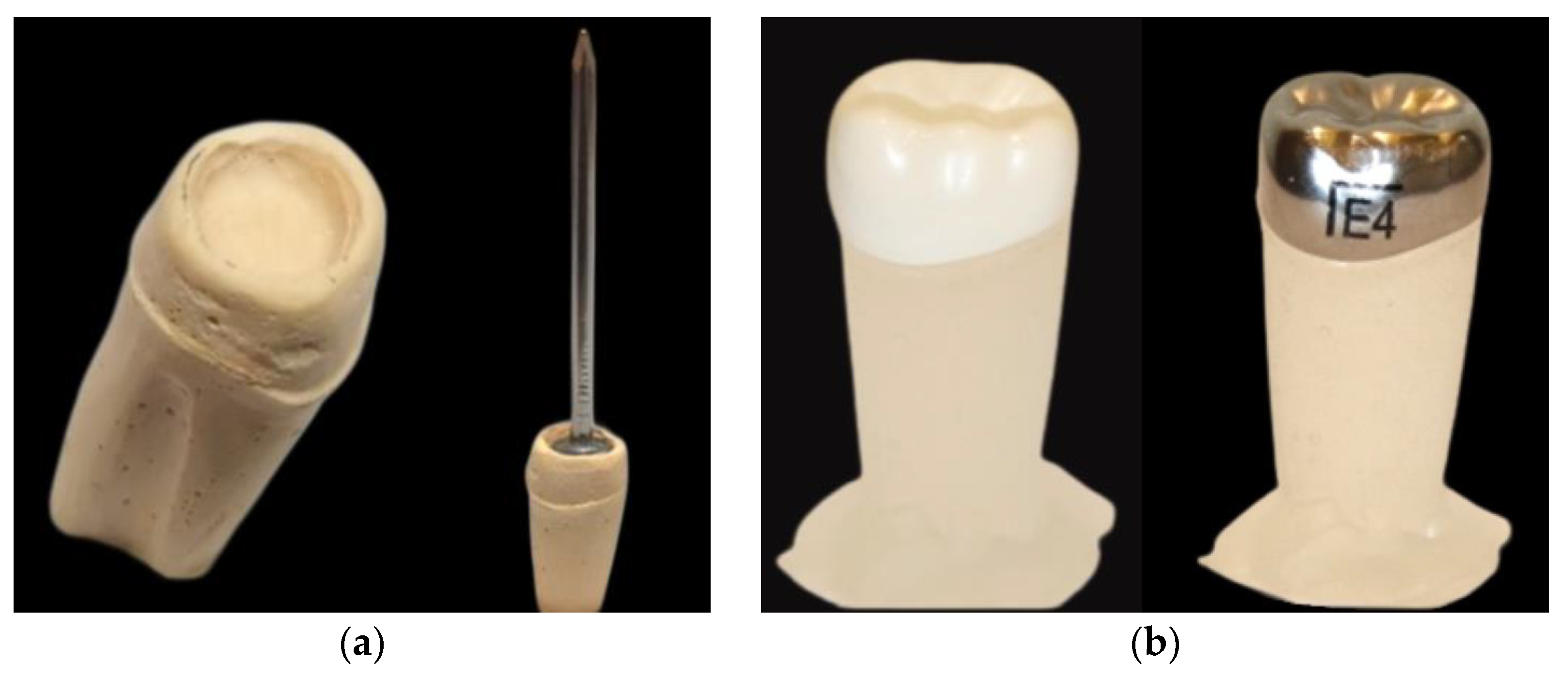
2.1. Die Preparation

A total of 160 resin dies were fabricated through the following procedure. First, the mandibular left second primary molar NuSmile® BioFlx® crown (BFC) (size 4; NuSmile Inc., Houston, TX, USA) was attached to the silicone-preformed mold, and the inner surface of the crown was separated using petroleum jelly (Vaseline) as a separating medium. Subsequently, a type IV dental die stone (GC Fujirock® EP, Leuven, Belgium) was poured into the BFC and allowed to set. Once the dental stone was set, the crown was removed from the stone die, and excess material was removed. Afterward, the stone die’s occlusal surface was deepened by carving to facilitate the placement of the nail head (Figure 1a). A silicone mold was made from this stone die, which is a negative replica of the crown, using Deguform® Plus (Dentsply International, Hanau, Germany) via the impression process. Subsequently, the resin dies were created from this silicone mold using Interacrylic Ortho Resin (Interacrylic Ortho, Interdent, Celje, Slovenia) and allowed to set for 24 h. Following that, the BFCs were tried on the resin dies to ensure a proper fit, and any excess material was removed with a finishing composite bur. The same resin dies of BFCs were utilized for the mandibular left second primary molars 3M™ ESPE SSCs (size 4; 3M™ ESPE, St. Paul, MN, USA), as the SSC was fitted precisely into the die (Figure 1b).

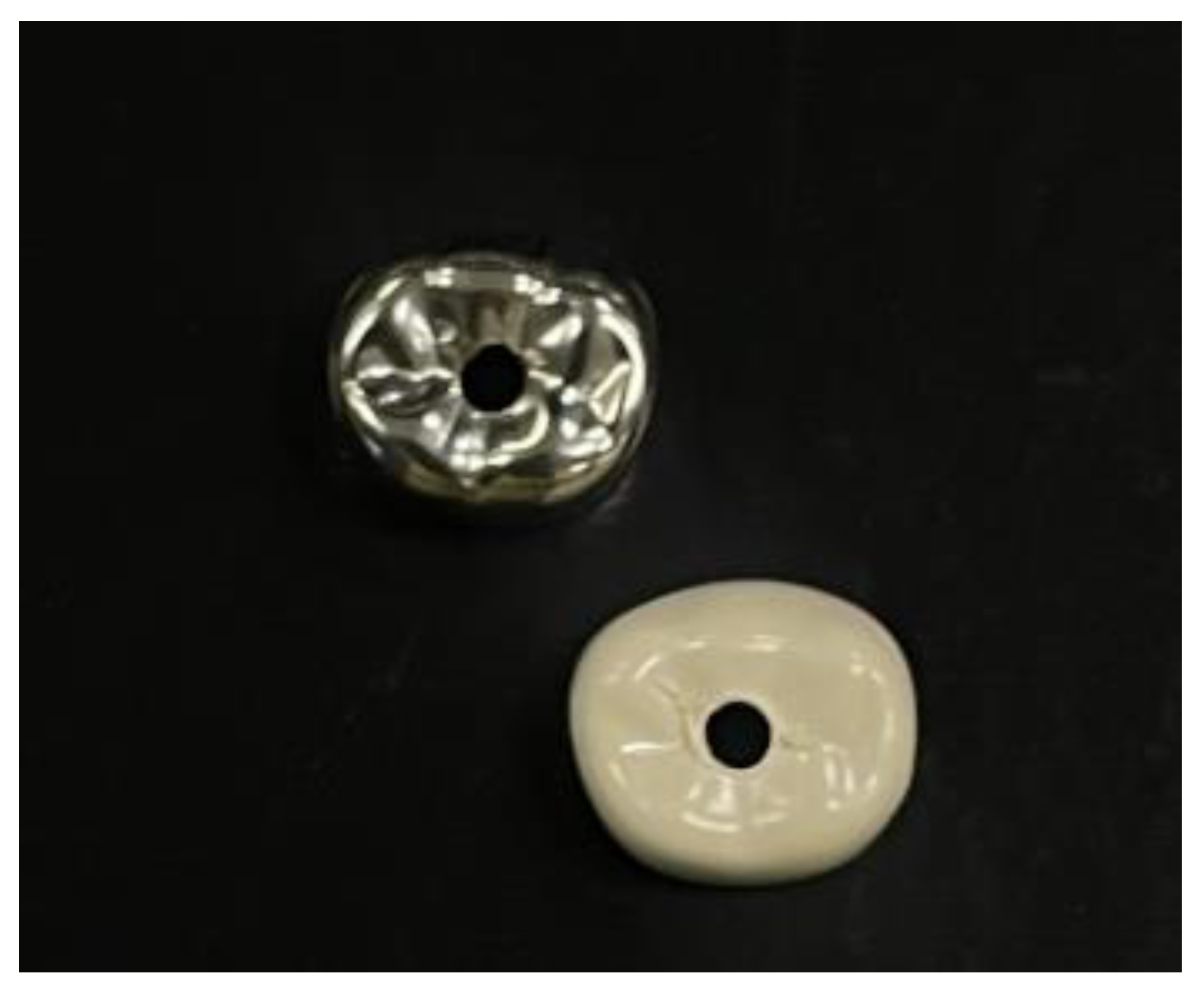
2.2. Crown Preparation

For all the crowns (BFCs and SSCs), a diamond round bur (FD-DS0447; Diaswiss SA, Nyon, Switzerland) with a head diameter of 1 mm was used to drill the central fossa (Figure 2). A nail with the following dimensions was inserted through the crown’s aperture: 25 mm in height, 1 mm in shank diameter, and 2.5 mm in flattened head diameter. This procedure enabled easy attachment of the specimens to the test machine.

2.3. Cementation

The standardized resin dies of each type of crown (80/group) were randomly allocated to one of the four experimental groups according to the type of luting cement used (20/group): Group I, GIC (3M™ Ketac™ Cem Aplicap™ 3M ESPE, Seefeld, Germany); Group II, RMGIC (RelyX Luting 2; 3M ESPE, St. Paul, MN, USA); Group III, SARC (RelyX™ U200; 3M ESPE, Seefeld, Germany); and Group IV, PXC (Poly-F®; Dentsply, Konstanz, Germany) (Figure S1). All the crowns were cemented according to the manufacturer’s instructions by a single trained pediatric dentist, who was trained during a pilot study to minimize variability. The luting cement was loaded onto two-thirds of the inner surface of each crown. Subsequently, the crown was correctly placed on the resin die using finger pressure, and an axial weight of 5 kg was applied to the crowns using a loading apparatus for 10 min to maintain the stability of the crowns during the cement setting process [33] (Figure S2a,b). Excess cement was removed from the cervical margin of the crown using a sharp-spoon excavator. The same protocol was applied to all the specimens. The crowns cemented with RMGIC and SARC were light-cured for 20 s on each side using a single-wave light-cure unit with a light intensity of 1200 mW/cm2 (Elipar S10, 3M™ ESPE™, St. Paul, MN, USA). Following that, all the specimens were immersed in distilled water and placed in an incubator (GI2 So-Low, Cincinnati, OH, USA) at 37 °C and 100% humidity for 24 h to ensure a complete set of the cement [23].

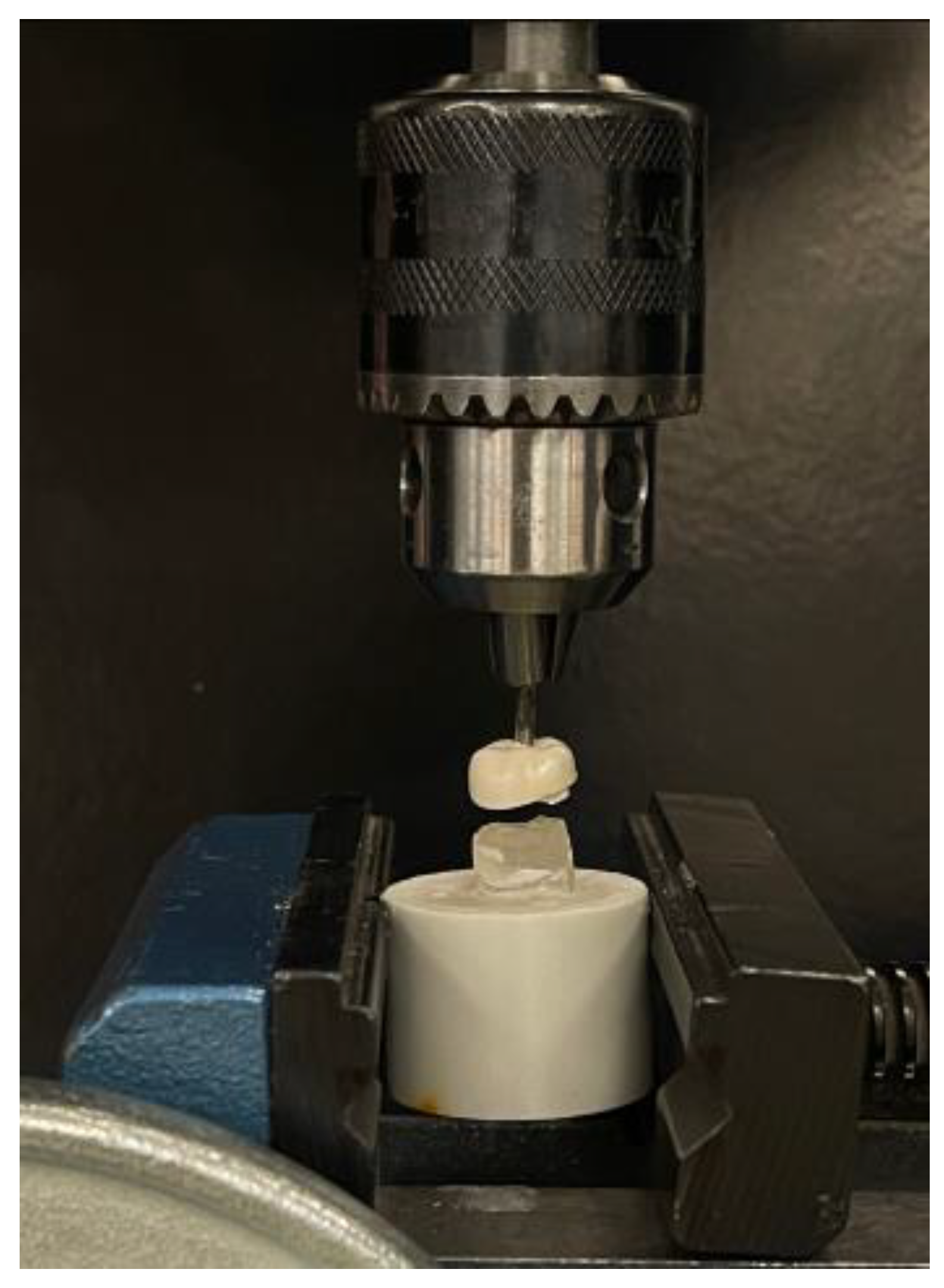
2.4. Retentive Strength Measurement

The retention strengths of the crowns were measured using a universal testing machine (Instron, model no. 8500; Illinois Tool Works, Inc., Norwood, MA, USA). The specimen was stabilized on the machine by directly engaging it with the lower crosshead, and the nail was positioned between the two surfaces of the upper crosshead. In the pullout test, the load was applied parallel to the long axis of the crown at a speed of 0.5 mm/min [34]. Load application was gradually increased from zero until the cemented crown was completely detached from the resin die; the retentive force was recorded in Newtons (N) (Figure 3). All the specimens were processed in the same manner.

2.5. Assessment of Cement Failure Pattern

Following the completion of the pullout test, specimens were visually assessed for cement failure patterns (CFPs) (Figure S2c) and scored according to Jing et al. (2019) (Table 1) [35].

2.6. Sample Size Calculation and Statistical Analysis
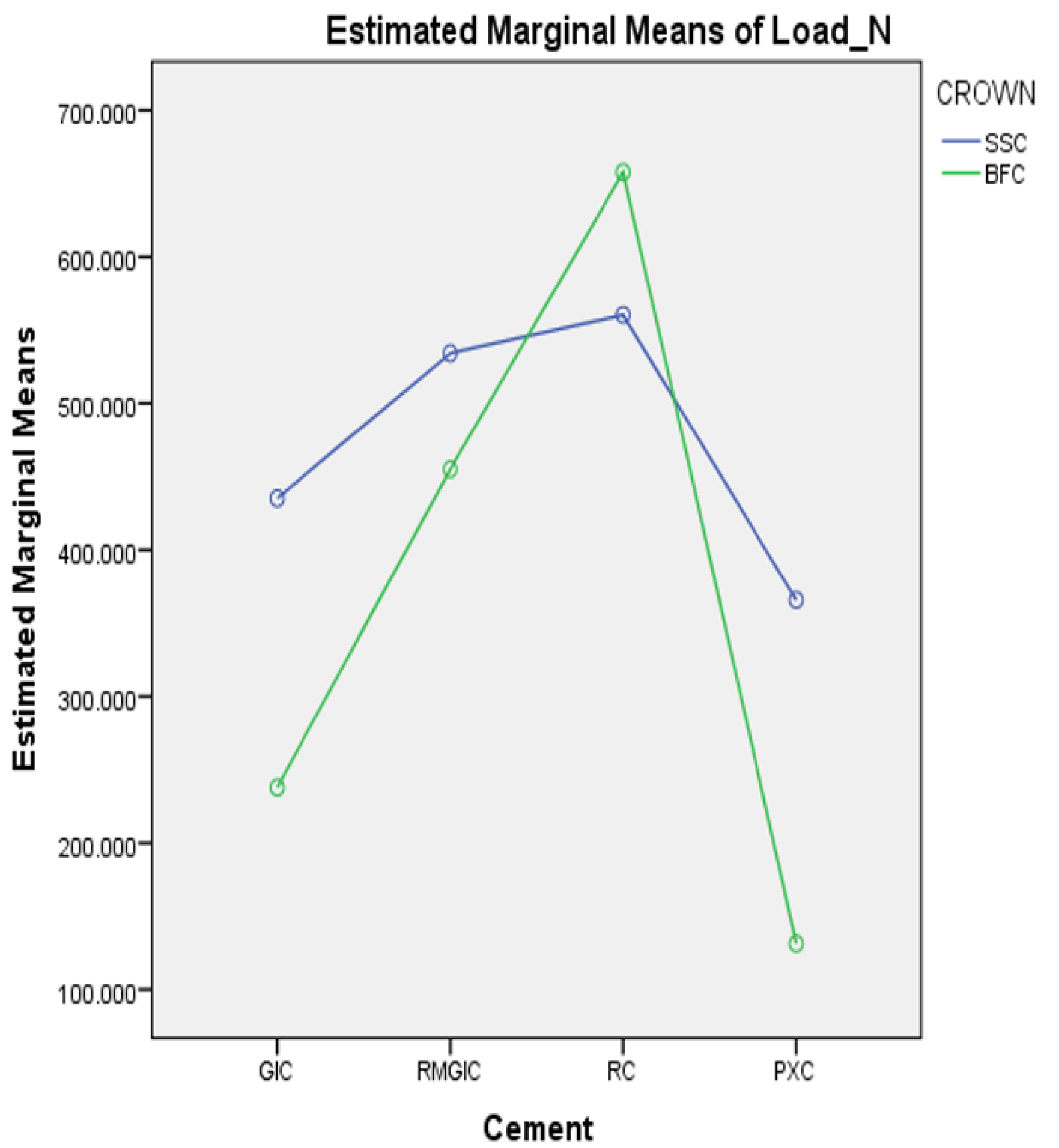
The G*Power 3.1.9.4 software (Heinrich-Heine-Universität Düsseldorf, Düsseldorf, Germany) was used to determine the required sample size; a minimum of 160 crowns were required (80/group) based on an effect size of 0.38 and a power of 0.9 at α = 0.05. Data were analyzed using the SPSS software version 24 (IBM Inc., Chicago, IL, USA). Two-way analysis of variance (ANOVA) was used to evaluate the interaction between the cement and the type of crown on the retentive strength, followed by an independent t-test. Furthermore, a one-way ANOVA was used to assess the impact of various types of luting cement on the retentive strength of each crown. First, the normality of the data distribution was verified using the Shapiro–Wilk test; no significant evidence of non-normality was found (p > 0.05). However, Levene’s test resulted in significant evidence that the assumption of homogeneity of variances was violated (p < 0.00). Thus, Welch’s ANOVA and Dunnett’s post hoc tests were employed to evaluate the cement within each crown group. Statistical significance was set at p < 0.05. The CFP was assessed by comparing the scores using descriptive statistics.

3. Results

Two-way ANOVA demonstrated a significant interaction between the types of luting cement and crowns regarding the retentive strength (p < 0.05) (Figure 4).
The SSCs exhibited significantly greater retention strength than BFCs across all cements, except in the SARC group, where BFCs demonstrated superior strength (p < 0.05) (Table 2; Figure 5).
Table 3 lists the effects of various luting cements on the retentive strength of each crown. A significant difference was observed in the retentive strength between all types of luting cements for the same type of crown (p < 0.05). The SARC exhibited the maximum retentive strength (560.29 ± 8.74 N; 657.72 ± 20.60 N), followed by RMGIC (534.20 ± 22.84 N; 454.90 ± 7.95 N), GIC (435.14 ± 8.66 N; 237.68 ± 9.37 N), and PXC (365.67 ± 19.11 N; 131.26 ± 5.37 N), for both SSCs and BFCs, respectively.
The CFP scores, including counts and percentages, are presented in Table 4. In the SSC group, a score of 1 was predominant in the SARC, RMGIC, and GIC groups (90%, 85%, and 65%, respectively), while a score of 4 was most common in the PXC group (75%). Conversely, the BFC group exhibited a more frequent score of 4 in the SARC and RMGIC groups (90% and 65%, respectively), whereas scores of 3 and 2 were more prevalent in the GIC and PXC groups (80% and 80%, respectively).

4. Discussion

The crown’s retention is a crucial element in assessing crown efficacy in primary teeth. It is achieved through both mechanical and chemical means. An appropriate size of crowns, contouring, and crimping provides mechanical retention, whereas chemical retention is achieved using a luting cement that seals the gap between the crown and abutment surfaces and maintains the crown until the time of exfoliation [16,33,36,37,38,39]. The choice of luting cement directly impacts the retention and longevity of preformed crowns. Various types of luting cements are utilized for the cementation of crowns. Over the past century, ZPCs have been commonly utilized for crown cementation. However, they lack chemical adhesion to tooth structure and depend entirely on mechanical retention, which leads to reduced adhesive strength. Despite having high compressive strength, they can be potentially harmful to vital pulp tissue because of their low pH [23]. Therefore, PXCs have been introduced as the cement that chemically adheres to enamel and dentin. They exhibit a greater adhesive strength compared to ZPCs and are compatible with vital tissue. However, they undergo significant plastic deformation when subjected to masticatory forces, and their high viscosity and low resistance to degradation in the oral cavity limit their application [23,40]. For that reason, GICs were introduced, and it was found that this type of cement can adhere to enamel and metal surfaces and is extensively preferred due to its ability to release fluoride, which assists in the prevention of recurrent caries [41]. Nevertheless, GICs possess certain limitations, such as an extended maturation period and susceptibility to moisture during the initial setting phase [42]. To overcome these shortcomings, RMGICs have been developed, effectively integrating the advantageous properties of traditional GICs, including fluoride release and adhesion, with improved physical properties, thus minimizing the likelihood of cohesive failure [43]. Recently, resin cements have become increasingly popular, typically using an etch-and-rinse or a self-etch adhesive system along with low-viscosity, dual-polymerizing resin cement. Nevertheless, the multi-step bonding process associated with these systems can be intricate, necessitating meticulous technique and considerable time. To simplify the complexities inherent to this process, a novel generation of self-adhesive resin cement has been introduced. These advanced cements eliminate the requirement for separate etching, priming, and bonding phases by integrating acidic monomers. The multifunctional phosphate-based acidic methacrylates present in these formulations are capable of reacting with the basic fillers within the luting cement, as well as with the hydroxyapatite found in hard dental tissue [44]. Recently, BFCs have been introduced as tooth-colored full-coverage restorations. However, there are limited data regarding the retentive strength of these crowns with various types of luting cement. Therefore, it is essential to assess the performance of common types of luting cement used in clinical settings with these new crowns and to compare them to the gold standard (SSCs).
Based on the results obtained in this study, the first and second null hypotheses were rejected, as there is a significant interaction between the types of luting cement and the retention strength of crowns. The SSCs were significantly superior to BFCs across all types of luting cement, except for SARC, where BFCs demonstrated superiority. These findings align with those of a randomized controlled study conducted by Agrawal et al. (2022), which demonstrated that 86.67% of PZCs and 100% of SSCs that were cemented with type I GIC were retained at 3 months following the restoration of primary molars [10]. Moreover, this corresponds with another clinical study conducted by Taran et al. (2018) that assessed the clinical success and periodontal health of primary molars restored with SSCs or PZCs cemented with GIC after 12 months. In their study, 2 out of 15 PZCs were lost within one month of placement, whereas all SSCs were retained for 12 months [45]. Kayal et al. (2023) conducted an ex-vivo study comparing the retention strength of SSCs and two types of PZCs cemented using GIC and found that the retention strength of SSCs was considerably greater than that of PZCs; however, no significant difference was observed between the two types of PZCs [46]. In addition, these results are consistent with those reported by Mandyala et al. (2024), who compared fiberglass (Figaro) crowns with SSCs using GIC and RMGIC and found that SSCs exhibited greater retention strength [47]. These findings are comparable to those of the present study, which demonstrated that SSCs exhibited superior retention to BFCs. This could be attributed to the snug fit of the SSC margin to the tooth surfaces, particularly in the undercut areas of the prepared tooth [41], as SSCs exhibit superior marginal sealing to BFCs [17] and other esthetic crowns [48,49] (Table S2).
Furthermore, the third null hypothesis was rejected, as the findings of the current study demonstrated a significant difference in the retentive strength among the different types of luting cement used with each crown. SARC exhibited the highest retentive strength, followed by RMGIC and GIC, whereas PXC demonstrated the lowest values for both crowns. This finding is consistent with those reported in previous in vitro studies, demonstrating the superiority of SARC to RMGIC [31,42,50] and GIC [18,27,42,50] in retaining SSCs on extracted teeth. The SARC exhibits superior properties, including enhanced strength, low film thickness, and extremely low solubility in oral settings [50]. It also contributes to both micromechanical retention and chemical interactions between the acidic phosphate groups of the monomer and the dentin/enamel (abutment surfaces) [51]. Consequently, these findings elucidate the reason for the superior retentive strength of SARC, which is attributable to the formation of polymer–polymer-linked connections between the cement and BFCs, which consist of hybrid resin polymer materials. The hybrid matrix reacts with the monomeric acidic groups to chemically and mechanically interlock the crown and cement. In the present study, RelyX™ Luting 2 and RelyX™ U200 were used for cementation. RelyX™ Luting 2 is a self-cure RMGIC; however, the manufacturer recommends optional light-curing to accelerate the setting process. RelyX™ U200, a dual-cure SARC, was selected for its ability to simplify the procedure by eliminating the need for substrate pretreatment. Both cements are suggested for the permanent cementation of metal and ceramic crowns. In this study, specimens were allowed to set for 10 min under axial force application to ensure proper adaptation. Traditional polymerization protocols were adhered to according to the manufacturer’s instructions to achieve complete cement polymerization and optimize the bonding performance.
Moreover, the better performance of RMGIC in comparison to GIC obtained in this study is consistent with the findings of other comparative in vitro studies that evaluated the retention of SSCs on primary molars using GIC and RMGIC [18,50,52]. Similarly, Walia et al. (2021) evaluated the retention of four different types of PZCs cemented on composite dies using GIC and two distinct RMGIC types and found that RMGICs considerably outperformed GIC in terms of crown retention [32]. Mandyala et al. (2024) reported that the retention of fiberglass (Figaro) crowns cemented on extracted primary molars with RMGIC was higher than that with GIC [47]. The RMGIC showed superior mechanical and physical properties than conventional GIC, including a lower modulus of elasticity and higher cohesive strength [53]. Additionally, the bond strength of RMGIC to tooth [54] and resin composites [55,56] is superior to that of conventional GIC. Furthermore, the hybrid resin polymer of the crown and the HEMA polymers in the cement may contribute to the mechanical interlocking between the RMGIC and BFC surfaces.
Additionally, the study findings align with those of other in vitro studies that evaluated and compared the retentive strength of SSCs cemented to natural teeth using PXC in comparison to GIC [23,27,30,50], RMGIC [50], and SARC [27,50]; SSCs cemented with PXC demonstrated markedly lower retention than the other cements. Generally, PXC adheres mainly to enamel; however, weaker adhesion to dentin occurs owing to the chelation reaction between the carboxyl groups and calcium in the tooth structure [57]. Conversely, GIC formed stronger bonds with the tooth structure and stainless steel than PXC [30]. Further, PXC possesses significantly higher plastic deformation, making it unsuitable for use in regions areas with high masticatory stress [23,40]. Therefore, the low retentive strength of PXC in this study can be explained by its fragility resulting from its thin-film thickness, chemical bonding, and plastic deformation.
However, in contrast to this sequence of luting cement performance, several laboratory and clinical studies have demonstrated that PZCs cemented with GIC display significantly higher retention than those cemented with RMGIC [34,58,59] and SARC [34]. This may be due to the subgingival tooth preparation for PZCs, which frequently leads to gingival hemorrhage and insufficient moisture control. Additionally, several in vitro studies have demonstrated that SSCs cemented with GIC exhibit greater retention than those cemented with RMGIC [42,47,60]. These differences may be attributed to the use of different techniques and various cement brands or noncompliance with manufacturing instructions.
As a part of this study, the CFP was assessed after crown detachment for four cement types. Based on the scores, cement failure can occur at the cement–crown interface (CFP score 1; adhesive failure), where the highest amount of cement remains on the die surface; at the die–cement interface (CFP score 4; adhesive failure), where the highest amount of residual cement is present on the internal surface of the crown; or at both interfaces (CFP scores 2 and 3; cohesive failure), where less than 75% of the cement is retained in either the die or crown. Among the SSCs, adhesive failure was predominant (occurring at the cement–crown interface [CFP score 1]) in the GIC, RMGIC, and SARC groups, whereas in the PXC group, it was observed at the die–cement interface (CFP score 4). These findings are comparable to those by Noffsinger et al. (1983), who reported cement–crown interface failure (adhesive) when GIC was used to cement the SSCs on natural teeth [23]. However, this contradicts the findings of Kameli et al. (2021), who used the same types of luting cement (GIC, RMGIC, SARC, and PXC) to cement the SSCs on primary teeth and demonstrated that failures predominantly occurred at both interfaces (cohesive failure) across all cement types [50]. Kayal et al. (2023) reported cement failure at the tooth–cement interface in the SSCs cemented on primary molars using GIC [46]. Discrepancies in failure patterns may result from various factors including the abutment surface, different scoring indices, crosshead speeds, and crown detachment techniques. The failure patterns of BFCs were primarily characterized by adhesive failure (CFP score 4) in RMGIC and SARC, which is associated with the majority adhering to the crown (adhesive failure). In contrast, cohesive failure was observed in GIC and PXC (CFP scores of 3 and 2, respectively); the cement primarily adhered to both the die and the crown. These findings align with those by Walia et al. (2021), who evaluated the failure modes of GIC and RMGIC for cementing other esthetic crowns (PZCs) on composite dies. The GIC predominantly demonstrated cohesive failure, whereas RMGIC exhibited adhesive failure at the die–cement interface [32]. Furthermore, the low retention of GIC and PXC may be attributed to the spontaneous cohesive fracture of the cement, the tension generated by contraction during the setting process, and low tensile strength and fracture toughness [61].
Currently, to the best of our knowledge, only one clinical trial conducted by Patil et al. (2024) has demonstrated that 96% of SSCs and 89.35% of BFCs cemented with type I GIC are retained for 12 months after restoring the primary molars [62]. This in vitro study provides baseline information regarding the retentive strength of BFCs with four different types of luting cements. Additionally, the CFP following detachment of the crowns was examined. Further details about all the materials used in this study are available in Table S2.
Nonetheless, this study has certain limitations that must be acknowledged. The present study is a laboratory study that lacks long-term follow-up and used resin die model replicas instead of primary teeth to standardize the specimens, which would have been challenging with extracted primary teeth. Moreover, it is important to recognize that the results from this study cannot be compared to those from other investigations on natural teeth, as cement may behave differently depending on the abutment surface. Furthermore, the clinical settings, including physiological and dynamic factors, have not been accurately replicated. It would be ideal to conduct a prospective clinical study to compare the retentive strength of BFCs using a variety of luting cements and evaluate the long-term performance of these crowns under physiological and dynamic conditions, including factors such as mastication forces, wear, moisture exposure, and oral pH and temperature. In addition, saliva contamination and gingival bleeding at the time of cementation, as well as oral habits and brushing, were evaluated. All these factors may influence the clinical retentive strength of crowns.

5. Conclusions

Considering the limitations of the present in vitro study, the following conclusions were drawn:
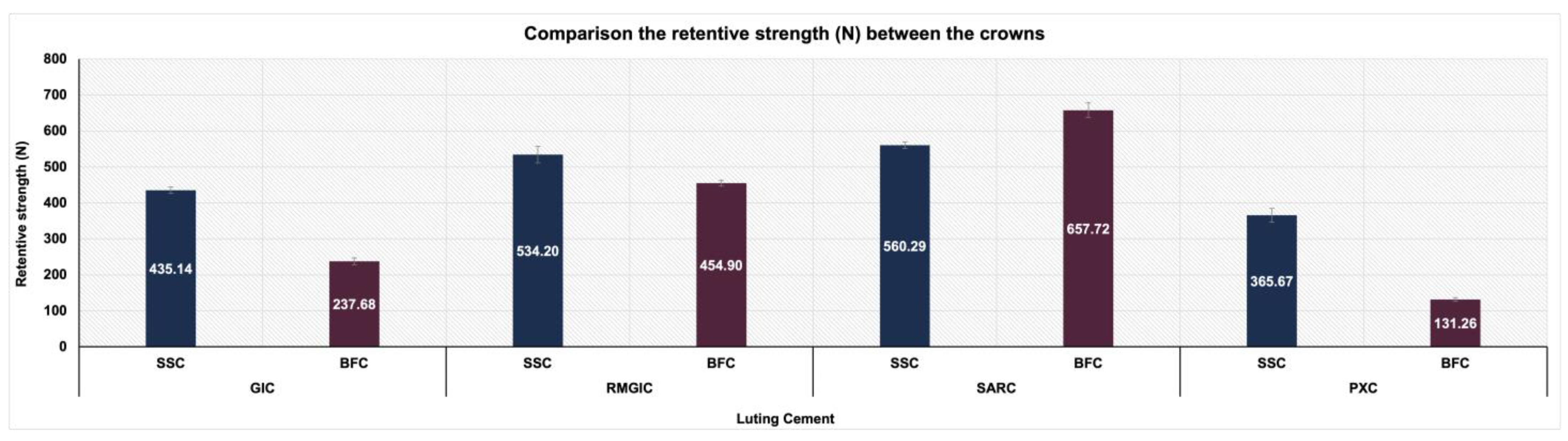
  • Across all types of cement, SSCs exhibited significantly greater retention strengths than BFCs, except for SARC, where BFCs exhibited superiority.
  • Among the four types of luting cement examined, SARC showed the highest retentive strength, whereas PXC demonstrated the lowest.
  • Adhesive failure was the predominant failure pattern in SSCs; however, both adhesive and cohesive failures were observed in BFCs.

Supplementary Materials

The following supporting information can be downloaded at https://www.mdpi.com/article/10.3390/ma18061287/s1. Table S1: Materials used in this study; Table S2: Comparison Between 3MTM stainless steel crowns and NuSmile® BioFlx® crowns; Figure S1: (a) SSC specimens’ preparation; (b) BFC specimens’ preparation; (c) assessment of CFP after pullout test; Figure S2: Experimental flow chart for the study.

Author Contributions

Conceptualization, A.A.A.M.; methodology, A.A.A.M., O.A.B. and A.S.A.; software, A.A.A.M. and O.A.B.; validation, A.M.S., L.A.A. and A.S.A.; formal analysis, A.A.A.M. and A.M.S.; investigation, A.A.A.M.; resources, O.A.B.; data curation, A.A.A.M.; writing—original draft, A.A.A.M. and O.A.B.; writing—review and editing, A.M.S., L.A.A. and A.S.A.; supervision, O.A.B. and A.S.A. All authors have read and agreed to the published version of the manuscript.

Funding

This research received no external funding.

Institutional Review Board Statement

The research project has been registered and approved by the College of Dentistry Research Center (CDRC) of King Saud University (CDRC No. PR0156, approval date: 8 January 2024).

Informed Consent Statement

Not applicable.

Data Availability Statement

The original contributions presented in this study are included in the article/Supplementary Materials. Further inquiries can be directed to the corresponding author.

Acknowledgments

The authors would like to thank the College of Dentistry Research Center at King Saud University for authorizing the use of the Physical Research Laboratory and its equipment for this research. We would also like to express our gratitude to Hassan F. Aldawsari, a laboratory technician, for his invaluable assistance in the fabrication of all dies used in this study, and Nassr S. AlMaflehi, a consultant statistician, for his help with the statistical analysis.

Conflicts of Interest

The authors declare no conflicts of interest.

References

  1. Mendes, F.M.; De Benedetto, M.S.; Zardetto, C.G.D.C.; Wanderley, M.T.; Correa, M.S.N.P. Resin Composite Restoration in Primary Anterior Teeth Using Short-Post Technique and Strip Crowns: A Case Report. Quintessence Int. 2004, 35, 689–692. [Google Scholar] [PubMed]
  2. Lucas-Rincón, S.E.; Robles-Bermeo, N.L.; Lara-Carrillo, E.; Scougall-Vilchis, R.J.; Pontigo-Loyola, A.P.; Rueda-Ibarra, V.; Loyola-Rodríguez, J.P.; Escoffié-Ramirez, M.; Medina-Solís, C.E. Interproximal Caries and Premature Tooth Loss in Primary Dentition as Risk Factors for Loss of Space in the Posterior Sector: A Cross-Sectional Study. Medicine 2019, 98, e14875. [Google Scholar] [CrossRef] [PubMed]
  3. Waggoner, W.F. Restoring Primary Anterior Teeth. Pediatr. Dent. 2002, 24, 511–516. [Google Scholar] [PubMed]
  4. American Academy of Pediatric Dentistry. Pediatric Restorative Dentistry. In The Reference Manual of Pediatric Dentistry; American Academy of Pediatric Dentistry: Chicago, IL, USA, 2024; pp. 452–465. Available online: https://www.aapd.org/globalassets/media/policies_guidelines/bp_restorativedent.pdf (accessed on 11 March 2025).
  5. Roberts, J.F.; Attari, N.; Sherriff, M. The Survival of Resin Modified Glass Ionomer and Stainless Steel Crown Restorations in Primary Molars, Placed in a Specialist Paediatric Dental Practice. Br. Dent. J. 2005, 198, 427–431. [Google Scholar] [CrossRef]
  6. Alzanbaqi, S.D.; Alogaiel, R.M.; Alasmari, M.A.; Al Essa, A.M.; Khogeer, L.N.; Alanazi, B.S.; Hawsah, E.S.; Shaikh, A.M.; Ibrahim, M.S. Zirconia Crowns for Primary Teeth: A Systematic Review and Meta-Analyses. Int. J. Environ. Res. Public. Health 2022, 19, 2838. [Google Scholar] [CrossRef]
  7. Abushanan, A.; Sharanesha, R.B.; Aljuaid, B.; Alfaifi, T.; Aldurayhim, A. Fracture Resistance of Primary Zirconia Crowns: An In Vitro Study. Children 2022, 9, 77. [Google Scholar] [CrossRef]
  8. Badr, S.Y.; Rayyan, M.; Elshiekh, M. Evaluation of Fracture Resistance and Color Stability of Innovative Esthetic Crowns for Primary Posterior Molars. Egypt. Dent. J. 2021, 67, 1879–1886. [Google Scholar] [CrossRef]
  9. Randall, R.C. Preformed Metal Crowns for Primary and Permanent Molar Teeth: Review of the Literature. Pediatr. Dent. 2002, 24, 489–500. [Google Scholar]
  10. Agrawal, R.; Khanduja, R.; Singhal, M.; Gupta, S.; Kaushik, M. Clinical Evaluation of Stainless Steel Crown versus Zirconia Crown in Primary Molars: An In Vivo Study. Int. J. Clin. Pediatr. Dent. 2022, 15, 15–19. [Google Scholar] [CrossRef]
  11. Pani, S.C.; Saffan, A.A.; AlHobail, S.; Bin Salem, F.; AlFuraih, A.; AlTamimi, M. Esthetic Concerns and Acceptability of Treatment Modalities in Primary Teeth: A Comparison between Children and Their Parents. Int. J. Dent. 2016, 2016, 3163904. [Google Scholar] [CrossRef]
  12. Holsinger, D.M.; Wells, M.H.; Scarbecz, M.; Donaldson, M. Clinical Evaluation and Parental Satisfaction with Pediatric Zirconia Anterior Crowns. Pediatr. Dent. 2016, 38, 192–197. [Google Scholar] [PubMed]
  13. Shah, P.V.; Lee, J.Y.; Wright, J.T. Clinical Success and Parental Satisfaction with Anterior Preveneered Primary Stainless Steel Crowns. Pediatr. Dent. 2004, 26, 391–395. [Google Scholar] [PubMed]
  14. Champagne, C.; Waggoner, W.; Ditmyer, M.; Casamassimo, P.S.; MacLean, J. Parental Satisfaction with Preveneered Stainless Steel Crowns for Primary Anterior Teeth. Pediatr. Dent. 2007, 29, 465–469. [Google Scholar]
  15. Mathew, M.G.; Roopa, K.B.; Soni, A.J.; Khan, M.M.; Kauser, A. Evaluation of Clinical Success, Parental and Child Satisfaction of Stainless Steel Crowns and Zirconia Crowns in Primary Molars. J. Family Med. Prim. Care. 2020, 9, 1418–1423. [Google Scholar] [CrossRef]
  16. Clark, L.; Wells, M.H.; Harris, E.F.; Lou, J. Comparison of Amount of Primary Tooth Reduction Required for Anterior and Posterior Zirconia and Stainless Steel Crowns. Pediatr. Dent. 2016, 38, 42–46. [Google Scholar]
  17. Abo-Elsoud, A.A.E.; Mohamady, E.M.; Fathi Abdou, N.E.S. Thermomechanical Aging Effects on Vertical Marginal Gap and Fracture Resistance: A Comparative Study of Bioflx and Traditional Pediatric Crowns. BMC Oral. Health 2024, 24, 1334. [Google Scholar] [CrossRef]
  18. Subramaniam, P.; Kondae, S.; Gupta, K.K. Retentive Strength of Luting Cements for Stainless Steel Crowns: An In Vitro Study. J. Clin. Pediatr. Dent. 2010, 34, 309–312. [Google Scholar] [CrossRef]
  19. Seale, N.S.; Randall, R. The Use of Stainless Steel Crowns: A Systematic Literature Review. Pediatr. Dent. 2015, 37, 145–160. [Google Scholar]
  20. Ram, D.; Fuks, A.B.; Eidelman, E. Long-Term Clinical Performance of Esthetic Primary Molar Crowns. Pediatr. Dent. 2003, 25, 582–584. [Google Scholar]
  21. Savide, N.L.; Caputo, A.A.; Luke, L.S. The Effect of Tooth Preparation on the Retention of Stainless Steel Crowns. ASDC J. Dent. Child. 1979, 46, 385–389. [Google Scholar]
  22. Myers, D.R.; Bell, R.A.; Barenie, J.T. The Effect of Cement Type and Tooth Preparation on the Retention of Stainless Steel Crowns. J. Pedod. 1981, 5, 275–280. [Google Scholar] [PubMed]
  23. Noffsinger, D.P.; Jedrychowski, J.R.; Caputo, A.A. Effects of Polycarboxylate and Glass Ionomer Cements on Stainless Steel Crown Retention. Pediatr. Dent. 1983, 5, 68–71. [Google Scholar]
  24. Garcia-Godoy, F. Clinical Evaluation of the Retention of Preformed Crowns Using Two Dental Cements. J. Pedod. 1984, 8, 278–281. [Google Scholar]
  25. Sener, I.; Turker, B.; Valandro, L.F.; Ozcan, M. Marginal Gap, Cement Thickness, and Microleakage of 2 Zirconia Crown Systems Luted with Glass Ionomer and MDP-Based Cements. Gen. Dent. 2014, 62, 67–70. [Google Scholar]
  26. Erdemci, Z.Y.; Cehreli, S.B.; Tirali, R.E. Hall Versus Conventional Stainless Steel Crown Techniques: In Vitro Investigation of Marginal Fit and Microleakage Using Three Different Luting Agents. Pediatr. Dent. 2014, 36, 286–290. [Google Scholar]
  27. Parisay, I.; Khazaei, Y. Evaluation of Retentive Strength of Four Luting Cements with Stainless Steel Crowns in Primary Molars: An In Vitro Study. Dent. Res. J. 2018, 15, 201–207. [Google Scholar] [CrossRef]
  28. Sahin, I.; Karayilmaz, H.; Çiftçi, Z.Z.; Kirzioglu, Z. Fracture Resistance of Prefabricated Primary Zirconium Crowns Cemented with Different Luting Cements. Pediatr. Dent. 2018, 40, 443–448. [Google Scholar]
  29. Khinda, V.I.S.; Grewal, N. Retentive [Correction of Preventive] Efficacy of Glass Ionomer, Zinc Phosphate and Zinc Polycarboxylate Luting Cements in Preformed Stainless Steel Crowns: A Comparative Clinical Study. J. Indian. Soc. Pedod. Prev. Dent. 2002, 20, 41–46. [Google Scholar] [PubMed]
  30. Reddy, M.; Reddy, V.; Basappa, N. A Comparative Study of Retentive Strengths of Zinc Phosphate, Polycarboxylate and Glass Ionomer Cements with Stainless Steel Crowns—An In Vitro Study. J. Indian. Soc. Pedod. Prev. Dent. 2010, 28, 245–250. [Google Scholar] [CrossRef]
  31. Pathak, S.; Shashibhushan, K. In Vitro Evaluation of Stainless Steel Crowns Cemented with Resin-Modified Glass Ionomer and Two New Self-Adhesive Resin Cements. Int. J. Clin. Pediatr. Dent. 2016, 9, 197–200. [Google Scholar] [CrossRef]
  32. Walia, T.; Brigi, C.; Ziadkhani, M.M.; Khayat, A.A.; Tabibzadeh, Z. Retention Force of Glass Ionomer Based Luting Cements with Posterior Primary Zirconium Crowns—A Comparative in Vitro Study. J. Clin. Pediatr. Dent. 2021, 45, 259–264. [Google Scholar] [CrossRef] [PubMed]
  33. Ali, S.N.A.H. In Vitro Comparison of Marginal and Internal Fit Between Stainless Steel Crowns and Esthetic Crowns of Primary Molars Using Different Luting Cements. Dent. Res. J. 2019, 16, 366–371. [Google Scholar] [CrossRef]
  34. Rosato, L.A.; Chen, J.W.; Roggenkamp, C.L.; Su, J.M. Retentive Strengths for Prefabricated Primary Molar Zirconia Crowns Using Five Different Cements. Taiwan J. Pediatr. Dent. 2018, 18, 127–135. [Google Scholar]
  35. Jing, L.; Chen, J.W.; Roggenkamp, C.; Suprono, M.S. Effect of Crown Preparation Height on Retention of a Prefabricated Primary Posterior Zirconia Crown. Pediatr. Dent. 2019, 41, 229–233. [Google Scholar] [PubMed]
  36. Yilmaz, Y.; Gurbuz, T.; Eyuboglu, O.; Belduz, N. The Repair of Preveneered Posterior Stainless Steel Crowns. Pediatr. Dent. 2008, 30, 429–435. [Google Scholar] [CrossRef]
  37. Innes, N.P.T.; Ricketts, D.; Chong, L.Y.; Keightley, A.J.; Lamont, T.; Santamaria, R.M. Preformed crowns for decayed primary molar teeth. Cochrane Database Syst. Rev. 2015, 12, CD005512. [Google Scholar] [CrossRef]
  38. Waggoner, W.F. Restoring Primary Anterior Teeth: Updated for 2014. Pediatr Dent. 2015, 37, 163–170. [Google Scholar]
  39. Kindelan, S.A.; Day, P.; Nichol, R.; Willmott, N.; Fayle, S.A.; British Society of Paediatric Dentistry. UK National Clinical Guidelines in Paediatric Dentistry: Stainless Steel Preformed Crowns for Primary Molars. Int. J. Paediatr. Dent. 2008, 18 (Suppl. S1), 20–28. [Google Scholar] [CrossRef]
  40. Lad, P.P.; Kamath, M.; Tarale, K.; Kusugal, P.B. Practical Clinical Considerations of Luting Cements: A Review. J. Int. Oral. Health 2014, 6, 116–120. [Google Scholar]
  41. Rector, J.A.; Mitchell, R.J.; Spedding, R.H. The Influence of Tooth Preparation and Crown Manipulation on the Mechanical Retention of Stainless Steel Crowns. ASDC J. Dent. Child. 1985, 52, 422–427. [Google Scholar]
  42. Yilmaz, Y.; Dalmis, A.; Gurbuz, T.; Simsek, S. Retentive Force and Microleakage of Stainless Steel Crowns Cemented with Three Different Luting Agents. Dent. Mater. J. 2004, 23, 577–584. [Google Scholar] [CrossRef]
  43. Rosenstiel, S.F.; Land, M.F.; Crispin, B.J. Dental Luting Agents: A Review of the Current Literature. J. Prosthet. Dent. 1998, 80, 280–301. [Google Scholar] [CrossRef] [PubMed]
  44. Hikita, K.; Van Meerbeek, B.; De Munck, J.; Ikeda, T.; Van Landuyt, K.; Maida, T.; Lambrechts, P.; Peumans, M. Bonding Effectiveness of Adhesive Luting Agents to Enamel and Dentin. Dent. Mater. 2007, 23, 71–80. [Google Scholar] [CrossRef] [PubMed]
  45. Taran, P.K.; Kaya, M.S. A Comparison of Periodontal Health in Primary Molars Restored with Prefabricated Stainless Steel and Zirconia Crowns. Pediatr. Dent. 2018, 40, 334–339. [Google Scholar] [PubMed]
  46. Kayal, S.; Kang, Y.; Finkelman, M.; Swee, G.; Loo, C. Retention of Zirconia Crowns Compared to Stainless Steel Crowns: An Ex-Vivo Study. Pediatr. Dent. 2023, 45, 142–146. [Google Scholar]
  47. Mandyala, V.; Padakandla, P.; Kola, N.; Kedari, S.; Putta, L.; Gandhasiri, K. Comparative Evaluation of Retentive Strength of Figaro Crowns and Stainless Steel Crowns Luted with Glass Ionomer Cement and Resin-Modified Glass Ionomer Cement—An In Vitro Study. J. Dr. YSR Univ. Health Sci. 2024, 13, 36–41. [Google Scholar] [CrossRef]
  48. Subramanian, E.M.G.; Kumar, A.S.; Lavanya, G. Comparative Evaluation of Marginal Leakage of SSC and Zirconia Crowns in Primary Teeth. Nveo-Nat. Volatiles Essent. Oils J. 2021, 8, 7063–7068. [Google Scholar]
  49. Elsayed, H.D.; Sherif, M.; Eldin, S.; Shaimaa Omer, M.M. Comparison Between Stainless-Steel, Zirconia, and Fiberglass Crowns as Full Coverage Restoration for Deciduous Molars of Children. Afr. J. Biol. Sci. 2024, 6, 51–59. [Google Scholar]
  50. Kameli, S.; Khani, F.; Bahraminasab, M.; Ghorbani, R.; Abbas, F.M. Bond Strength and Microleakage of Different Types of Cements in Stainless Steel Crown of Primary Molar Teeth. Dent. Res. J. 2021, 18, 58. [Google Scholar] [CrossRef]
  51. Radovic, I.; Monticelli, F.; Goracci, C.; Vulicevic, Z.R.; Ferrari, M. Self-Adhesive Resin Cements: A Literature Review. J. Adhes. Dent. 2008, 10, 251–258. [Google Scholar]
  52. Veerabadhran, M.M.; Reddy, V.; Nayak, U.A.; Rao, A.P.; Sundaram, M.A. The Effect of Retentive Groove, Sandblasting, and Cement Type on the Retentive Strength of Stainless Steel Crowns in Primary Second Molars: An In Vitro Comparative Study. J. Indian. Soc. Pedod. Prev. Dent. 2012, 30, 19–26. [Google Scholar] [CrossRef]
  53. Mathis, R.S.; Ferracane, J.L. Properties of a Glass-Ionomer/Resin-Composite Hybrid Material. Dent. Mater. 1989, 5, 355–358. [Google Scholar] [CrossRef] [PubMed]
  54. Wilson, A.D. Resin-Modified Glass-Ionomer Cements. Int. J. Prosthodont. 1990, 3, 425–429. [Google Scholar] [PubMed]
  55. Farah, C.S.; Orton, V.G.; Collard, S.M. Shear Bond Strength of Chemical and Light-Cured Glass Ionomer Cements Bonded to Resin Composites. Aust. Dent. J. 1998, 43, 81–86. [Google Scholar] [CrossRef] [PubMed]
  56. Li, J.; Liu, Y.; Liu, Y.; Söremark, R.; Sundström, F. Flexure Strength of Resin-Modified Glass Ionomer Cements and Their Bond Strength to Dental Composites. Acta Odontol. Scand. 1996, 54, 55–58. [Google Scholar] [CrossRef]
  57. Ladha, K.; Verma, M. Conventional and Contemporary Luting Cements: An Overview. J. Indian. Prosthodont. Soc. 2010, 10, 79–88. [Google Scholar] [CrossRef]
  58. Srinivasan, S.R.; Mathew, M.G.; Jayaraman, J. Comparison of Three Luting Cements for Prefabricated Zirconia Crowns in Primary Molar Teeth: A 36-month Randomized Clinical Trial. Pediatr. Dent. 2023, 45, 117–124. [Google Scholar]
  59. Azab, M.; Moheb, D.; El Shahawy, O.; Rashed, M. Influence of Luting Cement on the Clinical Outcomes of Zirconia Pediatric Crowns. A 3-Year Split-Mouth Randomized Controlled Trial. Int. J. Paediatr. Dent. 2019, 30, 314–322. [Google Scholar] [CrossRef]
  60. Yilmaz, Y.; Simsek, S.; Dalmis, A.; Gurbuz, T.; Kocogullari, M.E. Evaluation of Stainless Steel Crowns Cemented with Glass-Ionomer and Resin-Modified Glass-Ionomer Luting Cements. Am. J. Dent. 2006, 19, 106–110. [Google Scholar]
  61. Mitchell, C.A.; Douglas, W.H.; Cheng, Y.S. Fracture Toughness of Conventional, Resin-Modified Glass-Ionomer and Composite Luting Cements. Dent. Mater. 1999, 15, 7–13. [Google Scholar] [CrossRef]
  62. Patil, A.S.; Jain, M.; Choubey, S.; Patil, M.; Chunawala, Y. Comparative evaluation of clinical success of Stainless Steel and Bioflx crowns in primary molar—A 12 month split mouth prospective randomized clinical trial. J. Indian. Soc. Pedod. Prev. Dent. 2024, 42, 37–45. [Google Scholar] [CrossRef]
Figure 1. (a) A stone die was deepened by carving the occlusal surface to accommodate the nail head. (b) Crowns were tried on resin dies to ensure a proper fit.
Materials 18 01287 g001
Figure 2. Preparation of a hole in the central fossa of the crowns.
Materials 18 01287 g002
Figure 3. BioFlx crown during pullout test.
Materials 18 01287 g003
Figure 4. Interaction between the types of luting cement and crown materials in relation to retentive strength.
Materials 18 01287 g004
Figure 5. Comparison of the retentive strength (N) between SSCs and BFCs within each cement.
Materials 18 01287 g005
Table 1. Categorization of cement failure pattern.
ScoreDefinition
1More than 75% of cement left on the resin die.
2Between 50 and 75% of cement left on the resin die.
3Between 25 and 50% of cement left on the resin die.
4More than 75% of cement left on the crown.
Table 2. Comparison of the retentive strength (N) between SSCs and BFCs within each cement.
GICRMGICSARCPXC
SSC435.14 ± 8.66534.20 ± 22.84560.29 ± 8.74365.67 ± 19.11
BFC237.68 ± 9.37454.90 ± 7.95657.72 ± 20.60131.26 ± 5.37
p-Value *<0.001<0.001<0.001<0.001
Abbreviations used in this table: SSC = stainless steel crown; BFC = BioFlx crown; GIC = glass ionomer cement; RMGIC = resin-modified glass ionomer cement; SARC = self-adhesive resin cement; PXC = zinc polycarboxylate cement. * An-independent t-test showed statistically significant differences between the groups (p-value < 0.05).
Table 3. Distribution and comparison of mean retentive strength values (N) across all groups.
CrownCementnRetentive Strength (N)95% Confidence Interval for Meanp-Value *
LowerUpper
SSCGIC20435.14 ± 8.66 b431.08439.19<0.001
RMGIC20534.20 ± 22.84 c523.52544.89
SARC20560.29 ± 8.74 d556.20564.38
PXC20365.67 ± 19.11 a356.73374.62
BFCGIC20237.68 ± 9.37 b233.30242.07<0.001
RMGIC20454.90 ± 7.95 c451.18458.62
SARC20657.72 ± 20.60 d648.07667.36
PXC20131.26 ± 5.37 a128.75133.78
Abbreviations used in this table: SSC = stainless steel crown; BFC = BioFlx crown; GIC = glass ionomer cement; RMGIC = resin-modified glass ionomer cement; SARC = self-adhesive resin cement; PXC = zinc polycarboxylate cement. * Welch’s analysis of variance test showed statistically significant differences between groups (p-value < 0.05). The Dunnett T3 test showed statistically significant differences between groups with similar small letters (a < b < c < d).
Table 4. The CFPs among different types of luting cement groups within each crown.
CrownCementFrequencyCFP Score
1234
SSCGICn13700
%65%35%0%0%
RMGICn17300
%85%15%0%0%
SARCn18200
%90%10%0%0%
PXCn00515
%0%0%25%75%
BFCGICn00164
%0%0%80%20%
RMGICn00713
%0%0%35%65%
SARCn00218
%0%0%10%90%
PXCn41600
%20%80%0%0%
Abbreviations used in this table: SSC = stainless steel crown; BFC = BioFlx crown; CFP = cement failure pattern; GIC = glass ionomer cement; RMGIC = resin-modified glass ionomer cement; SARC = self-adhesive resin cement; PXC = zinc polycarboxylate cement.
Disclaimer/Publisher’s Note: The statements, opinions and data contained in all publications are solely those of the individual author(s) and contributor(s) and not of MDPI and/or the editor(s). MDPI and/or the editor(s) disclaim responsibility for any injury to people or property resulting from any ideas, methods, instructions or products referred to in the content.

