Article

In Vivo Evaluation of Laser-Textured Air Plasma in Osseointegration of Dental Implants

by
Larissa Azeredo da Silva Lessa Nicolau
1,
Suelen Cristina Sartoretto
2,3,
Pamella Santana Nunes
1,
Ezio Gheno
4,
Jose Mauro Granjeiro
2,5,
Domenico D’Angelo
6,
Federico Mussano
7,
Monica Diuana Calasans-Maia
2,*,
Olivio Della Bella
8,
Francesca Motta
8 and
Rafael Seabra Louro
3
1
Post-Graduation Program in Dentistry, Fluminense Federal University, Niteroi 24020-140, Brazil
2
Clinical Research Laboratory, Dentistry School, Universidade Federal Fluminense, Niteroi 24220-140, Brazil
3
Oral Surgery Department, Fluminense Federal University, Niteroi 24220-140, Brazil
4
Department of Surgical Sciences and Integrated Diagnostics, University of Genova, 16132 Genova, Italy
5
National Institute of Metrology, Quality and Technology (INMETRO), Duque de Caxias 25250-020, Brazil
6
Plasma Nano-Tech Lab, Environment Park S.p.A., 10144 Turin, Italy
7
Bone and Dental Bioengineering Laboratory, CIR Dental School, Department of Surgical Sciences, University of Turin, 10126 Turin, Italy
8
Biomec Srl, 23823 Colico, Italy
*
Author to whom correspondence should be addressed.
Materials 2025, 18(16), 3810; https://doi.org/10.3390/ma18163810
Submission received: 21 May 2025 / Revised: 16 July 2025 / Accepted: 31 July 2025 / Published: 14 August 2025
(This article belongs to the Section Biomaterials)

Abstract

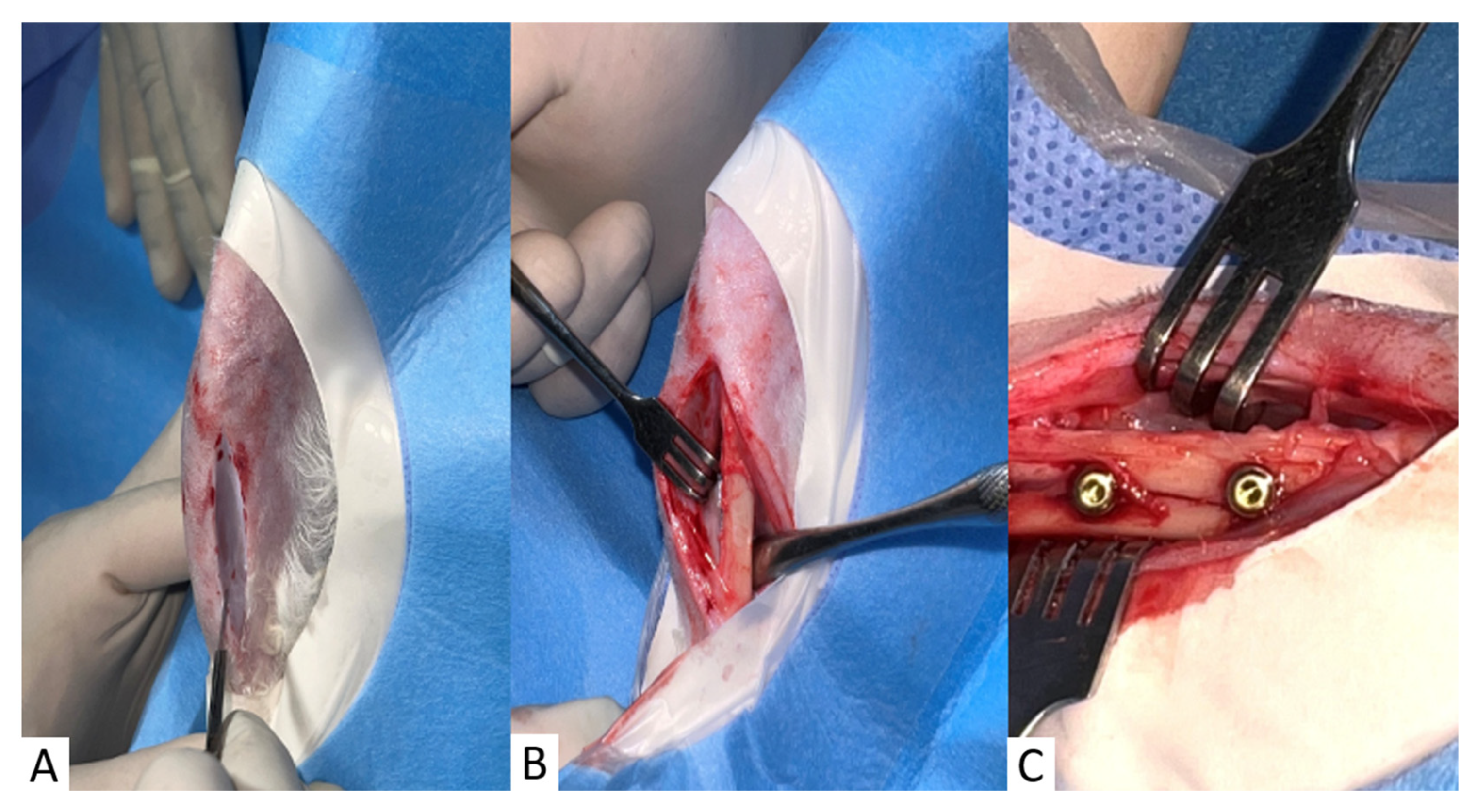
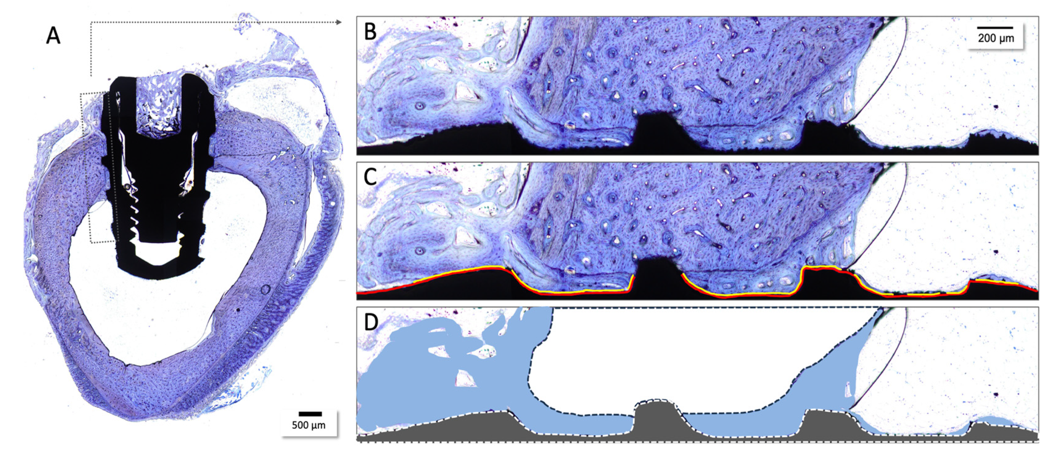
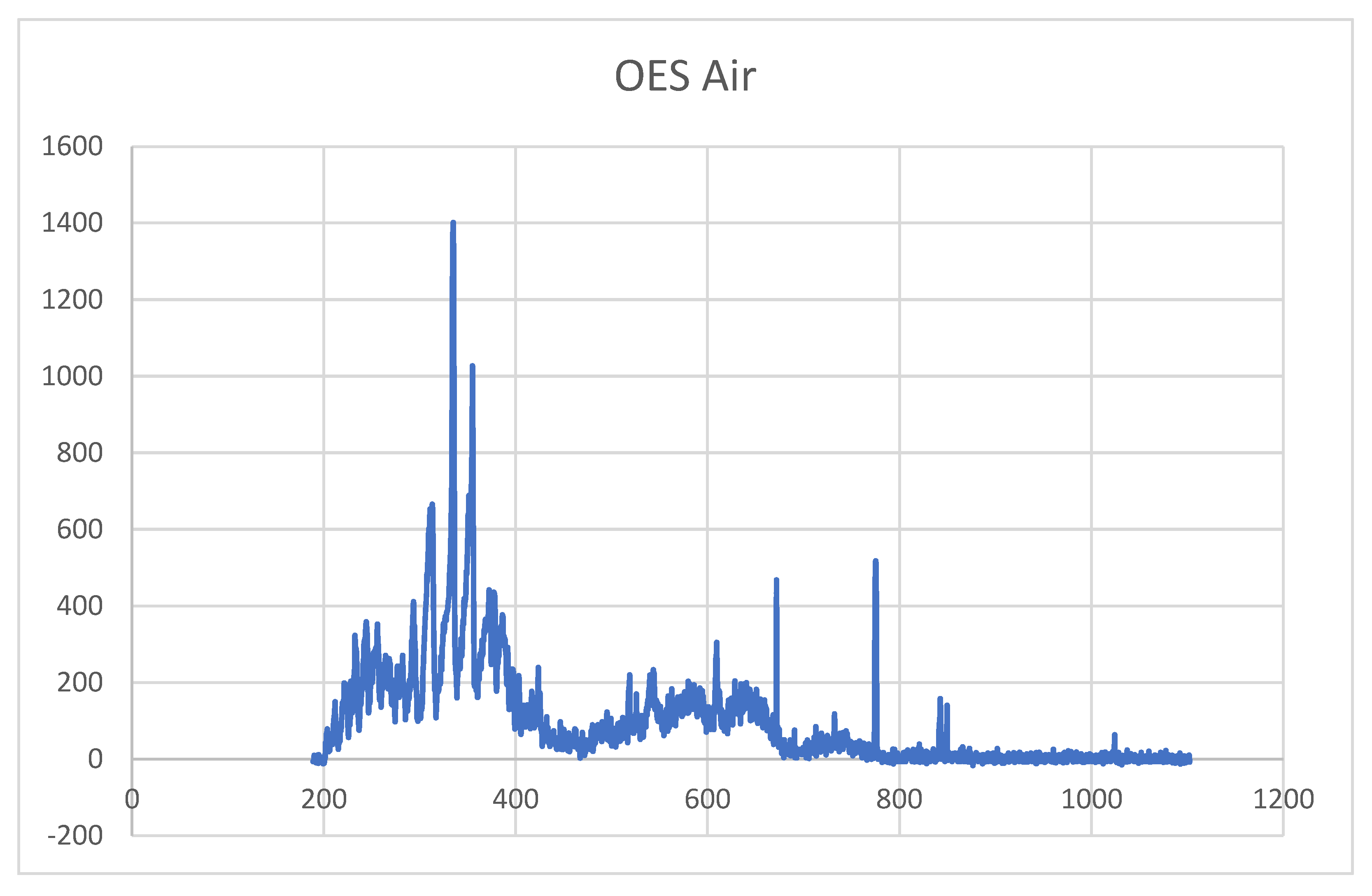
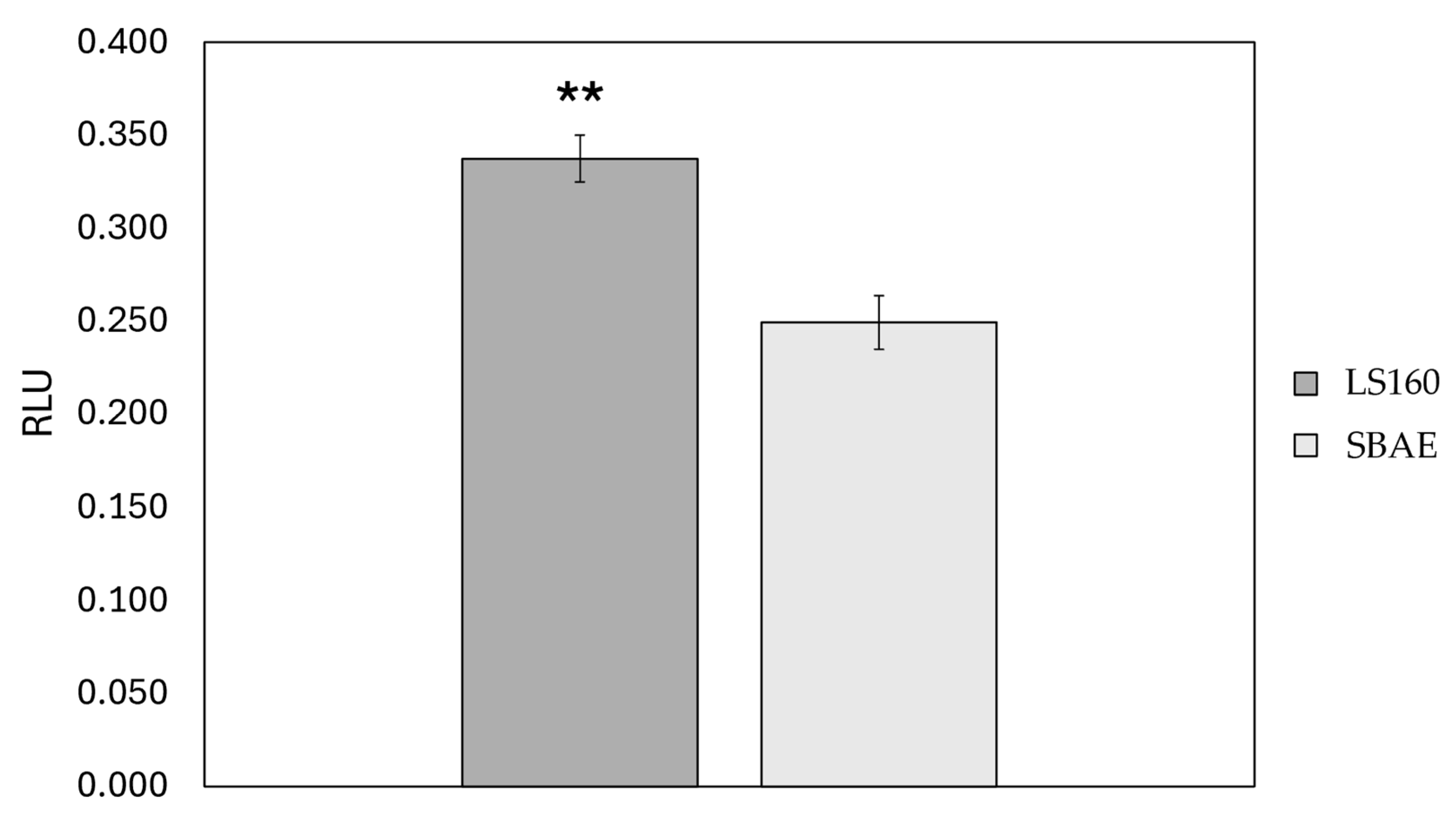
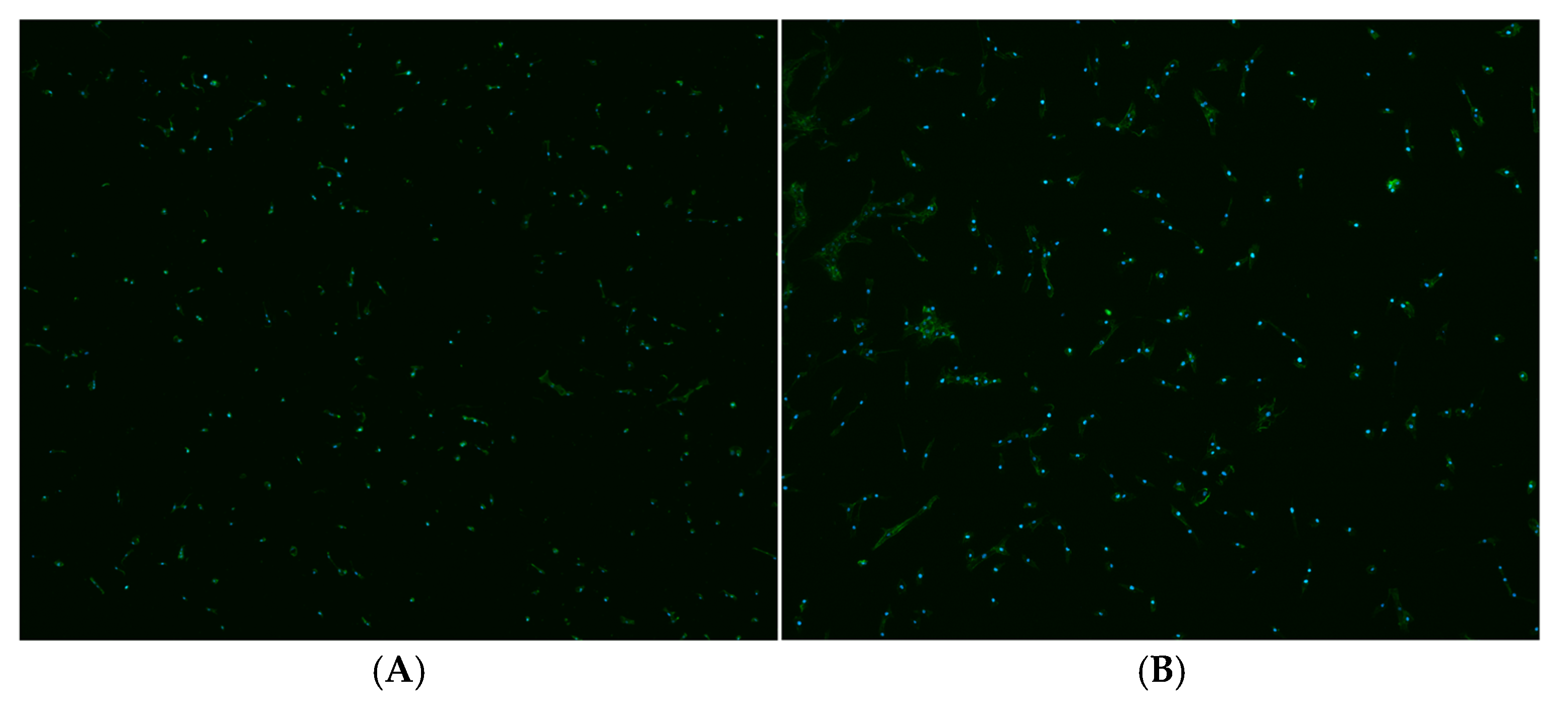
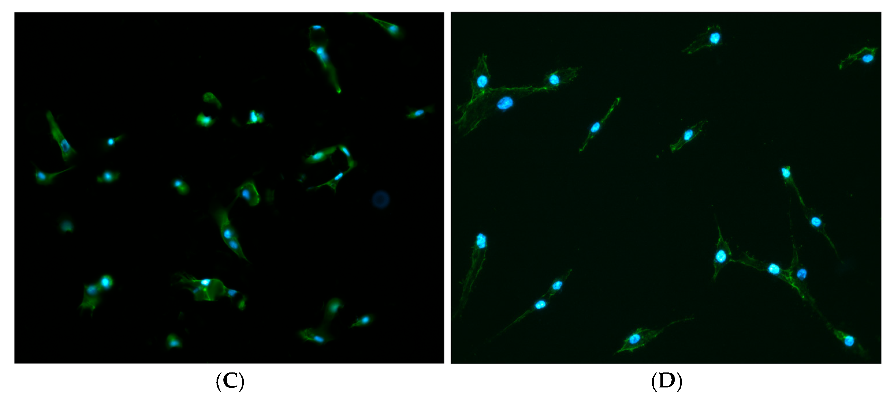
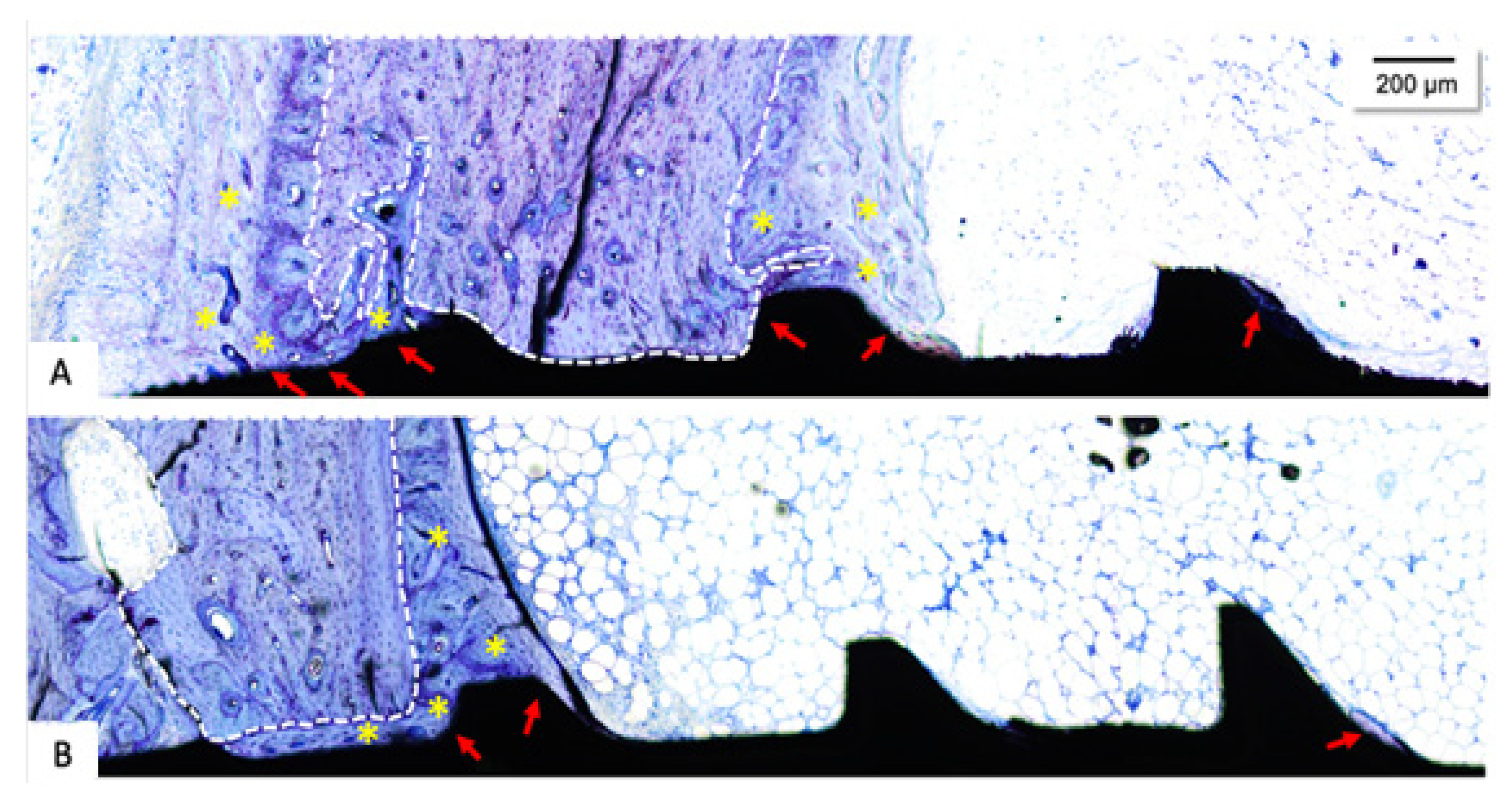
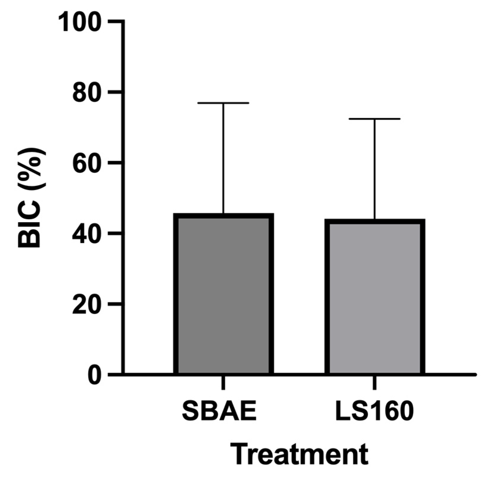
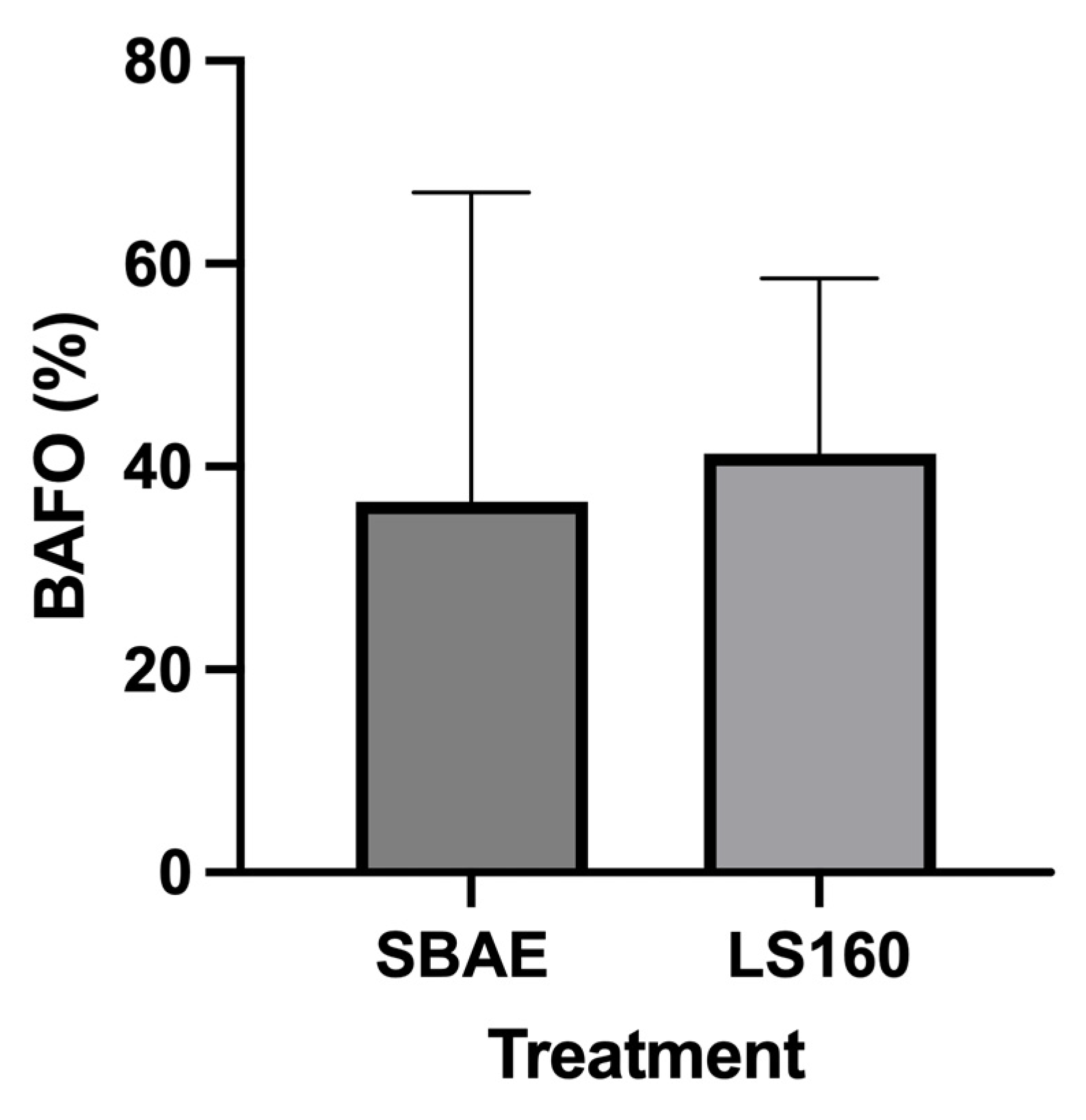
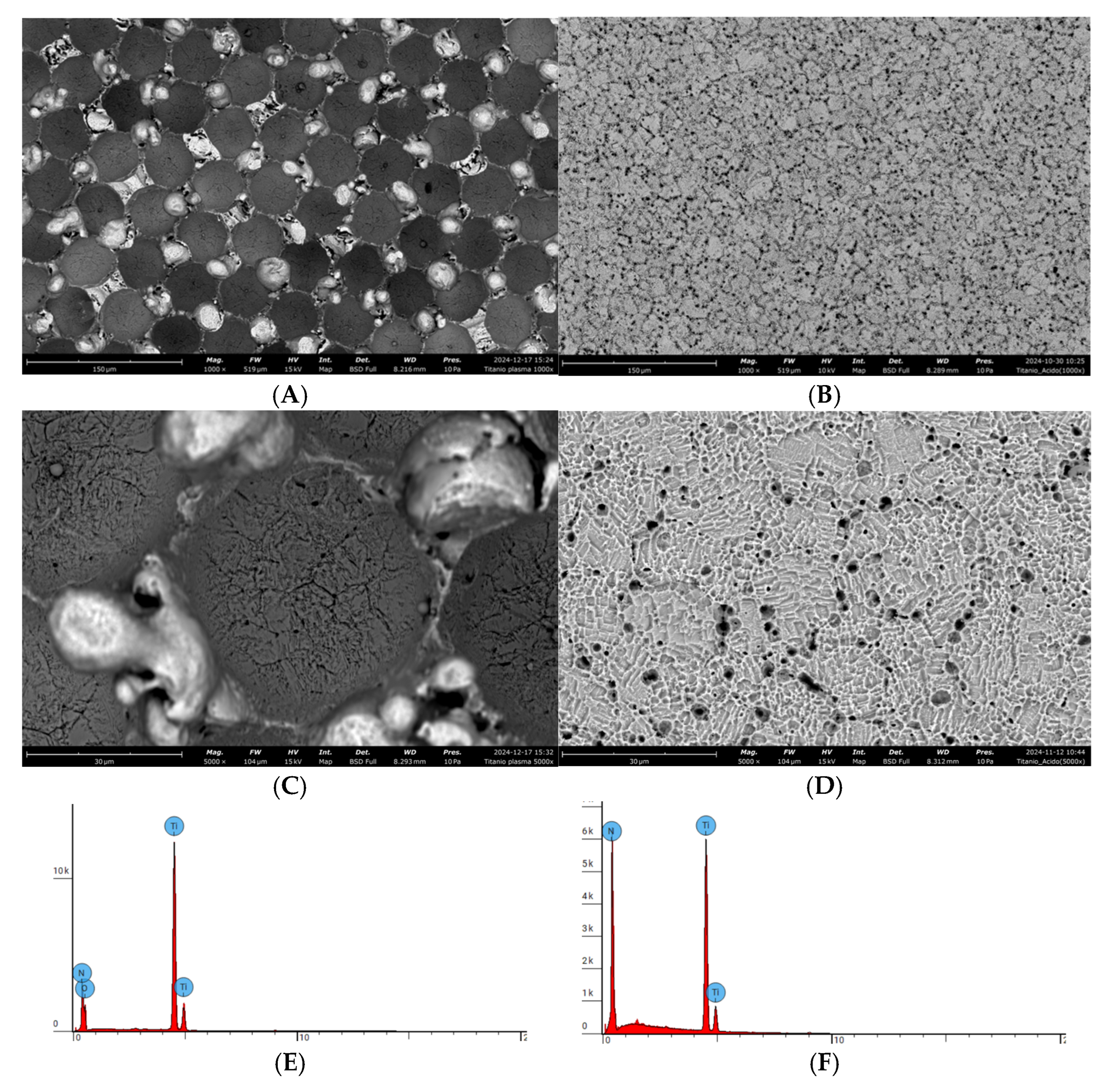
The different macro and micro geometries of dental implants are parameters that directly affect osseointegration, making them an important area for research. The objective of this preclinical study was to compare, through histological and histomorphometric analyses, the biological response of two different dental implant surfaces in osseointegration. Surface morphology and chemistry were characterized by SEM/EDX, optical-emission spectroscopy, protein adsorption (BSA), and adipose-derived stem-cell morphology. For the in vivo arm, ten commercially pure titanium implants (n = 5 LS160 + 5 SBAE) were placed bilaterally in the tibiae of five skeletally mature New Zealand rabbits (one implant of each surface per animal). After six weeks, undecalcified sections were prepared and bone-to-implant contact (BIC) and bone-area-fraction occupancy (BAFO) were quantified histomorphometrically. Data normality was confirmed with the Shapiro–Wilk test; paired two-tailed Student’s t-tests were applied (α = 0.05). Results: The descriptive histological analysis showed a fraction of pre-existing bone in all experimental groups, which probably ensured primary stability. Adjacent to this area, it was possible to observe peri-implant newformed bone in all tested groups. The results of the histomorphometric analysis of BIC and BAFO were considered normal by the Shapiro–Wilk test (p > 0.05); after six weeks of implantation, the BIC values for the LS160 and SBAE groups were 44.13 (15.83–72.43) and 39.24 (10.72–89.21), respectively. The analysis of variance (ANOVA and Tukey’s post-test) showed no statistical differences between the groups tested. Likewise, the bone volume density showed no statistical differences between the groups (ANOVA and Tukey’s post-test) with averages of 41.27 (C.I. 24.00–58.55) and 26.52 (C.I. −17.51–70.54) in the LS160 and SBAE groups, respectively. Although both surfaces showed similar osseointegration after six weeks, the new surface appears to be a promising, eco-friendly alternative to SBAE. Future studies with shorter time points and larger samples are needed to assess early biological responses.
Keywords: osseointegration; dental implants; rabbits; histomorphometric evaluation osseointegration; dental implants; rabbits; histomorphometric evaluation