Article

Enhanced Luminescence of Yb3+ Ions Implanted to ZnO through the Selection of Optimal Implantation and Annealing Conditions

1
National Centre for Nuclear Research, Soltana 7, 05-400 Otwock, Poland
2
Institute of Physics, Polish Academy of Sciences, Al. Lotnikow 32/46, 02-668 Warsaw, Poland
3
Helmholtz-Zentrum Dresden-Rossendorf, Institute of Ion Beam Physics and Materials Research, Bautzner Landstrasse 400, D-01328 Dresden, Germany
*
Author to whom correspondence should be addressed.
Materials 2023, 16(5), 1756; https://doi.org/10.3390/ma16051756
Submission received: 9 January 2023 / Revised: 10 February 2023 / Accepted: 16 February 2023 / Published: 21 February 2023
(This article belongs to the Section Optical and Photonic Materials)

Abstract

:
Rare earth-doped zinc oxide (ZnO:RE) systems are attractive for future optoelectronic devices such as phosphors, displays, and LEDs with emission in the visible spectral range, working even in a radiation-intense environment. The technology of these systems is currently under development, opening up new fields of application due to the low-cost production. Ion implantation is a very promising technique to incorporate rare-earth dopants into ZnO. However, the ballistic nature of this process makes the use of annealing essential. The selection of implantation parameters, as well as post-implantation annealing, turns out to be non-trivial because they determine the luminous efficiency of the ZnO:RE system. This paper presents a comprehensive study of the optimal implantation and annealing conditions, ensuring the most efficient luminescence of RE3+ ions in the ZnO matrix. Deep and shallow implantations, implantations performed at high and room temperature with various fluencies, as well as a range of post-RT implantation annealing processes are tested: rapid thermal annealing (minute duration) under different temperatures, times, and atmospheres (O2, N2, and Ar), flash lamp annealing (millisecond duration) and pulse plasma annealing (microsecond duration). It is shown that the highest luminescence efficiency of RE3+ is obtained for the shallow implantation at RT with the optimal fluence of 1.0 × 10¹⁵ RE ions/cm2 followed by a 10 min annealing in oxygen at 800 °C, and the light emission from such a ZnO:RE system is so bright that can be observed with the naked eye.

Graphical Abstract

1. Introduction

Zinc oxide (ZnO) is a promising transparent material, which belongs to the group of wide bandgap (WBG) semiconductors. Its bandgap (~3.37 eV at T = 300 K) is very close to that of gallium nitride (GaN ~3.39 eV), currently leading in device production, but the technology of growing ZnO crystals and films is much easier and cheaper than GaN.
The unique properties of this material place ZnO in the frontline of future applications in electronics (Schottky junctions dedicated to a new type of non-volatile crossbar memories, conductive electrodes, ultrasonic transducers, spintronic, and organic electronics) and sensing systems (biosensors, UV and nuclear radiation detectors) [1,2]. However, the high exciton binding energy (~60 meV at room temperature (RT), thus 2.4 times higher than that of GaN) predestinates ZnO for unrivaled optical and optoelectronic applications in phosphors, light-emitting diodes (LED), and display technology [3,4,5,6,7]. ZnO is also more radiation resistant compared to GaN [8].
The fundamental near-band emission from ZnO is located in the ultraviolet spectral range. However, doping with rare-earth (RE) metals can tune the optical emission into the visible spectral range [9,10,11,12,13]. Moreover, it is expected that due to its high bandgap, ZnO can be an especially efficient luminescent host material for a number of trivalent rare-earth ions, overcoming the temperature-quenching effect observed in other materials used as RE matrices (e.g., in Si) [14,15]. Therefore, the ZnO:RE systems seem to be attractive for future optoelectronic devices with emissions in the visible region working even in radiation-intense environments.
Implantation of RE ions provides precise control of the concentration and depth distribution of dopants and hence, it is a very promising technique for the production of such systems [16]. However, an important limitation of this technique is the build-up of lattice disorder due to the ballistic nature of the process. Typically, structural defects are undesirable because they adversely affect the lifetime of devices based on defective materials. On the other hand, they quite often act as emission traps, strongly influencing luminescence [17,18,19].
In the last few years, several papers on heavy ion-implanted ZnO have been published [9,17,20,21,22,23,24,25,26,27,28,29,30]. It has been established that ZnO cannot be driven amorphous even after heavy ion bombardment with fluences of up to 100 displacements per atom (DPA) [9]. The maximum level of produced damage exhibits negligible dependence on the ion beam flux [22], collision cascade density [23], and sample temperature in the range of 20–400 °C [24]. However, the anisotropy of ZnO irradiated with Gd ions has also been reported [25,26]. The effect of the RE dopant accumulation on the structure and defects of the crystal lattice as well as the neighborhood environment of Yb ions has been already reported [28,29,30,31]. It was found that in the first stage, nuclear collisions of penetrating ions with matrix atoms create mainly simple defects, such as vacancies and interstitials. Further accumulation of point defects upon increasing the ion fluence leads to the creation of different types of extended structural defects and their transformation into more complicated forms, making the accumulation process in ion-implanted compound crystals a multi-step phenomenon [28,29]. The HRXRD studies carried out to date [27,28] clearly show that the reason for the defect transformations is the tensile strain that grows up with an increase in ion fluence, according to Hooke’s law. When the critical yield of the stress is attained, then the plastic deformation due to dislocation slips takes place. Consequently, the stress is released, the dislocations become mobile and the defect structure transformation to a new form occurs. Additionally, the depth profile of the built-up damage in the ZnO lattice is atypical and proceeds differently than in other semiconductor compounds, e.g., in GaN [32,33]. The latter finding is supported by channeling Rutherford backscattering spectrometry (RBS/c) analysis [34,35], which clearly shows an additional damage peak in the aligned spectra [36,37,38].
It should be emphasized that in the case of ZnO implanted with RE ions at standard conditions (RT), most of the RE dopants remain optically inactive at the as-implanted stage, regardless of the fluences used [39]. Thus, post-implantation annealing has to be applied to induce structural recovery and optically activate the implanted dopants [39,40,41,42,43,44].
However, it has been demonstrated that the annealing process can only be successful for implantation fluences below the critical one, i.e., below the threshold of plastic deformation. The plastic deformation threshold for ZnO:RE systems was estimated at 1.5 × 10¹⁵ RE ions/cm2, which corresponds to the values of ~8 DPA and ~0.14 at. % [29]. Above this critical fluence, extended defects are formed, such as dislocation tangles and stacking faults [28]. A crystal mosaic [45] and large defect clusters related to RE and native defects of ZnO can also be observed [17,30,31,46,47]. It should be noted that such new forms of the defect and defect complexes, which form in the matrix for ion fluences higher than the plastic deformation threshold, are resistant to post-implantation thermal annealing [30,48].
Optical and electrical properties are strongly related to the threshold of structural transformation. It has been found that two effects take place at this threshold: luminescence quenching and resistivity lowering. Surprisingly, above this threshold, the photoluminescence (PL) intensity and resistivity increase again with the trend to saturation [29]. This means that the new form of defect forces changes in the location and environment of RE in the ZnO lattice. Nevertheless, because defects shorten the lifetime of devices based on such defective material, and the new form of defects cannot be removed by annealing, the optimal RE fluence applied to one-step implantation should be selected from the range of fluences below this plastic deformation edge.
The most typical lattice recovery is performed using conventional equilibrium thermal processing such as furnace annealing (FA) or rapid thermal annealing (RTA) in a variety of atmospheres such as Ar, and O2, as well as in a vacuum [39,40,41,42,43,44]. For ZnO samples implanted with RE ions with fluences below the plastic deformation threshold, such thermal annealing leads to effective recrystallization of the lattice damaged during ion implantation, as well as the relaxation of the out-of-plane lattice strain [38]. However, it is simultaneously associated with the RE atoms’ diffusion, and further, their agglomeration on the surface. As reported in ref. [39], the complete recovery is attained after thermal annealing at 1100 °C, but this apparent success is paid off with dramatic changes in the RE depth profile. The studies have shown that directly after RT-implantation approximately half of the RE ions are located at the substitution position in the ZnO lattice. During thermal annealing, the RE ions become mobile, initially moving to interstitial positions and further toward the sample surface. The RE depth distribution remains almost unchanged for temperatures up to 800 °C, but at 1100 °C all impurity atoms precipitate on the sample surface. This effect should be avoided since the precipitated ions become optically inactive [39].
The above overview accurately shows that the implantation and annealing of ZnO implanted with RE is non-trivial. From the point of view of applications in optoelectronics, an important issue is that, regardless of the technique of incorporation of RE into the crystal matrix, the luminescence quenching effect associated with the RE concentration takes place [49,50,51,52]. Secondly, after annealing, the diffusion effect of RE ions leads to their precipitation on the surface, where they become optically inactive. Therefore, in contrast to many previous works on ZnO implanted with RE, which have focused almost exclusively on the study of a few doses and one selected kind of annealing process, this paper presents a comprehensive study on the optimal implantation and annealing conditions, providing the most efficient luminescence of RE3+ ions in the ZnO matrix. We compare deep and shallow implantations, implantations performed with different fluencies and at different high temperatures (HT) as well as at standard RT. A range of post-implantation annealing processes such as rapid thermal annealing (RTA, minute time-duration) at different temperatures, times, and atmospheres (O2, N2, and Ar), flash lamp annealing (FLA, millisecond time-duration) and plasma pulse annealing (PPA, microsecond time-duration) are investigated as well. Post-implantation damages in the ZnO lattice, structure recovery after annealing, as well as RE depth profiles and their lattice site location in the matrix before and after annealing, were monitored by channeling Rutherford backscattering spectrometry (RBS/c), while the optical response was studied by photoluminescence spectroscopy (PL). The surface morphology was examined by atomic force microscopy (AFM) and scanning electron microscopy (SEM) with an energy-dispersive spectrometry (EDS) system. Most of the studies were performed on ZnO implanted with Yb because the optical response from Yb3+ occurs in the infrared (IR) region, where the characteristic, band edge, and defect-related PL emissions of ZnO are not present [17,39].
It is worth mentioning, that experimental PL results on ZnO:RE systems published so far are not fully consistent for samples obtained by different methods (or the same method, but grown under various conditions) [53,54], and also strongly depend on the methods and conditions of RE-doping [55]. Therefore, it is very difficult to complete a comprehensive picture of ZnO:RE systems based on the available literature. The current research aims to fill this gap. We hope that the results presented here will positively contribute to a deeper understanding of such optical systems.

2. Material and Methods

Epitaxial ZnO films grown by atomic layer deposition (ALD) at the Institute of Physics, Polish Academy of Science (IP PAS) on the GaN/Al2O3 substrate at 300 °C were used in the studies. The details of the growth process can be found in ref. [56]. The crystalline quality of the unmodified material was confirmed by XRD and RBS/c studies reported elsewhere [38,57]. The thickness of the deposited layers was about 1 µm.
The ZnO layers were implanted at RT, with a medium-energy 150 keV Yb+ ion beam and fluences ranging from 5 × 10¹⁴ up to 1 × 10¹⁶/cm2 at the Ion Beam Centre, Helmholtz-Zentrum Dresden-Rossendorf (HZDR), Germany. The selected implantation energy allows for obtaining a doped layer less than 100 nm thick. Subsequently, RTA at 800 °C for 10 min in an oxygen atmosphere was performed using an Accu Thermo AW-610 from Allwin21 Corporation equipment at IP PAS. In this system, the temperature of 800 °C is achieved in 20 s while cooling down until RT in a gas flow is achieved within about 300 s. To explore the subject, the set of RT-implanted samples with the 150 keV Yb ions at a fluence of 5 × 10¹⁴/cm2 were also annealed at 800 °C for 10 min in oxygen (O), nitrogen (N), and argon (Ar) atmospheres.
Post-implantation FLA was performed for 20 ms with a flash energy density of 110 J/cm2 in an oxygen-rich atmosphere using the FLA system located at HZDR. For PPA an “IBIS” plasma pulse generator operated at the National Centre for Nuclear Research (NCBJ) was employed. This machine produces plasma jets of a duration of about 1 μs (melt time) and an energy density of 1.17 J/cm2. Such pulses can melt a thin surface layer of about 0.7 μm of ZnO. The molten layer re-grows epitaxially with a recrystallization rate of ~1 m/s.
Another part of the ZnO layers was HT-implanted with the 150 keV Yb ions at a fluence of 1 × 10¹⁵/cm2 at temperatures of 600, 700, 800, and 900 °C. Additionally, some layers were HT-implanted at 600 °C with energies of 500 keV and 1000 keV, leading to the doped layer thickness of about 150 and 200 nm, respectively. HT implantations were performed at HZDR.
Crystal structure quality, damage recovery, as well as Yb depth profiles, and Yb ions location in the ZnO crystal lattice, were evaluated by RBS/c at the Ion Beam Centre, HZDR, using 1.7 MeV He+ ions. The backscattered particles were detected by a silicon surface barrier detector at a backscattering angle of 170° with a depth resolution <5 nm and an energy resolution <20 keV.
The optical properties of RE-implanted and annealed ZnO were studied by PL spectroscopy at the Semiconductor Materials Department, HZDR. The PL spectra were recorded at RT using a Jobin Yvon Triax 550 monochromator and a cooled InGaAs detector for measurements in the IR region. A UV laser with 325 nm wavelength and 8 mW power was used for sample excitation.
For a selected set of samples, the surface microstructure and elemental mapping were examined by a Zeiss EVO® MA10 scanning electron microscope (SEM) with an energy-dispersive spectrometry (EDS) system, located at NCBJ. For the studies the EDX BrukerXFlash Detector 5010 (Bruker Corp., Billerica, MA, USA) and dedicated Quantax 200, Esprit 1.9 code were applied. The SEM-EDS observations were performed for a variety of magnifications and operating voltage (Figures S1–S3). The surface morphology was also investigated by atomic force microscopy (AFM, Bruker Dimension Icon) using silicon nitride probes with sharp tips (a tip radius: 2 nm) in the peak force tapping mode at IP PAS. Surface roughness was determined by a root-mean-square (RMS) roughness of the AFM height measurements from images taken from a 10 × 10 μm2 region (Figures S4 and S5).

3. Results and Discussion

In many respects, the HT implantation process can be considered an alternative to RT implantation, followed by annealing. To investigate this issue in more detail, the set of samples, implanted at different temperatures (RT, 600, 700, 800, and 900 °C) with 150 keV of Yb ions to the fluence 1 × 10¹⁵/cm2 as well as the sample implanted at RT with the same fluencies and energy of the Yb- ions and subsequently annealed for 10 min at 800 °C in O2, were compared.
The chemical distributions (EDS mapping) of Yb, Zn and O, with 20,000× magnification and an accelerating voltage of 20 kV, are shown in Figure 1. Even though the concentration of Yb in the samples is very small (1 × 10¹⁵ ions/cm2 corresponds to 0.14 at. %), the EDS mapping, collected from a significantly greater depth than the modified implantation layer, detects the Yb signal. As seen in Figure 1, Yb atoms are distributed uniformly over the whole surface sample; however, SEM-EDS does not reveal any subtle differences between the samples prepared in different ways.
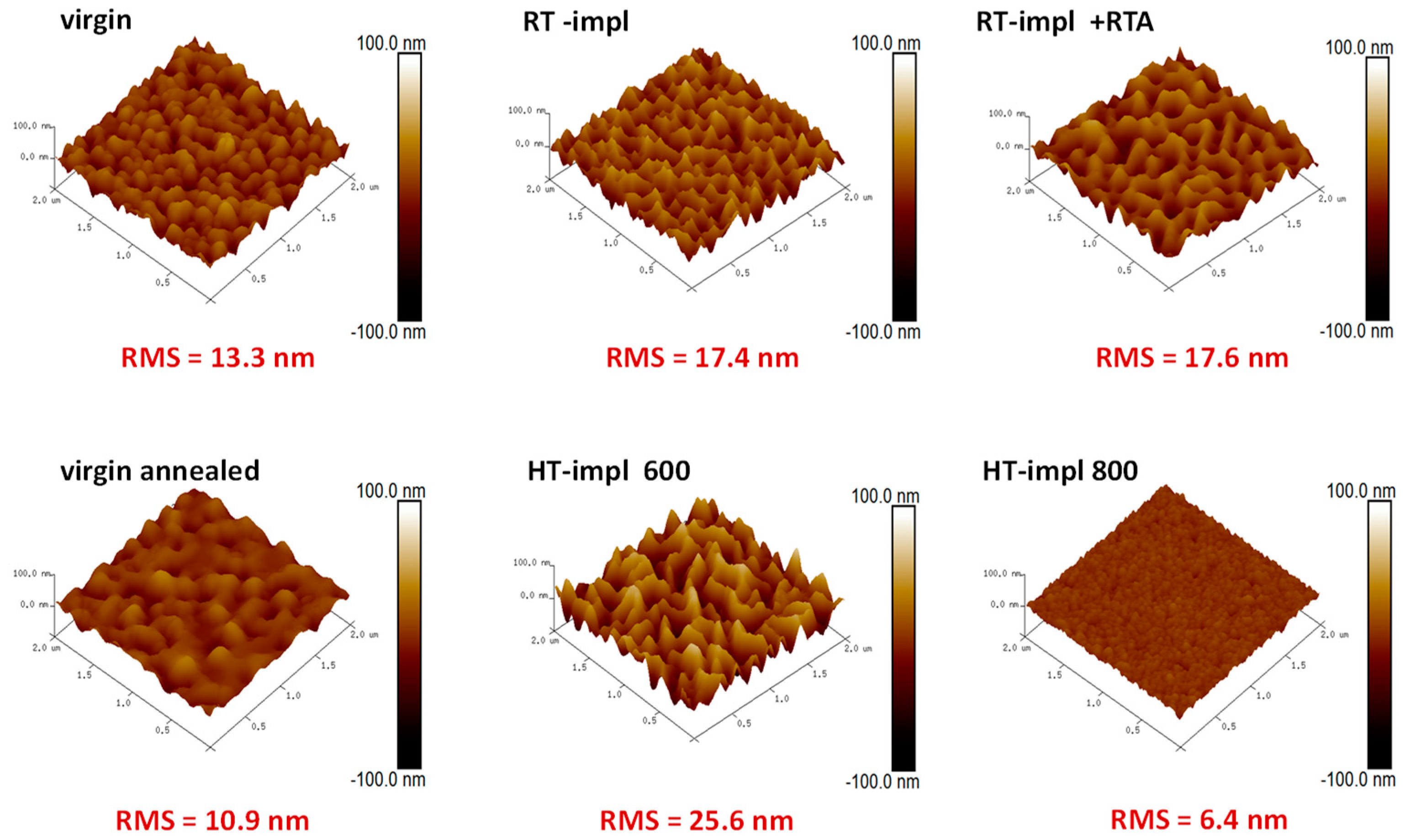
In turn, the AFM studies presented in Figure 2 show the positive contribution of temperature to the smoothness of the layers. An RMS value of ~13.3 nm is measured for the virgin samples, while for temperature-treated samples this value decreases by up to ~6.4 nm. Any other effects, e.g., the appearance of crystalline precipitation of Yb after temperature treatment in the ZnO films, are not detected.
The depth distributions of matrix and impurity atoms were specified by RBS. Moreover, thanks to the use of this method in the channeling mode, the depth distributions of defects caused by ion implantation as well as the Yb lattice site location were also determined. The random and aligned (i.e., taken in the channeling mode) RBS spectra obtained for ZnO before and after HT implantation at different temperatures with 150 keV Yb ions at the optimal fluence of 1 × 10¹⁵/cm2 [29] show separated signals coming from He ions backscattered on Zn and RE atoms (Figure 3). However, differences in the intensity of those signals are significant; therefore, for clarity, they are presented on a different scale (Figure 3a,b). Separate signals mean that crystal lattice defects and RE behavior can be simultaneously monitored, which is an advantage of the RBS/c technique in such analyses. The RBS studies show that with the increase of the implantation temperature, the heights of aligned spectra decrease, which means that the level of defects become smaller with the temperature of implantation. However, at the same time, the RE profile changed, reflecting the diffusion of RE ions toward the sample surface. For implantation at 800 °C, the perfect crystal structure recovery is observed (Figure 3a), but most of the RE ions precipitate on the surface, which results in a high RBS signal at an energy of about 1550 keV (Figure 3b).
Comparing the aligned spectra with the random one for the Yb signal observed in the energy region 1470–1570 keV, it can be remarked that for HT-implanted samples, the random and aligned RBS spectra of RE presented in Figure 3b have the same intensity, which means that the RE ions are not incorporated into the ZnO matrix.
The details of these observations can be listed numerically by the minimum yield (χmin) and substitutional fraction (fs) parameters [34] (Table 1). The χmin is the ratio of the backscattering yield of an aligned spectrum (calculated for a selected energy range) to the corresponding yield of the random one. It represents the level of structural defects in the Zn sublattice. The fs is defined as the relative amount of impurity atoms occupying lattice site positions. Table 1 specifies also the maximum value of the backscattering yield in the Yb peak, located around 1550 keV, which reflects the change in the amount of Yb ions precipitated on the surface (Figure 3b), as well as the PL intensity at 978 nm (Figure 3c).
Structural changes directly affect the optical properties of the samples. A typical PL spectrum of Yb ions in the 3+ oxidation state (Figure 3c), observed in the IR spectral range between 900 and 1150 nm, is not affected by ZnO characteristic emissions, coming from the near band edge and deep-level emission connected with defects, that appear in the visible region [17]. The IR PL emission from Yb3+ observed at about 980 nm corresponds to the main radiative transition from the excited 2F5/2 energy state to the 2F7/2 ground state of Yb3+. Broad peaks located between 1000 and 1150 nm correspond to the Yb3+ ion emission vibrionic band (2F5/22F7/2(n); n = 1,2,3,4) [58]. In contrast to RT implantation, the RE-ions after HT implantation are optically active (Figure 3c). However, as the implantation temperature increases to 700 °C, the luminescence coming from RE decreases drastically, and finally, for 800 °C, when all RE ions precipitate on the surface, the luminescence coming from Yb3+ completely disappears.
Other interesting conclusions have been drawn from the experiment, in which different energies of implanted ions are used, resulting in a different implantation depth. In the experiment, epitaxial ZnO films were implanted at 600 °C with Yb ions at energies of 150, 500, and 1000 keV, which results in ~100, 160, and 240 nm modified layers, respectively. The RBS-aligned spectra (Figure 4a,b), confirmed by simulations performed using the McChasy code [59,60], show that the number of point defects increases with the implantation energy, but the density of extended defects, such as dislocations, is smaller for the faster ions. Regardless of these subtle differences in the defects’ structure, the luminescence efficiency, presented in Figure 4c, is significantly lower for deeper implantations, probably due to the high-light absorption coefficient, which for ZnO is about 5 × 10⁴/cm [61].
To eliminate the undesirable RE out-diffusion effect, alternative annealing techniques are tested. The millisecond range FLA has already been successfully used to improve the crystalline quality of RT-implanted materials [62,63]. Furthermore, in the case of ZnO:RE implanted at RT with fluences below the plastic deformation threshold, the RE out-diffusion effect after FLA is not observed, and RE ions are mainly placed at substitutional positions. However, unfortunately, the RE ions after FLA remain optically inactive [39].
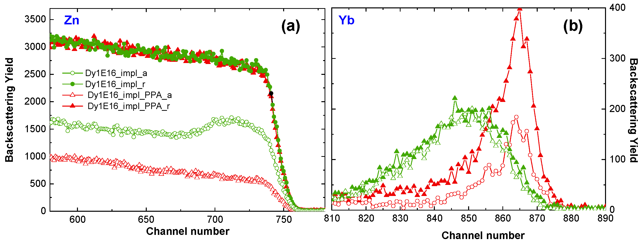
The crystalline structure recovery effect of ZnO:RE implanted at RT is also observed after PPA, as shown in Figure 5a, where the RBS spectra of ZnO implanted at RT with Dy ions and subsequently PPA annealed are presented. The above studies are conducted on the ZnO:Dy system; however, as previously reported, there is no difference in the level of structural damage after implantation with different RE ions at the same fluence [17]. Unfortunately, after this kind of annealing, the RE atoms diffuse to the sample surface (Figure 5b), and no light coming from RE3+ is observed, as in the high-temperature cases (see Figure 3c). This means that neither FLA nor PPA are useful for the optical activation of RE in ZnO:RE systems.
In the last part of the study, the optimal atmosphere of RTA post-RT implantation annealing is tested. A comparison of RBS-aligned spectra obtained for ZnO implanted at RT with a fluence of Yb ions below the plastic deformation threshold and subsequently annealed for 10 min at 800 °C in O2, N2, and Ar atmospheres is presented in Figure 6a,b. It has been reported that too long annealing time leads to an increase in the number of RE atoms on the surface, and thus to a decrease in PL efficiency. The 10 min RTA annealing time results in a balance between structure recovery and RE out-diffusion [39]. As can be seen in Figure 6a,b, and Table 2 as well, the spectra and all parameters vary significantly depending on the annealing atmosphere. That applies to both the thermal-induced structural recovery of ZnO and the migration of Yb to the interstitial site as well. The recovery level, as well as the Yb behavior after the RTA annealing in oxygen and nitrogen atmospheres, are very similar. The weakest recovery is observed after annealing in the Ar atmosphere. In turn, the luminescence response, to these structural changes, shows the most efficient luminescence for oxygen annealing, as presented in Figure 4c. Interestingly, the PL intensity is not directly correlated with the RMS value, which is 14.4 nm for oxygen annealing so between the values obtained for Ar and N2 annealing (Figure 7).
Finally, the optical response of Yb ions implanted at the optimum temperature (600 °C) is compared with the optical response of these ions implanted at RT and subsequently annealed at 800 °C in oxygen (Figure 8c). Although, as can be observed in the aligned RBS spectra (and Table 3), the crystal structure recovery is only slightly better for RT implantation and subsequent annealing (Figure 8a), the recorded luminescence efficiency for this sample is four times higher (Figure 8c), showing that this way of preparing samples is the most beneficial for the optical applications of ZnO:RE systems. This effect might be assigned to both better crystal structure recovery and a lower number of Yb ions on the crystal’s surface as can be deduced from Figure 8b. However, the role of oxygen in the luminescence of ZnO seems to be important. It may be assumed that oxygen diffuses into the sample during RTA and oxidizes RE to the 3+ state which increases PL [39], but the other scenarios are also possible [64].

4. Summary

The effects of implantation and subsequent annealing conditions on the surface morphology, defects in ZnO lattice and RE locations, as well as on the optical response coming from Yb ions in the 3+ oxidation state were studied systematically. The results of deep and shallow implantations, performed at high and room temperature, as well as a range of post-RT-implantation annealing processes were compared for ZnO:RE systems.
It is found that shallow implantation (with the energy of RE ions about 150 keV) with the RE ion fluence below the plastic deformation edge of ZnO crystals (less than 1.5 × 10¹⁵/cm2) is optimal for optical applications of ZnO:RE systems. It was also established that RE ions after HT implantation at a temperature lower than 700 °C are optically active; however, the temperature of 600 °C is optimal, ensuring a higher luminescence efficiency of Yb3+. In contrast, directly after RT implantation most of the Yb ions are optically inactive; therefore, post-implantation annealing is necessary. For the samples implanted at RT, the FLA, PPA, and RTA thermal annealing in O2, N2, and Ar atmospheres have been tested for RE activation. The highest luminescence efficiency of RE3+ was obtained after 10 min RTA annealing performed at 800 °C in oxygen. The comparison of the optical response of this sample with the ZnO:Yb system implanted at 600 °C with the same fluence and energy, shows over four times higher luminescence efficiency of the former system. The light emission from the ZnO:RE system prepared in the above way is so bright that it can be observed by the naked eye, which makes the ZnO:RE systems worth considering for applications in LED or display technologies.

Supplementary Materials

The following supporting information can be downloaded at: https://www.mdpi.com/article/10.3390/ma16051756/s1, Figure S1: SEM imaging of the surface morphology with a variety of magnifications and operating voltage for ZnO implanted at 800 °C with 150 keV Yb ions to the fluence of 1 × 10¹⁵/cm2; Figure S2: EDS mapping with a variety of magnifications and operating voltage for ZnO implanted at 800 °C with 150 keV Yb ions to the fluence of 1 × 10¹⁵/cm2; Figure S3: SEM imaging of the surface morphology for ZnO implanted with 150 keV Yb ions to the fluence of 1 × 10¹⁵/cm2 at different temperatures: 600 and 800 °C and RT and subsequently RTA-annealed for 10 min at 800 °C in an oxygen atmosphere; Figure S4: AFM imaging of the surface morphology for ZnO implanted with 150 keV Yb ions to the fluence of 1 × 10¹⁵/cm2 at different temperatures: 600 and 800 °C and RT and subsequently RTA-annealed for 10 min at 800 °C in an oxygen atmosphere; Figure S5: AFM imaging of the surface morphology for ZnO implanted with 150 keV Yb ions to the fluence 1 × 10¹⁵/cm2 and subsequently RTA-annealed for 10min at 800 °C in different atmospheres N2, Ar, O2.

Author Contributions

Conceptualization, R.R.; Methodology, R.R.; Software, C.M. and P.J.; Formal analysis, R.R.; Investigation, R.R., S.P., C.M., P.J., M.B., S.R. and S.G.; Resources, S.P., M.B., W.W., R.H. and U.K.; Writing—original draft, R.R. and E.G.; Writing—review & editing, R.R. and E.G.; Visualization, R.R. and P.J.; Supervision, E.G.; Project administration, R.R.; Funding acquisition, R.R. and S.F. All authors have read and agreed to the published version of the manuscript.

Funding

This research was carried out under the co-financed international project supported by the Polish Ministry of Education and Science (5134/HZDR/2020/0) and Helmholtz-Zentrum Dresden-Rossendorf (20001987-ST, 21002630-ST, 21002632-ST).

Institutional Review Board Statement

Not applicable.

Informed Consent Statement

Not applicable.

Data Availability Statement

Not applicable.

Conflicts of Interest

The authors declare no conflict of interest.

References

  1. Wang, X.; Sun, F.; Duan, Y.; Yin, Z.; Luo, W.; Huang, Y.; Chen, J.J. Highly sensitive, temperature-dependent gas sensor based on hierarchical ZnO nanorod arrays. Mater. Chem. C 2015, 3, 11397. [Google Scholar] [CrossRef]
  2. Richards, B. Luminescent layers for enhanced silicon solar cell performance: Down-conversion. Sol. Energy Mat. Sol. Cells 2006, 90, 1189. [Google Scholar] [CrossRef]
  3. Socol, M.; Preda, N.; Breazu, C.; Florica, C.; Costas, A.; Istrate, C.; Stanculescu, A.; Girtan, M.; Gherendi, F. Organic heterostructures obtained on ZnO/Ag/ZnO electrode. Vacuum 2018, 154, 366. [Google Scholar] [CrossRef]
  4. Jagadish, C.; Pearton, S.J. Thin Films and Nanostructures; Elsevier: Amsterdam, The Netherlands, 2006; ISBN 978-0-08-044722-3. [Google Scholar]
  5. Thomas, D.G. The Exciton Spectrum of Zinc Oxide. J. Phys. Chem. Solids 1960, 15, 86. [Google Scholar] [CrossRef]
  6. Özgür, Y.; Alivov, C.; Liu, A.; Teke, M.; Reshchikov, S.; Dogan, V.; Avrutin, S.-J.; Cho, H.; Morkoc, J. A comprehensive review of ZnO materials and devices. J. Appl. Phys. 2005, 98, 041301. [Google Scholar] [CrossRef] [Green Version]
  7. Mishra, Y.; Adelung, R. ZnO tetrapod materials for functional applications. Mater. Today 2018, 21, 631–651. [Google Scholar] [CrossRef]
  8. Rasmidi, R.; Duinong, M.; Chee, F.P. Radiation damage effects on zinc oxide (ZnO) based semiconductor devices—A review. Radiat. Phys. Chem. 2021, 184, 109455. [Google Scholar] [CrossRef]
  9. Wendler, E.; Wesch, W.; Azarov, A.Y.; Catarino, N.; Redondo-Cubero, A.; Alves, E.; Lorenz, K. Comparison of low- and room-temperature damage formation in Ar ion implanted GaN and ZnO. Nucl. Instr. Meth. Phys. Res. B 2013, 307, 394. [Google Scholar] [CrossRef]
  10. Kenyon, A.J. Recent developments in rare-earth doped materials for optoelectronics. Prog. Quantum El. 2002, 26, 225. [Google Scholar] [CrossRef]
  11. Favennec, P.N.; L’Haridon, H.; Salvi, M.; Moutonnet, D.; Le Guillou, Y. Luminescence of erbium implanted in various semiconductors: IV, III-V and II-VI materials. Electron. Lett. 1989, 25, 718–719. [Google Scholar] [CrossRef]
  12. Jadwisienczak, W.; Lozykowski, H.; Xu, A.; Patel, B.J. Visible emission from ZnO doped with rare-earth ions. Electron. Mater. 2002, 31, 776. [Google Scholar] [CrossRef]
  13. Vařák, P.; Mrázek, J.; Jasim, A.; Bysakh, S.; Dhar, A.; Kamrádek, M.; Podrazký, O.; Kašík, I.; Bartoň, I.; Nekvindová, P. Thermal stability and photoluminescence properties of RE-doped (RE = Ho, Er, Tm) alumina nanoparticles in bulk and fiber-optic silica glass. Opt. Mater. 2021, 118, 111239. [Google Scholar] [CrossRef]
  14. Singh, A.; Arya, P.; Choudhary, D.; Kumar, S.; Srivastava, A.; Singh, I.B. Cost-effective ZnO–Eu3+ films with efficient energy transfer between host and dopant. SN Appl. Sci. 2020, 2, 870. [Google Scholar] [CrossRef] [Green Version]
  15. Kumar, R.; Mishra, S. Enhancement in the luminescence of green-emission from emissive surface defects of Dy3+doped ZnO nanoluminophores: A simple, mass-scale productive approach for optoelectronic devices. Appl. Surf. Sci. Adv. 2022, 9, 100256. [Google Scholar] [CrossRef]
  16. Poate, J.; Williams, S. Ion Implantation and Beam Processing; Williams, J.S., Poate, J.M., Eds.; Academic Press: Sydney, Astralia, 1984; p. 13. ISBN 0127569804. [Google Scholar]
  17. Ratajczak, R.; Guziewicz, E.; Prucnal, S.; Łuka, G.; Böttger, R.; Heller, R.; Mieszczynski, C.; Wozniak, W.; Turos, A. Luminescence in the Visible Region from Annealed Thin ALD-ZnO Films Implanted with Different Rare Earth Ions. Phys. Status Solidi A 2018, 215, 1700889. [Google Scholar] [CrossRef]
  18. Zhou, X.; Ning, L.; Qiao, J.; Zhao, Y.; Xiong, P.; Xia, Z. Interplay of defect levels and rare earth emission centers in multimode luminescent phosphors. Nat. Commun. 2022, 13, 7589. [Google Scholar] [CrossRef]
  19. Masai, H.; Okada, G.; Kawaguchi, N.; Yanagida, T. Relationship between defect formation by X-ray irradiation and thermally stimulated luminescence of binary zinc phosphate glasses. Opt. Mater. Express 2019, 9, 2037–2045. [Google Scholar] [CrossRef]
  20. Kucheyev, S.O.; Williams, J.S.; Jagadish, C.; Zou, J.; Evans, C.; Nelson, A.J.; Hamza, A.V. Ion-beam-produced structural defects in ZnO. Phys. Rev. B 2003, 67, 094115. [Google Scholar] [CrossRef] [Green Version]
  21. Wendler, E.; Bilani, O.; Gaertner, K.; Wesch, W.; Hayes, M.; Auret, F.D.; Lorenz, K.; Alves, E. Radiation damage in ZnO ion implanted at 15 K. Nucl. Instrum. Methods B 2009, 267, 2708. [Google Scholar] [CrossRef] [Green Version]
  22. Azarov, A.Y.; Titov, A.I.; Karaseov, P.A.; Kucheyev, S.O.; Hallen, A.; Kuznetsov, A.Y.; Svenson, B.G.; Pathak, A.P. Structural damage in ZnO bombarded by heavy ions. Vacuum 2010, 84, 1058. [Google Scholar] [CrossRef]
  23. Azarov, A.Y.; Kucheyev, S.; Titov, A.; Karaseov, P.J. Effect of the density of collision cascades on ion implantation damage in ZnO. Appl. Phys. 2007, 102, 083547. [Google Scholar] [CrossRef]
  24. Azarov, A.; Wendler, E.; Kuznetsov, A.Y.; Svenson, B. Crucial role of implanted atoms on dynamic defect annealing in ZnO. Appl. Phys. Lett. 2014, 104, 052101. [Google Scholar] [CrossRef]
  25. Jagerová, A.; Malinsky, P.; Miksova, R.; Nekvindova, P.; Cajzl, J.; Akhmadaliev, S.; Holy, V.; Mackova, A. Distinct defect appearance in Gd implanted polar and nonpolar ZnO surfaces in connection to ion channeling effect. J. Vac. Sci. Technol. A 2019, 37, 061406. [Google Scholar] [CrossRef]
  26. Nekvindová, P.; Cajzl, J.; Macková, A.; Malinský, P.; Oswald, J.; Böttger, R.; Yatskiv, R. Er implantation into various cuts of ZnO – experimental study and DFT modelling. J. Alloys Compd. 2020, 816, 152455. [Google Scholar] [CrossRef]
  27. Turos, A.; Jóźwik, P.; Wójcik, M.; Gaca, J.; Ratajczak, R.; Stonert, A. Mechanism of damage buildup in ion bombarded ZnO. Acta Mater. 2017, 134, 249. [Google Scholar] [CrossRef]
  28. Jozwik, P.; Magalhães, S.; Ratajczak, R.; Mieszczynski, C.; Sequeira, M.; Turos, A.; Böttger, R.; Heller, R.; Lorenz, K.; Alves, E. RBS/C, XRR, and XRD Studies of Damage Buildup in Er-Implanted ZnO. Phys. Status Solidi B 2019, 256, 1800364. [Google Scholar] [CrossRef]
  29. Ratajczak, R.; Mieszczynski, C.; Prucnal, S.; Krajewski, T.A.; Guziewicz, E.; Wozniak, W.; Kopalko, K.; Heller, R.; Akhmadaliev, S. Correlations between the structural transformations and concentration quenching effect for RE-implanted ZnO systems. Appl. Surf. Sci. 2020, 521, 146421. [Google Scholar] [CrossRef]
  30. Demchenko, I.N.; Melikhov, Y.; Konstantynov, P.; Ratajczak, R.; Turos, A.; Guziewicz, E. Resonant Photoemission Spectroscopy Study on the Contribution of the Yb 4f States to the Electronic Structure of ZnO. Acta Phys. Pol. A 2018, 133, 907. [Google Scholar] [CrossRef]
  31. Anjana, R.; Jayaraj, M.K.; Yadav, A.K.; Jha, S.N.; Bhattacharyya, D.J. Investigating the evolution of local structure around Er and Yb in ZnO:Er and ZnO:Er, Yb on annealing using X-ray absorption spectroscopy. App. Phys. 2018, 123, 153102. [Google Scholar] [CrossRef]
  32. Myers, M.; Charnvanichborikarn, S.; Shao, L.; Kucheyev, S. Effect of the surface on ion-beam damage build-up in ZnO. Scr. Mater. 2012, 67, 65. [Google Scholar] [CrossRef]
  33. Myers, M.T.; Charnvanichborikarn, S.; Wei, C.C.; Luo, Z.P.; Aitkaliyeva, A.; Shao, L.; Kucheyev, S.O. Defect microstructure in heavy-ion-bombarded (0 0 0 1) ZnO. Acta Mater. 2012, 60, 6086. [Google Scholar] [CrossRef]
  34. Chu, W.; Mayer, J.; Nicolet, M. Backscattering Spectrometry; Academic Press: Orlando, FL, USA, 1978; ISBN 1-12-173850-7. [Google Scholar]
  35. Feldman, L.; Mayer, J.; Picraux, S. Materials Analysis by Ion Channeling; Academic Press: New York, NY, USA, 1982; ISBN 9780323139816. [Google Scholar]
  36. Azarov, A.; Galeckas, A.; Hallen, A.; Kuznetsov, A.; Monakhov, E.; Svensson, B.G. Optical activity and defect/dopant evolution in ZnO implanted with Er. J. Appl. Phys. 2015, 118, 125703. [Google Scholar] [CrossRef]
  37. Azarov, A.; Titov, A.I.; Kucheyev, S.O. Effect of pre-existing disorder on surface amorphization in GaN. Appl. Phys. 2010, 108, 033505. [Google Scholar] [CrossRef]
  38. Ratajczak, R.; Mieszczynski, C.; Prucnal, S.; Guziewicz, E.; Stachowicz, M.; Snigurenko, D.; Gaca, J.; Böttger, R.; Wojcik, M.; Heller, R.; et al. Structural and optical studies of Pr implanted ZnO films subjected to a long-time or ultra-fast thermal annealing. Thin Solid Film. 2017, 643, 24. [Google Scholar] [CrossRef]
  39. Ratajczak, R.; Prucnal, S.; Guziewicz, E.; Mieszczynski, C.; Snigurenko, D.; Stachowicz, M.; Skorupa, W.; Turos, A.J. The photoluminescence response to structural changes of Yb implanted ZnO crystals subjected to non-equilibrium processing. App. Phys. 2017, 121, 075101. [Google Scholar] [CrossRef]
  40. Miranda, S.; Peres, M.; Monteiro, T.; Alves, E.; Sun, H.; Geruschke, T.; Vianden, R.; Lorenz, K. Rapid thermal annealing of rare earth implanted ZnO epitaxial layers. Opt. Mater. 2011, 33, 1139. [Google Scholar] [CrossRef]
  41. Alves, E.; Rita, E.; Wahl, U.; Correia, J.; Monteiro, T.; Soares, J.; Boemare, C. Lattice site location and optical activity of Er implanted ZnO. Nucl. Instr. Meth. Phys. Res. B 2003, 206, 1047. [Google Scholar] [CrossRef]
  42. Murmu, P.; Kennedy, J.; Ruck, B.J.; Williams, G.V.M.; Markowitz, A.; Rubanov, S.; Suvorov, A.A. Correlation between microstructural and magnetic properties of Tb implanted ZnO. AIP Conf. Proc. 2013, 1525, 300. [Google Scholar] [CrossRef] [Green Version]
  43. Rita, E.; Alves, E.; Wahl, U.; Correia, J.G.; Neves, A.J.; Soares, M.J.; Monteiro, T. Optical doping of ZnO with Tm by ion implantation. Physica B 2003, 340, 235. [Google Scholar] [CrossRef]
  44. Azarov, A.; Hallén, A.; Du, X.; Liu, Z.; Svensson, B.; Kuznetsov, A. Thermally induced surface instability in ion-implanted MgxZn1-xO films. Phys. Rev. B. 2011, 84, 014114. [Google Scholar] [CrossRef] [Green Version]
  45. Dolabella, S.; Borzì, A.; Dommann, A.; Neels, A. Lattice Strain and Defects Analysis in Nanostructured Semiconductor Materials and Devices by High-Resolution X-Ray Diffraction: Theoretical and Practical Aspects. Small Methods 2022, 6, 2100932. [Google Scholar] [CrossRef]
  46. Knutsen, K.; Galeckas, A.; Zubiaga, A.; Tuomisto, F.; Farlow, G.; Svensson, B.; Kuznetsov, A.Y. Zinc vacancy and oxygen interstitial in ZnO revealed by sequential annealing and electron irradiation. Phys. Rev. B 2012, 86, 121203. [Google Scholar] [CrossRef] [Green Version]
  47. Meng, Z.; Mo, X.; Cheng, X.; Zhou, Y.; Tao, X.; Ouyang, Y. Interactions between Er dopant and intrinsic point defects of ZnO: A first-principles study. Mater. Res. Express 2017, 4, 035903. [Google Scholar] [CrossRef]
  48. Krajewski, T.; Ratajczak, R.; Kobyakov, S.; Wozniak, W.; Kopalko, K.; Guziewicz, E. Electrical properties of ZnO films implanted with rare earth and their relationship with structural and optical parameters. Mater. Sci. Eng. B 2022, 275, 115526. [Google Scholar] [CrossRef]
  49. Gehring, P.; Weng, Y.; Wu, Z.; Strunk, H.J. Photoluminescence from Alx In1−xN layers doped with Tb3+ ions. Phys. Conf. Ser. 2011, 281, 012014. [Google Scholar] [CrossRef]
  50. Ajimsha, R.; Das, A.; Singh, B.; Misra, P.; Kukreja, L.M. Structural, electrical and optical properties of Dy doped ZnO thin films grown by buffer assisted pulsed laser deposition. Phys. E 2010, 42, 1838. [Google Scholar] [CrossRef]
  51. Benz, F.; Guerra, J.; Weng, Y.; Zanatta, A.; Weingärtner, R.; Strunk, H.J. Concentration quenching of the green photoluminescence from terbium ions embedded in AlN and SiC matrices. J. Lumin 2013, 137, 73. [Google Scholar] [CrossRef]
  52. Benz, F.; Gonser, A.; Völker, R.; Walther, T.; Mosebach, J.-T.; Schwanda, B.; Mayer, N.; Richter, G.; Strunk, H.J. Concentration quenching of the luminescence from trivalent thulium, terbium, and erbium ions embedded in an AlN matrix. J. Lumin 2014, 145, 855. [Google Scholar] [CrossRef]
  53. Zhao, L.; Xu, C.; Liu, Y.; Shao, C.; Li, X. A new approach to white light emitting diodes of p-GaN/i-ZnO/n-ZnO heterojunctions. Appl. Phys. B 2008, 92, 185–188. [Google Scholar] [CrossRef]
  54. Ayoub, I.; Kumar, V.; Abolhassani, R.; Sehgal, R.; Sharma, V.; Sehgal, R.; Swart, H.; Mishra, Y. Advances in ZnO: Manipulation of defects for enhancing their technological potentials. Nanotechnol. Rev. 2022, 11, 575. [Google Scholar] [CrossRef]
  55. Hansen, D.M.; Zhang, R.; Perkins, N.R.; Safvi, S.; Zhang, L.; Bray, K.L.; Kuech, T.F. Photoluminescence of erbium-implanted GaN and in situ-doped GaN:Er. Appl. Phys. Lett. 1998, 72, 1244. [Google Scholar] [CrossRef] [Green Version]
  56. Guziewicz, E.; Ratajczak, R.; Stachowicz, M.; Snigurenko, D.; Krajewski, T.; Mieszczynski, C.; Mazur, K.; Witkowski, B.; Dluzewski, P.; Morawiec, K. Atomic layer deposited ZnO films implanted with Yb: The influence of Yb location on optical and electrical properties. Thin Solid Film. 2017, 643, 7–15. [Google Scholar] [CrossRef]
  57. Sarwar, M.; Ratajczak, R.; Ivanov, V.; Mishra, S.; Turek, M.; Wierzbicka, A.; Woźniak, W.; Guziewicz, E. Crystal Lattice Damage and Recovery of Rare-Earth implanted Wide Bandgap Oxides. Adv. Sci. Technol. Res. J. 2022, 16, 147–154. [Google Scholar] [CrossRef]
  58. Lchi-Ghazaani, M.; Parvin, P. Fiber Laser; Mukul Chandra Paul: London, UK, 2016. [Google Scholar] [CrossRef] [Green Version]
  59. Jozwik, P.; Nowicki, L.; Ratajczak, R.; Stonert, A.; Mieszczynski, C.; Turos, A.; Morawiec, K.; Lorenz, K.; Alves, E. Monte Carlo simulations of ion channeling in crystals containing dislocations and randomly displaced atoms. J. Appl. Phys. 2019, 126, 195107. [Google Scholar] [CrossRef]
  60. Jozwik, P.; Nowicki, L.; Ratajczak, R.; Mieszczynski, C.; Stonert, A.; Turos, A.; Lorenz, K.; Alves, E. Advanced Monte Carlo Simulations for Ion-Channeling Studies of Complex Defects in Crystals. In Theory, and Simulation in Physics for Materials Applications; Levchenko, E.V., Dappe, Y.J., Ori, G., Eds.; Springer: Cham, Switzerland, 2020; pp. 133–160. [Google Scholar]
  61. Mursal; Irhamni; Bukhari; Jalil, Z.J. Structural and Optical Properties of Zinc Oxide (ZnO) based Thin Films Deposited by Sol-Gel Spin Coating Method. Phys. Conf. Ser. 2018, 1116, 032020. [Google Scholar] [CrossRef]
  62. Lindberg, P.; Bregolin, F.L.; Wiesenhütter, K.; Wiesenhütter, U.; Riise, H.; Vines, L.; Prucnal, S.; Skorupa, W.; Svensson, B.G.; Monakhov, E.J. The effect of millisecond flash lamp annealing on electrical and structural properties of ZnO:Al/Si structures. Appl. Phys. 2016, 119, 185305. [Google Scholar] [CrossRef] [Green Version]
  63. Rebohle, L.; Prucnal, S.; Reichel, D. Flash Lamp Annealing—from Basics to Application; Springer Series in Materials Science (SSMATERIALS): New York, NY, USA, 2019; Volume 288, ISBN 978-3-030-23298-6. [Google Scholar]
  64. Naliwajko, P.; Peppel, T.; Strunk, J. Thermal and light induced infrared blackening of ZnO revisited: Rediscovery of fundamental scientific knowledge. React. Kinet. Mech. Catal. 2022, 135, 2291–2305. [Google Scholar] [CrossRef]
Figure 1. EDS mapping of Yb (pink), Zn (red), and O (green) for ZnO implanted with 150 keV Yb ions to the fluence of 1 × 10¹⁵/cm2 was performed for samples implanted at different temperatures: 600 °C (HT-impl 600), 800 °C. (HT-impl 800), and RT (RT-impl) as well as implanted at RT and subsequently RTA-annealed for 10 min at 800 °C in an oxygen atmosphere (RT-impl +RTA).
Materials 16 01756 g001
Figure 2. AFM imaging of the surface morphology for ZnO before (virgin) and after implantation at different temperatures with 150 keV Yb ions to the fluence of 1 × 10¹⁵/cm2 as well as RT-implanted samples and subsequently RTA-annealed for 10 min at 800 °C in O2.
Materials 16 01756 g002
Figure 3. Random (solid symbols) and aligned (open symbols) RBS spectra (a,b) obtained for ZnO implanted at different temperatures with 150keV Yb ions at the fluence of 1 × 10¹⁵/cm2. The corresponding PL spectra (c).
Materials 16 01756 g003
Figure 4. Random (solid symbols) and aligned (open symbols) RBS spectra (a,b) obtained for ZnO implanted with different energies of Yb ions at the fluence of 1 × 10¹⁵ /cm2 at 600 °C. The corresponding PL spectra (c).
Materials 16 01756 g004
Figure 5. (a,b) Random and aligned RBS spectra obtained for ZnO implanted with 150 keV Dy ions to the fluence of 1 × 10¹⁶/cm2 and subsequently annealed using plasma pulse annealing with a pulse energy of 1.17 J/cm2.
Materials 16 01756 g005
Figure 6. Random (open symbols) and aligned (solid symbols) RBS spectra (a,b) obtained for ZnO implanted at RT with 150 keV Yb ions to the fluence 5 × 10¹⁴ /cm2 and subsequently RTA-annealed for 10min at 800 °C in different atmospheres. The corresponding PL spectra (c).
Materials 16 01756 g006
Figure 7. AFM imaging of the surface morphology for ZnO implanted with 150 keV Yb ions to the fluence 5 × 10¹⁴/cm2 and subsequently RTA-annealed for 10 min at 800 °C in different atmospheres N2, Ar, O2.
Materials 16 01756 g007
Figure 8. Aligned RBS spectra (a,b) obtained for ZnO implanted with 150 keV Yb ions to the fluence of 1 × 10¹⁵/cm2 at 600 °C and RT and subsequently RTA-annealed for 10 min at 800 °C in an oxygen atmosphere. The corresponding PL spectra (c).
Materials 16 01756 g008
Table 1. Numerical evaluation of the RBS and PL spectra of ZnO implanted at different high temperatures with 150 keV Yb ions at the fluence of 1 × 10¹⁵/cm2.
RBSPL
χmin
(%)
fs
(%)
The Backscattering Yield of
Yb Ions on the Surface (Counts)
Intensity
(a.u.)
virgin3.2---
HT-impl 60027.020.6881.00
HT-impl 7006.65.62050.30
HT-impl 8004.23.52480.03
Table 2. Numerical evaluation of the RBS and PL spectra of ZnO implanted at RT with 150 keV Yb ions at the fluence of 5x10¹⁴ /cm2 and RTA-annealed.
RBSPL
χmin
(%)
fs
(%)
The Backscattering Yield of
Yb Ions on the Surface (Counts)
Intensity
(a.u.)
Virgin2.9---
RT-impl20.464.970.02
RT-impl+RTA O2 6.240.1201.00
RT-impl+RTA N2 5.655.9160.56
RT-impl+RTA Ar 12.016.5200.16
Table 3. Numerical evaluation of the RBS and PL spectra of ZnO implanted at RT and 600 °C with 150 keV Yb ions at the fluence of 1 × 10¹⁵/cm2.
RBSPL
χmin
(%)
fs
(%)
The Backscattering Yield of
Yb Ions on the Surface (Counts)
χmin
(%)
virgin3.2---
HT 60027.020.6881.00
RT 28.649.1250.04
RT+RTA23.726.5644.59
Disclaimer/Publisher’s Note: The statements, opinions and data contained in all publications are solely those of the individual author(s) and contributor(s) and not of MDPI and/or the editor(s). MDPI and/or the editor(s) disclaim responsibility for any injury to people or property resulting from any ideas, methods, instructions or products referred to in the content.