Article

Clinical and Radiographic Outcomes of Zirconia Dental Implants—A Clinical Case Series Study

1
Oral and Maxillofacial Surgery Department, Universitat Internacional de Catalunya, 08017 Barcelona, Spain
2
Department of Periodontics and Oral Medicine, School of Dentistry, University of Michigan, Ann Arbor, MI 48109, USA
3
Clínica Dental, 08036 Barcelona, Spain
*
Author to whom correspondence should be addressed.
Materials 2022, 15(7), 2437; https://doi.org/10.3390/ma15072437
Submission received: 11 February 2022 / Revised: 22 March 2022 / Accepted: 23 March 2022 / Published: 25 March 2022
(This article belongs to the Special Issue Dental Implants: Materials and Design)

Abstract

:
The purpose of this clinical series was to evaluate the clinical and radiographic outcomes of 20 zirconia dental implants, with a minimum follow-up of two years. Patients with at least one zirconia dental implant were included, with a mean follow-up of 34.05 months. The patient complaints such as pain and foreign body sensation, as well as clinical parameters including peri-implant infections with suppuration, implant mobility, gingival index (GI), modified plaque index (mPI), modified sulcus bleeding index (mBI), probing depth (PD) and radiological distance between the implant shoulder to the closest mesial and distal bone-to-implant contact (BIC), were assessed. All zirconia implants were successfully integrated without any pain or foreign body sensation. No mobility was detected in any of the 20 implants. Clinical examination revealed a mean PD of 2.56 mm and a radiological mean distance between the implant shoulder and the initial site of visible bone-to-implant contact (BIC) of 1.44 mm. In addition, GI, mPI and mBI indicated minimal to no inflammation. Results obtained from this series suggest that one-piece zirconia dental implants achieve good clinical and radiographic outcomes over a mean follow-up of 34 months and may be deemed a good option in patients with favorable bone conditions.

1. Introduction

Since the 1970s, titanium (Ti) has been one of the most well-documented biomaterials in dental implantology. Due to its bioinert nature, strength, superiority over other metals in reducing Tresca stress and ability to osseointegrate, it has been regarded as the best material for manufacturing dental implants [1]. However, Ti does pose some challenges, such as a poor aesthetic appearance especially in areas of insufficient gingival and buccal bone thickness. Although rare, Ti has also been associated with allergic reactions [2]. Furthermore, Ti implants may generate some Ti particles which have been noted in peri-implant tissue. These Ti particles have recently been linked to the development of peri-implantitis [3] as well as to soft tissue lesions [4,5]. Moreover, Ti particles and ions could lead to the release of proinflammatory cytokines such as TNF-α and IL-1β, thereby playing a possible role in the inflammatory response [6,7,8]. Consequently, the need to establish an alternative material to Ti has been suggested.
Zirconium dioxide (ZrO2) or zirconia (Zr) was initially introduced as an alternative dental implant material due to the superior aesthetic effect of its color, particularly in areas characterized by a thin tissue phenotype [9,10], and it has also been related to a lesser tendency to accumulate plaque compared to Ti implants [10,11,12]. In addition, Zr implants show a 74–98% implant survival rate over 12 to 56 months and a 79.6–91.6% success rate over 6 to 12 months [13].
Surface characteristics are among the primary influencing factors of the osseointegration process [14,15], and there have been important developments in Ti implant surfaces over the years. Similarly, several preclinical investigations have demonstrated that Zr implants show similar bone-to-implant contact (BIC) and equivalent removal torque as Ti implants [16,17,18]. Zirconia surface modifications via acid etching and the creation of a microrough surface seem to enhance the biological properties of Zr implants, stimulating osseointegration and being comparable to the modified Ti surface [19,20]. Nonetheless, Zr dental implants seem to require longer healing times compared to titanium implants [21].
Furthermore, Zr dental implants can be found in one-piece or two-piece modalities. One-piece implants include the transepithelial abutment in the same implant fixation unit, avoiding the implant–abutment microgap and canceling transepithelial micromovements [22]. In contrast, one-piece implants exclude the possibility of abutment modification and make it necessary to perform cemented restorations.
According to Cionca et al. [23], some patients are aware of the existence of titanium dental implant alternatives on the market, and professionals must be prepared to respond to the patient demands. In a systematic review and meta-analysis, Elnayef et al. [24] concluded that in specific clinical scenarios, particularly in esthetic areas with a thin tissue phenotype, a Zr-based implant may be a good alternative to a Ti implant. However, Gahlert et al. [25] reported an unacceptable survival rate of 82.4% at three years with one-piece zirconia dental implants, and some studies involving one-piece Zr implants do not have follow-up periods of more than a year—though good results have been reported in terms of bleeding and plaque index, probing depth and marginal bone loss [26,27]. In contrast, few studies have evaluated Zr implants beyond one year of follow-up [28].
Thus, further studies are needed to ascertain Zr dental implant behavior involving rough surfaces and one- or two-piece modalities, with more than one year of follow-up.
The aim of the present case series was to document the clinical and radiographic outcomes of one-piece zirconia dental implants in healthy patients with favorable anatomical bone conditions over at least two years of follow-up after final restoration.

2. Materials and Methods

2.1. Sample Description

The study protocol was approved by the Clinical Research Ethics Committee of the Universitat Internacional de Catalunya (Study code: CIR-ELC-2021-10).
The inclusion criteria were one-piece Zr implants (Straumann® Pure Ceramic implant system; Straumann Holding AG, Basel, Switzerland) placed with an either early or delayed approach [29] and with a minimum follow-up period of two years after final crown delivery. Healthy patients were included, with optimal bone conditions (≥6 mm bucco-lingual bone width and more than 8 mm vertical length) for adequate primary implant stability. Guided bone regeneration (GBR) was allowed to enhance peri-implant bone contour if needed and was always performed simultaneously using demineralized bovine bone grafts (Bio-Oss, Geistlich Pharma AG, Wolhusen, Switzerland) and a collagen membrane (Bio-Gide, Geistlich Pharma AG, Wolhusen, Switzerland). We excluded two-piece Zr implants, as well as implants other than Zr implants (including titanium implants), bone deficiencies requiring bone augmentation procedures before placing of the implants, cases of extensive bone regeneration procedures carried out simultaneously to implant placement and one-piece Zr implants definitely restored less than two years ago.
Dental records from January 2013 to December 2019 corresponding to patients who underwent one-piece zirconia implantation were carefully screened. Then, the eligible patient records were reviewed to assess follow-up status. Patients who fulfilled the inclusion criteria were invited by phone to participate in the study.
All subjects were aware that a ceramic implant was being placed and that regular check-ups would be made to assess the clinical and radiographic conditions of the implant. All of the patients provided informed consent before participating in the study, and the ALARA (as low as is reasonably achievable) radiographic principles were followed.

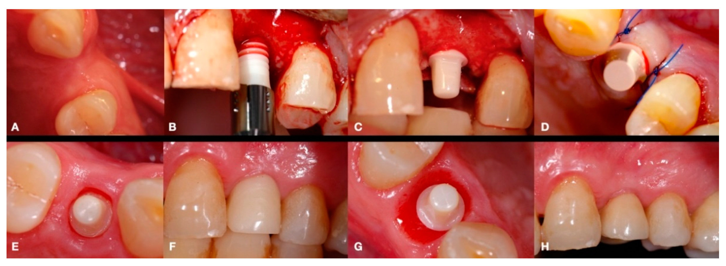
2.2. Surgical Procedures

Digital 3D cone-beam computed tomography (CBCT) presurgical planning (Orthophos XG, Dentsply Sirona, York, PA, USA) was made for each patient. All patients were treated using a full thickness envelope flap with a crestal incision in the center of the alveolar ridge and with a marginal incision around the adjacent neighboring teeth in order to sufficiently expose the crestal bone of the edentulous space. Following this, a conventional drilling sequence was performed according to the instructions of the manufacturer (Straumann® Pure Ceramic implant system, Straumann Holding AG, Basel, Switzerland). The bone drilling sequence involved an initial stainless steel round drill 2.3 mm in diameter, followed by twist drills measuring 2.2 and 2.8 mm in diameter. In the case that a 4.1 mm diameter implant had to be placed, an additional 3.5 mm diameter twist drill was also used. An indicator of the direction of the bone ostectomy (and height of the pillar) was used after the 2.8 mm and 3.5 mm diameter drills to ensure proper positioning of the implant and adequate height of the abutment. Finally, the bone drilling sequence was completed with a stainless-steel profile drill for the 3.5 or 4.1 mm diameter implants. A ceramic implant measuring 8 or 10 mm in length and 3.3 or 4.1 mm in diameter was then installed. Figure 1 illustrates the entire surgical and prosthodontic workflow, and a detailed intraoperative view of a Zr monotype implant placement before flap closure is shown in Figure 2.
Afterwards, GBR procedures were performed if needed, a plastic abutment protector was placed, and flaps were closed with single sutures to ensure transmucosal implant healing. An intraoral radiograph was taken immediately after surgery (Figure 3). Postoperative medication was then prescribed in the form of amoxicillin 500 mg (or clindamycin 300 mg in patients allergic to penicillin) every 8 h for 7 days, ibuprofen 600 mg every 8 h for four days, and chlorhexidine rinses twice a day for 15 days.

2.3. Prosthetic Restoration

Immediately after implant placement surgery, conventional impressions using the manufacture impression copings were performed. A laboratory-processed resin was used to fabricate a temporary crown and was inserted to protect the implant abutment and modulate the gingival contour (Figure 4).
The temporary crown was placed in infraocclusion, ascertaining the absence of occlusal contacts in maximum intercuspidation and during lateral mandibular movements. The gingival implant contours were further adapted by chair-side modifying of the temporary crown. Three months after implant placement, definitive impressions were made using conventional manufacture impression copings. An acrylic try-in was tested and, after laboratory finishing and personalizing, a monolithic zirconia crown was finally cemented using an elastomeric resin cement (Premier® Implant Cement™, Plymouth, MA, USA). Excess cement was carefully removed. Annual follow-up appointments were then scheduled to perform the clinical and radiographic measurements. Figure 5 and Figure 6 illustrate a definitive ceramic nonmetal restoration and the corresponding intraoral X-ray image, respectively, after three years and three months of follow-up.
Lastly, Figure 7 and Figure 8 show a definitive ceramic restoration in another case located in the maxillary left first premolar space and its corresponding intraoral X-ray view, after three years of follow-up.

2.4. Clinical and Radiographic Parameters

Clinical parameters were considered based on the Buser criteria [1]: (a) presence of subjective complaints related to the area of treatment; (b) recurrent infections in the peri-implant region, accompanied by suppuration; (c) mobility of the implant fixture (0 or 1 corresponding to no or yes, respectively); (d) gingival index (GI) at four sites around the implant (mesial, distal, buccal and lingual); (e) amount of plaque detected on the implant via the modified plaque index (mPI), at four sites around the implant (mesial, distal, buccal and lingual); (f) amount of sulcus bleeding according to the modified sulcus bleeding index (mBI), taken at two sites around the implant (buccal and lingual) and (g) peri-implant probing depth (PD) at four sites (mesial, distal, buccal and lingual), measured in millimeters using the North Carolina periodontal probe (Hu-Friedy Mfg. Co., Chicago, IL, USA). The peri-implant indexes (GI, mPI and MBI) are detailed in Table 1.
Additionally, radiological examination comprised an intraoral radiograph of the region of interest using the paralleling technique and the Rinn-type film holder (Kerr Corporation, Bioggio, Switzerland). To evaluate the distance between the implant shoulder and the closest mesial and distal BIC, a calibrated image using special software (Sidexis, Dentsply Sirona, York, PA, USA) was employed. Intraexaminer calibration was performed by taking all measurements two times on different days [30]: the mean of the mesial and distal distance values was calculated per implant.
Thus, each included implant was categorized as success or failure using the collected clinical and radiographic data, based on the predetermined success criteria suggested by Buser et al. [1].

3. Results

3.1. Sample Analysis

A total of 20 dental implants (seven in women and 13 in men) in 10 healthy patients were included in the study. The mean patient age was 60.75 years (range 45–79). Eight patients were nonsmokers, while two patients smoked <5 cigarettes per day. Five implants (25%) were 3.3 mm in diameter and 15 (75%) were 4.1 mm in diameter. Eleven implants were placed in the maxilla (55%) and nine in the mandible (45%). All implants were located in the posterior area (six in premolars and 14 in molars): five in superior premolars (two in the right second premolar and three in the left first premolar); six in the superior molars (two in the right first molar, one in the right second molar, two in the left first molar and one in the left second molar); one in the second left premolar and eight in the inferior molars (three in the right first molar, one in the right second molar, three in the first left molar and one in the second left molar). All implants achieved primary stability and were installed with an insertion torque of at least of 30 Ncm. Additionally, guided bone regeneration was employed simultaneously to implant placement in 12 implants (60%) in order to enhance the implant bone contour. The mean follow-up was 34.05 months (range 24–84). Overall, the definite prosthetic restoration was placed after an average of 2.67 months (range 1.5–7). The detailed sample data are provided in Table 2.

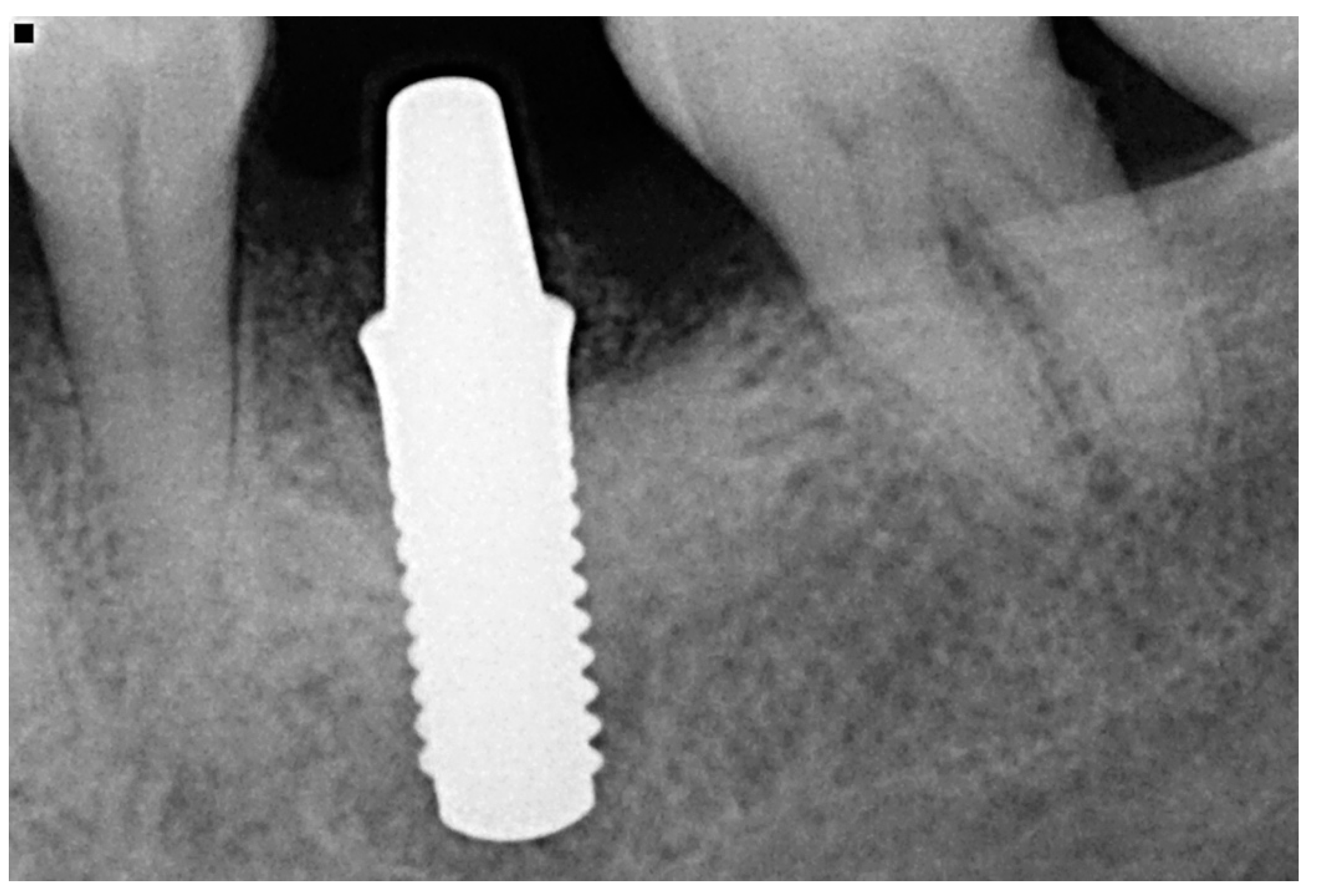
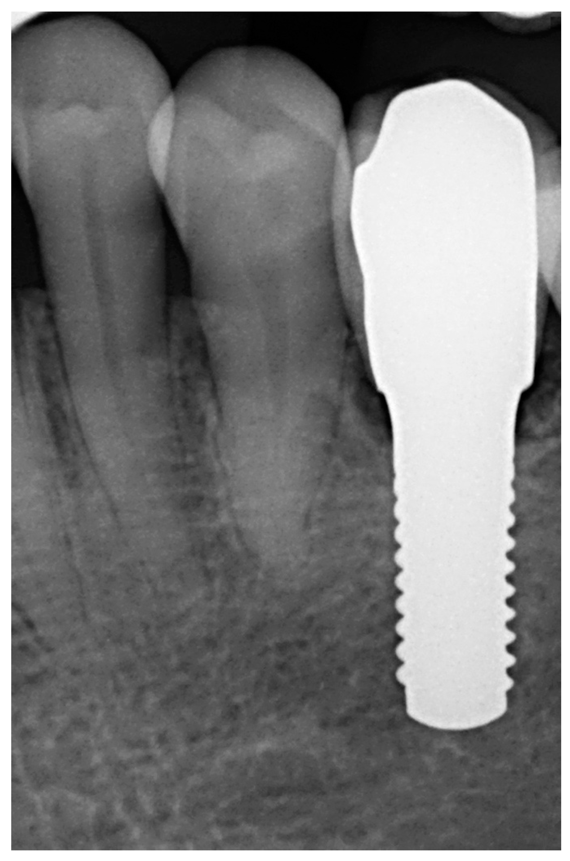
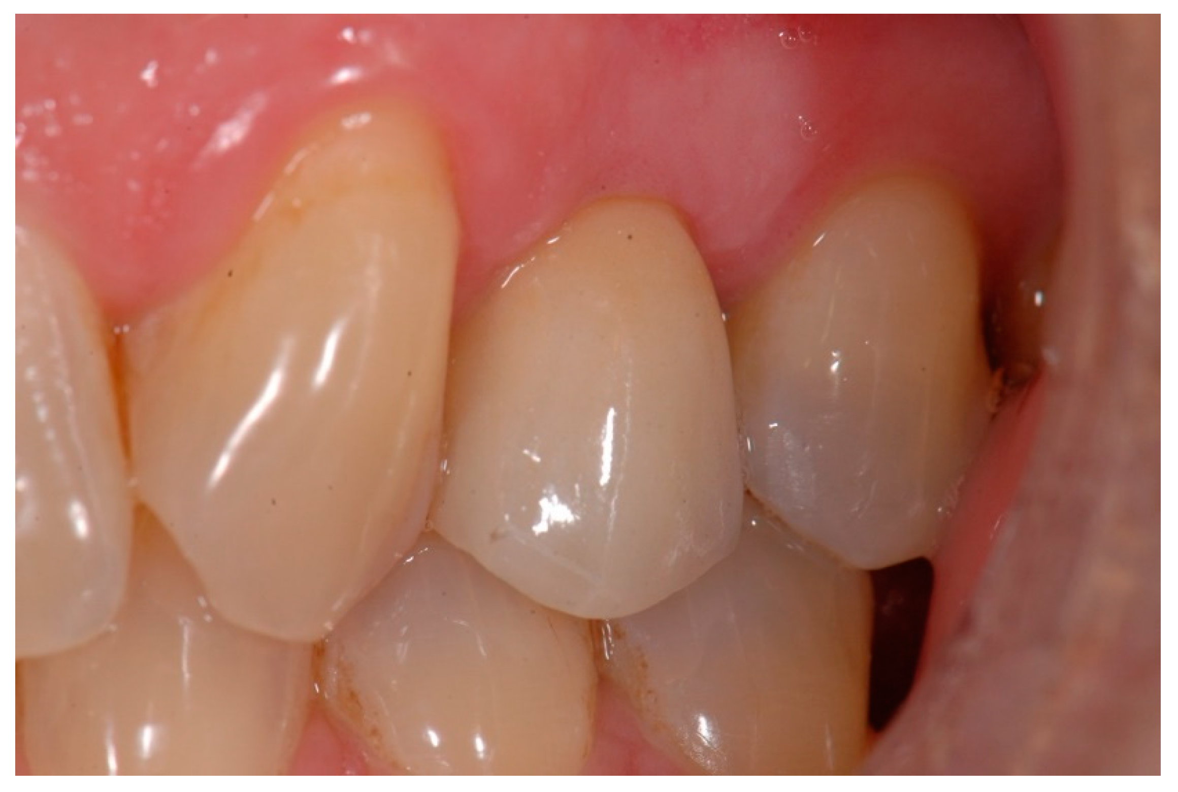
3.2. Clinical and Radiological Outcomes

All implants were classified as successful according to the criteria of Buser et al. [1], with no patient-reported complaints of pain, foreign body sensation or dysesthesia. Implant mobility was not observed in any implant. The mean gingival index, modified plaque index and modified sulcus bleeding index values were 0.38, 0.33 and 1.075, respectively, indicating scarce gingival inflammation and plaque accumulation. A peri-implant soft-tissue bleeding score of close to 1 also represents little bleeding. Nonetheless, bleeding signs were observed in some cases (Table 3). The overall mean probing depth (PD) was 2.56 mm (range 0–5). Lastly, the overall radiological mean distance between the implant shoulder and the initial site of visible bone-to-implant contact (BIC) was 1.44 mm (range 0.17–2.94). The detailed results are reported in Table 3.

4. Discussion

Zirconia implants have been shown to be a plausible alternative to Ti implants due to their biocompatibility and resistance to fracture and compression, being lighter in color and featuring considerably lower bacterial adherence [10,31]. However, some authors have reported lower implant survival rates with Zr implants in comparison to Ti implants [32]. Manzano et al. (2014) showed that a treated Zr implant surface could increase the BIC values, resulting in a reduced occurrence of early failure and reversal torque commensurate versus that of Ti implants [12]. In addition, a meta-analysis [24] has reported Zr implants to have a 91.5% survival rate and a 91.6% success rate over a mean follow-up period of 42.37 months. This study also suggested that Zr-based dental implants may be advantageous in thin gingival phenotypes, especially in the aesthetic zone, compared to Ti implants, due to their white color [33]. The premolar area is less challenging than the canine or incisor zone for one-piece implants, due to the lesser aesthetic demands involved. Two-piece implants may result in a better aesthetic outcome.
Our clinical series displayed optimal clinical and radiological results. All the implants exhibited adequate bone levels and healthy peri-implant tissues at final assessment. These findings are in line with the data presented by Borgonovo et al. [26] and Vilor-Fernández et al. [27], who reported no bleeding, minimal plaque index and hard and soft tissue stability after at least 6 months or 1 year of follow-up, respectively, with one-piece Zr implants, and the clinical attachment level of implants of this kind was seen to remain stable after three years of follow-up, as reported by Balmer et al. [28].
Thus, evaluation of the peri-implant soft-tissue parameters was certainly positive, supporting the observations of previous authors regarding effective adaptation at the gingiva–Zr interface [34]. Furthermore, the absent or scarce plaque accumulation and little sulcus bleeding observed across the cases may be explained by the previously demonstrated lesser bacterial attachment related to Zr implants [35]—ultimately resulting in reduced inflammation and a lesser prevalence of mucositis and/or peri-implantitis as compared to traditional Ti implants. However, these results are to be interpreted with caution, due to the limited sample size and considering that all implants were being evaluated in healthy patients with good oral hygiene and suitable bone volume. In addition, in order to avoid deficient analysis as a consequence of the small data differences found in the clinical and radiological parameters, comparisons of baseline and follow-up measurements were not contemplated.
The overall mean PD of 2.56 mm found in this series may certainly be attributed to the distinct microdesign of the implant, involving a rough surface with a 1.8 mm smooth coronal surface. This specific tissue-level implant design has formerly displayed positive tissue stability in long-term studies [36]. These PD outcomes are slightly lower than those reported by Borgonovo et al. [26] and Balmer et al. [28], who found mean PDs of 3.19 mm and 3.52 mm after at least 6 months or 3 years of follow-up, respectively, both using the same one-piece Zr implant. Furthermore, we must take into account that PD of one-piece Zr implants tends to increase over time, according to different studies [27,28].
All these findings are consistent with the radiographic parameters. The radiological mean distance between the implant shoulder and the initial site of visible bone-to-implant contact was 1.4 mm, indicating good stability of the marginal bone. One-piece implants have been reported to exhibit marginal bone stability superior to that of a two-piece implant by avoiding the implant–abutment microgap and micromovements [22]. Rodriguez et al. [37] combined one- and two-piece Zr implants and recorded similar clinical and radiological outcomes, though with a lower success rate.
With regard to marginal bone loss (MBL), the present study does not address this issue, but Borgonomo et al. observed a MBL of 1.2 mm after four years of follow-up with one-piece Zr implants [38], and Balmer et al. [28] reported 0.72 mm of MBL from implant insertion for three years of follow-up after the final restoration. Additionally, Elnayef et al. reported 0.14 mm more MBL with Zi versus Ti implants [24].
The limitations of our study include (but are not limited to) its small sample size and the lack of comparison with the baseline parameters. Patient recruitment was limited to the cases that fulfilled the inclusion criteria, and this certainly may have had an impact upon data compilation. Although such recruitment implied good homogeneity regarding the general patient conditions and local bone environment, it would be strongly advisable to have a larger sample size. Additionally, some cases were treated with GBR while others were not; one-piece implants rendered abutment modification impossible, this being a major challenge in esthetic zones; and lastly, the cemented restorations with cement margins were determined by the manufacturer. On the other hand, low temperature degradation of zirconia is also a debated phenomenon, though some investigations indicate that ceramics and stabilized zirconia are favorable materials for use in the oral environment in this regard [39,40]. Thus, the results presented in our case series should be interpreted with caution, and larger randomized controlled clinical trials are needed to evaluate the long-term clinical suitability of Zr implants.

5. Conclusions

One-piece zirconia implants seem to be a feasible option in patients with favorable bone anatomy, offering optimal clinical and radiographic outcomes after a mean follow-up period of 34.05 months (range 24–84), showing hard and soft tissue stability at the last follow-up visit. Nevertheless, further research is imperative in order to evaluate the long-term performance of zirconia implants, involving challenging situations such as bone deficiencies and considering the peri-implant clinical and radiologic parameters and the biomechanical behavior of both two-and one-piece zirconia implants.

Author Contributions

Conceptualization, J.G.-A. and K.B.; methodology, J.G.-A. and H.-L.W.; software, J.G.-A. and K.B.; validation, J.G.-A. and H.-L.W.; formal analysis, J.G.-A. and H.-L.W.; investigation, J.G.-A. and K.B.; resources, J.G.-A., K.B. and H.-L.W.; data compilation, J.G.-A. and K.B.; writing—original draft preparation, J.G.-A.; writing—review and editing, J.G.-A.; visualization, J.G.-A.; supervision, H.-L.W.; project administration, J.G.-A. and K.B. All authors have read and agreed to the published version of the manuscript.

Funding

This research received no external funding.

Institutional Review Board Statement

The study present was conducted according to the guidelines of the Declaration of Helsinki and was approved by the Clinical Research Ethics Committee of the Universitat Internacional de Catalunya (study code: CIR-ELC-2021-10) on 20 January 2022.

Informed Consent Statement

Informed consent was obtained from all subjects involved in the study.

Data Availability Statement

The data presented in this study are available upon request to the corresponding author.

Conflicts of Interest

The authors declare that they have no conflict of interest with respect to the publication of this study or the materials described in the manuscript.

References

  1. Buser, D.; Janner, S.F.; Wittneben, J.G.; Bragger, U.; Ramseier, C.A.; Salvi, G.E. 10-year survival and success rates of 511 titanium implants with a sandblasted and acid-etched surface: A retrospective study in 303 partially edentulous patients. Clin. Implant. Dent. Relat. Res. 2012, 14, 839–851. [Google Scholar] [CrossRef] [PubMed]
  2. Sicilia, A.; Cuesta, S.; Coma, G.; Arregui, I.; Guisasola, C.; Ruiz, E.; Maestro, A. Titanium allergy in dental implant patients: A clinical study on 1500 consecutive patients. Clin. Oral Impants Res. 2008, 19, 823–835. [Google Scholar] [CrossRef] [PubMed]
  3. Safioti, L.M.; Kotsakis, G.A.; Pozhitkov, A.E.; Chung, W.O.; Daubert, D.M. Increased levels of dissolved titanium are associated with peri-implantitis—A cross-sectional study. J. Periodontol. 2017, 88, 436–442. [Google Scholar] [CrossRef]
  4. Halperin-Sternfeld, M.; Sabo, E.; Akrish, S. The pathogenesis of implant-related reactive lesions: A clinical, histologic and polarized light microscopy study. J. Periodontol. 2016, 87, 502–510. [Google Scholar] [CrossRef] [PubMed]
  5. Olmedo, D.G.; Paparella, M.L.; Brandizzi, D.; Cabrini, R.L. Reactive lesions of peri-implant mucosa associated with titanium dental implants: A report of 2 cases. Int. J. Oral Maxillofac. Surg. 2010, 39, 503–507. [Google Scholar] [CrossRef]
  6. Liu, F.X.; Wu, C.L.; Zhu, Z.A.; Li, M.Q.; Mao, Y.Q.; Liu, M.; Wang, X.Q.; Yu, D.G.; Tang, T.T. Calcineurin/nfat pathway mediates wear particle-induced tnf-alpha release and osteoclastogenesis from mice bone marrow macrophages in vitro. Acta Pharmacol. Sin. 2013, 34, 1457–1466. [Google Scholar] [CrossRef] [PubMed] [Green Version]
  7. Nishimura, K.; Kato, T.; Ito, T.; Oda, T.; Sekine, H.; Yoshinari, M.; Yajima, Y. Influence of titanium ions on cytokine levels of murine splenocytes stimulated with periodontopathic bacterial lipopolysaccharide. Int. J. Oral Maxillofac. Implants 2014, 29, 472–477. [Google Scholar]
  8. Pettersson, M.; Kelk, P.; Belibasakis, G.N.; Bylund, D.; Molin Thoren, M.; Johansson, A. Titanium ions form particles that activate and execute interleukin-1beta release from lipopolysaccharide-primed macrophages. J. Periodontal Res. 2017, 52, 21–32. [Google Scholar] [CrossRef] [Green Version]
  9. Van Brakel, R.; Noordmans, H.J.; Frenken, J.; de Roode, R.; de Wit, G.C.; Cune, M.S. The effect of zirconia and titanium implant abutments on light reflection of the supporting soft tissues. Clin. Oral Implants Res. 2011, 22, 1172–1178. [Google Scholar] [CrossRef] [Green Version]
  10. Andreiotelli, M.; Wenz, H.J.; Kohal, R.J. Are ceramic implants a viable alternative to titanium implants? A systematic literature review. Clin. Oral Implants Res. 2009, 20 (Suppl. 4), 32–47. [Google Scholar] [CrossRef]
  11. Payer, M.; Arnetzl, V.; Kirmeier, R.; Koller, M.; Arnetzl, G.; Jakse, N. Immediate provisional restoration of single-piece zirconia implants: A prospective case series—Results after 24 months of clinical function. Clin. Oral Implants Res. 2013, 24, 569–575. [Google Scholar] [CrossRef] [PubMed]
  12. Manzano, G.; Herrero, L.R.; Montero, J. Comparison of clinical performance of zirconia implants and titanium implants in animal models: A systematic review. Int. J. Oral Maxillofac. Implants 2014, 29, 311–320. [Google Scholar] [CrossRef] [PubMed] [Green Version]
  13. Depprich, R.; Naujoks, C.; Ommerborn, M.; Schwarz, F.; Kubler, N.R.; Handschel, J. Current findings regarding zirconia implants. Clin. Implant. Dent. Relat. Res. 2014, 16, 124–137. [Google Scholar] [CrossRef] [PubMed]
  14. Ferguson, S.J.; Langhoff, J.D.; Voelter, K.; von Rechenberg, B.; Scharnweber, D.; Bierbaum, S.; Schnabelrauch, M.; Kautz, A.R.; Frauchiger, V.M.; Mueller, T.L.; et al. Biomechanical comparison of different surface modifications for dental implants. Int. J. Oral Maxillofac. Implants 2008, 23, 1037–1046. [Google Scholar]
  15. Langhoff, J.D.; Voelter, K.; Scharnweber, D.; Schnabelrauch, M.; Schlottig, F.; Hefti, T.; Kalchofner, K.; Nuss, K.; von Rechenberg, B. Comparison of chemically and pharmaceutically modified titanium and zirconia implant surfaces in dentistry: A study in sheep. Int. J. Oral Maxillofac. Surg. 2008, 37, 1125–1132. [Google Scholar] [CrossRef]
  16. Koch, F.P.; Weng, D.; Kramer, S.; Biesterfeld, S.; Jahn-Eimermacher, A.; Wagner, W. Osseointegration of one-piece zirconia implants compared with a titanium implant of identical design: A histomorphometric study in the dog. Clin. Oral Implants Res. 2010, 21, 350–356. [Google Scholar] [CrossRef]
  17. Gahlert, M.; Gudehus, T.; Eichhorn, S.; Steinhauser, E.; Kniha, H.; Erhardt, W. Biomechanical and histomorphometric comparison between zirconia implants with varying surface textures and a titanium implant in the maxilla of miniature pigs. Clin. Oral Implants Res. 2007, 18, 662–668. [Google Scholar] [CrossRef]
  18. Sennerby, L.; Dasmah, A.; Larsson, B.; Iverhed, M. Bone tissue responses to surface-modified zirconia implants: A histomorphometric and removal torque study in the rabbit. Clin. Implant. Dent. Relat. Res. 2005, 7, S13–S20. [Google Scholar] [CrossRef]
  19. Bosshardt, D.D.; Chappuis, V.; Buser, B. Osseointegration of titanium, titanium alloy and zirconia dental implants: Current knowledge and open questions. Periodontology 2000 2017, 73, 22–40. [Google Scholar] [CrossRef]
  20. Gahlert, M.; Röhling, S.; Wieland, M.; Eichhorn, S.; Küchenhoff, H.; Kniha, H. A comparison study of the osseointegration of zirconia and titanium dental implants. A biomechanical evaluation in the maxilla of pigs. Clin. Implant. Dent. Relat. Res. 2010, 12, 297–305. [Google Scholar] [CrossRef]
  21. Krautwald, L.; Smeets, R.; Stolzer, C.; Rutkowski, R.; Guo, L.; Reitmeier, A.; Gosau, M.; Henningsen, A. Osseointegration of zirconia implants after UV-light or cold atmospheric plasma surface treatment in vivo. Materials 2022, 15, 496. [Google Scholar] [CrossRef] [PubMed]
  22. Vohra, F.; Al-Kheraif, A.A.; Ab Ghani, S.M.; Abu Hassan, M.I.; Alnassar, T.; Javed, F. Crestal bone loss and periimplant inflammatory parameters around zirconia implants: A systematic review. J. Prosthet. Dent. 2015, 114, 351–357. [Google Scholar] [CrossRef] [PubMed]
  23. Cionca, N.; Hashim, D.; Mombelli, A. Zirconia dental implants: Where are we now, and where are we heading? Periodontol. 2000 2017, 73, 241–258. [Google Scholar] [CrossRef] [PubMed]
  24. Elnayef, B.; Lazaro, A.; Suarez-Lopez Del Amo, F.; Galindo-Moreno, P.; Wang, H.L.; Gargallo-Albiol, J.; Hernandez-Alfaro, F. Zirconia implants as an alternative to titanium: A systematic review and meta-analysis. Int. J. Oral Maxillofac. Implants 2017, 32, e125–e134. [Google Scholar] [CrossRef] [PubMed] [Green Version]
  25. Gahlert, M.; Burtscher, D.; Pfundstein, G.; Grunert, I.; Kniha, H.; Roehling, S. Dental zirconia implants up to three years in function: A retrospective clinical study and evaluation of prosthetic restorations and failures. Int. J. Oral Maxillofac. Implants 2013, 28, 896–904. [Google Scholar] [CrossRef] [Green Version]
  26. Borgonovo, A.E.; Censi, R.; Vavassori, V.; Dolci, M.; Calvo-Guirado, J.L.; Delgado Ruiz, R.A.; Maiorana, C. Evaluation of the success criteria for zirconia dental Implants: A four-year clinical and radiological study. Int. J. Dent. 2013, 2013, 463073. [Google Scholar] [CrossRef]
  27. Vilor-Fernández, V.; García-de-la-Fuente, A.M.; Marichalar-Mendia, X.; Estefanía-Fresco, R.; Aguirre-Zorzano, L.A. Single tooth restoration in the maxillary esthetic zone using a one-piece ceramic implant with 1 year of follow-up: Case series. Int. J. Implant Dent. 2021, 7, 26. [Google Scholar] [CrossRef]
  28. Balmer, M.; Spies, B.C.; Vach, K.; Ralf-Joachim, K.; Hämmerle, C.H.F.; Jung, R.E. Three-year analysis of zirconia implants used for single-tooth replacement and three unit fixed dental prostheses: A prospective multicenter study. Clin. Oral Implants Res. 2018, 29, 290–299. [Google Scholar] [CrossRef] [Green Version]
  29. Hammerle, C.H.; Chen, S.T.; Wilson, T.G., Jr. Consensus statements and recommended clinical procedures regarding the placement of implants in extraction sockets. Int. J. Oral Maxillofac. Implants 2004, 19, 26–28. [Google Scholar]
  30. Bornstein, M.M.; Lauber, R.; Sendi, P.; von Arx, T. Comparison of periapical radiography and limited cone-beam computed tomography in mandibular molars for analysis of anatomical landmarks before apical surgery. J. Endod. 2011, 37, 151–157. [Google Scholar] [CrossRef]
  31. Kohal, R.J.; Knauf, M.; Larsson, B.; Sahlin, H.; Butz, F. One-piece zirconia oral implants: One-year results from a prospective cohort study. 1. Single tooth replacement. J. Clin. Periodontol. 2012, 39, 590–597. [Google Scholar] [CrossRef] [PubMed]
  32. Roehling, S.; Woelfler, H.; Hicklin, S.; Kniha, H.; Gahlert, M. A retrospective clinical study with regard to survival and success rates of zirconia implants up to and after 7 years of loading. Clin. Implant. Dent. Relat. Res. 2016, 18, 545–558. [Google Scholar] [CrossRef] [PubMed]
  33. Belser, U.; Buser, D.; Higginbottom, F. Consensus statements and recommended clinical procedures regarding esthetics in implant dentistry. Int. J. Oral Maxillofac. Implants 2004, 19, 73–74. [Google Scholar] [PubMed]
  34. Glauser, R.; Sailer, I.; Wohlwend, A.; Studer, S.; Schibli, M.; Scharer, P. Experimental zirconia abutments for implant-supported single-tooth restorations in esthetically demanding regions: 4-year results of a prospective clinical study. Int. J. Prosthodont. 2004, 17, 285–290. [Google Scholar] [PubMed]
  35. Rimondini, L.; Cerroni, L.; Carrassi, A.; Torricelli, P. Bacterial colonization of zirconia ceramic surfaces: An in vitro and in vivo study. Int. J. Oral Maxillofac. Implants 2002, 17, 793–798. [Google Scholar] [PubMed]
  36. Chappuis, V.; Buser, R.; Bragger, U.; Bornstein, M.M.; Salvi, G.E.; Buser, D. Long-term outcomes of dental implants with a titanium plasma-sprayed surface: A 20-year prospective case series study in partially edentulous patients. Clin. Implant Dent. Relat. Res. 2013, 15, 780–790. [Google Scholar] [CrossRef]
  37. Rodriguez, A.E.; Monzavi, M.; Yokoyama, C.L.; Nowzari, H. Zirconia dental implants: A clinical and radiographic evaluation. J. Esthet. Restor. Dent. 2018, 30, 538–544. [Google Scholar] [CrossRef]
  38. Borgonovo, A.E.; Fabbri, A.; Vavassori, V.; Censi, R.; Maiorana, C. Multiple teeth replacement with endosseous one-piece yttrium-stabilized zirconia dental implants. Med. Oral Patol. Oral Cir. Bucal. 2012, 17, e981–e987. [Google Scholar] [CrossRef]
  39. Kohorst, P.; Borchers, L.; Strempel, J.; Stiesch, M.; Hassel, T.; Bach, F.W.; Hubsch, C. Low-temperature degradation of different zirconia ceramics for dental applications. Acta Biomater. 2012, 8, 1213–1220. [Google Scholar] [CrossRef]
  40. Lughi, V.; Sergo, V. Low temperature degradation -aging- of zirconia: A critical review of the relevant aspects in dentistry. Dent. Mater. 2010, 26, 807–820. [Google Scholar] [CrossRef]
Figure 1. Surgical and prosthodontic workflow: (A) preoperative occlusal view; (B) one-piece Zr implant placement; (C) Zr transepithelial abutment image before flap closure; (D) immediate postoperative view of the abutment plastic protector and suture; (E) occlusal view of the Zr transepithelial abutment before temporary restoration; (F) lateral view of the temporary restoration; (G) occlusal view of the Zr transepithelial abutment before definitive restoration (note the soft tissue contour modulation using the temporary crown); (H) lateral view of the final restoration.
Materials 15 02437 g001
Figure 2. Intraoperative view of a one-piece zirconia implant placed in the mandibular left second premolar position.
Materials 15 02437 g002
Figure 3. Immediate postoperative intraoral X-ray view after one-piece zirconia implant placement.
Materials 15 02437 g003
Figure 4. Laboratory-made long-term temporary crown performed to modulate the gingival contour.
Materials 15 02437 g004
Figure 5. Definitive ceramic restoration (at 3 years and 3 months of follow-up).
Materials 15 02437 g005
Figure 6. Intra-oral X-ray image after 3 years and 3 months follow-up.
Materials 15 02437 g006
Figure 7. Definitive ceramic restoration in a maxillary left first premolar (after 3 years of follow-up).
Materials 15 02437 g007
Figure 8. Intraoral X-ray view after three years of follow-up.
Materials 15 02437 g008
Table 1. Peri-implant index details based on the Buser criteria [1].
NIndex ScoreParameters
Gingival index (GI)1Normal gingiva: natural coral pink with no inflammation
2Mild inflammation: slight changes in color, slight edema. No bleeding on probing
3Moderate inflammation: redness, edema and glazing
Modified plaque index (mPI)0No detection of plaque
1Plaque only recognized by running a probe across the smooth marginal surface of the implant
2Plaque can be seen by the naked eye
3Abundance of soft matter
Modified sulcus bleeding index (mBI)0No bleeding
1Isolated bleeding spots visible
2Blood forms a confluent red line on margin
3Heavy or profuse bleeding
Table 2. General overview and characteristics of the included cases.
NGenderAgeSmoking (>10 cig/Day)Implant PositionImplant ∅ (mm)Implant Length (mm)Bone Augmentation Definitive Restoration Placement (Months)
1M70Y154.110N2.5
2M70Y264.110Y2.5
3F47N364.112Y3
4F47N464.112Y3
5M50N164.18N3
6M50N464.110N3
7F45N364.110N2.5
8M63N174.110N1.5
9M63N163.310Y1.5
10M63N274.110N1.5
11M63N264.110Y1.5
12M63N373.310Y1.5
13M63N363.310Y1.5
14M63N473.310Y1.5
15M63N463.310Y1.5
16F61N244.112N3
17M75N244.112N4
18F48N154.112Y4
19F69N354.110Y4.0
20F79N244.110Y7.0
Mean 60.75 2.67
Table 3. Clinical and radiological outcomes (GI: gingival index; mPI: modified plaque index; mSBI: modified sulcus bleeding index; PD: probing depth; B: buccal; L: lingual; M: mesial; D: distal; BIC: bone-to-implant contact).
NFollow-Up (Months)GI (B, L, M, D)Mean GImPI (B, L, M, D)Mean mPImSB (B, L)Mean mSBPD (B, L, M, D)Mean PDImplant Shoulder-First BIC (M; D)Mean Implant Shoulder-First BIC
1240, 0, 1, 00.250, 0, 0, 000, 000, 0, 0, 20.50.54; 0.170.355
2240, 1, 0, 10.50, 0, 0, 000, 10.50, 2, 3, 42.251.8; 2.11.95
3240, 2, 0, 10.750, 1, 2, 21.252, 11.53, 4, 3, 43.52.2; 2.22.2
4240, 0, 2, 10.750, 0, 1, 10.50, 10.52, 3, 5, 43.52.3; 2.42.35
5240, 0, 1, 00.250, 0, 1, 10.50, 10.51, 2, 1, 11.251.53; 1.841.685
6240, 0, 0, 10.250, 0, 1, 10.50, 10.51, 2, 1, 21.52.32; 2.152.235
7241, 0, 1, 00.51, 1, 0, 00.50, 002, 1, 4, 32.51.66; 2.011.835
8240, 0, 1, 00.251, 0, 1, 00.52, 32.54, 2, 3, 22.750.81; 0.90.855
9240, 1, 0, 00.250, 1, 0, 10.52, 223, 3, 2, 22.50.45; 0.290.37
10240, 0, 0, 10.250, 0, 0, 002, 11.54, 3, 5, 33.752.2; 1.491.845
11240, 1, 0, 00.250, 1, 0, 00.252, 224, 4, 3, 23.250.15; 2.881.515
12240, 0, 1, 10.50, 1, 1, 10.751, 324, 3, 4, 43.750.43; 0.350.39
13241, 0, 1, 10.750, 0, 0, 10.252, 32.54, 4, 4, 442.75; 0.921.835
14240, 0, 0, 20.50, 0, 0, 10.252, 32.55, 3, 3, 33.50.48; 0.740.61
15240, 0, 1, 10.51, 0, 0, 10.52, 32.55, 4, 4, 342.2; 2.942.57
16360, 1, 1, 10.750, 0, 0, 000, 002, 2, 3, 32.51.9; 2.12
17720, 0, 0, 000, 0, 0, 000, 001, 2, 1, 11.251.8; 2.11.95
18721, 1, 0, 00.50, 0, 0, 000, 001, 2, 1, 11.250.9; 1.81.35
19840, 0, 0, 001, 0, 0, 10.51, 00.52, 1, 1, 11.251.5–1.81.65
20570, 0, 0, 000, 0, 0, 000, 002, 3, 2, 32.50.3–0.40.35
Mean34.05 0.38 0.33 1.075 2.56 1.44
Publisher’s Note: MDPI stays neutral with regard to jurisdictional claims in published maps and institutional affiliations.

