
Novel Trends in Dental Color Match Using Different Shade Selection Methods: A Systematic Review and Meta-Analysis

 ,
,  ,
,  ,
,  ,
,  ,
, 
 and
and 
Abstract
:1. Introduction
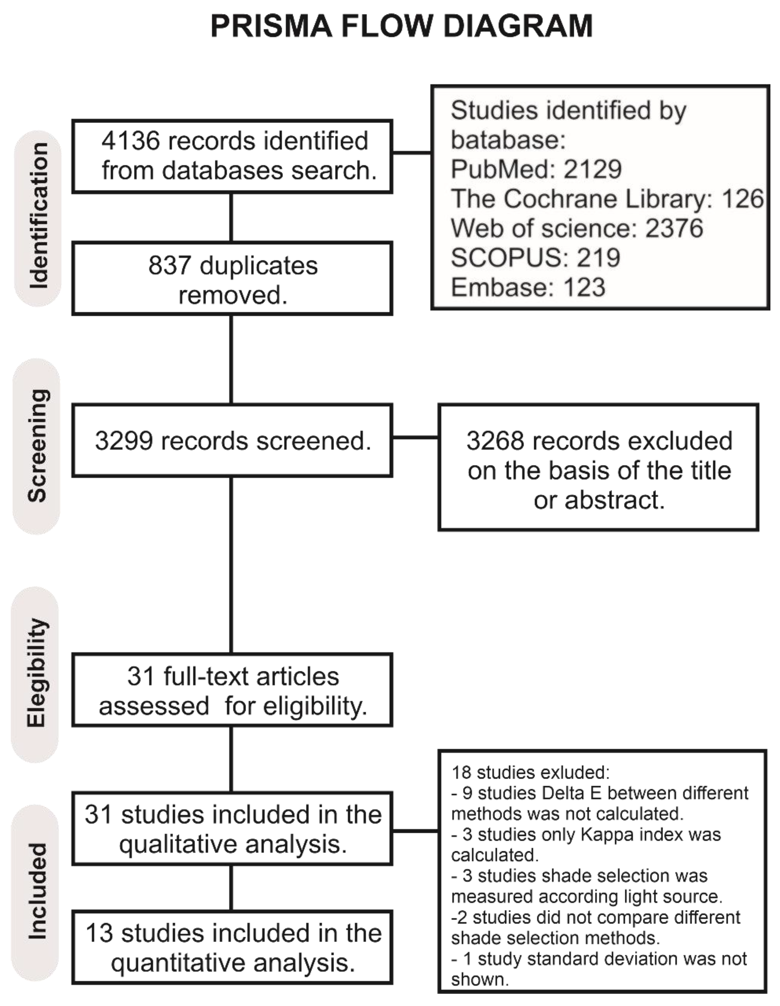
2. Materials and Methods
2.1. Literature Search
2.2. Study Selection
2.3. Data Extraction
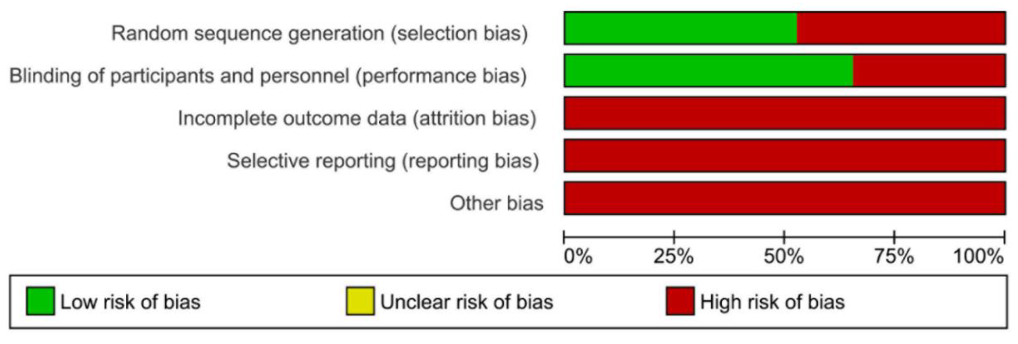
2.4. Quality Assessment
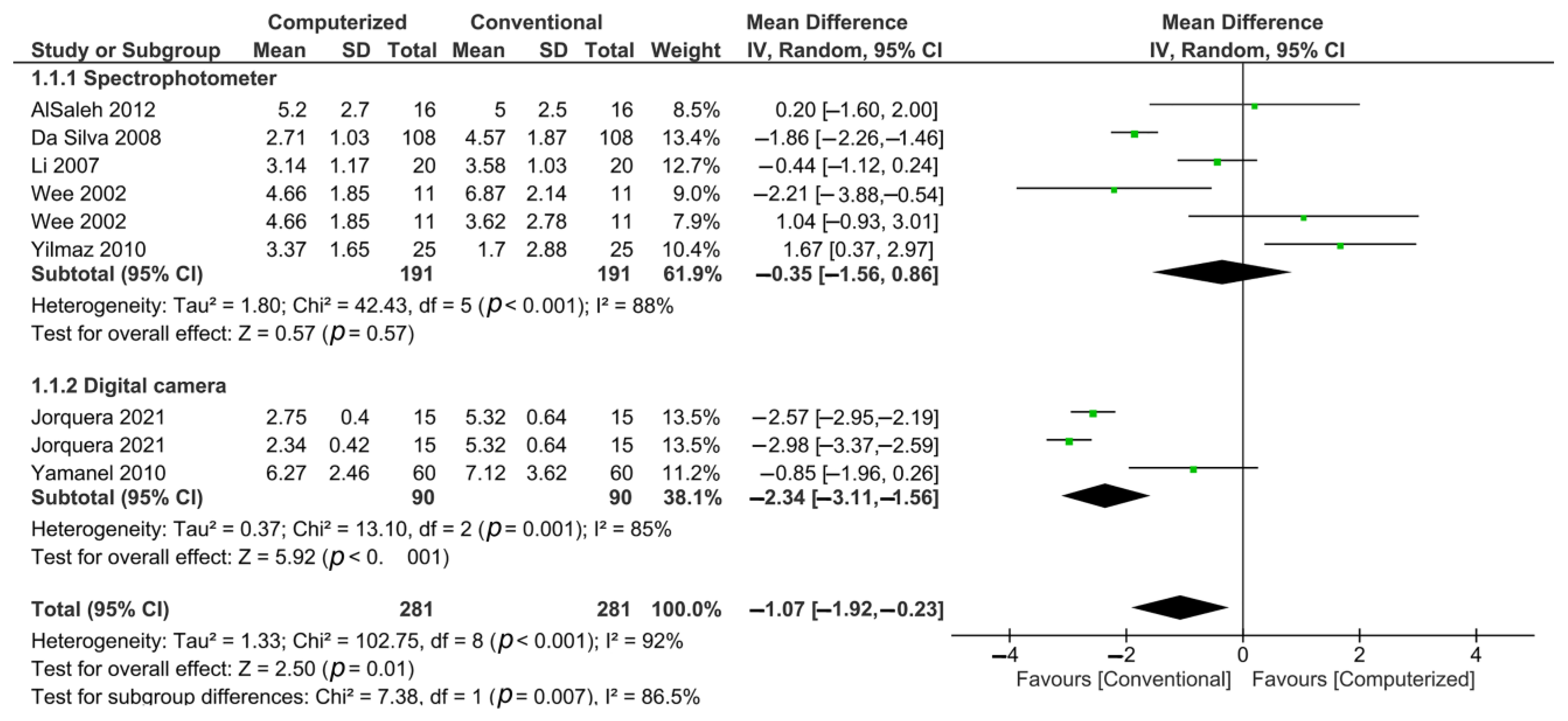
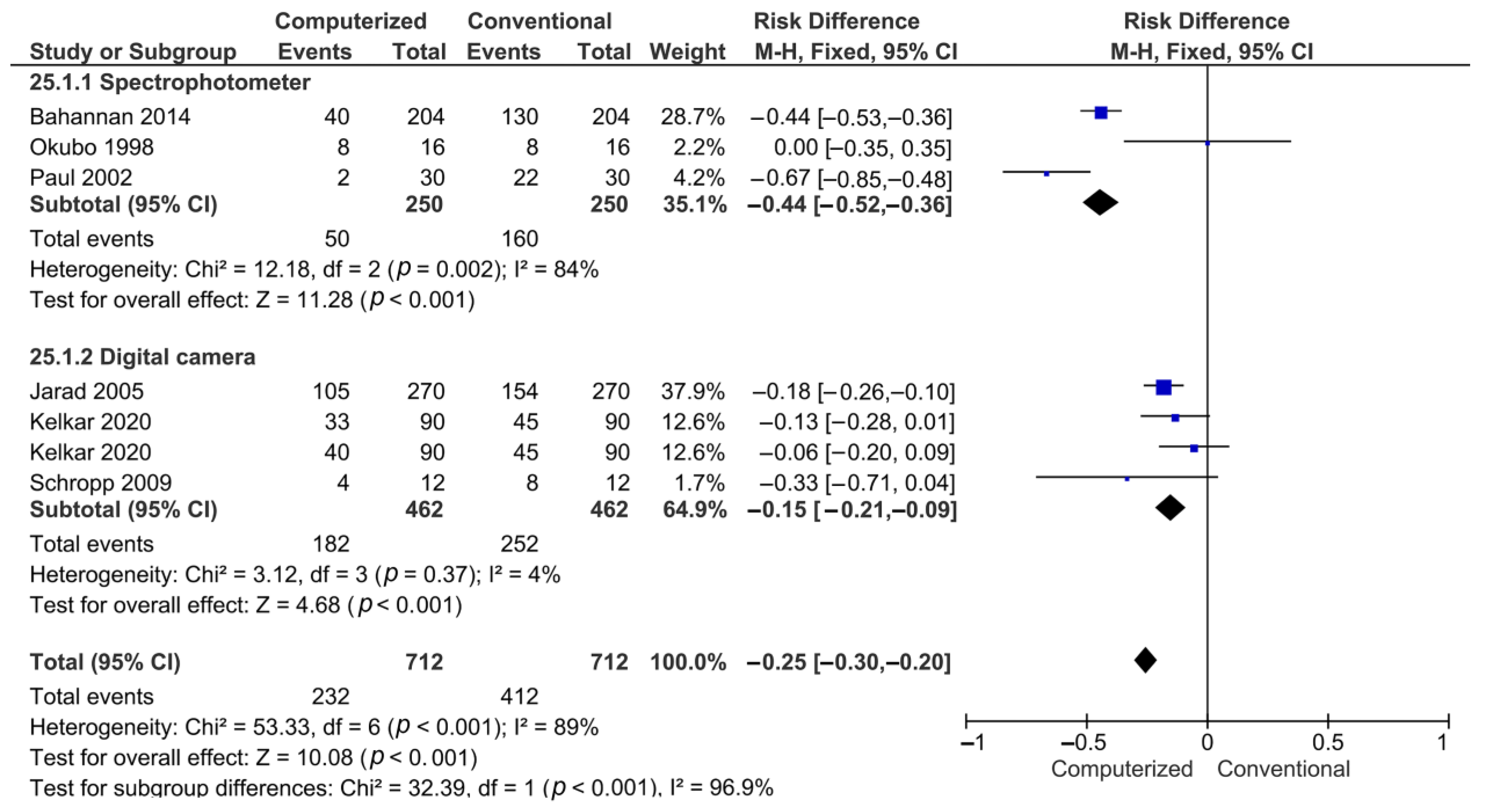
2.5. Statistical Analysis
3. Results
4. Discussion
5. Conclusions
Supplementary Materials
Author Contributions
Funding
Data Availability Statement
Acknowledgments
Conflicts of Interest
References
- Gasparik, C.; Grecu, A.G.; Culic, B.; Badea, M.E.; Dudea, D. Shade-matching Performance Using a New Light-correcting Device. J. Esthet. Res. Dent. 2015, 27, 285–292. [Google Scholar] [CrossRef] [PubMed]
- Öngül, D.; Şermet, B.; Balkaya, M.C. Visual and instrumental evaluation of color match ability of 2 shade guides on a ceramic system. J. Prosthet. Dent. 2012, 108, 9–14. [Google Scholar] [CrossRef]
- Horn, D.J.; Bulan-Brady, J.; Hicks, M.L. Sphere Spectrophotometer versus Human Evaluation of Tooth Shade. J. End. 1998, 24, 786–790. [Google Scholar] [CrossRef]
- Takatsui, F.; de Andrade, M.F.; Neisser, M.P.; Barros, L.A.B.; de Loffredo, L.C.M. CIE L* A* B*: Comparison of Digital Images Obtained Photographically by Manual and Automatic Modes. Braz. Oral Res. 2012, 26, 578–583. [Google Scholar] [CrossRef]
- van der Burgt, T.P.; ten Bosch, J.J.; Borsboom, P.C.F.; Kortsmit, W. A Comparison of New and Conventional Methods for Quantification of Tooth Color. J. Prosthet. Dent. 1990, 63, 155–162. [Google Scholar] [CrossRef]
- Gokce, H.S.; Piskin, B.; Ceyhan, D.; Gokce, S.M.; Arisan, V. Shade Matching Performance of Normal and Color Vision-Deficient Dental Professionals with Standard Daylight and Tungsten Illuminants. J. Prosthet. Dent. 2010, 103, 139–147. [Google Scholar] [CrossRef]
- Preethi Suganya, S.; Manimaran, P.; Saisadan, D.; Dhinesh Kumar, C.; Abirami, D.; Monnica, V. Evaluation of Shade Selection with Digital and Visual Methods. J. Pharm. Bioallied. Sci. 2020, 12, 319–323. [Google Scholar] [CrossRef] [PubMed]
- Tabatabaian, F.; Beyabanaki, E.; Alirezaei, P.; Epakchi, S. Visual and digital tooth shade selection methods, related effective factors and conditions, and their accuracy and precision: A literature review. J. Esthet. Restor. Dent. 2021, 33, 1084–1104. [Google Scholar] [CrossRef]
- Brewer, J.D.; Wee, A.; Seghi, R. Advances in Color Matching. Dent. Clin. 2004, 48, 341–358. [Google Scholar] [CrossRef] [PubMed]
- Douglas, R.D.; Przybylska, M. Predicting Porcelain Thickness Required for Dental Shade Matches. J. Prosthet. Dent. 1999, 82, 143–149. [Google Scholar] [CrossRef]
- Blaes, J. Today’s Technology Improves the Shade-Matching Problems of Yesterday. J. Indiana Dent. Assoc. 2002, 81, 17–19. [Google Scholar] [PubMed]
- Czigola, A.; Róth, I.; Vitai, V.; Fehér, D.; Hermann, P.; Borbély, J. Comparing the effectiveness of shade measurement by intraoral scanner, digital spectrophotometer, and visual shade assessment. J. Esthet. Restor. Dent. 2021, 33, 1166–1174. [Google Scholar] [CrossRef]
- Dietschi, D.; Fahl, N., Jr. Shading concepts and layering techniques to master direct anterior composite restorations: An update. Br. Dent. J. 2016, 221, 765–771. [Google Scholar] [CrossRef]
- Page, M.; McKenzie, J.; Bossuyt, P.; Boutron, I.; Hoffman, T.; Mulrow, C.; Shamseer, L.; Tetzlaff, J.; Akl, E.; Brennan, S.; et al. The PRISMA 2020 Statement: An Updated Guideline for Reporting Systematic Reviews. BMJ 2021, 372, n71. [Google Scholar] [CrossRef]
- Bourgi, R.; Hardan, L.; Rivera-Gonzaga, A.; Cuevas-Suárez, C.E. Effect of Warm-Air Stream for Solvent Evaporation on Bond Strength of Adhesive Systems: A Systematic Review and Meta-Analysis of in Vitro Studies. Int. J. Adhes. Adhes. 2021, 105, 102794. [Google Scholar] [CrossRef]
- Matis, B.A.; Wang, Y.; Jiang, T.; Eckert, G.J. Extended At-Home Bleaching of Tetracycline-Stained Teeth with Different Concentrations of Carbamide Peroxide. Quintessence Int. 2002, 33, 645–655. [Google Scholar] [PubMed]
- Matis, B.A.; Mousa, H.N.; Cochran, M.A.; Eckert, G.J. Clinical Evaluation of Bleaching Agents of Different Concentrations. Quintessence Int. 2000, 31, 303–310. [Google Scholar] [PubMed]
- Mahn, E.; Tortora, S.C.; Olate, B.; Cacciuttolo, F.; Kernitsky, J.; Jorquera, G. Comparison of Visual Analog Shade Matching, a Digital Visual Method with a Cross-Polarized Light Filter, and a Spectrophotometer for Dental Color Matching. J. Prosthet. Dent. 2021, 125, 511–516. [Google Scholar] [CrossRef]
- Hein, S.; Zangl, M. The Use of a Standardized Gray Reference Card in Dental Photography to Correct the Effects of Five Commonly Used Diffusers on the Color of 40 Extracted Human Teeth. Int. J. Esthet. Dent. 2016, 11, 246–259. [Google Scholar]
- Kröger, E.; Matz, S.; Dekiff, M.; Tran, B.L.; Figgener, L.; Dirksen, D. In Vitro Comparison of Instrumental and Visual Tooth Shade Determination under Different Illuminants. J. Prosthet. Dent. 2015, 114, 848–855. [Google Scholar] [CrossRef]
- He, W.H.; Park, C.J.; Byun, S.; Tan, D.; Lin, C.Y.; Chee, W. Evaluating the Relationship between Tooth Color and Enamel Thickness, Using Twin Flash Photography, Cross-Polarization Photography, and Spectrophotometer. J. Esthet. Dent. 2020, 32, 91–101. [Google Scholar] [CrossRef]
- Matis, B.A.; Wang, Y.; Jiang, T.; Eckert, G.J. A Clinical Evaluation of a Bleaching Agent Used With and Without Reservoirs. Oper. Dent. 2002, 27, 5–11. [Google Scholar] [PubMed]
- Miyajiwala, J.; Kheur, M.; Patankar, A.; Lakha, T. Comparison of Photographic and Conventional Methods for Tooth Shade Selection: A Clinical Evaluation. J. Indian Prosthodont. Soc. 2017, 17, 273–281. [Google Scholar]
- Wang, P.; Wei, J.; Li, Q.; Wang, Y. Evaluation of an optimized shade guide made from porcelain powder mixtures. J. Prosthet. Dent. 2014, 112, 1553–1558. [Google Scholar] [CrossRef] [PubMed]
- Tung, O.H.; Lai, Y.L.; Ho, Y.C.; Chou, I.C.; Lee, S.Y. Development of Digital Shade Guides for Color Assessment Using a Digital Camera with Ring Flashes. Clin. Oral Investig. 2011, 15, 49–56. [Google Scholar] [CrossRef]
- Olms, C.; Setz, J.M. The Repeatability of Digital Shade Measurement-a Clinical Study. Clin. Oral Investig. 2013, 17, 1161–1166. [Google Scholar] [CrossRef]
- Sampaio, C.S.; Atria, P.J.; Hirata, R.; Jorquera, G. Variability of Color Matching with Different Digital Photography Techniques and a Gray Reference Card. J. Prosthet. Dent. 2019, 121, 333–339. [Google Scholar] [CrossRef]
- Pimentel, W.; Tiossi, R. Comparison between Visual and Instrumental Methods for Natural Tooth Shade Matching. Gen. Dent. 2014, 62, 47–49. [Google Scholar]
- Gómez-Polo, C.; Gómez-Polo, M.; Celemin-Viñuela, A.; Martínez Vázquez De Parga, J.A. Differences between the Human Eye and the Spectrophotometer in the Shade Matching of Tooth Colour. J. Dent. 2014, 42, 742–745. [Google Scholar] [CrossRef] [PubMed]
- Matis, B.; Nousa, H.N.; Cochran, M.A.; Eckert, G. Clinical Evaluation of In-Office and at-Home Bleaching Treatments Dental Trauma View Project Polymerization Pattern within a Resin-Based Composite View Project. Esth. Dent. 2000, 31, 303–310. [Google Scholar]
- Chitrarsu, V.K.; Chidambaranathan, A.S.; Balasubramaniam, M. Analysis of Shade Matching in Natural Dentitions Using Intraoral Digital Spectrophotometer in LED and Filtered LED Light Sources. J. Prosth. 2019, 28, e68–e73. [Google Scholar] [CrossRef] [Green Version]
- Lakhanpal, S.; Neelima, M.S. Accuracy of Three Shade-Matching Devices in Replicating the Shade of Metal Ceramic Restorations: An in Vitro Study. J. Contemp. Dent. Pract. 2016, 17, 1003–1008. [Google Scholar] [CrossRef]
- Baharin, S.A.; Tey, Y.D.; Tan, W.J. Anterior Tooth Shade Selection Procedure: Influence of Light Sources and Patient’s Position. Sains Malays. 2013, 42, 7–11. [Google Scholar]
- Kelkar, K.C.; Dogra, E.S.; Bhat, V.; Prasad, D.K.; Hegde, C. A Comparison between Visual, Digital Photography and Polarizing Filter Photography for Shade Selection. Indian J. Dent. Res. 2020, 31, 712. [Google Scholar]
- Jorquera, G.J.; Atria, P.J.; Galán, M.; Feureisen, J.; Imbarak, M.; Kernitsky, J.; Cacciuttolo, F.; Hirata, R.; Sampaio, C.S. A Comparison of Ceramic Crown Color Difference between Different Shade Selection Methods: Visual, Digital Camera, and Smartphone. J. Prosthet. Dent. 2021. [Google Scholar] [CrossRef]
- Yamanel, K.; Caglar, A.; Özcan, M.; Gulsah, K.; Bagis, B. Assessment of Color Parameters of Composite Resin Shade Guides Using Digital Imaging versus Colorimeter. J. Esthet. Dent. 2010, 22, 379–388. [Google Scholar] [CrossRef] [PubMed]
- Li, Q.; Wang, Y.N. Comparison of Shade Matching by Visual Observation and an Intraoral Dental Colorimeter. J. Oral Rehabil. 2007, 34, 848–854. [Google Scholar] [CrossRef] [PubMed]
- Alsaleh, S.; Labban, M.; Alhariri, M.; Tashkandi, E. Evaluation of Self Shade Matching Ability of Dental Students Using Visual and Instrumental Means. J. Dent. 2012, 40, e82–e87. [Google Scholar] [CrossRef]
- da Silva, J.D.; Park, S.E.; Weber, H.-P.; Dent, M.; Ishikawa-Nagai, S.; Silva, D. Clinical Performance of a Newly Developed Spectrophotometric System on Tooth Color Reproduction. J. Prosthet. Dent. 2008, 99, 361–368. [Google Scholar] [CrossRef]
- Yilmaz, B.; Karaagaclioglu, L. In Vitro Evaluation of Color Replication of Metal Ceramic Specimens Using Visual and Instrumental Color Determinations. J. Prosthet. Dent. 2010, 105, 21–27. [Google Scholar] [CrossRef]
- Paul, S.; Peter, A.; Pietrobon, N.; Hämmerle, C.H.F. Visual and Spectrophotometric Shade Analysis of Human Teeth. J. Dent. Res. 2002, 81, 578–582. [Google Scholar] [CrossRef]
- Schropp, L. Shade Matching Assisted by Digital Photography and Computer Software. J. Prosthodont. 2009, 18, 235–241. [Google Scholar] [CrossRef] [PubMed]
- Jarad, F.D.; Russell, M.D.; Moss, B.W. The Use of Digital Imaging for Colour Matching and Communication in Restorative Dentistry. Br. Dent. J. 2005, 199, 43–49. [Google Scholar] [CrossRef] [Green Version]
- Bahannan, S.A. Shade Matching Quality among Dental Students Using Visual and Instrumental Methods. J. Dent. 2014, 42, 48–52. [Google Scholar] [CrossRef]
- Okubo, S.R.; Kanawati, A.; Richards, M.W.; Childressd, S. Evaluation of Visual and Instrument Shade Matching. J. Prosthet. Dent. 1998, 80, 642–648. [Google Scholar] [CrossRef]
- Wee, A.G.; Monaghan, P.; Johnston, W.M. Variation in Color between Intended Matched Shade and Fabricated Shade of Dental Porcelain. J. Prosthet. Dent. 2002, 87, 657–666. [Google Scholar] [CrossRef]
- Alghazali, N.; Burnside, G.; Moallem, M.; Smith, P.; Preston, A.; Jarad, F.D. Assessment of Perceptibility and Acceptability of Color Difference of Denture Teeth. J. Dent. 2012, 40, e10–e17. [Google Scholar] [CrossRef] [PubMed]
- Bergman, B.; Nilson, H.; Andersson, M. A Longitudinal Clinical Study of Procera Ceramic-Veneered Titanium Copings. Int. J. Prosthodont. 1999, 12, 135–139. [Google Scholar]
- Paolone, G.; Orsini, G.; Manauta, J.; Devoto, W.; Putignano, A. Composite shade guides and color matching. Int. J. Esthet Dent. 2014, 9, 164–182. [Google Scholar]
- Haddad, H.J.; Jakstat, H.A.; Arnetzl, G.; Borbely, J.; Vichi, A.; Dumfahrt, H.; Renault, P.; Corcodel, N.; Pohlen, B.; Marada, G.; et al. Does gender and experience influence shade matching quality? J. Dent. 2009, 37, 40–44. [Google Scholar] [CrossRef]
- Tam, W.K.; Lee, H.J. Dental Shade Matching Using a Digital Camera. J. Dent. 2012, 40, e3–e10. [Google Scholar] [CrossRef]
- Clary, J.A.; Ontiveros, J.C.; Cron, S.G.; Paravina, R.D. Influence of Light Source, Polarization, Education, and Training on Shade Matching Quality. J. Prosthet. Dent. 2016, 116, 91–97. [Google Scholar] [CrossRef]
- Alomari, M.; Chadwick, R.G. Factors influencing the shade matching performance of dentists and dental technicians when using two different shade guides. Br. Dent. J. 2011, 211, 23. [Google Scholar] [CrossRef] [PubMed] [Green Version]
- Hardan, L. Protocols for Mobile Dental Photography with Auxiliary Lighting. J. Craniomandib. Sleep Pract. 2021, 39, 181. [Google Scholar]
- Tam, W.-K.; Lee, H.-J. Accurate Shade Image Matching by Using a Smartphone Camera. J. Prosthodont. Res. 2017, 61, 168–176. [Google Scholar] [CrossRef]
- Sampaio, C.S.; Gurrea, J.; Gurrea, M.; Bruguera, A.; Atria, P.J.; Janal, M.; Bonfante, E.A.; Coelho, P.G.; Hirata, R. Dental Shade Guide Variability for Hues B, C, and D Using Cross-Polarized Photography. Int. J. Periodontics Restor. Dent. 2018, 38, s113–s118. [Google Scholar] [CrossRef] [PubMed] [Green Version]
- Gurrea, J.; Gurrea, M.; Bruguera, A.; Sampaio, C.S.; Janal, M.; Bonfante, E.; Coelho, P.G.; Hirata, R. Evaluation of Dental Shade Guide Variability Using Cross-Polarized Photography. Int. J. Periodontics Restor. Dent. 2016, 36, e76–e81. [Google Scholar] [CrossRef] [PubMed] [Green Version]
- Johnston, W.M.; Kao, E.C. Assessment of Appearance Match by Visual Observation and Clinical Colorimetry. J. Dent. Res. 1989, 68, 819–822. [Google Scholar] [CrossRef]
- Wee, A.G.; Lindsey, D.T.; Kuo, S.; Johnston, W.M. Color Accuracy of Commercial Digital Cameras for Use in Dentistry. Dent. Mater. 2006, 22, 553–559. [Google Scholar] [CrossRef]
- Monterubbianesi, R.; Tosco, V.; Bellezze, T.; Giuliani, G.; Özcan, M.; Putignano, A.; Orsini, G. A Comparative Evaluation of Nanohydroxyapatite-Enriched Hydrogen Peroxide Home Bleaching System on Color, Hardness and Microstructure of Dental Enamel. Materials 2021, 14, 3072. [Google Scholar] [CrossRef]
- Chu, S.J.; Trushkowsky, R.D.; Paravina, R.D. Dental color matching instruments and systems. Review of clinical and research aspects. J. Dent. 2010, 38, 2–16. [Google Scholar] [CrossRef] [PubMed]
- Hardan, L.S.; Moussa, C. Mobile Dental Photography: A Simple Technique for Documentation and Communication. Quintessence Int. 2020, 51, 510–518. [Google Scholar] [PubMed]
- Moussa, C.; Hardan, L.; Kassis, C.; Bourgi, R.; Devoto, W.; Jorquera, G.; Panda, S.; Abou Fadel, R.; Cuevas-Suárez, C.E.; Lukomska-Szymanska, M. Accuracy of Dental Photography: Professional vs. Smartphone’s Camera. BioMed Res. Int. 2021, 2021, 3910291. [Google Scholar] [CrossRef] [PubMed]

| Population | Color of the Tooth |
| Intervention | Spectroscopic or camera method for color determination |
| Control | Clinical perception using a shade tab guide |
| Outcome | Shade match |
| Study design | Randomized clinical trials and in vitro studies |
| #1 | Color OR Color measurement OR Colorimeters OR Spectrophotometers OR spectrophotometer OR CIE L*a*b* OR Tooth Color OR Color Shade OR dental color OR spectrophotometry OR colorimetry OR color perception OR color matching OR color accuracy OR spectroradiometry OR color* |
| #2 | Smartphone OR Mobile dental photography OR Digital camera OR dental photography OR digital photography OR DSLR camera OR Mobile camera OR Cell Phone Use OR photography OR photography* OR digital dentistry OR polarizing filter OR light OR light* |
| #3 | Shade communication OR shade matching OR shade selection OR shade guide OR shade OR dental shade OR shade determination OR dental shade OR shade selection program OR visual shade matching |
| #4 | #1 and #2 and #3 |
| Study | Method Used for Shade Match | Materials Used | Type of Room Light | Main Outcome | Main Results |
|---|---|---|---|---|---|
| Jorquera, 2021 |
| Human tooth | The room had ambient light between 5500 K and 6500 K. The clothing of the volunteers was covered with a neutral color cloth. | E | Digital shade choice using both a digital camera and a smartphone displayed a threshold within the adequate values (ΔE < 3.7). Visual shade choice displayed an average ∆E above the level for acceptable values (ΔE > 3.7). |
| Mahn, 2021 |
| Human tooth | The room had ambient light between 5500 K and 6500 K. The clothing of the volunteers was covered with a neutral color cloth. | ΔE | No statistically significant differences within digital images and spectrophotometer. The visual shade process led to enormous variances compared to the other approaches beneath study. |
| Sampaio, 2019 |
| Human tooth | For comparison, the measurements were made in the same spots. | E | Using a cross-polarizing filter consequences in more color-standardized photographs; however, using an iPhone 7 and a ring flash system resulted in fewer standardized pictures. |
| Zekonis, 2002 |
| Human tooth | Non-specified | E | At-home (10% carbamide peroxide) treatment sides were meaningfully distinctive from the in-office (35% hydrogen peroxide) treatment sides through all active treatment stages and during follow-up visits conferring to all three color assessment approaches. |
| Matis, 2002 |
| Human tooth | Non-specified | E | Shade guide and slide photography data displayed no meaningful variances between teeth lightened with agent with or without reservoirs. |
| Jarad, 2003 |
| Porcelain shade tabs | All background lighting in the room was maintained at a consistent level for all sessions. (Colour temperature was set at 6500 K). | CIELab | The viewers’ shade-harmonizing performance was significantly improved using the computer method compared to the conventional one. |
| Matis, 2000 |
| Human tooth | Non-specified | CIELab and ΔE | All three methods of evaluation revealed a significant difference in the tooth lightness. |
| Matis, 2000 |
| Human tooth | Non-specified | CIELab | This study suggests that when a higher concentra-tion of carbamide peroxide was used, the further the lightness value and ΔE altered. |
| Gómez-Polo, 2014 |
| Human tooth | Recordings were made under fluorescent tubes with daylight and an intensity of 1200–1500 lux, in the same room under standardized lighting conditions. | Lightness Chroma Hue | This research showed differences between the measurement of color using the spectrophotometry tool and the visual shade selection technique. |
| Kröger, 2015 |
| Human tooth | A room with dimmed fluorescent ceiling light. | CIELab | The spectrophotometer provided higher reproducibility. |
| Wang, 2014 |
| Human tooth | Northern daylight | CIELab and ΔE | An optimized shade guide improved the performance of color selection |
| He, 2019 |
| Human tooth | Non-specified | CIELab and ΔE | Combining non-polarized photography, cross-polarization photography, and spectrophotometer approaches were considered reasonable for shade matching. |
| AlSaleh, 2012 |
| Human tooth | A light grey wall in a room away from all windows. | CIELab and ΔE | Analysis using the spectrophotometric shade was considered more accurate in comparison to human shade evaluation. |
| Baharin, 2013 |
| Human tooth | Non-specified | Accuracy | This sudy revealed that for the anterior tooth, the patient’s position, lighting condition and number of readings acquired does impact the outcome of shade selection. |
| Bahannan, 2014 |
| Human tooth | Daylight | Daylight illuminator (GTI Graphic Technology, NY, USA) | The conventional visual method was significantly inferior compared to the shade assessment method device. |
| Chitrarsu, 2017 |
| Natural dentitions | Daylight, incandescent light, LED, and filtered LED. | CIELab | Vita Toothguide 3D-Master showed statistically important variances in shade matching in comparison to the intraoral digital spectrophotometer. |
| Da Silva, 2008 |
| Tooth color for anterior metal ceramic restorations | Under daylight and color temperature of 6500 °K. | ΔE | For anterior metal ceramic restorations, using a spectrophotometric method is an effective device for imitating and communicating the color of the tooth. |
| Hein, 2016 |
| Extracted human teeth | Non-specified | CIELab and ΔE | The use of a white balance reference card with acknowledged color coordinates can be suggested when diffusers are used for dental photography. |
| Miyajiwala, 2017 |
| Human tooth | Daylight | CIELab | Clinically, for shade selection, the use of the digital photography method can appear as a viable alternative to the use of spectrophotometric method. |
| Li, 2007 |
| Human tooth | Northern daylight | CIELab | The consistency of shade matching cannot be guaranteed by either the visual method or the colorimeter approach. |
| Pimentel, 2014 |
| Natural tooth | Controlled illumination | Accuracy | Shade match using the instrumental method presented more agreement than shade match using the visual method. |
| Okubo, 1998 |
| Ceramic shade guide teeth | Northern daylight | CIELab and ΔE | Color matching using the visual process is unpredictable. However, instrumental measurement of tooth color would deliver objective measured data to match the color of the natural teeth. |
| Olms, 2013 |
| Ceramic veneer | Ceiling lighting (Philips Master TLD 36 W) and a dental lamp (KaVo Dental GmbH, Germany). | CIELab | This study confirmed that worthy outcomes in terms of the repeatability and precision were obtained with the help of the VITA Easyshade measurements. |
| Paul, 2002 |
| Human tooth | Light source (6500 K). | ΔE | Human shade assessment is less accurate and less reproducible when compared to spectrophotometric shade evaluation. |
| Schropp, 2008 |
| Phantom head | Daylight lamps with a color temperature of 4800 K. | CIE LCh | Digital photographs and computer software were significantly more trustworthy than conventional visual approach for shade-matching analysis. |
| Tung, 2010 |
| Ceramic disks | The background lighting in the room was subdued and maintained at a constant level during the entire experiment. | CIELab | Digital images were more influenced by the illuminants and camera’s white balance setups when testing the reliability of color match. |
| Wee, 2002 |
| Dental porcelain | Color-corrected D65 lighting. | CIELab | The largest mean ∆E was noted for the Vitapan 3D-Master system, which was considerably distinct from the Vita Lumin and Shofu ShadeEye systems. |
| Yamanel, 2010 |
| Composite resin shade guides | Two 6500-K fluorescent tubes were combined with two 2700 K fluorescent tubes. | CIELab | The mean ∆E values verified a statistically significant difference with the colorimeter method. However, there was no significant change when using the digital imaging approach. |
| Yilmaz, 2010 |
| Metal ceramic | Non-specified | CIELab | Color imitation using instrumental shade method for the specimens made of metal ceramic was less accurate when compared to the visual shade approach. |
| Lakhanpal, 2016 |
| Extracted non-carious premolars | Dark room setup. | CIELab | A statistically important association was found to exist with the spectrophotometer and the polarization dental imaging modality for all CIE Lab color coordinates |
| Kelkar, 2020 |
| VITAPAN | Daylight | Individual Matching Ability of each Observer | For obtaining an aesthetic outcome, the use of digital photographic approach was considered most accurate among the three shade selection approaches. |
| Study | Specimen Randomization | Single Operator | Operator Blinded | Control Group | Standardized Specimens | Sample Size Calculation | Risk of Bias |
|---|---|---|---|---|---|---|---|
| Jarad, 2003 | YES | YES | NO | YES | YES | NO | Medium |
| Kröger, 2015 | YES | NO | NO | YES | YES | YES | Medium |
| He, 2019 | NO | NO | NO | YES | YES | NO | High |
| Hein, 2016 | YES | NO | NO | YES | YES | NO | Medium |
| Schropp, 2008 | NO | NO | YES | YES | YES | YES | Medium |
| Tung, 2010 | YES | NO | YES | YES | YES | NO | Medium |
| Wee, 2002 | YES | NO | NO | YES | YES | NO | Medium |
| Yamanel, 2010 | NO | NO | NO | YES | YES | NO | High |
| Yilmaz, 2010 | YES | NO | YES | YES | YES | YES | Low |
| Lakhanpal, 2016 | NO | NO | NO | YES | YES | NO | High |
| Kelkar, 2020 | YES | NO | NO | YES | YES | YES | Medium |
Publisher’s Note: MDPI stays neutral with regard to jurisdictional claims in published maps and institutional affiliations. |
© 2022 by the authors. Licensee MDPI, Basel, Switzerland. This article is an open access article distributed under the terms and conditions of the Creative Commons Attribution (CC BY) license (https://creativecommons.org/licenses/by/4.0/).
Share and Cite
Hardan, L.; Bourgi, R.; Cuevas-Suárez, C.E.; Lukomska-Szymanska, M.; Monjarás-Ávila, A.J.; Zarow, M.; Jakubowicz, N.; Jorquera, G.; Ashi, T.; Mancino, D.; et al. Novel Trends in Dental Color Match Using Different Shade Selection Methods: A Systematic Review and Meta-Analysis. Materials 2022, 15, 468. https://doi.org/10.3390/ma15020468
Hardan L, Bourgi R, Cuevas-Suárez CE, Lukomska-Szymanska M, Monjarás-Ávila AJ, Zarow M, Jakubowicz N, Jorquera G, Ashi T, Mancino D, et al. Novel Trends in Dental Color Match Using Different Shade Selection Methods: A Systematic Review and Meta-Analysis. Materials. 2022; 15(2):468. https://doi.org/10.3390/ma15020468
Chicago/Turabian StyleHardan, Louis, Rim Bourgi, Carlos Enrique Cuevas-Suárez, Monika Lukomska-Szymanska, Ana Josefina Monjarás-Ávila, Maciej Zarow, Natalia Jakubowicz, Gilbert Jorquera, Tarek Ashi, Davide Mancino, and et al. 2022. "Novel Trends in Dental Color Match Using Different Shade Selection Methods: A Systematic Review and Meta-Analysis" Materials 15, no. 2: 468. https://doi.org/10.3390/ma15020468
APA StyleHardan, L., Bourgi, R., Cuevas-Suárez, C. E., Lukomska-Szymanska, M., Monjarás-Ávila, A. J., Zarow, M., Jakubowicz, N., Jorquera, G., Ashi, T., Mancino, D., Kharouf, N., & Haikel, Y. (2022). Novel Trends in Dental Color Match Using Different Shade Selection Methods: A Systematic Review and Meta-Analysis. Materials, 15(2), 468. https://doi.org/10.3390/ma15020468