
Color and Translucency Stability of Three-Dimensional Printable Dental Materials for Crown and Bridge Restorations

 , ,
, ,  ,
, 

Abstract
1. Introduction
2. Materials and Methods
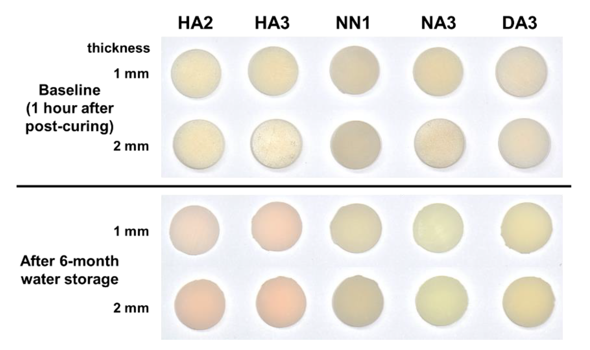
2.1. Specimen Preparation
2.2. Measurement of Color and Translucency
2.3. Calculation of Color and Translucency Differences
2.4. Statistical Analysis
3. Results
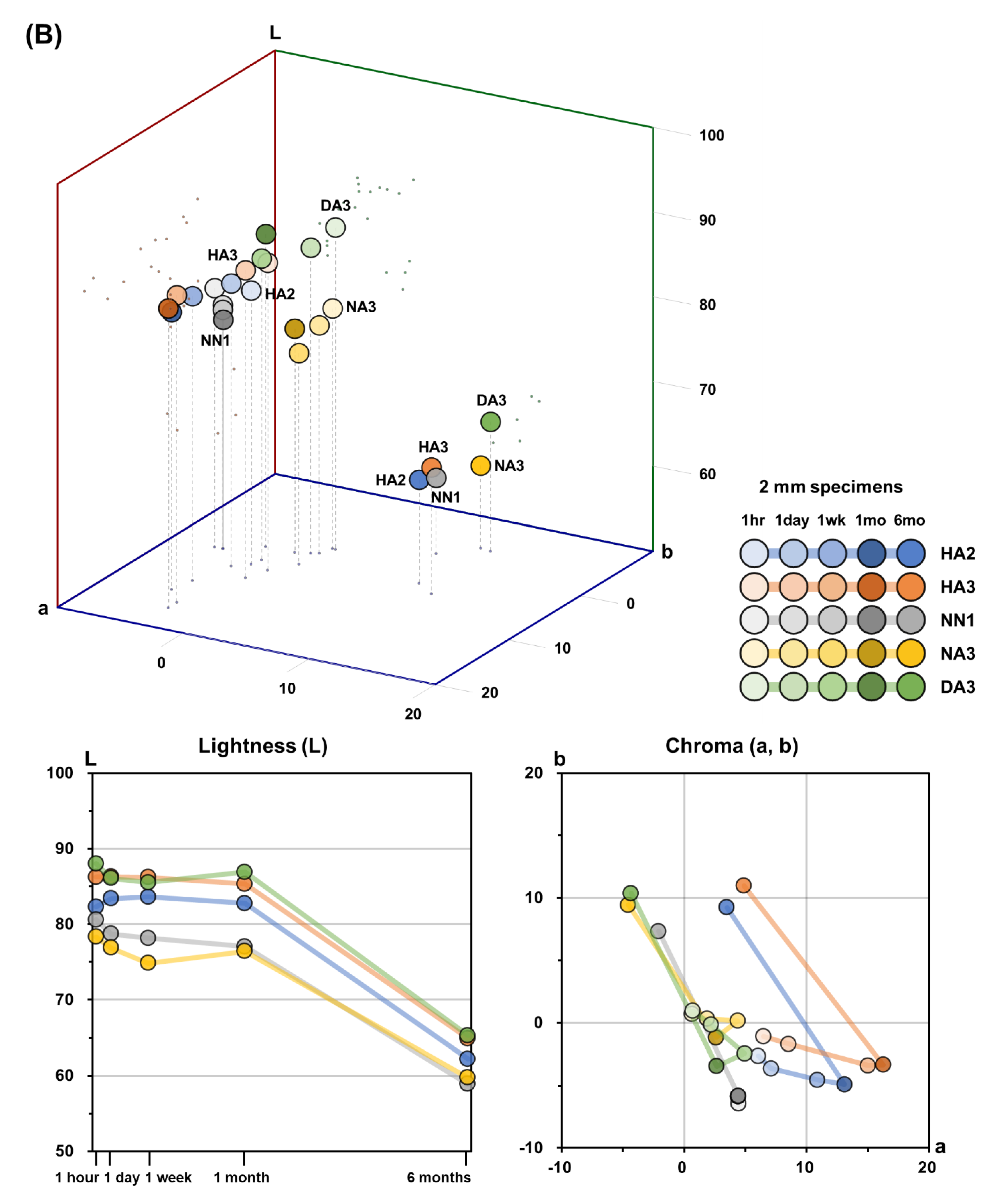
3.1. Color
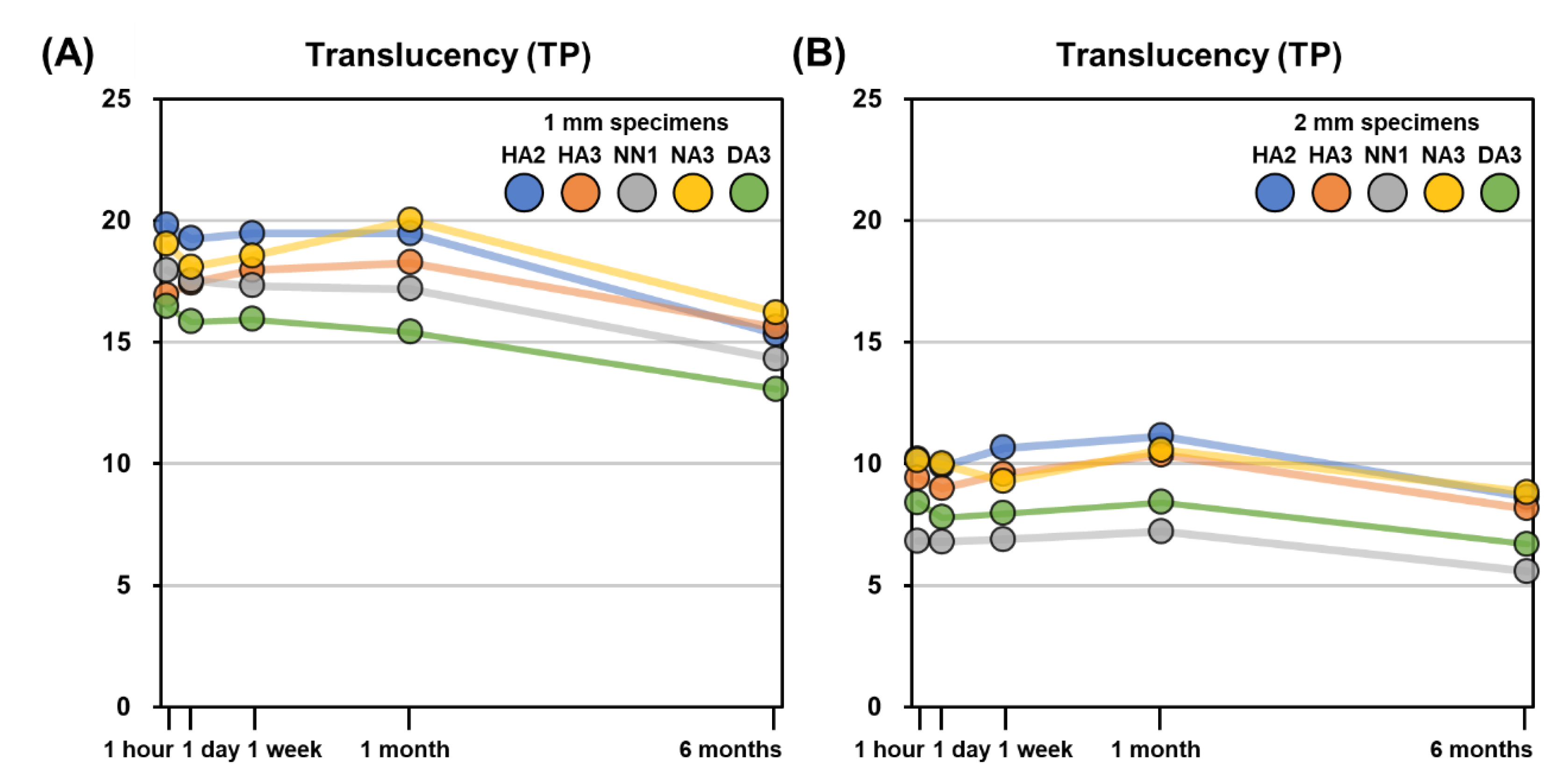
3.2. Translucency
4. Discussion
5. Conclusions
Author Contributions
Funding
Data Availability Statement
Conflicts of Interest
References
- Dawood, A.; Marti Marti, B.; Sauret-Jackson, V.; Darwood, A. 3D printing in dentistry. Br. Dent. J. 2015, 219, 521–529. [Google Scholar] [CrossRef] [PubMed]
- Kessler, A.; Hickel, R.; Reymus, M. 3D Printing in Dentistry-State of the Art. Oper. Dent. 2020, 45, 30–40. [Google Scholar] [CrossRef] [PubMed]
- Cousley, R.R. Introducing 3D printing in your orthodontic practice. J. Orthod. 2020, 47, 265–272. [Google Scholar] [CrossRef] [PubMed]
- Joo, H.S.; Park, S.W.; Yun, K.D.; Lim, H.P. Complete-mouth rehabilitation using a 3D printing technique and the CAD/CAM double scanning method: A clinical report. J. Prosthet. Dent. 2016, 116, 3–7. [Google Scholar] [CrossRef]
- Louvrier, A.; Marty, P.; Barrabé, A.; Euvrard, E.; Chatelain, B.; Weber, E.; Meyer, C. How useful is 3D printing in maxillofacial surgery? J. Stomatol. Oral Maxillofac. Surg. 2017, 118, 206–212. [Google Scholar] [CrossRef]
- Anderson, J.; Wealleans, J.; Ray, J. Endodontic applications of 3D printing. Int. Endod. J. 2018, 51, 1005–1018. [Google Scholar] [CrossRef]
- Tahayeri, A.; Morgan, M.; Fugolin, A.P.; Bompolaki, D.; Athirasala, A.; Pfeifer, C.S.; Ferracane, J.L.; Bertassoni, L.E. 3D printed versus conventionally cured provisional crown and bridge dental materials. Dent. Mater. 2018, 34, 192–200. [Google Scholar] [CrossRef]
- Gratton, D.G.; Aquilino, S.A. Interim restorations. Dent. Clin. N. Am. 2004, 48, 487–497. [Google Scholar] [CrossRef]
- Galindo, D.; Soltys, J.L.; Graser, G.N. Long-term reinforced fixed provisional restorations. J. Prosthet. Dent. 1998, 79, 698–701. [Google Scholar] [CrossRef]
- Munsell, A.H. A Color Notation; G. H. Ellis Company: Boston, MA, USA, 1905. [Google Scholar]
- Terry, D.A.; Geller, W.; Tric, O.; Anderson, M.J.; Tourville, M.; Kobashigawa, A. Anatomical form defines color: Function, form, and aesthetics. Pract. Proced. Aesthet. Dent. 2002, 14, 59–67. [Google Scholar]
- Winter, R. Visualizing the natural dentition. J. Esthet. Dent. 1993, 5, 102–117. [Google Scholar] [CrossRef] [PubMed]
- Johnston, W.M.; Ma, T.; Kienle, B.H. Translucency parameter of colorants for maxillofacial prostheses. Int. J. Prosthodont. 1995, 8, 79–86. [Google Scholar] [PubMed]
- Lee, Y.K. Influence of scattering/absorption characteristics on the color of resin composites. Dent. Mater. 2007, 23, 124–131. [Google Scholar] [CrossRef]
- Lee, Y.K. Translucency of human teeth and dental restorative materials and its clinical relevance. J. Biomed. Opt. 2015, 20, 045002. [Google Scholar] [CrossRef] [PubMed]
- Durand, L.B.; Ruiz-Lopez, J.; Perez, B.G.; Ionescu, A.M.; Carrillo-Perez, F.; Ghinea, R.; Perez, M.M. Color, lightness, chroma, hue, and translucency adjustment potential of resin composites using CIEDE2000 color difference formula. J. Esthet. Restor. Dent. 2020. [Google Scholar] [CrossRef]
- Sulaiman, T.A.; Rodgers, B.; Suliman, A.A.; Johnston, W.M. Color and translucency stability of contemporary resin-based restorative materials. J. Esthet. Restor. Dent. 2020. [Google Scholar] [CrossRef]
- Subaşı, M.G.; Alp, G.; Johnston, W.M.; Yilmaz, B. Effect of thickness on optical properties of monolithic CAD-CAM ceramics. J. Dent. 2018, 71, 38–42. [Google Scholar] [CrossRef]
- Arif, R.; Yilmaz, B.; Johnston, W.M. In vitro color stainability and relative translucency of CAD-CAM restorative materials used for laminate veneers and complete crowns. J Prosthet. Dent. 2019, 122, 160–166. [Google Scholar] [CrossRef]
- Revilla-León, M.; Umorin, M.; Özcan, M.; Piedra-Cascón, W. Color dimensions of additive manufactured interim restorative dental material. J. Prosthet. Dent. 2020, 123, 754–760. [Google Scholar] [CrossRef]
- Shin, J.W.; Kim, J.E.; Choi, Y.J.; Shin, S.H.; Nam, N.E.; Shim, J.S.; Lee, K.W. Evaluation of the Color Stability of 3D-Printed Crown and Bridge Materials against Various Sources of Discoloration: An In Vitro Study. Materials 2020, 13, 5359. [Google Scholar] [CrossRef]
- Commission Internationale de l’Eclairage. Colorimetry: Technical Report; Central Bureau of the CIE: Vienna, Austria, 2004. [Google Scholar]
- Johnston, W.M. Color measurement in dentistry. J. Dent. 2009, 37 (Suppl. 1), e2–e6. [Google Scholar] [CrossRef]
- Joiner, A.; Hopkinson, I.; Deng, Y.; Westland, S. A review of tooth colour and whiteness. J. Dent. 2008, 36 (Suppl. 1), S2–S7. [Google Scholar] [CrossRef]
- Khashayar, G.; Bain, P.A.; Salari, S.; Dozic, A.; Kleverlaan, C.J.; Feilzer, A.J. Perceptibility and acceptability thresholds for colour differences in dentistry. J. Dent. 2014, 42, 637–644. [Google Scholar] [CrossRef] [PubMed]
- Lee, Y.K. Criteria for clinical translucency evaluation of direct esthetic restorative materials. Restor. Dent. Endod. 2016, 41, 159–166. [Google Scholar] [CrossRef] [PubMed]
- Geissberger, M. Esthetic Dentistry in Clinical Practice; John Wiley & Sons: Hoboken, NJ, USA, 2013. [Google Scholar]
- Barutcigil, C.; Harorli, O.T.; Yildiz, M.; Ozcan, E.; Arslan, H.; Bayindir, F. The color differences of direct esthetic restorative materials after setting and compared with a shade guide. J. Am. Dent. Assoc. 2011, 142, 658–665. [Google Scholar] [CrossRef] [PubMed]
- Hassel, A.J.; Grossmann, A.C.; Schmitter, M.; Balke, Z.; Buzello, A.M. Interexaminer reliability in clinical measurement of L*C*h* values of anterior teeth using a spectrophotometer. Int. J. Prosthodont. 2007, 20, 79–84. [Google Scholar]
- Sailer, I.; Holderegger, C.; Jung, R.E.; Suter, A.; Thiévent, B.; Pietrobon, N.; Gebhard-Achilles, W.; Hämmerle, C.H. Clinical study of the color stability of veneering ceramics for zirconia frameworks. Int. J. Prosthodont. 2007, 20, 263–269. [Google Scholar]
- Haddad, H.J.; Salameh, Z.; Sadig, W.; Aboushelib, M.; Jakstat, H.A. Allocation of color space for different age groups using three-dimensional shade guide systems. Eur. J. Esthet. Dent. 2011, 6, 94–102. [Google Scholar]
- Johnston, W.M.; Kao, E.C. Assessment of appearance match by visual observation and clinical colorimetry. J. Dent. Res. 1989, 68, 819–822. [Google Scholar] [CrossRef]
- Ragain, J.C., Jr.; Johnston, W.M. Color acceptance of direct dental restorative materials by human observers. Color Res. Appl. 2000, 25, 278–285. [Google Scholar] [CrossRef]
- Peutzfeldt, A. Resin composites in dentistry: The monomer systems. Eur. J. Oral Sci. 1997, 105, 97–116. [Google Scholar] [CrossRef] [PubMed]
- Szczesio-Wlodarczyk, A.; Sokolowski, J.; Kleczewska, J.; Bociong, K. Ageing of Dental Composites Based on Methacrylate Resins-A Critical Review of the Causes and Method of Assessment. Polymers 2020, 12, 882. [Google Scholar] [CrossRef] [PubMed]
- Aromaa, M.K.; Vallittu, P.K. Delayed post-curing stage and oxygen inhibition of free-radical polymerization of dimethacrylate resin. Dent. Mater. 2018, 34, 1247–1252. [Google Scholar] [CrossRef] [PubMed]
- Engler, M.L.P.D.; Güth, J.-F.; Keul, C.; Erdelt, K.; Edelhoff, D.; Liebermann, A. Residual monomer elution from different conventional and CAD/CAM dental polymers during artificial aging. Clin. Oral Investig. 2020, 24, 277–284. [Google Scholar] [CrossRef]
- Larsen, I.B.; Munksgaard, E.C. Effect of human saliva on surface degradation of composite resins. Scand. J. Dent. Res. 1991, 99, 254–261. [Google Scholar] [CrossRef]
- Berli, C.; Thieringer, F.M.; Sharma, N.; Müller, J.A.; Dedem, P.; Fischer, J.; Rohr, N. Comparing the mechanical properties of pressed, milled, and 3D-printed resins for occlusal devices. J. Prosthet. Dent. 2020, 124, 780–786. [Google Scholar] [CrossRef]
- Karabela, M.M.; Sideridou, I.D. Effect of the structure of silane coupling agent on sorption characteristics of solvents by dental resin-nanocomposites. Dent. Mater. 2008, 24, 1631–1639. [Google Scholar] [CrossRef]
- Shin, D.H.; Rawls, H.R. Degree of conversion and color stability of the light curing resin with new photoinitiator systems. Dent. Mater. 2009, 25, 1030–1038. [Google Scholar] [CrossRef]
- Salgado, V.E.; Albuquerque, P.P.; Cavalcante, L.M.; Pfeifer, C.S.; Moraes, R.R.; Schneider, L.F. Influence of photoinitiator system and nanofiller size on the optical properties and cure efficiency of model composites. Dent. Mater. 2014, 30, e264–e271. [Google Scholar] [CrossRef]
- Ikemura, K.; Endo, T. A review of the development of radical photopolymerization initiators used for designing light-curing dental adhesives and resin composites. Dent. Mater. J. 2010, 29, 481–501. [Google Scholar] [CrossRef]
- Hadis, M.A.; Shortall, A.C.; Palin, W.M. Competitive light absorbers in photoactive dental resin-based materials. Dent. Mater. 2012, 28, 831–841. [Google Scholar] [CrossRef] [PubMed]
- Bertolo, M.V.; Moraes, R.C.; Pfeifer, C.; Salgado, V.E.; Correr, A.R.; Schneider, L.F. Influence of Photoinitiator System on Physical-Chemical Properties of Experimental Self-Adhesive Composites. Braz. Dent. J. 2017, 28, 35–39. [Google Scholar] [CrossRef] [PubMed]
- Yu, B.; Ahn, J.S.; Lee, Y.K. Measurement of translucency of tooth enamel and dentin. Acta Odontol. Scand. 2009, 67, 57–64. [Google Scholar] [CrossRef] [PubMed]
- Pop-Ciutrila, I.S.; Ghinea, R.; Colosi, H.A.; Dudea, D. Dentin translucency and color evaluation in human incisors, canines, and molars. J. Prosthet. Dent. 2016, 115, 475–481. [Google Scholar] [CrossRef]
- Shortall, A.C.; Palin, W.M.; Burtscher, P. Refractive index mismatch and monomer reactivity influence composite curing depth. J. Dent. Res. 2008, 87, 84–88. [Google Scholar] [CrossRef]
- Ota, M.; Ando, S.; Endo, H.; Ogura, Y.; Miyazaki, M.; Hosoya, Y. Influence of refractive index on optical parameters of experimental resin composites. Acta Odontol. Scand. 2012, 70, 362–367. [Google Scholar] [CrossRef]
- Lee, Y.K. Influence of filler on the difference between the transmitted and reflected colors of experimental resin composites. Dent. Mater. 2008, 24, 1243–1247. [Google Scholar] [CrossRef]
- Kim, D.; Shim, J.S.; Lee, D.; Shin, S.H.; Nam, N.E.; Park, K.H.; Shim, J.S.; Kim, J.E. Effects of Post-Curing Time on the Mechanical and Color Properties of Three-Dimensional Printed Crown and Bridge Materials. Polymers 2020, 12, 2762. [Google Scholar] [CrossRef]
- Szczepanska, J.; Poplawski, T.; Synowiec, E.; Pawlowska, E.; Chojnacki, C.J.; Chojnacki, J.; Blasiak, J. 2-hydroxylethyl methacrylate (HEMA), a tooth restoration component, exerts its genotoxic effects in human gingival fibroblasts trough methacrylic acid, an immediate product of its degradation. Mol. Biol. Rep. 2012, 39, 1561–1574. [Google Scholar] [CrossRef]
- Örtorp, A.; Jönsson, D.; Mouhsen, A.; Vult von Steyern, P. The fit of cobalt-chromium three-unit fixed dental prostheses fabricated with four different techniques: A comparative in vitro study. Dent. Mater. 2011, 27, 356–363. [Google Scholar] [CrossRef]
- Gutiérrez, T.J. Reactive and Functional Polymers Volume One—Biopolymers, Polyesters, Polyurethanes, Resins and Silicones; Springer: Berlin/Heidelberg, Germany, 2020; pp. 293–300. [Google Scholar]
- Luis, E.; Pan, H.M.; Sing, S.L.; Bajpai, R.; Song, J.; Yeong, W.Y. 3D Direct Printing of Silicone Meniscus Implant Using a Novel Heat-Cured Extrusion-Based Printer. Polymers 2020, 12, 1031. [Google Scholar] [CrossRef] [PubMed]
- Zafar, M.S.; Khurshid, Z. Dental Implants: Materials, Coatings, Surface Modifications and Interfaces with Oral Tissues; Woodhead Publishing: Cambridge, UK, 2020. [Google Scholar]


| Code | Product Name | Shade | Manufacturer | Component * |
|---|---|---|---|---|
| HA2 HA3 | DT-1 | A2 A3 | Hephzibah, Incheon, South Korea | Urethane acrylate, silicon oxides, pigments |
| NN1 | NextDent C&B MFH | N1 | NextDent, Soesterburg, Netherlands | Methacrylic oligomers, methacrylate monomer, inorganic filler, phosphine oxides, pigments |
| NA3 | NextDent C&B | A3.5 | NextDent, Soesterburg, Netherlands | > 90% methacrylic oligomers, methacrylate monomer, < 3% phosphine oxides, pigments |
| DA3 | DIOnavi C&B | A3 | DIO Inc, Busan, South Korea | > 90% methacrylic oligomers, < 10% phosphine oxides, pigments |
| Specimen Thickness | Storage Period | HA2 | HA3 | NN1 | NA3 | DA3 | ||||||||||
|---|---|---|---|---|---|---|---|---|---|---|---|---|---|---|---|---|
| Mean | SD | Mean | SD | Mean | SD | Mean | SD | Mean | SD | |||||||
| 1 mm | 1 hour | 93.44 | 2.17 | a | 95.41 | 2.13 | a | 90.02 | 1.95 | a | 91.07 | 1.81 | a | 96.62 | 2.46 | a |
| 1 day | 92.58 | 2.08 | a | 95.53 | 2.26 | a | 88.82 | 2.55 | ab | 88.65 | 2.24 | a | 94.53 | 2.81 | a | |
| 1 week | 93.23 | 2.09 | a | 95.24 | 2.07 | a | 89.08 | 2.00 | ab | 89.28 | 2.15 | a | 96.28 | 2.46 | a | |
| 1 month | 92.41 | 1.87 | a | 94.76 | 2.26 | a | 87.30 | 2.31 | b | 90.50 | 3.15 | a | 95.63 | 2.28 | a | |
| 6 months | 68.92 | 1.88 | b | 71.97 | 1.62 | b | 66.44 | 1.67 | c | 68.79 | 1.84 | b | 70.79 | 1.97 | b | |
| 2 mm | 1 hour | 82.29 | 2.03 | a | 86.24 | 1.88 | a | 80.59 | 0.86 | a | 78.34 | 1.42 | a | 88.01 | 1.39 | a |
| 1 day | 83.37 | 1.81 | a | 86.27 | 1.43 | a | 78.71 | 1.07 | b | 76.87 | 1.49 | ab | 86.06 | 1.11 | bc | |
| 1 week | 83.60 | 1.77 | a | 86.22 | 1.06 | a | 78.15 | 0.87 | b | 74.85 | 1.90 | c | 85.52 | 1.00 | c | |
| 1 month | 82.75 | 2.40 | a | 85.33 | 1.70 | a | 77.04 | 0.89 | c | 76.42 | 1.65 | bc | 86.91 | 1.05 | ab | |
| 6 months | 62.20 | 1.83 | b | 64.90 | 1.08 | b | 58.89 | 0.62 | d | 59.73 | 1.09 | d | 65.30 | 0.81 | d |
| Specimen Thickness | Storage Period | HA2 | HA3 | NN1 | NA3 | DA3 | ||||||||||
|---|---|---|---|---|---|---|---|---|---|---|---|---|---|---|---|---|
| Mean | SD | Mean | SD | Mean | SD | Mean | SD | Mean | SD | |||||||
| 1 mm | 1 hour | 7.02 | 0.45 | d | 7.14 | 0.44 | c | 5.09 | 0.19 | a | 3.61 | 0.88 | b | 2.44 | 0.56 | c |
| 1 day | 7.95 | 0.70 | c | 11.13 | 1.11 | b | 5.11 | 0.25 | a | 4.58 | 0.65 | a | 3.79 | 0.62 | a | |
| 1 week | 11.91 | 0.65 | b | 18.46 | 0.63 | a | 5.17 | 0.16 | a | 4.82 | 0.27 | a | 3.80 | 0.43 | a | |
| 1 month | 14.74 | 0.72 | a | 17.97 | 1.17 | a | 5.02 | 0.21 | a | 3.31 | 0.33 | b | 2.99 | 0.33 | b | |
| 6 months | 4.33 | 0.53 | e | 6.37 | 0.89 | c | −1.76 | 0.06 | b | −3.48 | 0.29 | c | −3.43 | 0.22 | d | |
| 2 mm | 1 hour | 6.03 | 0.28 | d | 6.46 | 0.32 | d | 4.47 | 0.12 | a | 0.64 | 0.67 | d | 0.69 | 0.43 | d |
| 1 day | 7.08 | 0.61 | c | 8.52 | 0.69 | c | 4.38 | 0.09 | a | 1.85 | 0.51 | c | 2.19 | 0.56 | c | |
| 1 week | 10.86 | 0.87 | b | 14.99 | 0.76 | b | 4.40 | 0.07 | a | 4.37 | 0.14 | a | 4.93 | 0.31 | a | |
| 1 month | 13.08 | 0.65 | a | 16.24 | 0.86 | a | 4.42 | 0.10 | a | 2.60 | 0.20 | b | 2.64 | 0.12 | b | |
| 6 months | 3.49 | 0.47 | e | 4.87 | 0.64 | e | −2.10 | 0.04 | b | −4.57 | 0.11 | e | −4.34 | 0.05 | e |
| Specimen Thickness | Storage Period | HA2 | HA3 | NN1 | NA3 | DA3 | ||||||||||
|---|---|---|---|---|---|---|---|---|---|---|---|---|---|---|---|---|
| Mean | SD | Mean | SD | Mean | SD | Mean | SD | Mean | SD | |||||||
| 1 mm | 1 hour | −4.89 | 0.47 | b | −1.97 | 0.88 | b | −3.92 | 0.51 | b | −1.16 | 0.87 | b | 1.74 | 1.11 | b |
| 1 day | −5.50 | 0.50 | c | −3.03 | 0.92 | c | −3.72 | 0.65 | b | −1.31 | 0.91 | b | 0.69 | 0.75 | c | |
| 1 week | −6.53 | 0.50 | d | −5.02 | 0.88 | d | −4.04 | 0.49 | b | −0.72 | 0.74 | b | −1.76 | 1.07 | d | |
| 1 month | −7.36 | 0.28 | e | −4.95 | 0.67 | d | −4.97 | 0.78 | c | −3.54 | 1.06 | c | −2.93 | 0.77 | e | |
| 6 months | 8.83 | 0.44 | a | 11.14 | 0.44 | a | 9.69 | 0.31 | a | 10.10 | 0.26 | a | 11.86 | 0.63 | a | |
| 2 mm | 1 hour | −2.65 | 0.37 | b | −1.08 | 0.59 | b | −6.47 | 0.37 | c | 0.68 | 0.26 | b | 0.97 | 0.70 | b |
| 1 day | −3.64 | 0.42 | c | −1.68 | 0.43 | c | −5.87 | 0.38 | b | 0.33 | 0.16 | c | −0.15 | 0.58 | c | |
| 1 week | −4.55 | 0.43 | d | −3.40 | 0.26 | d | −5.85 | 0.29 | b | 0.16 | 0.41 | c | −2.45 | 0.69 | d | |
| 1 month | −4.91 | 0.55 | d | −3.33 | 0.16 | d | −5.81 | 0.31 | b | −1.18 | 0.28 | d | −3.46 | 0.42 | e | |
| 6 months | 9.23 | 0.29 | a | 10.98 | 0.26 | a | 7.34 | 0.21 | a | 9.44 | 0.32 | a | 10.37 | 0.31 | a |
| Specimen Thickness | Storage Period | HA2 | HA3 | NN1 | NA3 | DA3 | ||||||||||
|---|---|---|---|---|---|---|---|---|---|---|---|---|---|---|---|---|
| Mean | SD | Mean | SD | Mean | SD | Mean | SD | Mean | SD | |||||||
| 1 mm | 1 hour | 19.81 | 2.09 | a | 16.93 | 1.69 | ab | 17.95 | 1.89 | a | 19.02 | 2.27 | ab | 16.46 | 2.21 | a |
| 1 day | 19.25 | 1.69 | a | 17.43 | 2.25 | ab | 17.53 | 2.09 | a | 18.08 | 3.35 | ab | 15.83 | 2.51 | a | |
| 1 week | 19.47 | 1.60 | a | 17.95 | 1.86 | a | 17.31 | 2.13 | a | 18.54 | 2.83 | ab | 15.93 | 2.44 | a | |
| 1 month | 19.47 | 1.61 | a | 18.27 | 1.86 | a | 17.18 | 2.35 | a | 20.01 | 3.52 | a | 15.41 | 2.03 | ab | |
| 6 months | 15.32 | 1.57 | b | 15.60 | 1.54 | b | 14.30 | 1.36 | b | 16.21 | 2.39 | b | 13.06 | 1.65 | b | |
| 2 mm | 1 hour | 10.20 | 1.55 | ab | 9.39 | 1.41 | ab | 6.82 | 0.48 | a | 10.13 | 0.85 | ab | 8.40 | 0.80 | a |
| 1 day | 9.89 | 1.52 | ab | 8.98 | 1.23 | ab | 6.78 | 0.53 | a | 10.00 | 0.78 | ab | 7.78 | 0.64 | a | |
| 1 week | 10.65 | 1.75 | a | 9.58 | 1.01 | ab | 6.89 | 0.41 | a | 9.28 | 0.89 | bc | 7.96 | 0.63 | a | |
| 1 month | 11.14 | 1.57 | a | 10.36 | 1.63 | a | 7.22 | 0.89 | a | 10.56 | 1.23 | a | 8.41 | 0.63 | a | |
| 6 months | 8.63 | 1.25 | b | 8.14 | 1.03 | b | 5.57 | 0.57 | b | 8.82 | 0.87 | c | 6.68 | 0.46 | b |
Publisher’s Note: MDPI stays neutral with regard to jurisdictional claims in published maps and institutional affiliations. |
© 2021 by the authors. Licensee MDPI, Basel, Switzerland. This article is an open access article distributed under the terms and conditions of the Creative Commons Attribution (CC BY) license (http://creativecommons.org/licenses/by/4.0/).
Share and Cite
Kim, J.-E.; Choi, W.-H.; Lee, D.; Shin, Y.; Park, S.-H.; Roh, B.-D.; Kim, D. Color and Translucency Stability of Three-Dimensional Printable Dental Materials for Crown and Bridge Restorations. Materials 2021, 14, 650. https://doi.org/10.3390/ma14030650
Kim J-E, Choi W-H, Lee D, Shin Y, Park S-H, Roh B-D, Kim D. Color and Translucency Stability of Three-Dimensional Printable Dental Materials for Crown and Bridge Restorations. Materials. 2021; 14(3):650. https://doi.org/10.3390/ma14030650
Chicago/Turabian StyleKim, Jong-Eun, Won-Huy Choi, Dasun Lee, Yooseok Shin, Sung-Ho Park, Byoung-Duck Roh, and Dohyun Kim. 2021. "Color and Translucency Stability of Three-Dimensional Printable Dental Materials for Crown and Bridge Restorations" Materials 14, no. 3: 650. https://doi.org/10.3390/ma14030650
APA StyleKim, J.-E., Choi, W.-H., Lee, D., Shin, Y., Park, S.-H., Roh, B.-D., & Kim, D. (2021). Color and Translucency Stability of Three-Dimensional Printable Dental Materials for Crown and Bridge Restorations. Materials, 14(3), 650. https://doi.org/10.3390/ma14030650