Surface Characteristics of Esthetic Nickel–Titanium and Beta-Titanium Orthodontic Archwires Produced by Plasma Electrolytic Oxidation (PEO)—Primary Results

 ,
,  and
and 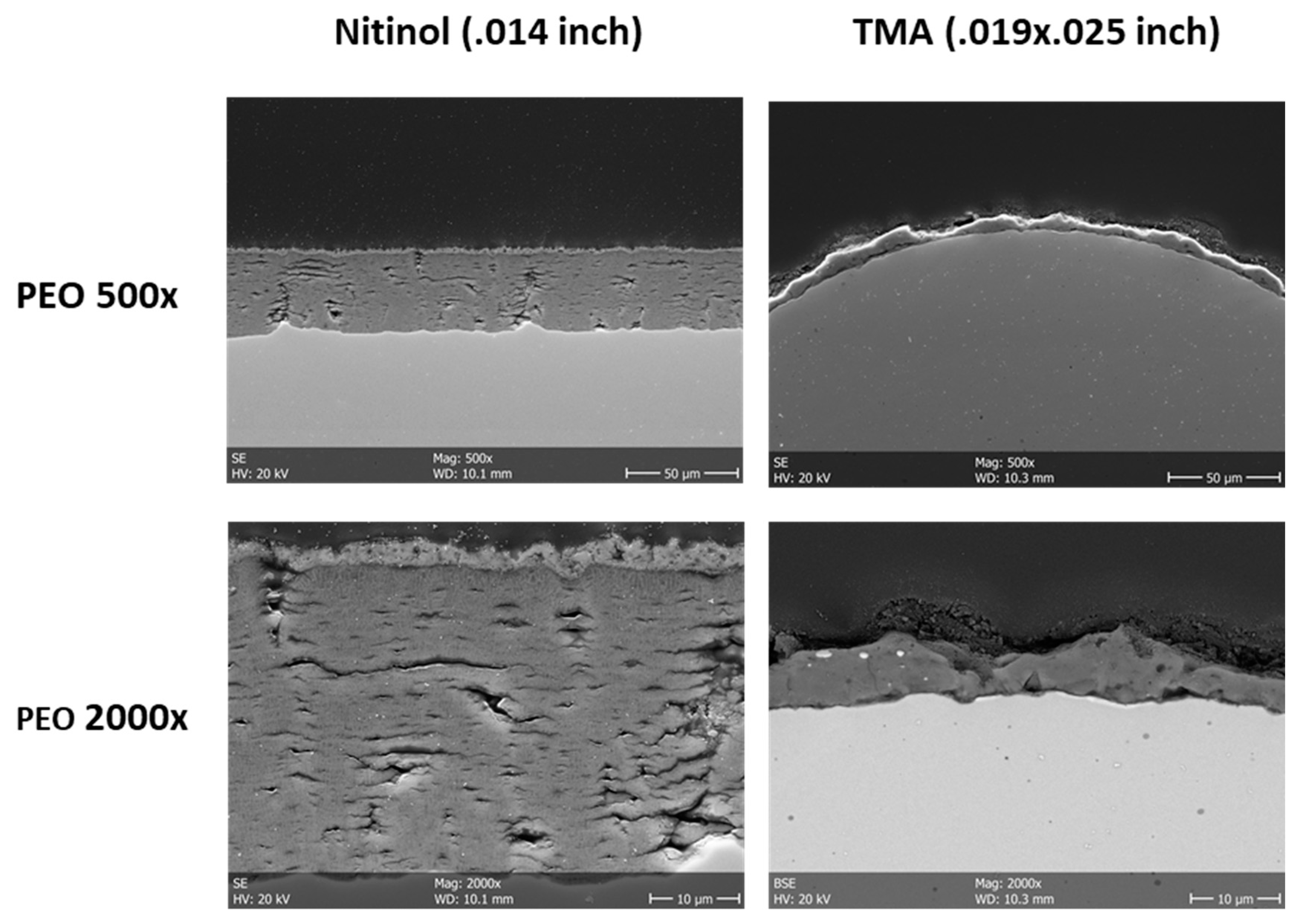
Abstract
1. Introduction
2. Materials and Methods
2.1. Biomaterial Preparation and Physiochemical Analysis
2.2. Cytocompatibility Analyses
2.3. Ex Vivo Analyses
2.4. Statistical Analyses
3. Results
3.1. Material Analyses
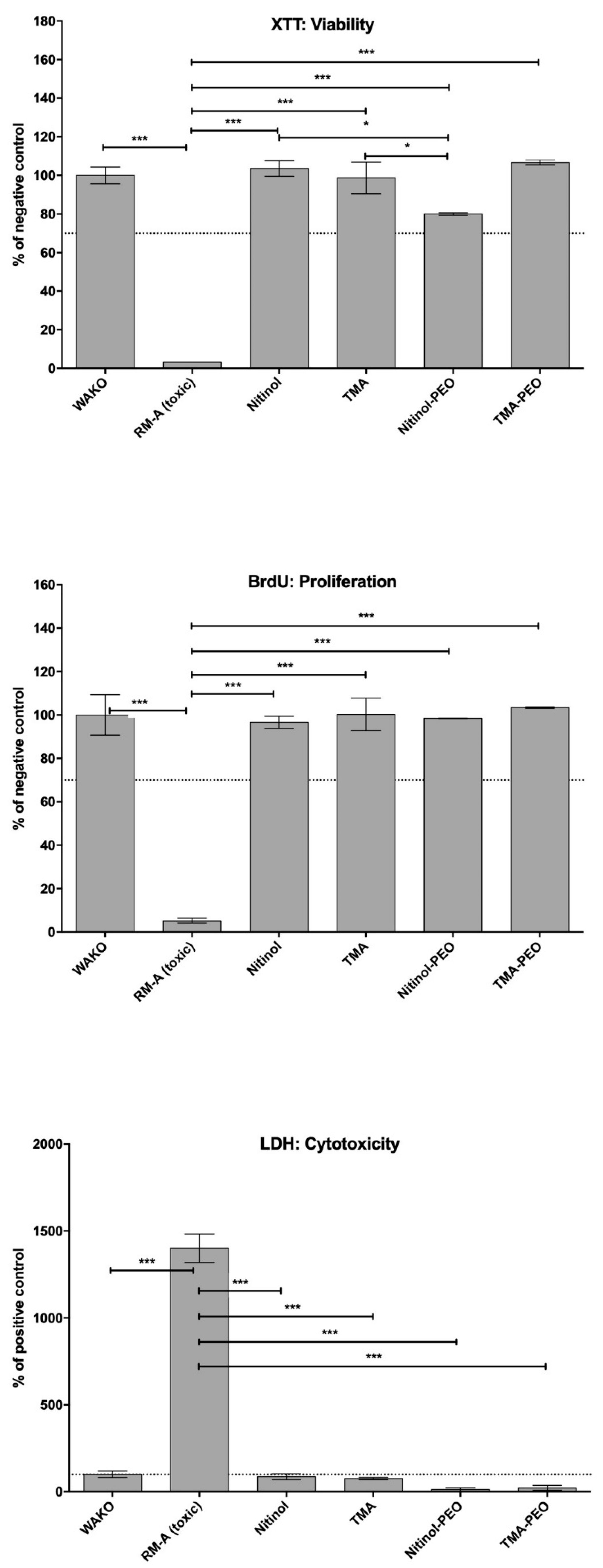
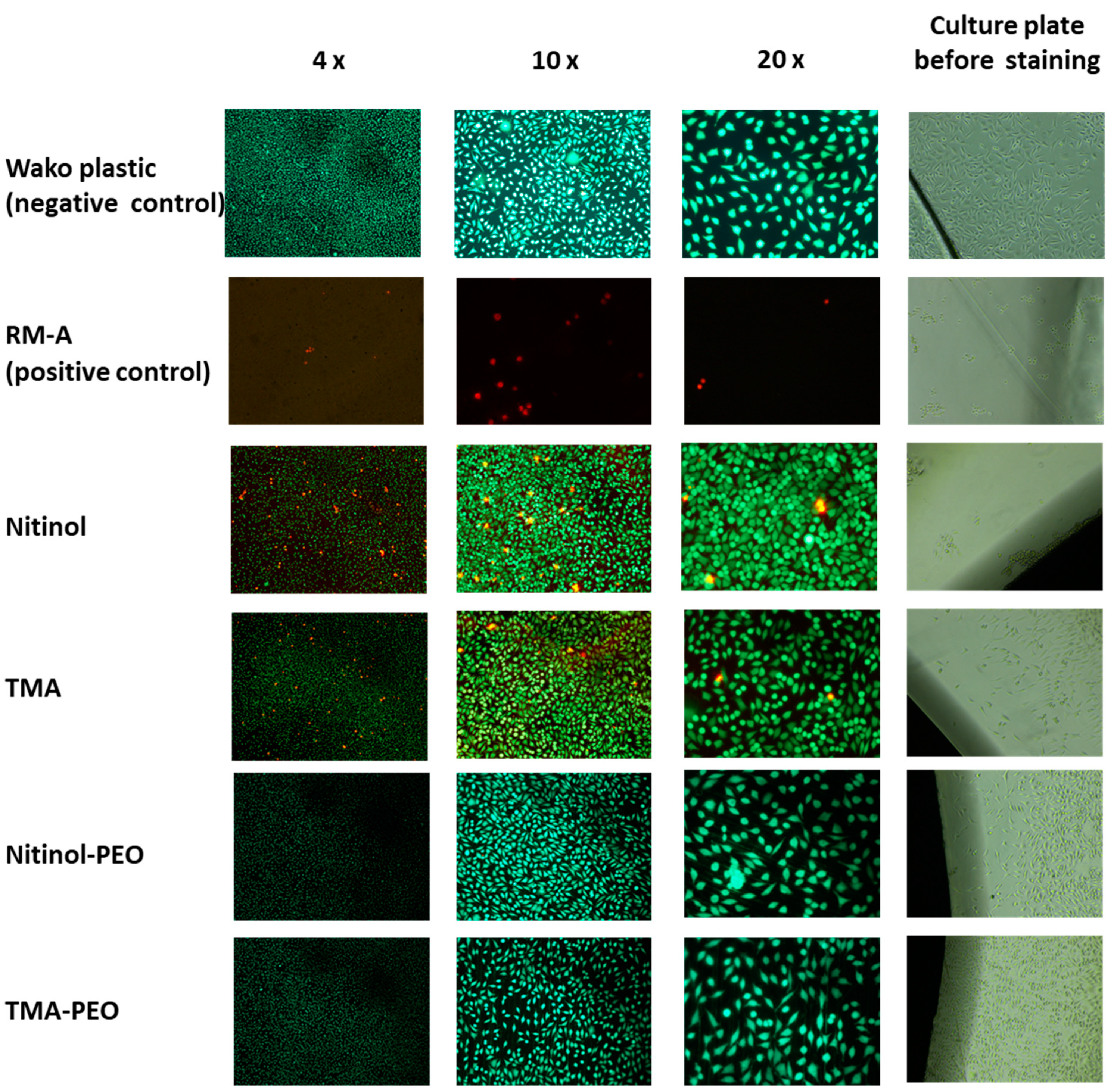
3.2. Cytocompatibility
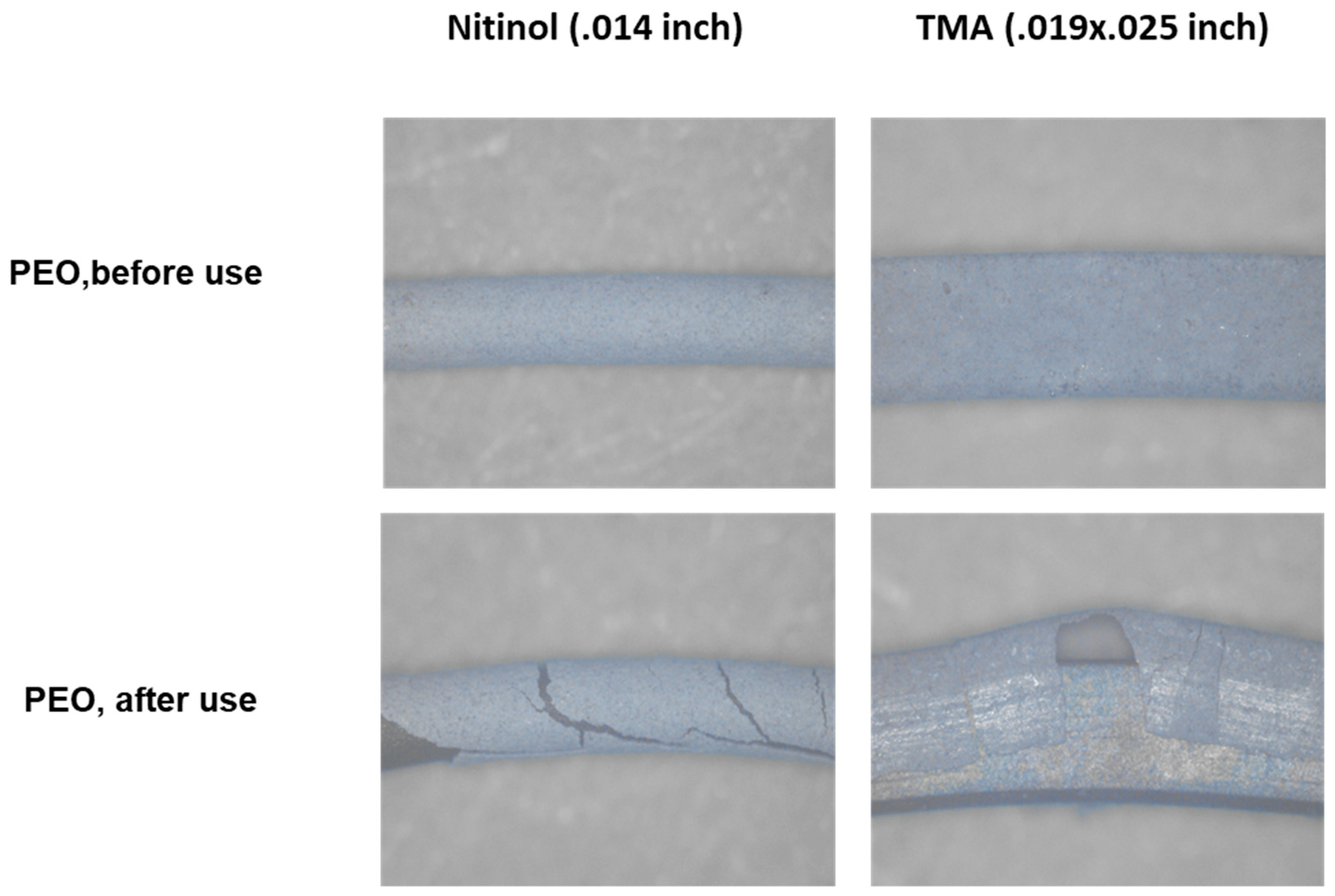
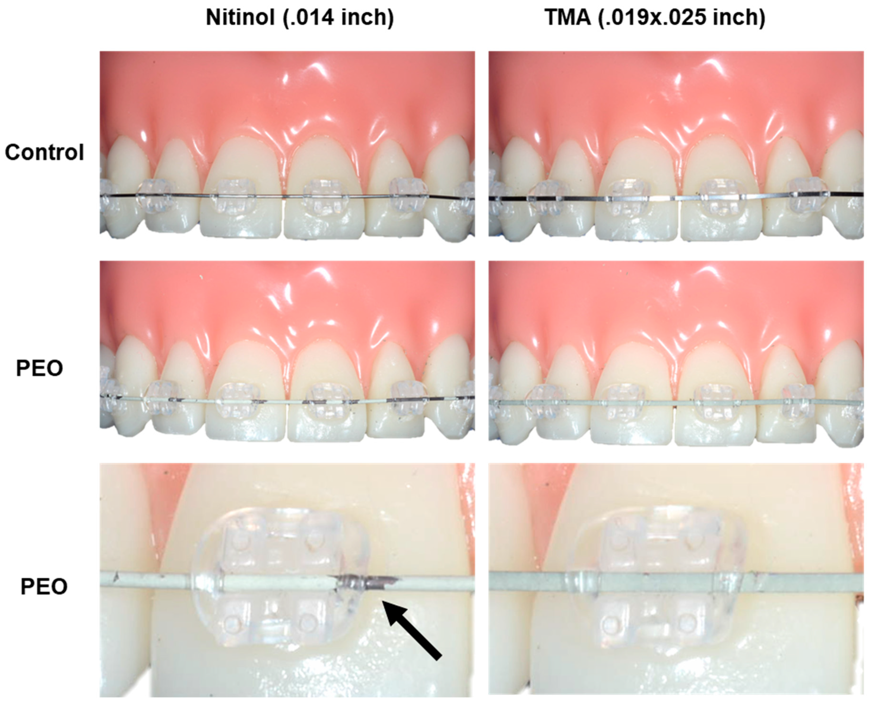
3.3. Ex Vivo Analysis
4. Discussion
Author Contributions
Funding
Conflicts of Interest
References
- Rossini, G.; Parrini, S.; Castroflorio, T.; Deregibus, A.; Debernardi, C.L. Efficacy of clear aligners in controlling orthodontic tooth movement: A systematic review. Angle Orthod. 2015, 85, 881–889. [Google Scholar] [CrossRef] [PubMed]
- Hennessy, J.; Al-Awadhi, E.A. Clear aligners generations and orthodontic tooth movement. J. Orthod. 2016, 43, 68–76. [Google Scholar] [CrossRef] [PubMed]
- Ata-Ali, F.; Ata-Ali, J.; Ferrer-Molina, M.; Cobo, T.; De Carlos, F.; Cobo, J. Adverse effects of lingual and buccal orthodontic techniques: A systematic review and meta-analysis. Am. J. Orthod. Dentofac. Orthop. 2016, 149, 820–829. [Google Scholar] [CrossRef]
- Rosvall, M.D.; Fields, H.W.; Ziuchkovski, J.; Rosenstiel, S.F.; Johnston, W.M. Attractiveness, acceptability, and value of orthodontic appliances. Am. J. Orthod. Dentofac. Orthop. 2009, 135, 276–277. [Google Scholar] [CrossRef]
- Aldossary, M.S.; Abu Hajia, S.S.; Santini, A. Light energy transmission through six different makes of ceramic orthodontic brackets. Int. Orthod. 2018, 16, 638–651. [Google Scholar] [CrossRef]
- Rongo, R.; Ametrano, G.; Gloria, A.; Spagnuolo, G.; Galeotti, A.; Paduano, S.; Valletta, R.; D’Anto, V. Effects of intraoral aging on surface properties of coated nickel-titanium archwires. Angle Orthod. 2014, 84, 665–672. [Google Scholar] [CrossRef]
- Bradley, T.G.; Berzins, D.W.; Valeri, N.; Pruszynski, J.; Eliades, T.; Katsaros, C. An investigation into the mechanical and aesthetic properties of new generation coated nickel-titanium wires in the as-received state and after clinical use. Eur. J. Orthod. 2014, 36, 290–296. [Google Scholar] [CrossRef]
- Scribante, A.; Vallittu, P.K.; Ozcan, M. Fiber-Reinforced Composites for Dental Applications. Biomed. Res. Int. 2018, 2018, 4734986. [Google Scholar] [CrossRef]
- Huang, Z.M.; Gopal, R.; Fujihara, K.; Ramakrishna, S.; Loh, P.L.; Foong, W.C.; Ganesh, V.K.; Chew, C.L. Fabrication of a new composite orthodontic archwire and validation by a bridging micromechanics model. Biomaterials 2003, 24, 2941–2953. [Google Scholar] [CrossRef]
- Kopp, A.; Derra, T.; Müther, M.; Jauer, L.; Schleifenbaum, J.H.; Voshage, M.; Jung, O.; Smeets, R.; Kröger, N. Influence of design and postprocessing parameters on the degradation behavior and mechanical properties of additively manufactured magnesium scaffolds. Acta Biomater. 2019. (Epub ahead of print). [Google Scholar] [CrossRef]
- Jung, O.; Smeets, R.; Kopp, A.; Porchetta, D.; Hiester, P.; Heiland, M.; Friedrich, R.E.; Precht, C.; Hanken, H.; Grobe, A.; et al. PEO-generated Surfaces Support Attachment and Growth of Cells In Vitro with No Additional Benefit for Micro-roughness in Sa (0.2-4 mum). In Vivo 2016, 30, 27–33. [Google Scholar]
- Fischerauer, S.F.; Kraus, T.; Wu, X.; Tangl, S.; Sorantin, E.; Hanzi, A.C.; Loffler, J.F.; Uggowitzer, P.J.; Weinberg, A.M. In vivo degradation performance of micro-arc-oxidized magnesium implants: A micro-CT study in rats. Acta Biomater. 2013, 9, 5411–5420. [Google Scholar] [CrossRef]
- Jung, O.; Smeets, R.; Hartjen, P.; Schnettler, R.; Feyerabend, F.; Klein, M.; Wegner, N.; Walther, F.; Stangier, D.; Henningsen, A.; et al. Improved In Vitro Test Procedure for Full Assessment of the Cytocompatibility of Degradable Magnesium Based on ISO 10993-5/-12. Int. J. Mol. Sci. 2019, 20, 255. [Google Scholar] [CrossRef]
- Joseph, J.; Ninan, V.S.; Abraham, M.E.; John, J.; Cherian, K.K.; Thomas, R.M. Arch Expansion Efficiency of Coaxial Tubular Superelastic Nickel-Titanium in Comparison to Single-Stranded Superelastic Nickel-Titanium While Relieving Mandibular Anterior Crowding: A Randomized Controlled Study. J. Int. Soc. Prev. Community Dent. 2019, 9, 60–64. [Google Scholar] [CrossRef]
- Chang, J.H.; Berzins, D.W.; Pruszynski, J.E.; Ballard, R.W. The effect of water storage on the bending properties of esthetic, fiber-reinforced composite orthodontic archwires. Angle Orthod. 2014, 84, 417–423. [Google Scholar] [CrossRef]
- Montasser, M.A.; El-Bialy, T.; Keilig, L.; Reimann, S.; Jager, A.; Bourauel, C. Force loss in archwire-guided tooth movement of conventional and self-ligating brackets. Eur. J. Orthod. 2014, 36, 31–38. [Google Scholar] [CrossRef]
- Jung, O.; Smeets, R.; Porchetta, D.; Kopp, A.; Ptock, C.; Muller, U.; Heiland, M.; Schwade, M.; Behr, B.; Kroger, N.; et al. Optimized in vitro procedure for assessing the cytocompatibility of magnesium-based biomaterials. Acta Biomater. 2015, 23, 354–363. [Google Scholar] [CrossRef]
- Balloni, S.; Locci, P.; Lumare, A.; Marinucci, L. Cytotoxicity of three commercial mouthrinses on extracellular matrix metabolism and human gingival cell behaviour. Toxicol. In Vitro 2016, 34, 88–96. [Google Scholar] [CrossRef]
- Pagano, S.; Chieruzzi, M.; Balloni, S.; Lombardo, G.; Torre, L.; Bodo, M.; Cianetti, S.; Marinucci, L. Biological, thermal and mechanical characterization of modified glass ionomer cements: The role of nanohydroxyapatite, ciprofloxacin and zinc l-carnosine. Mater. Sci. Eng. C Mater. Biol. Appl. 2019, 94, 76–85. [Google Scholar] [CrossRef]
- DIN EN ISO 10993-12-2012-10. Biological Evaluation of Medical Devices—Part 12: Sample Preparation and Reference Materials. Available online: https://www.beuth.de/de/norm/din-en-iso-10993-12/148200806 (accessed on 23 April 2019).
- DIN EN ISO 10993-5-2009-10. Biological Evaluation of Medical Devices—Part 5: Tests for In Vitro Cytotoxicity. Available online: https://www.beuth.de/de/norm/din-en-iso-10993-5/113571989 (accessed on 23 April 2019).
- Alford, T.J.; Roberts, W.E.; Hartsfield, J.K., Jr.; Eckert, G.J.; Snyder, R.J. Clinical outcomes for patients finished with the SureSmile method compared with conventional fixed orthodontic therapy. Angle Orthod. 2011, 81, 383–388. [Google Scholar] [CrossRef]
- Liu, F.; Xu, J.; Yu, D.; Wang, F.; Zhao, L. Wear resistance of micro-arc oxidation coatings on biomedical NiTi alloy. J. Alloy. Compd. 2009, 487, 391–394. [Google Scholar] [CrossRef]
- Chung, C.J.; Su, R.T.; Chu, H.J.; Chen, H.T.; Tsou, H.K.; He, J.L. Plasma electrolytic oxidation of titanium and improvement in osseointegration. J. Biomed. Mater. Res. B Appl. Biomater. 2013, 101, 1023–1030. [Google Scholar] [CrossRef] [PubMed]
- Hartjen, P.; Hoffmann, A.; Henningsen, A.; Barbeck, M.; Kopp, A.; Kluwe, L.; Precht, C.; Quatela, O.; Gaudin, R.; Heiland, M.; et al. Plasma Electrolytic Oxidation of Titanium Implant Surfaces: Microgroove-Structures Improve Cellular Adhesion and Viability. In Vivo 2018, 32, 241–247. [Google Scholar] [CrossRef] [PubMed]
- Kazek-Kesik, A.; Krok-Borkowicz, M.; Pamula, E.; Simka, W. Electrochemical and biological characterization of coatings formed on Ti-15Mo alloy by plasma electrolytic oxidation. Mater. Sci. Eng. C Mater. Biol. Appl. 2014, 43, 172–181. [Google Scholar] [CrossRef]


© 2019 by the authors. Licensee MDPI, Basel, Switzerland. This article is an open access article distributed under the terms and conditions of the Creative Commons Attribution (CC BY) license (http://creativecommons.org/licenses/by/4.0/).
Share and Cite
Jung, O.; Becker, J.-P.; Smeets, R.; Gosau, M.; Becker, G.; Kahl-Nieke, B.; Jung, A.-K.; Heiland, M.; Kopp, A.; Barbeck, M.; et al. Surface Characteristics of Esthetic Nickel–Titanium and Beta-Titanium Orthodontic Archwires Produced by Plasma Electrolytic Oxidation (PEO)—Primary Results. Materials 2019, 12, 1403. https://doi.org/10.3390/ma12091403
Jung O, Becker J-P, Smeets R, Gosau M, Becker G, Kahl-Nieke B, Jung A-K, Heiland M, Kopp A, Barbeck M, et al. Surface Characteristics of Esthetic Nickel–Titanium and Beta-Titanium Orthodontic Archwires Produced by Plasma Electrolytic Oxidation (PEO)—Primary Results. Materials. 2019; 12(9):1403. https://doi.org/10.3390/ma12091403
Chicago/Turabian StyleJung, Ole, Jean-Philippe Becker, Ralf Smeets, Martin Gosau, Germain Becker, Bärbel Kahl-Nieke, Anne-Kathrin Jung, Max Heiland, Alexander Kopp, Mike Barbeck, and et al. 2019. "Surface Characteristics of Esthetic Nickel–Titanium and Beta-Titanium Orthodontic Archwires Produced by Plasma Electrolytic Oxidation (PEO)—Primary Results" Materials 12, no. 9: 1403. https://doi.org/10.3390/ma12091403
APA StyleJung, O., Becker, J.-P., Smeets, R., Gosau, M., Becker, G., Kahl-Nieke, B., Jung, A.-K., Heiland, M., Kopp, A., Barbeck, M., & Koehne, T. (2019). Surface Characteristics of Esthetic Nickel–Titanium and Beta-Titanium Orthodontic Archwires Produced by Plasma Electrolytic Oxidation (PEO)—Primary Results. Materials, 12(9), 1403. https://doi.org/10.3390/ma12091403