Article

Self-Cleaning Ceramic Tiles Produced via Stable Coating of TiO2 Nanoparticles

1
Department of Mechanical Engineering, McMaster University, 1280 Main Street West, Hamilton, ON L8S 4L7, Canada
2
School of Biomedical Engineering, McMaster University, 1280 Main Street West, Hamilton, ON L8S 4L8, Canada
3
Nanophyll Inc., 175 Longwood Rd South, Hamilton, ON L8P 0A1, Canada
4
Institute for Infectious Disease Research (IIDR), McMaster University, 1280 Main Street West, Hamilton, ON L8S 4L8, Canada
*
Author to whom correspondence should be addressed.
Materials 2018, 11(6), 1003; https://doi.org/10.3390/ma11061003
Submission received: 17 May 2018 / Revised: 3 June 2018 / Accepted: 8 June 2018 / Published: 13 June 2018
(This article belongs to the Special Issue Materials: 10th Anniversary)

Abstract

:
The high photocatalytic power of TiO2 nanoparticles has drawn great attention in environmental and medical applications. Coating surfaces with these particles enables us to benefit from self-cleaning properties and decomposition of pollutants. In this paper, two strategies have been introduced to coat ceramic tiles with TiO2 nanoparticles, and the self-cleaning effect of the surfaces on degradation of an organic dye under ultraviolent (UV) exposure is investigated. In the first approach, a simple one-step heat treatment method is introduced for coating, and different parameters of the heat treatment process are examined. In the second method, TiO2 nanoparticles are first aminosilanized using (3-Aminopropyl)triethoxysilane (APTES) treatment followed by their covalently attachment onto CO2 plasma treated ceramic tiles via N-(3-Dimethylaminopropyl)-N′-ethylcarbodiimide hydrochloride (EDC) and N-Hydroxysuccinimide (NHS) chemistry. We monitor TiO2 nanoparticle sizes throughout the coating process using dynamic light scattering (DLS) and characterize developed surfaces using X-ray photoelectron spectroscopy (XPS). Moreover, hydrophilicity of the coated surfaces is quantified using a contact angle measurement. It is shown that applying a one-step heat treatment process with the optimum temperature of 200 °C for 5 h results in successful coating of nanoparticles and rapid degradation of dye in a short time. In the second strategy, the APTES treatment creates a stable covalent coating, while the photocatalytic capability of the particles is preserved. The results show that coated ceramic tiles are capable of fully degrading the added dyes under UV exposure in less than 24 h.

1. Introduction

TiO2 is one of the most well-known photocatalysts, which has been widely used for photodegradation of organic compounds and decomposition of pollutants [1,2,3]. While TiO2 with the bulk bandgap of ~3.2 eV (in anatase phase) is transparent to visible light, it could be activated under ultraviolet (UV) light illumination and create photo-generated charge carriers [4,5]. The electrons, which are excited by UV absorption, bring about reduction of oxygen molecules in air and produce superoxide radicals (O2). The superoxide radicals are further reduced to form hydrogen peroxide (H2O2) and subsequently hydroxyl radicals (OH•) [6,7,8]. On the other hand, oxidation of water molecules by electron holes at the surface of the UV excited TiO2 particles can also lead to the formation of hydroxyl radicals and hydrogen peroxide [7,9]. These reactive oxygen species can drive decomposition of organic pollutants and inactivation of micro-organisms, such as Escherichia coli cell [10,11,12].
In addition to the photochemical power, TiO2 has several other advantages, such as chemical stability, nontoxicity, hydrophilicity and cost, which make it a promising candidate for many industrial and environmental applications, including air purification, water purification, and creation of self-cleaning tiles or glasses used in constructions [9,13,14,15]. From a biomedical perspective, photoexcited TiO2 nanoparticles (NPs) have been utilized for cancer cell therapeutics [16,17], self-sterilizing of catheters [18] and antibacterial surfaces, as well as photocatalytic disinfection [19,20].
So far, various physical and chemical approaches have been employed in order to coat TiO2 NPs onto different types of surfaces. One of the most commonly used coating methods is the sol-gel deposition technique, which enables a uniform thin TiO2 layer on different substrates [21,22,23,24]. Applying organic and inorganic binding agents capable of creating strong adhesion of TiO2 is the other widely used strategy to directly coat different substrates with pre-synthesized TiO2 particles [25]. Sopyan et al. have added TiO2 particles to a paint-like sol composed of fluoropolymer resin and an organotitanium coupling agent. The sol was coated onto a glass substrate and cured at 120 °C [26]. Lin et al. have coated polyvinyl chloride (PVC) polymeric substrate with TiO2 particles via dip-coating the substrate into Tetrahydrofuran (THF)-PVC-TiO2 suspension [27]. In this method, THF solvent can melt the PVC, providing a strong physical attachment between TiO2 particles and the substrate. In another research carried out by Pal et al., TiO2 particles were combined with the inorganic binder of potassium silicate, which can create chemical bonds with substrates by a silicification process, and were then applied onto the fired bricks [28].
Silanization of inorganic surfaces with (3-Aminopropyl)triethoxysilane (APTES) is a widely known procedure to functionalize the surfaces with amino terminal groups, which could then be used to anchor different biomolecules (i.e., antibodies and proteins) via creating covalent peptide bonds [29,30,31]. The silanization process creates self-assembled monolayers (SAMs) of desired functional groups onto the surfaces and can be performed via chemical vapor deposition [32,33], liquid phase deposition (LPD) [34] and micro-contact printing [35,36,37]. APTES treatment has also been applied on TiO2 particles for various purposes, such as enhancement of proteins immobilization [38], preventing the UV photobleaching [39], and grafting with thermoresponsive polymers which enables TiO2 particles to have self-flocculation and temperature-controlled switching photocatalytic properties [40]. However, so far, this method has not been utilized to coat construction materials, such as ceramic tiles with TiO2 particles.
Heat treatment is also a determining factor in many TiO2 coating techniques. It is possible to precisely control the morphology, distribution, and size of the NPs in TiO2 coating via adjusting the heat treatment parameters such as temperature and time [41,42,43]. In addition, heat treatment method has a prominent role in doping TiO2 crystal structure with different dopants such as nitrogen and carbon for the sake of photocatalytic activity [44,45].
In this paper, two different strategies were introduced to create a stable covalent coating of TiO2 NPs onto ceramic tiles to induce self-cleaning capability. In the first approach, a single heat treatment step was used to make a strong physical bond between the TiO2 particles and the ceramic tile. In the second procedure, we used the APTES treatment technique to functionalize TiO2 particles, and then the particles were covalently bonded to CO2 plasma treated ceramic tiles. In both approaches, we have shown that the photocatalytic power of particles was preserved, the coating was stable, and the surfaces were able to thoroughly degrade the dye that was used as an organic pollutant.

2. Materials and Methods

2.1. Materials

TiO2 particles grade SSP-20 (Sakai Chemical Industry Co., Osaka, Japan) with a fully anatase crystal structure, a pink food dye (A Preema Quality Product, Ingredients: Sodium Chloride and E122 Carmosine), glazed ceramic tiles with the dimension of ~ 3 × 3 cm2 (The Home Depot, Hamilton, ON, Canada), triethylamine (Sigma-Aldrich, Oakville, ON, Canada), ammonium hydroxide (Sigma-Aldrich, Oakville, ON, Canada), APTES (Sigma Aldrich, Oakville, ON, Canada), toluene (Sigma-Aldrich, Oakville, ON, Canada), N-(3-Dimethylaminopropyl)-N′-ethylcarbodiimide hydrochloride (EDC) (Sigma-Aldrich, Oakville, ON, Canada), and N-Hydroxysuccinimide (NHS) (Sigma-Aldrich, Oakville, ON,, Canada) were used as received.

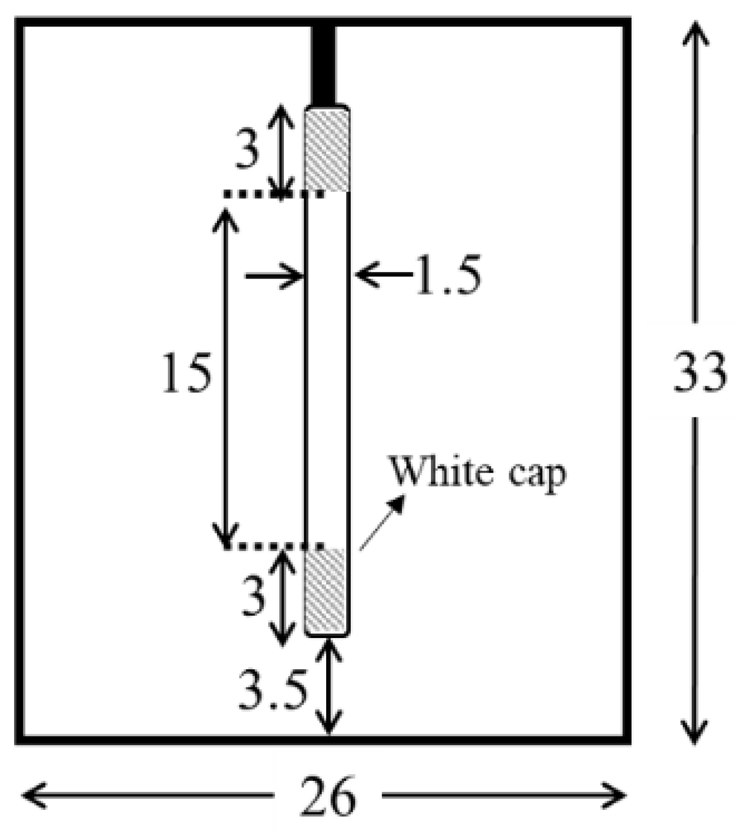
2.2. Dye-Degradation Measurements

Dye-degradation experiments were conducted using a custom-made UV chamber (Figure 1). We used a 10-watts Ultraviolet (UV) lamp (Ster-L-Ray, Preheat Germicidal Ultraviolet Lamps, Atlantic Ultraviolet Corporation, Hauppauge, NY, USA) with a total UV output of 2.7 W, a length of 21 cm, and a diameter of ~1.5 cm. The chamber dimensions were 26 × 33 × 25 cm3, and the distance from the lamp to the samples was around 3.5 cm. The chamber was covered with an aluminum foil to enhance the reflection.

2.3. Dye-Degradation Measurement of TiO2 Nanoparticles

The Ultraviolet-Visible (UV-VIS) spectrum of the dye was first measured to determine the optimal wavelength. This was done by using a 1.0 mg/mL concentration of the dye in deionized water and absorbances were read using a plate reader (Infinite M200 Pro, Tecan, Männedorf, Switzerland). The optimal wavelength was determined, based on results obtained using a range of excitation wavelengths. This wavelength was then used to produce a calibration curve to determine the concentration of the dye during the process of cleaning. The calibration graph was produced by measuring the absorbances for dye concentrations of 1, 0.85, 0.70, 0.55, 0.40, 0.25, and 0.1 mg/mL, respectively. Then, TiO2 NPs with a concentration of 1 mg/mL in deionized water were mixed with 1 mg/mL of the coloring dye. The suspension was then kept in the UV chamber while stirring to provide consistency with the reaction. A small sample was retrieved from the reaction at 5, 15, 30, 60, 180, and 360 min, and put into a microcentrifuge tube. The tube was then spun at 14,800 rpm for 10 min to pellet the TiO2 NPs out of the suspension. Next, 100 μL of the supernatant was gently pipetted from the microcentrifuge tube to the well plate. The well plate was then scanned using the plate reader, and the data obtained was graphed and interpreted.

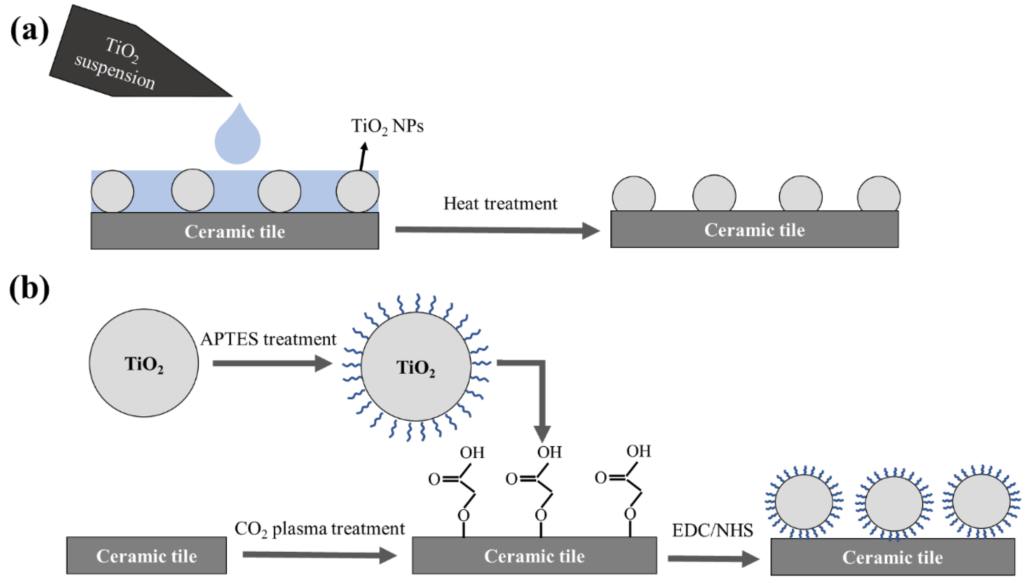
2.4. Coating TiO2 Nanoparticles Using Heat Treatment Technique

Thirty microliter of 1 mg/mL TiO2 NPs suspension was added onto the ceramic tiles’ surfaces and spread to cover the whole surface using a simple pipetting system, and then left to dry at room temperature. The surfaces were then placed in a tube furnace or a vacuum oven for lower temperatures. The heat treatments were done in different durations of 2 h and 5 h, and at different temperatures of 100, 200, 400, 500, 700 and 900 °C. The rate of the temperature gradient in all experiments was 5 °C/min. Following heat treatment, the surfaces were rinsed and wiped to remove any unattached particle (Figure 2a). Five microliter of 1 mg/mL of dye was added onto each surface and placed in the UV chamber to evaluate the amount of dye degradation in different time intervals.

2.5. APTES Treatment Technique

TiO2 NPs were homogenized with triethylamine (for improving the particle dispersion [46]) and ethanol at 60 °C. Further, ammonium hydroxide, deionized water (DI) water, and APTES (~1%) were added to the suspension and left to stir overnight. [40]. Ammonium hydroxide as a catalyst can increase the rate of APTES hydrolysis reactions [47]. Finally, the suspension was centrifuged at 4500 rpm for 15 min. The supernatant was discarded, and the pellet was mixed with ethanol. The washing step was repeated 3 times and the obtained powder was stored in 50 mL of toluene.
In order to coat ceramic tiles with the APTES treated particles, the cleaned surfaces were CO2 plasma treated (Harrick Plasma, Ithaca, NY, USA) to create carboxylic groups onto the surface. EDC-NHS chemistry (a molar ratio of EDC:NHS: ~ 1:1) was used to activate the carboxylic groups on the surfaces. The solution was rinsed off the surface with DI water, and then the APTES treated TiO2 suspension was added to the surfaces and left for 2 h. Finally, the surfaces were rinsed with DI water and dried at room temperature (Figure 2b). Five microliter of 1 mg/mL dye was added to the surfaces and left to dry. The surfaces were placed in the UV chamber and imaged at different time intervals.

2.6. Characterization Techniques

High-resolution XPS was used to investigate the amount of carboxylic groups induced by CO2 plasma treatment of glass slides for different time durations. XPS measurements were carried out at binding energy between ~280 to ~300 (eV) to capture bond of O–C=O, C=O, C–C, and C–O as counts per second. The size distribution profile of the TiO2 NPs in suspension was determined using the dynamic light scattering method (DLS, Beckman Coulter, Indianapolis, IN, USA). In DLS analysis, 10 acquisitions were performed for every experiment, and each acquisition was 15 s long. All TiO2 NPs were diluted in DI water with the concentration of 0.01 mg/mL and sonicated for 5 min. For the APTES treated sample, we took 5 μL of the suspension of NPs in toluene and diluted in DI water (10 mL) to achieve the desired concentration in order to exclude the effect of toluene in DLS measurements. Contact angles were measured by an optical contact angle (OCA 35) (Future Digital Scientific Corp., Garden City, NY, USA) using 5 μL droplet of DI water. Fourier-transform infrared spectroscopy (FT-IR) (Bruker, Karlsruhe, Germany) was used to confirm the presence of the TiO2 coating on the ceramic tiles’ surfaces after the washing steps as well as the effect of APTES treatment on the TiO2 NPs. FT-IR was conducted on different samples and different spots on the surfaces for each experiment. ImageJ was utilized to quantify the photos achieved from the dye-degradation results. For better comparison, the graphs were normalized, so that all the graphs started at the same point. Atomic force microscopy (AFM) (BioScope Catalyst, Bruker, Milton, ON, Canada) was performed to analyze surface topography and roughness within the scan size of 1 × 1 μm2. A ScanAsyst mode was utilized in the measurements using a probe with the spring constant of 0.4 N/m.

3. Results and Discussion

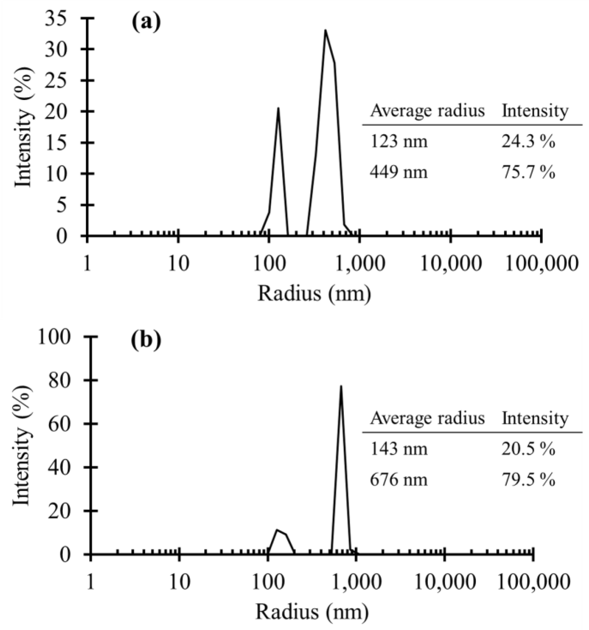
3.1. TiO2 NPs Size Measurements

Figure 3 demonstrates the average particle sizes of untreated TiO2 NPs and APTES treated TiO2 NPs versus the particles population percentage. As is shown, the average radius of the untreated NPs was mostly distributed in two different sizes of 123 nm (24.3% of particle number) and 449 nm (75.7% of particle number). The existence of the larger particle sizes could be due to the NPs agglomeration even after the sonication. In Figure 3b, there were also two main particle size distributions associated with the APTES treated NPS. At the first peak, the average radius was almost the same as the untreated NPS. However, the number of NPs with that radius decreased, compared to the untreated NPs. Moreover, APTES treatment caused the second distribution to go up in terms of average radius as well as population density of the NPs. Consequently, APTES treatment of the particles caused a slight increase in the particles agglomeration, although this was not significant.

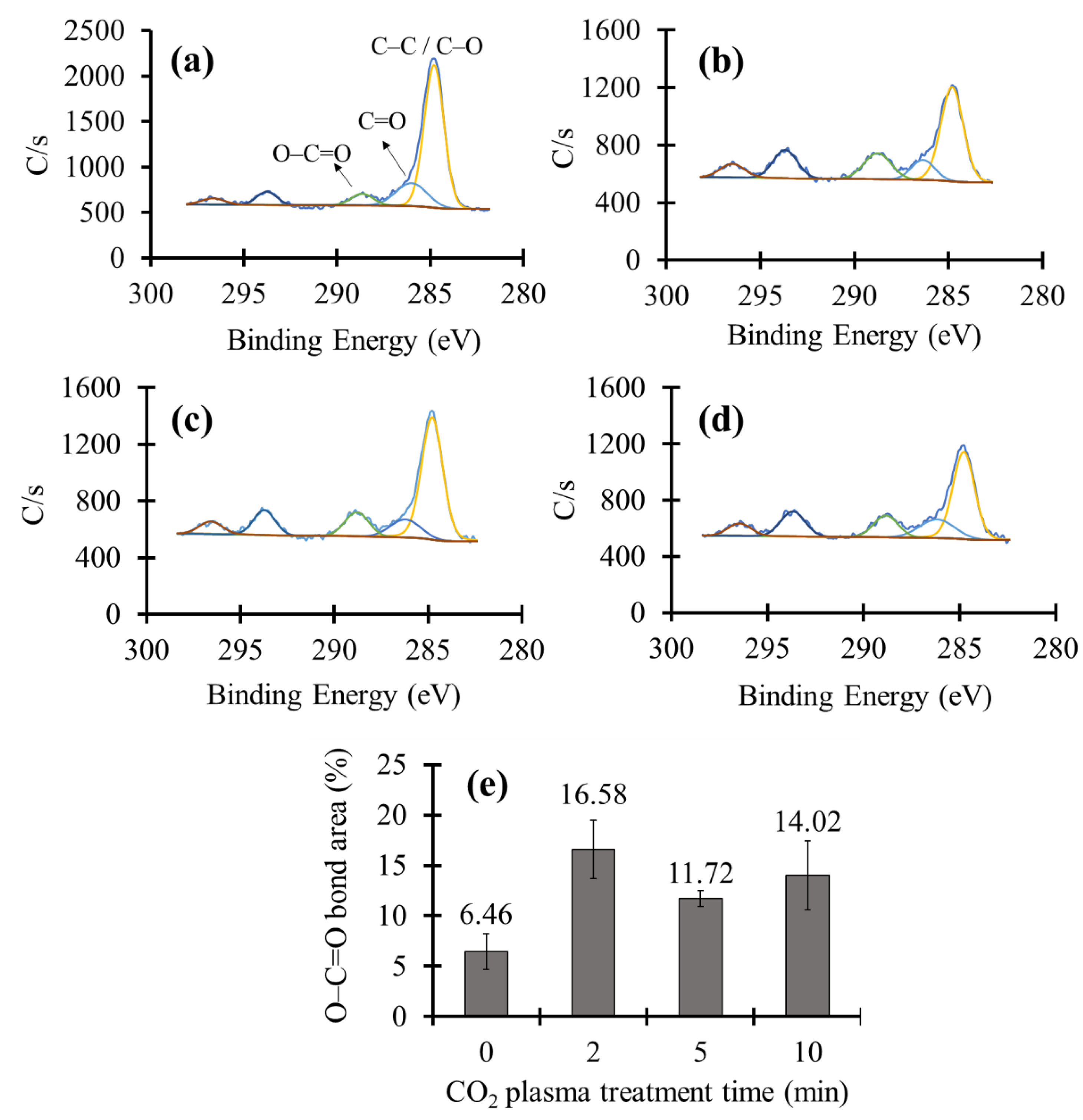
3.2. XPS Measurement of the CO2 Plasma Treated Ceramic Tiles

The second approach that we employed to covalently bind TiO2 NPs to the surface of ceramic tiles was aminosilanization of TiO2 NPs and CO2 plasma treatment of ceramic tiles. APTES treatment of TiO2 NPs formed a monolayer of hydrolyzed and condensed APTES molecules on the surface providing free terminal NH2 groups. Schematic presentation of this functionalization process is shown in Figure 2b. On the other hand, CO2 plasma treatment of ceramic tiles can produce carboxylic groups (COOH) on the surface, which can subsequently be activated using EDC/NHS chemistry. The functional carboxylic groups can go through a chemical reaction with amino groups of the APTES treated particles and form covalent peptide bonds. Consequently, we would be able to covalently bind the TiO2 particles to the ceramic tiles.
To investigate the carboxylic group formation on the surface, a high-resolution XPS test was performed. Figure 4a depicts the XPS results of an untreated sample. XPS graphs of CO2 plasma treated samples for different time periods are shown in Figure 4b–d. The peaks at binding energies around 288.66 (eV), 286.02 (eV), and 284.8 (eV) were assigned to O–C=O, C=O, and C–C as well as C–O, respectively [48]. Figure 4e demonstrates the variation in the amount of O–C=O groups by increasing the CO2 plasma treatment time. It could be seen that after 2 min of plasma treatment, the O–C=O band area in XPS patterns has risen from 6.46% to 16.58%, indicating the well formation of carboxylic groups via the CO2 plasma treatment. Increasing the plasma treatment time more than 2 min did not significantly change the O–C=O % area.

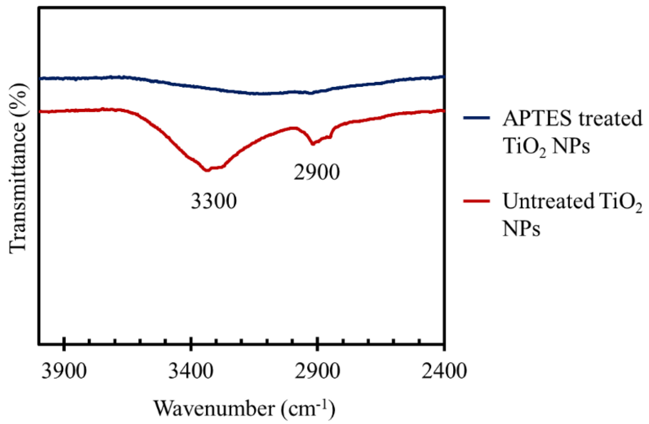
3.3. FT-IR Studies of the TiO2 Coated Tiles

FT-IR spectra of plain ceramic tiles, as well as the tiles coated with TiO2 NPs using heat treatment and APTES treatment methods, are plotted in Figure 5. In FT-IR measurements, the surface of a plain ceramic tile was considered to be the background, and all other spectra were normalized based on that. As a control, we applied untreated TiO2 NPs on ceramic tiles and then performed the regular washing steps. It can be seen that the obtained spectrum was a flat line similar to the background, indicating a complete removal of the TiO2 NPs during the washing process. Nevertheless, the spectra of the TiO2 coated ceramic tiles using the heat treatment protocol had a number of peaks in the absorption band between 400 to 1200 cm−1, mainly at 400 cm−1, 790 cm−1, and 900 cm−1. This absorption band could be ascribed to the Ti–O stretching and Ti–O–Ti bridging stretching modes [49,50,51,52,53]. Thus, the results demonstrated the presence of TiO2 NPs onto the surface of the tiles after performing the washing steps. This is due to the strong adhesion of TiO2 NPs to the surface induced via the heat treatment at 200 °C for 5 h (the optimum heat treatment condition based on the dye-degradation results in Figure 10). Figure 5 also depicts the same FT-IR spectra for the APTES treated NPs bonded to the plasma treated ceramic tiles. Moreover, the transmittance percentages of the peaks were almost the same as those of the heat-treated samples, which roughly illustrated the equal mass of TiO2 coating. The results confirmed the stable bond formation between TiO2 NPs and the substrate that retained the NPs onto the surface during the washing steps.
In Figure 6, the FT-IR spectra of APTES treated TiO2 NPs and untreated NPs were compared at the wavenumber between 2400 to 4000 cm−1 to confirm the efficacy of APTES functionalization. There are two main peaks at ~2900 and ~3300 in the transmittance spectrum of untreated TiO2 NPs, which indicate the presence of hydroxyl groups on the surface of the NPs. The first peak could be associated with both symmetric and asymmetric CH2 stretching vibrations in –CH2–OH compound [54], and the second peak was attributed to hydroxyl group symmetric and asymmetric stretching vibrations in Ti–OH compounds [49]. The hydroxyl groups can be formed due to the chemical and physical adsorptions of atmosphere moisture on the surface of NPs [49,55]. As shown in Figure 6, for the APTES treated particles, the peaks related to hydroxyl groups almost disappeared, and a broad peak between 2800 to 3600 cm−1 was formed in the spectrum of APTES treated NPs instead. This is because of the hydrolysis and condensation reactions of the silane head groups in APTES molecules and anchoring to the hydroxyl groups of the NPs surfaces to form oxane (Si–O–M) bonds [56]. The broad peak could be caused by OH stretching in silanol (Si–OH) groups (3200–3700 cm−1) formed via hydrolysis of APTES, primary amine (3160–3450 cm−1), as well as alkane C–H stretching vibrations in –CH3 and –CH2– (2840–2975 cm−1) due to the presence of APTES molecules [54].

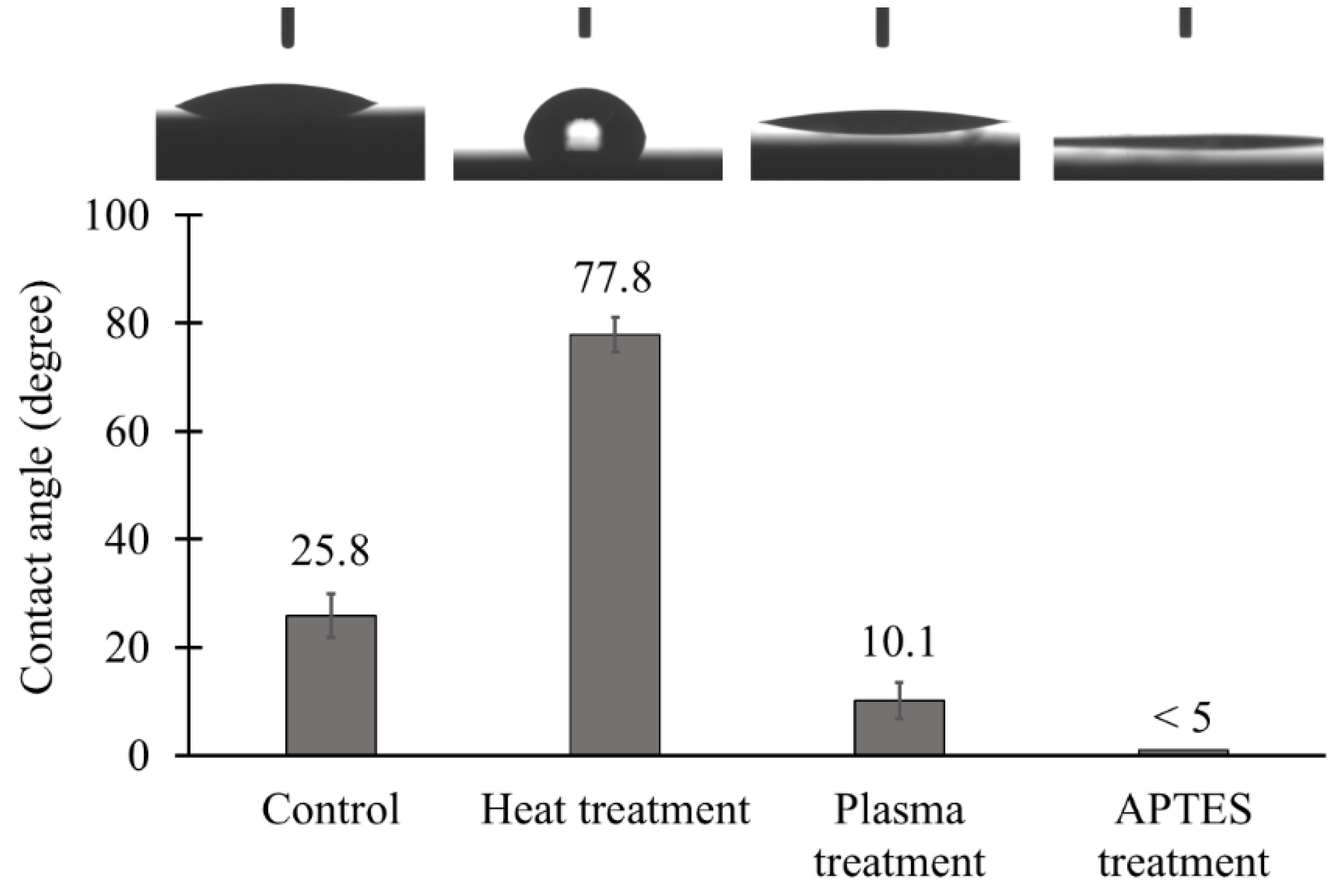
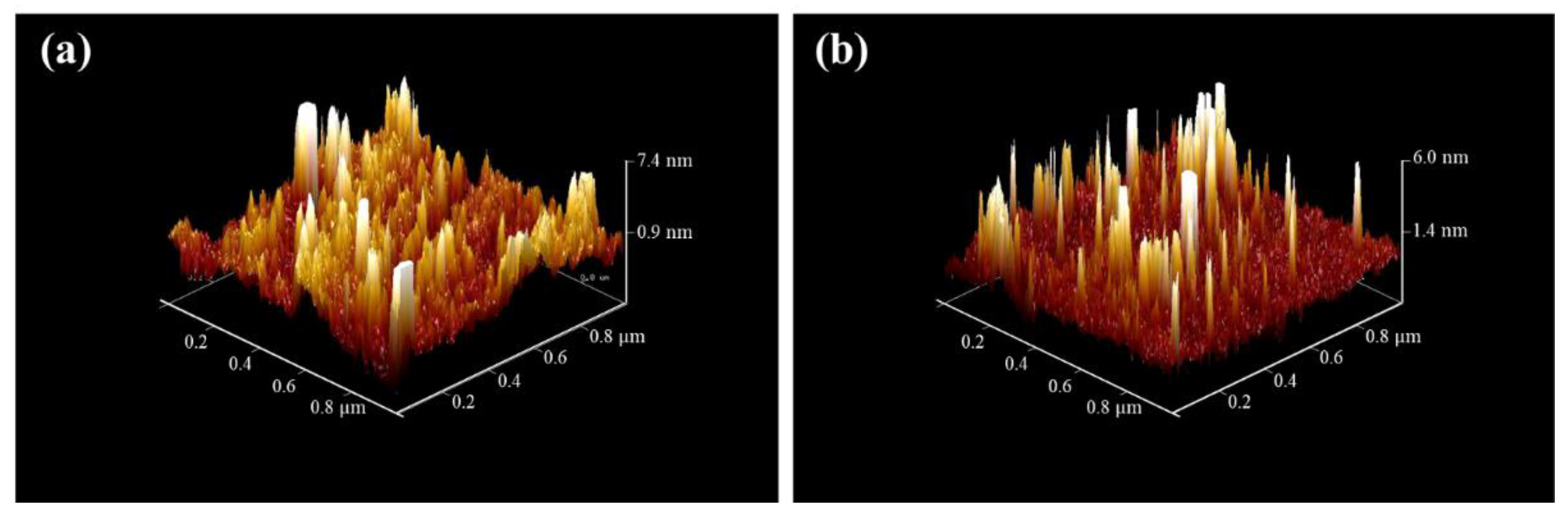
3.4. Hydrophilicity and Surface Topography of the TiO2 Coated Tiles

The results of the contact angle measurement are shown in Figure 7. The contact angle of plain ceramic tiles was about 25.8 ± 4.0° indicating the hydrophilic behavior of the surfaces. By performing heat treatment, although surfaces remained hydrophilic, the contact angle rose to 77.8 ± 3.2°. This unexpected increase in contact angle could be due to the surface roughness generated by partial diffusion of TiO2 NPs into the surface, which can compensate for the inherent hydrophilicity of TiO2 [57]. The APTES treatment method, on the other hand, resulted in more hydrophilic TiO2 coated surfaces with the contact angle of less than 5°. A possible explanation for that is CO2 plasma treatment step during the APTES treatment protocol, which can greatly increase hydrophilicity as well as the APTES coating on the surface of the NPs [58].
AFM images of the TiO2 coated ceramic tiles are illustrated in Figure 8. The density of the NPs in the heat-treated sample (Figure 8a) was higher, compared to the sample coated with the APTES treatment protocol (Figure 8b). Furthermore, the root-mean-square (RMS) surface roughness of the ceramic tile increased from 0.676 nm for a plain ceramic to 1.26 nm for TiO2 coated ceramic using the APTES treatment method. The RMS parameter of the TiO2 coated ceramic tiles with the heat treatment process was 1.82 nm, which was more than that for tiles with the APTES treatment protocol due to the higher number of NPs on the surface.

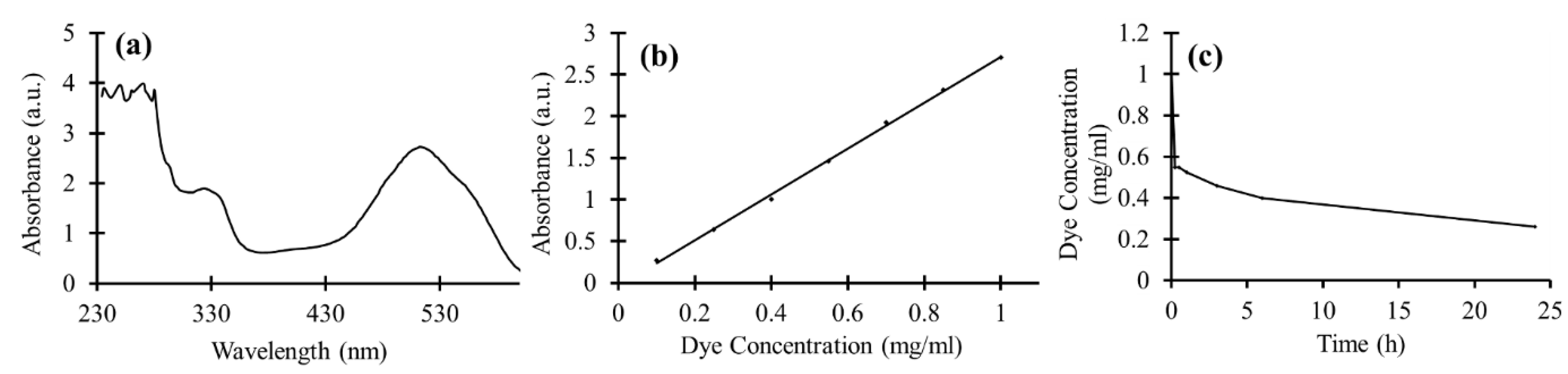
3.5. Dye Degradation of TiO2 NPs in Suspension

In order to achieve the proper wavelength for reading the absorbances of the dye with a plate reader, the full absorbance spectra of 1 mg/mL concentration of the dye diluted in deionized water was measured. As shown in Figure 9a, the sharp peak at the wavelength of 512 nm, which can be easily distinguished, was used as a reference wavelength for the next absorbance measurements. The calibration curve for dye absorbance as a function of concentration at the wavelength of 512 nm is illustrated in Figure 9b. The obtained scatter diagram has a linear trend line. To measure the photocatalytic power of TiO2 NPs, the particles were added to the 1 mg/mL dye solution and were kept in the UV light, under a constant stirring condition. The amount of dye-degradation induced by TiO2 photocatalyst could be calculated based on Figure 9b. The dye concentration results versus UV exposure time are shown in Figure 9c. As is shown, a considerable reduction in the dye concentration was achieved after 15 min where almost half of the dye was degraded, and the concentration reached 0.55 mg/mL. Afterwards, the rate of dye degradation became slower and after 1 day of UV irradiation, the dye concentration decreased down to 0.26 mg/mL. Thus, the TiO2 nanoparticles possessed a considerable photocatalytic power in suspension. The reason why the expected exponential decay to zero was not observed in our sample could be attributed to the absorption of the partial degradation products on the photocatalyst surface and/or the pH variation of the suspension leading to deterioration in the dye-degradation process.

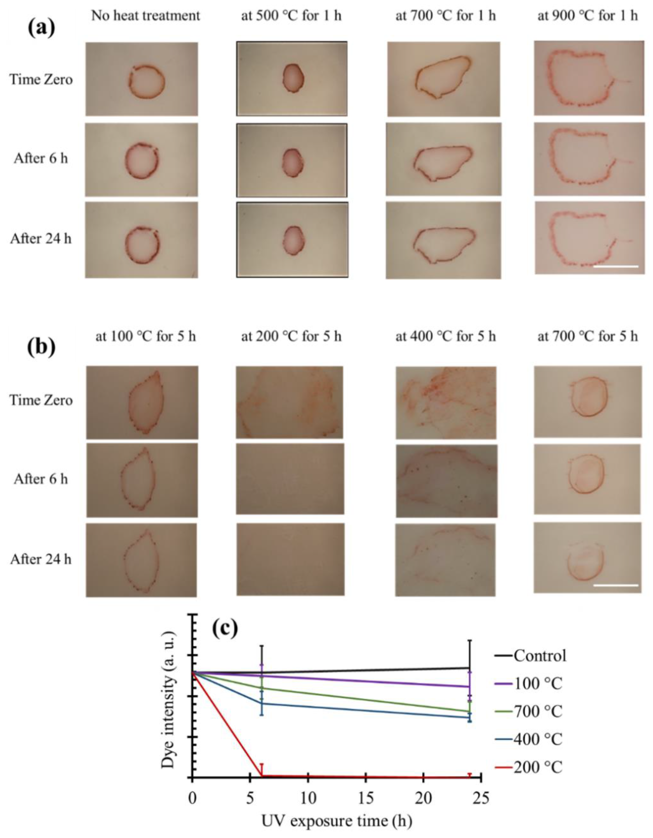
3.6. Dye Degradation on TiO2 Coated Tiles Produced Using Heat Treatment

Figure 10 demonstrates the dye-degradation properties of ceramic tiles coated with TiO2 nanoparticles using the heat treatment approach at different temperatures and incubation time (1 and 5 h). Control ceramic tiles were incubated with a suspension of TiO2 NPs with the same incubation time, but no heat treatment was involved (labeled by “no heat treatment” in Figure 10). Following the washing step, all TiO2 NPs were detached from the surface of control tiles and therefore, no dye degradation was observed. Moreover, when the samples were heat treated for 1 h (Figure 10a), no dye-degradation properties were recorded at any heat treatment temperatures. One major problem with high temperatures of 700 °C and 900 °C is that since the temperatures were above the phase transition temperature of TiO2 from anatase to rutile [59,60,61,62], the formed rutile phase could have deteriorated the photocatalytic activity of the TiO2 particles even if the particles were successfully attached to the ceramic tiles. It has been proved that TiO2 exhibits a higher photocatalytic activity in anatase phase compared to fully rutile phase, which may be due to larger band gap in anatase compared to rutile TiO2 [4]. Thus, the optimal temperature for heat treatment should not change the structure of TiO2. According to the composite phase diagram of TiO2, the anatase-rutile phase transition takes place at around 600 °C at atmospheric pressure [59].
Therefore, we increased the heat treatment duration from 1 h to 5 h to investigate the effect of heat treatment time on the coating process, specially the possibility of successful and functional coating at lower temperatures. Figure 10b exhibits the dye-degradation trend for the ceramic tile samples, which were heat treated at varying temperatures for 5 h. In Figure 10c, the results were also quantified for better comparison. The samples that were heat treated at 400 °C degraded the dye at a lower rate than heat treatment at 200 °C, although both temperatures were below the anatase-rutile phase transition temperature. This can be caused by slightly more agglomeration of the TiO2 NPs at 400 °C in comparison to 200 °C, leading to a small increase in the average particle size and a reduction in the overall surface area of the particles, thereby diminishing the photocatalytic power. The other possible explanation could be attributed to the higher diffusion of NPs inside the glossy surface of the ceramic tiles at 400 °C. Therefore, the smaller area of the particles would get in touch with the dye, causing a decrease in the dye-degradation rate. Performing the heat treatment at a more cost-effective temperature of 200 °C caused the dye to be fully degraded after 6 h. Nevertheless, 100 °C heat treatment for 5 h appeared to be not enough for the TiO2 particles to be completely attached to the surface of ceramic tiles, thereby resulting in less dye degradation under UV exposure. Therefore, a 5 h heat treatment at 200 °C provided the best dye-degradation properties for the ceramic tiles.

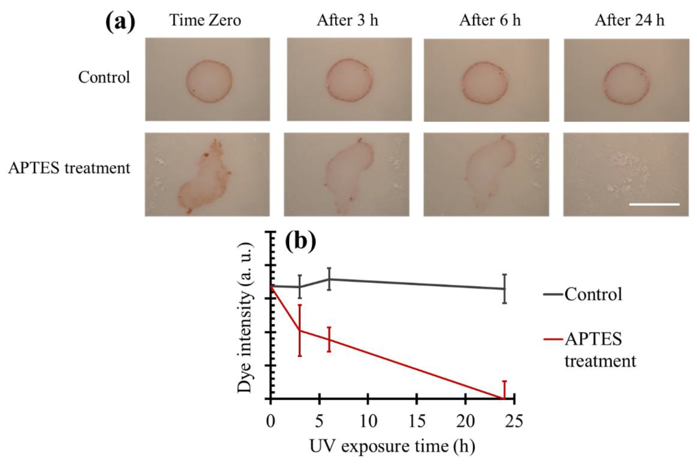
3.7. Dye Degradation on TiO2 Coated Tiles Produced Using Aminosilanization

Figure 11a illustrates the dye-degradation results of the ceramic tiles coated with the APTES treatment method. Quantifications of the obtained results are plotted in Figure 11b. We applied untreated TiO2 particles onto the ceramic tiles as control samples. Most of the particles were removed from the surface during the washing process, and the dye was not degraded after 1 day in control samples. However, when APTES treated particles were used, the dye was almost invisible after 24 h, demonstrating excellent effectivity of the APTES treatment process for coating ceramic tiles with TiO2 NPs. In comparison to the heat treatment at 200 °C for 5 h, coating the surfaces via the APTES treatment resulted in a lower rate of dye degradation due to two main reasons: a smaller number of particles on the surface which was confirmed with AFM test, and covering the particles via a silane coupling agent which can reduce the photoactivity of the particles [63]. Nevertheless, using the APTES treatment protocol has two important advantages. First, the method for functionalization using APTES is much simpler and does not require high temperatures compared to the heat treatment. Second, since this technique creates covalent bonds between TiO2 NPs and surfaces, the durability of the coating would be higher in the long term.

4. Conclusions

The photocatalytic power of TiO2 NPs measured inside the suspension showed a proper dye-degradation effect under UV exposure. We introduced two strategies to coat ceramic tiles with the TiO2 NPs. Both protocols led to transparent TiO2 coating on ceramic tiles without any adverse effect on the tile’s physical appearance. In the heat treatment approach, it was shown that applying a simple one-step heat treatment with the optimum temperature of 200 °C for 5 h resulted in rapid degradation of dye in a short time. In the second strategy, the APTES treatment method was employed to covalently bond the TiO2 particles to the CO2 treated ceramic tiles, while the photocatalytic power of the particles was preserved. The success of the covalent bonding protocol suggests a possible longer duration of the coating in harsh environments. In future, we will focus on enhancement of the self-cleaning behavior, as well as studying the durability of the coating by performing proper weathering tests.

Author Contributions

T.F.D. conceived the research and supervised the experiments. A.S. performed the experiments, data validation, data visualization, and analysis with help from D.Y. A.S. wrote the manuscript with input from T.F.D. and M.S. M.B. conducted the FT-IR tests. S.M.I. performed the AFM analyses. All authors have read, commented and approved the final version of the paper.

Funding

This project was supported by an ENGAGE-OCE-VIPI grant from the Natural Science and Engineering Research Council of Canada (NSERC, Ontario Centers of Excellence (OCE) and Nanophyll Inc.

Conflicts of Interest

M.S. holds equity in Nanophyll Inc.

References

  1. Dong, H.; Zeng, G.; Tang, L.; Fan, C.; Zhang, C.; He, X.; He, Y. An overview on limitations of TiO2-based particles for photocatalytic degradation of organic pollutants and the corresponding countermeasures. Water Res. 2015, 79, 128–146. [Google Scholar] [CrossRef] [PubMed]
  2. Weon, S.; Kim, J.; Choi, W. Dual-components modified TiO2 with Pt and fluoride as deactivation-resistant photocatalyst for the degradation of volatile organic compound. Appl. Catal. B Environ. 2018, 220, 1–8. [Google Scholar] [CrossRef]
  3. Li, S.; Ishikawa, S.; Huang, C.; Zhang, W.; Tanizaki, T.; Higuchi, T.; Haraga, H. Production of active intermediates and decomposition behaviors of organic compounds in the ultraviolet ray/supersonic wave reactions with TiO2 photocatalyst. Sustain. Environ. Res. 2015, 25, 67–72. [Google Scholar]
  4. Luttrell, T.; Halpegamage, S.; Tao, J.; Kramer, A.; Sutter, E.; Batzill, M. Why is anatase a better photocatalyst than rutile? Model studies on epitaxial TiO2 films. Sci. Rep. 2015, 4, 4043. [Google Scholar] [CrossRef] [PubMed]
  5. Hashimoto, K.; Irie, H.; Fujishima, A. TiO2 photocatalysis: A historical overview and future prospects. Jpn. J. Appl. Phys. 2005, 44, 8269–8285. [Google Scholar] [CrossRef]
  6. Nosaka, Y.; Nosaka, A.Y. Identification and roles of the active species generated on various photocatalysts. In Photocatalysis and Water Purification; Wiley-VCH Verlag GmbH & Co. KGaA: Weinheim, Germany, 2013. [Google Scholar]
  7. Fujishima, A.; Zhang, X.; Tryk, D.A. TiO2 photocatalysis and related surface phenomena. Surf. Sci. Rep. 2008, 63, 515–582. [Google Scholar] [CrossRef]
  8. Fujishima, A.; Rao, T.N.; Tryk, D.A. Titanium dioxide photocatalysis. J. Photochem. Photobiol. C Photochem. Rev. 2000, 1, 1–21. [Google Scholar] [CrossRef]
  9. Nakata, K.; Fujishima, A. TiO2 photocatalysis: Design and applications. J. Photochem. Photobiol. C Photochem. Rev. 2012, 13, 169–189. [Google Scholar] [CrossRef]
  10. Marugán, J.; van Grieken, R.; Pablos, C.; Satuf, M.L.; Cassano, A.E.; Alfano, O.M. Photocatalytic inactivation of escherichia coli aqueous suspensions in a fixed-bed reactor. Catal. Today 2015, 252, 143–149. [Google Scholar] [CrossRef]
  11. Liu, H.-L.; Yang, T.C.-K. Photocatalytic inactivation of escherichia coli and lactobacillus helveticus by ZnO and TiO2 activated with ultraviolet light. Process Biochem. 2003, 39, 475–481. [Google Scholar] [CrossRef]
  12. Padervand, M.; Vossoughi, M.; Yousefi, H.; Salari, H.; Gholami, M.R. An experimental and theoretical study on the structure and photoactivity of XFe2O4 (X = Mn, Fe, Ni, Co, and Zn) structures. Russ. J. Phys. Chem. A 2014, 88, 2451–2461. [Google Scholar] [CrossRef]
  13. Banerjee, S.; Dionysiou, D.D.; Pillai, S.C. Self-cleaning applications of TiO2 by photo-induced hydrophilicity and photocatalysis. Appl. Catal. B Environ. 2015, 176–177, 396–428. [Google Scholar] [CrossRef]
  14. Klein, M.; Grabowska, E.; Zaleska, A. Noble metal modified TiO2 for photocatalytic air purification. Physicochem. Probl. Miner. Process. 2015, 51, 49–57. [Google Scholar]
  15. Song, J.; Wang, X.; Yan, J.; Yu, J.; Sun, G.; Ding, B. Soft Zr-doped TiO2 nanofibrous membranes with enhanced photocatalytic activity for water purification. Sci. Rep. 2017, 7, 1636. [Google Scholar] [CrossRef] [PubMed]
  16. Imani, R.; Dillert, R.; Bahnemann, D.W.; Pazoki, M.; Apih, T.; Kononenko, V.; Repar, N.; Kralj-Iglič, V.; Boschloo, G.; Drobne, D.; et al. Multifunctional gadolinium-doped mesoporous TiO2 nanobeads: Photoluminescence, enhanced spin relaxation, and reactive oxygen species photogeneration, beneficial for cancer diagnosis and treatment. Small 2017, 13, 1–11. [Google Scholar] [CrossRef] [PubMed]
  17. Seo, J.W.; Chung, H.; Kim, M.Y.; Lee, J.; Choi, I.H.; Cheon, J. Development of water-soluble single-crystalline TiO2 nanoparticles for photocatalytic cancer-cell treatment. Small 2007, 3, 850–853. [Google Scholar] [CrossRef] [PubMed]
  18. Ohko, Y.; Utsumi, Y.; Niwa, C.; Tatsuma, T.; Kobayakawa, K.; Satoh, Y.; Kubota, Y.; Fujishima, A. Self-sterilizing and self-cleaning of silicone catheters coated with TiO2 photocatalyst thin films: A preclinical work. J. Biomed. Mater. Res. 2001, 58, 97–101. [Google Scholar] [CrossRef]
  19. Suketa, N.; Sawase, T.; Kitaura, H.; Naito, M.; Baba, K.; Nakayama, K.; Wennerberg, A.; Atsuta, M. An antibacterial surface on dental implants, based on the photocatalytic bactericidal effect. Clin. Implant Dent. Relat. Res. 2005, 7, 105–111. [Google Scholar] [CrossRef] [PubMed]
  20. Kim, D.S.; Kwak, S.-Y. Photocatalytic inactivation of E. coli with a mesoporous TiO2 coated film using the film adhesion method. Environ. Sci. Technol. 2009, 43, 148–151. [Google Scholar] [CrossRef] [PubMed]
  21. Behpour, M.; Mehrzad, M.; Hosseinpour-Mashkani, S.M. TiO2 thin film: Preparation, characterization, and its photocatalytic degradation of basic yellow 28 dye. J. Nanostructures 2015, 5, 183–187. [Google Scholar]
  22. Vera, M.L.; Leyva, G.; Litter, M.I. Simple TiO2 coatings by sol–gel techniques combined with commercial TiO2 particles for use in heterogeneous photocatalysis. J. Nanosci. Nanotechnol. 2017, 17, 4946–4954. [Google Scholar] [CrossRef]
  23. Justicia, I.; Garcia, G.; Vázquez, L.; Santiso, J.; Ordejón, P.; Battiston, G.; Gerbasi, R.; Figueras, A. Self-doped titanium oxide thin films for efficient visible light photocatalysis: An example: Nonylphenol photodegradation. Sens. Actuators B Chem. 2005, 109, 52–56. [Google Scholar] [CrossRef]
  24. Justicia, I.; Ordejón, P.; Canto, G.; Mozos, J.L.; Fraxedas, J.; Battiston, G.A.; Gerbasi, R.; Figueras, A. Designed self-doped titanium dioxide thin films for efficient visible-light photocatalysis. Adv. Mater. 2002, 14, 1399–1402. [Google Scholar] [CrossRef]
  25. Yemmireddy, V.K.; Farrell, G.D.; Hung, Y.C. Development of titanium dioxide (TiO2) nanocoatings on food contact surfaces and method to evaluate their durability and photocatalytic bactericidal property. J. Food Sci. 2015, 80, N1903–N1911. [Google Scholar] [CrossRef] [PubMed]
  26. Sopyan, I.; Watanabe, M.; Murasawa, S.; Hashimoto, K.; Fujishima, A. A film-type photocatalyst incorporating highly active TiO2 powder and fluororesin binder: Photocatalytic activity and long-term stability. J. Electroanal. Chem. 1996, 415, 183–186. [Google Scholar] [CrossRef]
  27. Lin, H.; Xu, Z.; Wang, X.; Long, J.; Su, W.; Fu, X.; Lin, Q. Photocatalytic and antibacterial properties of medical-grade PVC material coated with TiO2 film. J. Biomed. Mater. Res. Part B Appl. Biomater. 2008, 87, 425–431. [Google Scholar] [CrossRef] [PubMed]
  28. Pal, S.; Contaldi, V.; Licciulli, A.; Marzo, F. Self-cleaning mineral paint for application in architectural heritage. Coatings 2016, 6, 48. [Google Scholar] [CrossRef]
  29. Gunda, N.S.K.; Singh, M.; Norman, L.; Kaur, K.; Mitra, S.K. Optimization and characterization of biomolecule immobilization on silicon substrates using (3-aminopropyl)triethoxysilane (APTES) and glutaraldehyde linker. Appl. Surf. Sci. 2014, 305, 522–530. [Google Scholar] [CrossRef]
  30. Landoulsi, J.; Genet, M.J.; Kirat, K.E.; Richard, C.; Pulvin, S.; Rouxhe, P.G. Silanization with APTES for controlling the interactions between stainless steel and biocomponents: Reality vs. expectation. Biomater. Phys. Chem. 2011, 5, 99–126. [Google Scholar]
  31. Li, G.; Yang, P.; Qin, W.; Maitz, M.F.; Zhou, S.; Huang, N. The effect of coimmobilizing heparin and fibronectin on titanium on hemocompatibility and endothelialization. Biomaterials 2011, 32, 4691–4703. [Google Scholar] [CrossRef] [PubMed]
  32. Badv, M.; Jaffer, I.H.; Weitz, J.I.; Didar, T.F. An omniphobic lubricant-infused coating produced by chemical vapor deposition of hydrophobic organosilanes attenuates clotting on catheter surfaces. Sci. Rep. 2017, 7, 11639. [Google Scholar] [CrossRef] [PubMed]
  33. Villegas, M.; Cetinic, Z.; Shakeri, A.; Didar, T.F. Fabricating smooth PDMS microfluidic channels from low-resolution 3D printed molds using an omniphobic lubricant-infused coating. Anal. Chim. Acta 2018, 1000, 248–255. [Google Scholar] [CrossRef] [PubMed]
  34. Didar, T.F.; Cartwright, M.J.; Rottman, M.; Graveline, A.R.; Gamini, N.; Watters, A.L.; Leslie, D.C.; Mammoto, T.; Rodas, M.J.; Kang, J.H.; et al. Improved treatment of systemic blood infections using antibiotics with extracorporeal opsonin hemoadsorption. Biomaterials 2015, 67, 382–392. [Google Scholar] [CrossRef] [PubMed]
  35. Didar, T.F.; Tabrizian, M. Generating multiplex gradients of biomolecules for controlling cellular adhesion in parallel microfluidic channels. Lab Chip 2012, 12, 4363–4371. [Google Scholar] [CrossRef] [PubMed]
  36. Didar, T.F.; Bowey, K.; Almazan, G.; Tabrizian, M. A miniaturized multipurpose platform for rapid, label-free, and simultaneous separation, patterning, and in vitro culture of primary and rare cells. Adv. Healthc. Mater. 2014, 3, 253–260. [Google Scholar] [CrossRef] [PubMed]
  37. Shakeri, A.; Sun, N.; Badv, M.; Didar, T.F. Generating 2-dimensional concentration gradients of biomolecules using a simple microfluidic design. Biomicrofluidics 2017, 11. [Google Scholar] [CrossRef] [PubMed]
  38. Kim, W.J.; Kim, S.; Lee, B.S.; Kim, A.; Ah, C.S.; Huh, C.; Sung, G.Y.; Yun, W.S. Enhanced protein immobilization efficiency on a TiO2 surface modified with a hydroxyl functional group. Langmuir 2009, 25, 11692–11697. [Google Scholar] [CrossRef] [PubMed]
  39. Cheng, F.; Sajedin, S.M.; Kelly, S.M.; Lee, A.F.; Kornherr, A. UV-stable paper coated with APTES-modified P25 TiO2 nanoparticles. Carbohydr. Polym. 2014, 114, 246–252. [Google Scholar] [CrossRef] [PubMed]
  40. Gong, Z.; Tang, D.; Guo, Y. The fabrication and self-flocculation effect of hybrid TiO2 nanoparticles grafted with poly(N-isopropylacrylamide) at ambient temperature via surface-initiated atom transfer radical polymerization. J. Mater. Chem. 2012, 22, 16872. [Google Scholar] [CrossRef]
  41. Xu, Z.; Shang, J.; Liu, C.; Kang, C.; Guo, H.; Du, Y. The preparation and characterization of TiO2 ultrafine particles. Mater. Sci. Eng. B 1999, 63, 211–214. [Google Scholar] [CrossRef]
  42. Yu, J.-G.; Yu, H.-G.; Cheng, B.; Zhao, X.-J.; Yu, J.C.; Ho, W.-K. The effect of calcination temperature on the surface microstructure and photocatalytic activity of TiO2 thin films prepared by liquid phase deposition. J. Phys. Chem. B 2003, 107, 13871–13879. [Google Scholar] [CrossRef]
  43. Lopez, N.; Nørskov, J.K.; Janssens, T.V.W.; Carlsson, A.; Puig-Molina, A.; Clausen, B.S.; Grunwaldt, J.-D. The adhesion and shape of nanosized Au particles in a Au/TiO2 catalyst. J. Catal. 2004, 225, 86–94. [Google Scholar] [CrossRef]
  44. Kang, I.-C.; Zhang, Q.; Yin, S.; Sato, T.; Saito, F. Preparation of a visible sensitive carbon doped TiO2 photo-catalyst by grinding TiO2 with ethanol and heating treatment. Appl. Catal. B Environ. 2008, 80, 81–87. [Google Scholar] [CrossRef]
  45. Yamada, K.; Yamane, H.; Matsushima, S.; Nakamura, H.; Ohira, K.; Kouya, M.; Kumada, K. Effect of thermal treatment on photocatalytic activity of N-doped TiO2 particles under visible light. Thin Solid Films 2008, 516, 7482–7487. [Google Scholar] [CrossRef]
  46. Widegren, J.; Bergström, L. The effect of acids and bases on the dispersion and stabilization of ceramic particles in ethanol. J. Eur. Ceram. Soc. 2000, 20, 659–665. [Google Scholar] [CrossRef]
  47. Kardys, A.Y.; Bharali, D.J.; Mousa, S.A. Amino-functionalized silica nanoparticles: In vitro evaluation for targeted delivery and therapy of pancreatic cancer. J. Nanotechnol. 2013, 2013, 768724. [Google Scholar] [CrossRef]
  48. Chen, L.-X.; Zheng, J.-N.; Wang, A.-J.; Wu, L.-J.; Chen, J.-R.; Feng, J.-J. Facile synthesis of porous bimetallic alloyed PdAg nanoflowers supported on reduced graphene oxide for simultaneous detection of ascorbic acid, dopamine, and uric acid. Analyst 2015, 140, 3183–3192. [Google Scholar] [CrossRef] [PubMed]
  49. Praveen, P.; Viruthagiri, G.; Mugundan, S.; Shanmugam, N. Structural, optical and morphological analyses of pristine titanium di-oxide nanoparticles—Synthesized via sol-gel route. Spectrochim. Acta Part A Mol. Biomol. Spectrosc. 2014, 117, 622–629. [Google Scholar] [CrossRef] [PubMed]
  50. Lu, H.B.; Zhou, Y.Z.; Vongehr, S.; Tang, S.C.; Meng, X.K. Effects of hydrothermal temperature on formation and decoloration characteristics of anatase TiO2 nanoparticles. Sci. China Technol. Sci. 2012, 55, 894–902. [Google Scholar] [CrossRef]
  51. Watson, S.; Beydoun, D.; Scott, J.; Amal, R. Preparation of nanosized crystalline TiO2 particles at low temperature for photocatalysis. J. Nanoparticle Res. 2004, 6, 193–207. [Google Scholar] [CrossRef]
  52. Chen, Y.F.; Lee, C.Y.; Yeng, M.Y.; Chiu, H.T. The effect of calcination temperature on the crystallinity of TiO2 nanopowders. J. Cryst. Growth 2003, 247, 363–370. [Google Scholar] [CrossRef]
  53. Chellappa, M.; Anjaneyulu, U.; Manivasagam, G.; Vijayalakshmi, U. Preparation and evaluation of the cytotoxic nature of TiO2 nanoparticles by direct contact method. Int. J. Nanomed. 2015, 10, 31–41. [Google Scholar]
  54. Socrates, G. Infrared and Raman Characteristic Group Frequencies, 3rd ed.; John Wiley & Sons Ltd.: Chichester, UK, 2001. [Google Scholar]
  55. Olurode, K.; Neelgund, G.M.; Oki, A.; Luo, Z. A facile hydrothermal approach for construction of carbon coating on TiO2 nanoparticles. Spectrochim. Acta Part A Mol. Biomol. Spectrosc. 2012, 89, 333–336. [Google Scholar] [CrossRef] [PubMed]
  56. Kango, S.; Kalia, S.; Celli, A.; Njuguna, J.; Habibi, Y.; Kumar, R. Surface modification of inorganic nanoparticles for development of organic-inorganic nanocomposites—A review. Prog. Polym. Sci. 2013, 38, 1232–1261. [Google Scholar] [CrossRef]
  57. Yoshimitsu, Z.; Nakajima, A.; Watanabe, T.; Hashimoto, K. Effects of surface structure on the hydrophobicity and sliding behavior of water droplets. Langmuir 2002, 18, 5818–5822. [Google Scholar] [CrossRef]
  58. Han, J.-B.; Wang, X.; Wang, N.; Wei, Z.-H.; Yu, G.-P.; Zhou, Z.-G.; Wang, Q.-Q. Effect of plasma treatment on hydrophilic properties of TiO2 thin films. Surf. Coat. Technol. 2006, 200, 4876–4878. [Google Scholar] [CrossRef]
  59. Nie, X.; Zhuo, S.; Maeng, G.; Sohlberg, K. Doping of TiO2 polymorphs for altered optical and photocatalytic properties. Int. J. Photoenergy 2009, 2009, 1–22. [Google Scholar] [CrossRef]
  60. Ren, R.; Yang, Z.; Shaw, L.L. Polymorphic transformation and powder characteristics of TiO2 during high energy milling. J. Mater. Sci. 2000, 35, 6015–6026. [Google Scholar] [CrossRef]
  61. Mei, Z.-G.; Wang, Y.; Shang, S.; Liu, Z.-K. First-principles study of the mechanical properties and phase stability of TiO2. Comput. Mater. Sci. 2014, 83, 114–119. [Google Scholar] [CrossRef]
  62. Hanaor, D.A.H.; Sorrell, C.C. Review of the anatase to rutile phase transformation. J. Mater. Sci. 2011, 46, 855–874. [Google Scholar] [CrossRef]
  63. Zhao, J.; Milanova, M.; Warmoeskerken, M.M.C.G.; Dutschk, V. Surface modification of TiO2 nanoparticles with silane coupling agents. Colloids Surfaces A Physicochem. Eng. Asp. 2012, 413, 273–279. [Google Scholar] [CrossRef]
Figure 1. Schematic drawing of the custom-made UV chamber with a UV lamp at the center. All dimensions are in cm.
Materials 11 01003 g001
Figure 2. Schematic representation of (a) a heat treatment method and (b) an APTES treatment method for coating TiO2 NPs on ceramic tiles.
Materials 11 01003 g002
Figure 3. DLS results of the (a) untreated TiO2 NPs and (b) APTES treated TiO2 NPs. The vertical axes indicate the particle number percentages. The insect tables show the average radius of the NPs and related particle number intensities.
Materials 11 01003 g003
Figure 4. High-resolution XPS plots of CO2 plasma treated glass surfaces for (a) 0 min; (b) 2 min; (c) 5 min; and (d) 10 min; (e) Variation of the amount of O–C=O bonds versus plasma treatment time.
Materials 11 01003 g004
Figure 5. FT-IR spectra of a plain ceramic tile indicated as the background, a control ceramic tile in which untreated particles were applied to the substrate and then the washing steps were conducted, a TiO2 coated ceramic tile using the heat treatment method at 200 °C for 5 h, and a TiO2 coated ceramic tile using an APTES treatment protocol.
Materials 11 01003 g005
Figure 6. FT-IR spectra of the APTES treated TiO2 NPs compared to untreated NPs at the wavenumber between 2400 to 4000 cm−1.
Materials 11 01003 g006
Figure 7. Contact angles of plain ceramic tiles (control), TiO2 coated ceramic tiles via the heat treatment method at 200 °C for 5 h, CO2 plasma treated ceramic tiles, and TiO2 coated ceramic tiles via the APTES treatment method (n = 3).
Materials 11 01003 g007
Figure 8. 3D AFM images of ceramic tile surfaces coated with TiO2 NPs using (a) heat treatment at 200 °C for 5 h and (b) the APTES treatment method.
Materials 11 01003 g008
Figure 9. (a) UV–VIS spectra of 1 mg/mL concentration of the dye; (b) Absorbance curve as a function of dye concentration at the wavelength of 512 nm; (c) Dye concentration mixed with TiO2 NPs suspension as a function of UV exposure time.
Materials 11 01003 g009
Figure 10. Dye-degradation results of the heat-treated ceramic tiles under UV exposure. Heat treatment was performed at different temperatures for 1 h (a) and 5 h (b). Scale bars are 1 cm. (c) Quantification of the dye degradation on the heat-treated samples for 5 h at different temperatures and the control sample without any heat treatment.
Materials 11 01003 g010
Figure 11. (a) Dye-degradation results of the ceramic tiles coated with TiO2 particles using the APTES treatment technique. Scale bar is 1 cm. (b) Quantification of the dye-degradation images.
Materials 11 01003 g011

