Article

Magnetron-Sputtered and Rapid-Thermally Annealed NiO:Cu Thin Films on 3D Porous Substrates for Supercapacitor Electrodes

by
Seongha Oh
1,
Young-Kil Jun
2 and
Nam-Hoon Kim
1,*
1
Department of Electrical Engineering, Chosun University, Gwangju 61452, Republic of Korea
2
Hydrogen Industry Team, Green Energy Institute, Mokpo 58656, Republic of Korea
*
Author to whom correspondence should be addressed.
Energies 2025, 18(11), 2704; https://doi.org/10.3390/en18112704
Submission received: 22 April 2025 / Revised: 21 May 2025 / Accepted: 21 May 2025 / Published: 23 May 2025
(This article belongs to the Section D2: Electrochem: Batteries, Fuel Cells, Capacitors)

Abstract

:
The performance of NiO-based supercapacitor electrodes for energy storage systems was enhanced by doping Cu into NiO thin films (200 nm) using radio-frequency magnetron co-sputtering on 3D porous Ni foam substrates, followed by rapid thermal annealing. The Hall effect measurements demonstrated enhanced electrical conductivity, with resistivity values of 1.244 × 10⁻⁴ Ω·cm. The 3D porous NiO:Cu electrodes significantly increased the specific capacitance and achieved a value of 1809.2 Fg−1, with the NiO:Cu (10 at% Cu) thin films at a scan rate of 5 mVs−1, which is a 2.67-fold increase compared with the undoped NiO films on a glass substrate. The 3D porous NiO:Cu electrodes significantly improved the electrochemical properties of the NiO-based electrode, which resulted in a higher specific capacitance for enhancing the energy storage performance during grid stabilization.

1. Introduction

Energy storage systems (ESSs) play a vital role in stabilizing power grids and improving the energy efficiency to effectively harness renewable energy sources, which have inherent variability and load response characteristics [1,2,3,4]. These systems store the excess power generated during low-demand periods and release it during peak demand times, thereby addressing the intermittent nature of renewable resources such as solar and wind energy. As the global demand for clean and sustainable energy increases, ESSs are becoming essential for ensuring the stability of power grids and maximizing the efficiency of energy use. Among various energy storage options, supercapacitors have gained significant attention owing to their unique advantages, such as high power density, rapid charge–discharge cycles, and long cycle life [5]. These features render supercapacitors promising candidates for grid stabilization and power optimization applications [6]. Supercapacitors can deliver large bursts of power over short durations, either independently or in combination with batteries. They are used in a wide range of applications, ranging from industrial mobile equipment to electric vehicles [7]. Compared with batteries, supercapacitors offer superior output characteristics and exhibit higher energy densities than conventional capacitors. They are being explored as alternatives or complements to batteries in advanced ESSs [8,9,10]. The integration of advanced nanomaterials engineered into innovative nanostructures with improved electrochemical properties has driven the recent advancements in supercapacitor technology. These materials include metal oxides, activated carbon, carbon nanotubes (CNTs), graphene, and conductive polymers, which enhance the specific capacitance, stability, and lifespan of supercapacitors [11]. One such material is RuO2, which exhibits excellent capacitance; however, it is limited by its high cost and environmental impact, prompting the search for more affordable and environmentally friendly alternatives like metal oxides [12,13,14,15,16,17]. Nickel oxide (NiO) has emerged as a highly promising candidate because of its impressive electrochemical properties, chemical stability, and non-toxic nature. NiO with p-type conductivity and a bandgap of approximately 3.6–4 eV is particularly suitable for supercapacitor applications. It has been used in various devices, including sensors and ESSs [18]. Furthermore, NiO thin films can be fabricated using several deposition techniques such as solution growth, spin coating, electrochemical deposition, and sputtering [19,20]. In particular, sputtering offers advantages such as high deposition rates, strong material adhesion, scalability, and precise control over film thickness [21,22,23,24,25].
Despite these advantages, NiO has relatively low electrical conductivity, which restricts its performance as a supercapacitor electrode. To address this challenge, recent studies have focused on enhancing the electrical conductivity, photoconductivity, and specific capacitance of NiO by incorporating composite materials such as graphene [26,27], CNTs [28], and core–shell carbon structures [29,30]. These composites combine the superior conductivity and structural benefits of carbon-based materials with the electrochemical properties of NiO, which results in a remarkable improvement in performance for energy storage applications with the specific capacitance ranging from a few hundred to approximately 1400 Fg−1 [26,27,28]. This study investigates the potential of NiO thin films as supercapacitor electrodes, focusing on enhancing their electrochemical performance and efficiency in ESS. This approach involves doping metals into NiO films using radio-frequency (RF) co-sputtering and incorporating three-dimensional (3D) porous substrates. Copper doping of NiO thin films is expected to improve its electrical conductivity by increasing the charge carrier concentration through oxygen vacancies and Ni3+ formation, narrowing the band gap, reducing grain boundary scattering by improving crystallinity, and improving the engineering defects to provide more free charge carriers [31,32]. Furthermore, the deposition of NiO:Cu thin films onto a Ni foam substrate is expected to yield an enhanced electrical conductivity. This is primarily because of the highly conductive metallic nature and the 3D interconnected structure of the Ni foam, which serves as an efficient current collector with a maximized interfacial contact area, thereby minimizing the internal resistance and facilitating rapid electron transport within the electrode architecture [33].

2. Materials and Methods

NiO thin films were fabricated by RF sputtering (IDT Engineering Co., Gyeonggi, Republic of Korea) on 20 × 20 mm2 ITO substrates [34]. The deposition process used a NiO target (99.99% purity, 2-inch diameter; LTS Chemical Inc., Orangeburg, NY, USA). The sputtering conditions were as follows: Ar gas flow rate, 50 sccm; base pressure, 10 × 10−6 Torr; distance from the substrate, 5.0 cm; RF sputtering power, 40 W; pre-sputtering time, 3 min; and vacuum pressure, 7.5 × 10⁻³ Torr. The deposition was performed for 100 min to achieve a thin-film thickness of 200 nm. For doping, Cu was co-sputtered with NiO using a Cu target (99.99% purity, 2-inch diameter; RND Korea Corp., Gyeonggi, Republic of Korea). The Cu doping was controlled by adjusting the deposition time based on the deposition rate of each target, while fixing the sputtering power of the NiO and Cu targets at 40 and 15 W, respectively. The Cu co-sputtering deposition times were adjusted to 0, 400, 800, 1200, and 1600 s for total deposition times of 100, 95, 90, 85, and 80 min, respectively, to control the thickness of the NiO:Cu thin film to approximately 200 nm. Additionally, porous NiO thin films were deposited on Ni foam (10 × 20 mm2) through RF sputtering. After deposition, the films underwent a rapid annealing process using a GRT-100 (GD-TECH Co., Ltd., Gyeongsangbuk, Republic of Korea) rapid thermal annealing (RTA) system [18], at 400 °C under a vacuum pressure of 30 × 10−3 Torr with the partial nitrogen (N2) pressure set to 40 sccm.
Field-emission scanning electron microscopy (FESEM; S-4700, Hitachi, Tokyo, Japan) was used to analyze the surface morphologies of the thin films. The elemental compositions of the thin films were determined by energy-dispersive X-ray spectroscopy (EDS; INCA, Oxford Instruments, Abingdon, Oxfordshire, UK). The crystal structure of the thin thin films was examined through X-ray diffraction (XRD, Cu Kα = 0.15405 nm, 40 kV, 30 mA; X’pert-PRO-MRD, PANalytical B.V., Almelo, The Netherlands). The optical properties were measured using a Cary500 ultraviolet–visible (UV-Vis) spectroscopy (Varian Techtron, Mulgrave, Australia) in the wavelength range of 300–1500 nm. For electrical characterization, Hall effect measurements were recorded using an HL5500PC system (Accent Optical Technologies, Bend, OR, USA). The electrochemical performance was assessed through galvanostatic charge–discharge (GCD) tests using a multichannel potentiostat (WBCS 3000Le, WonA Tech, Seoul, Republic of Korea) with a 20 × 20 mm2 NiO:Cu thin film, 20 × 20 mm2 Pt electrode, and a saturated calomel electrode (SCE) as the working, counter, and reference electrodes, respectively. The electrolyte was a 1 M KOH (99%, DAEJUNG, Gyeonggi, Republic of Korea) aqueous solution at 25 °C.

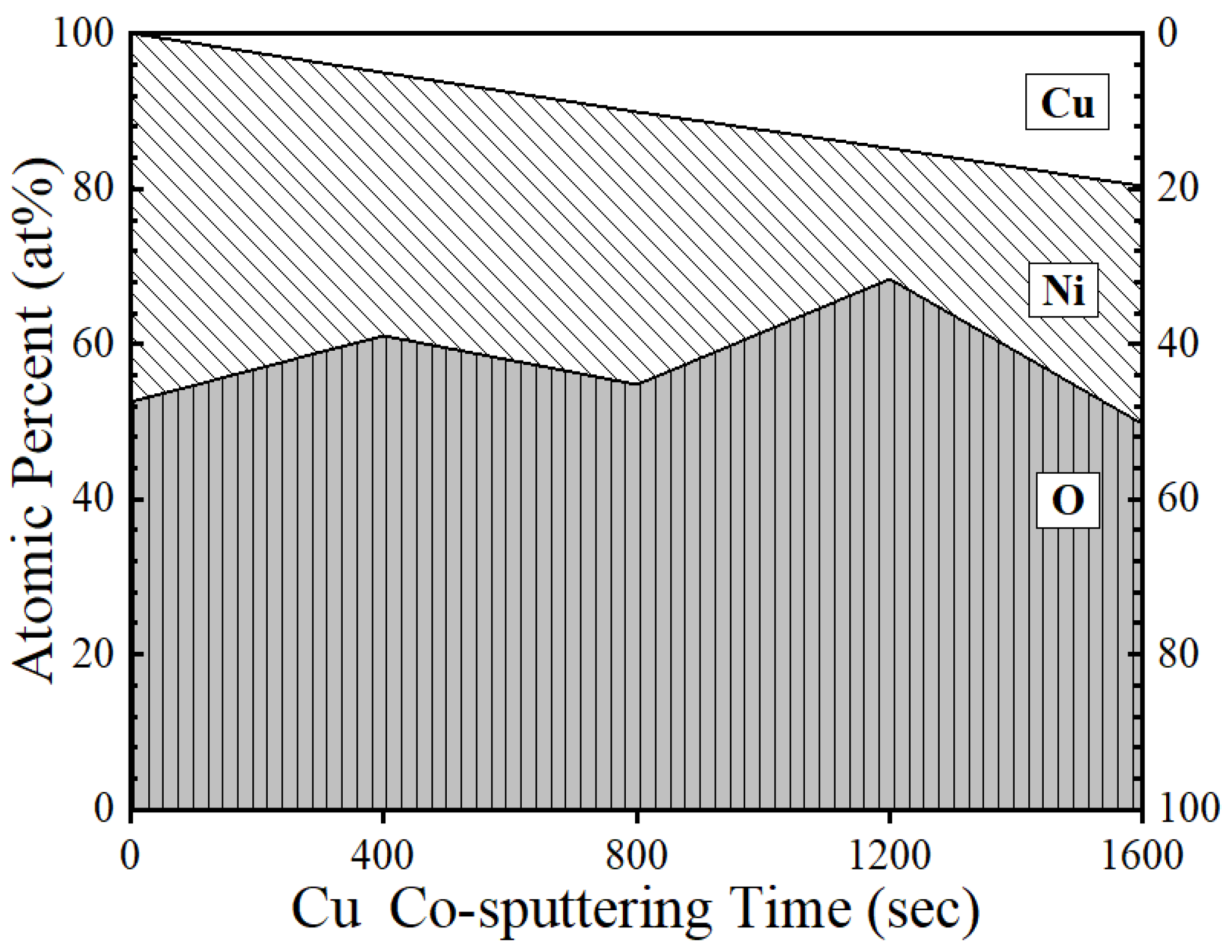
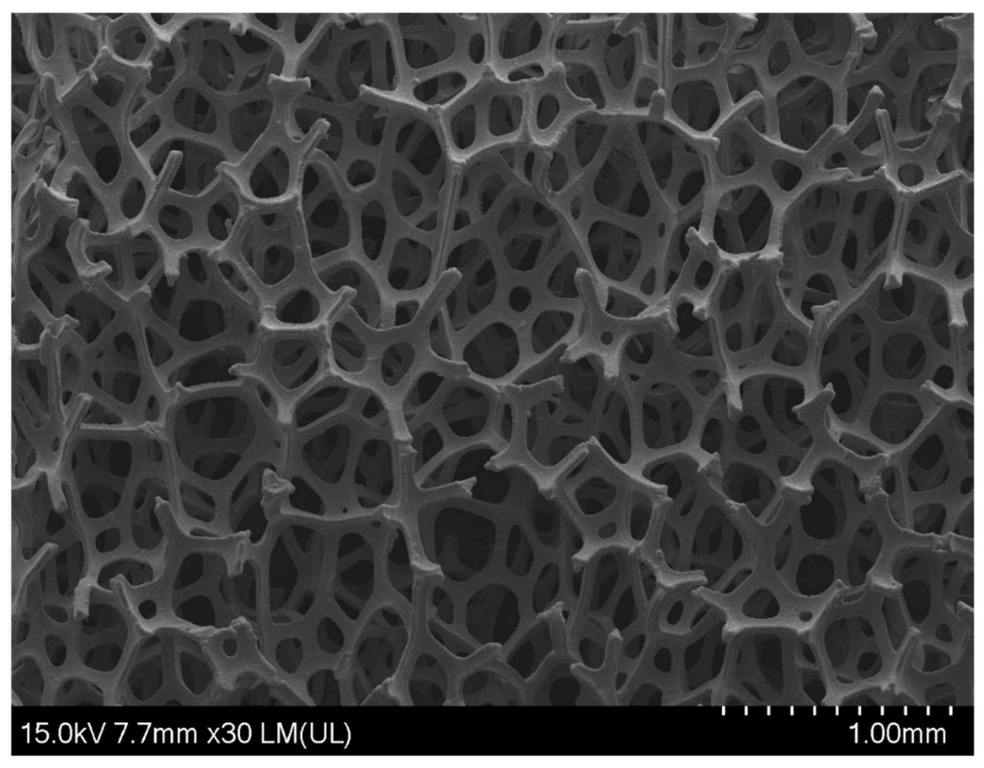
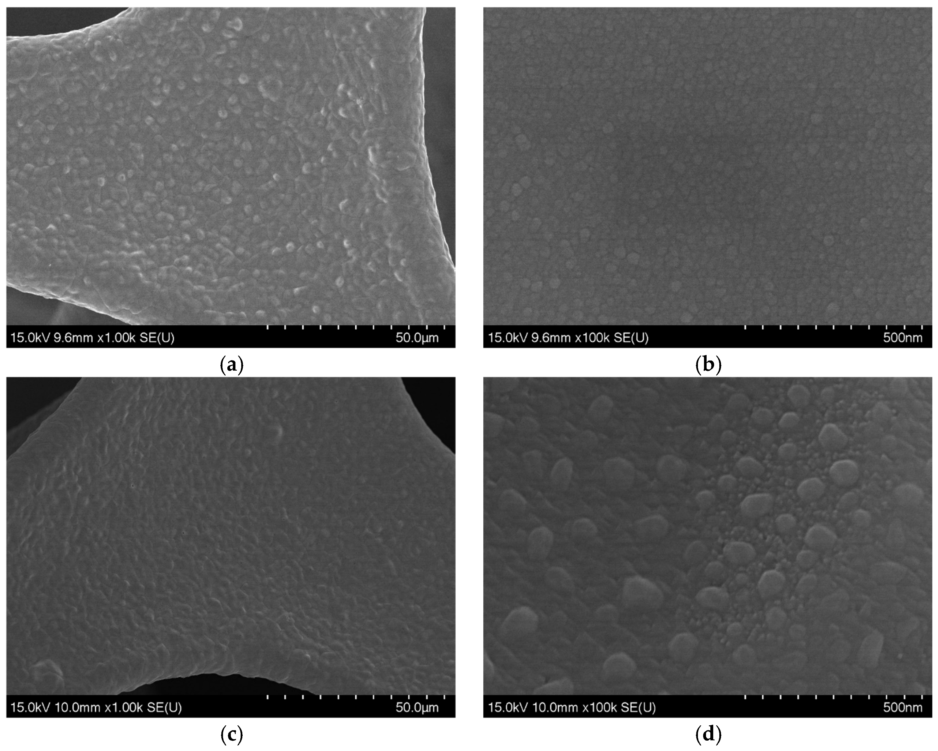
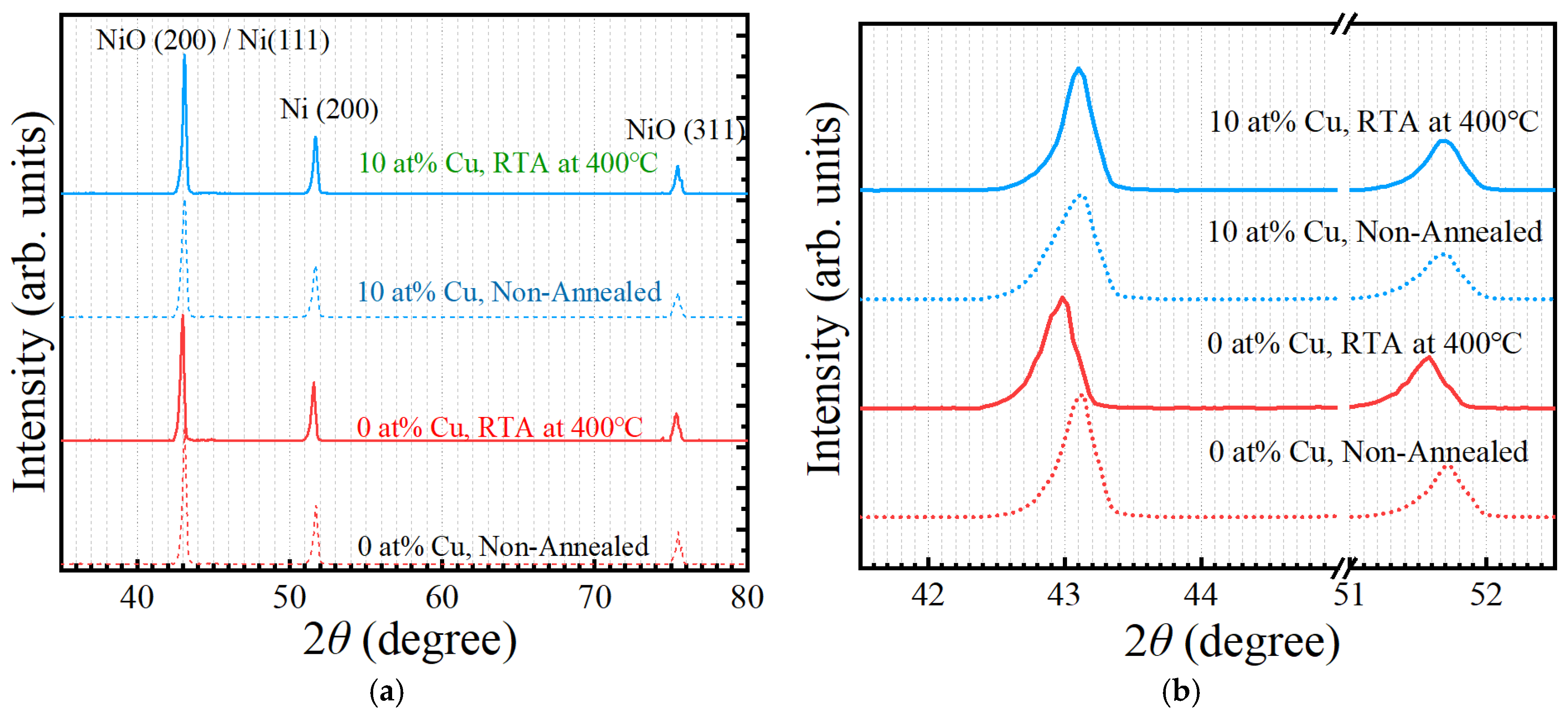
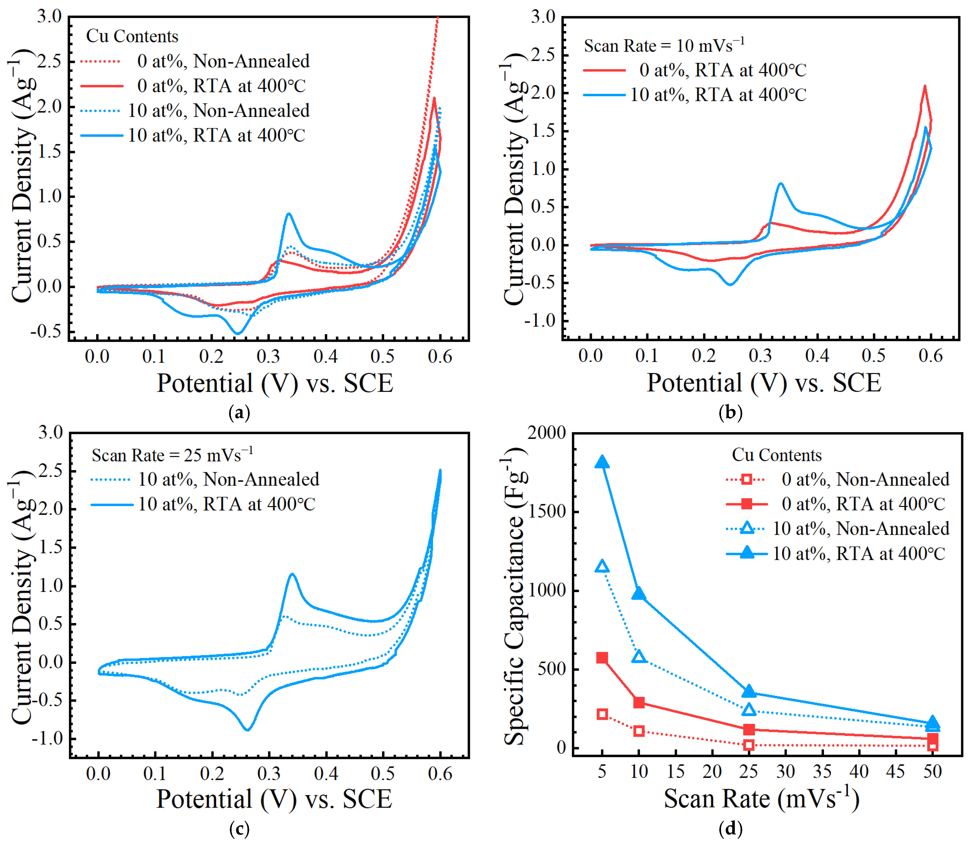
3. Results and Discussion

The relationship between the Cu doping ratio and the atomic composition of the NiO:Cu thin films, deposited by the co-sputtering method using NiO and Cu targets, was verified through EDS analysis. The FESEM cross-sectional images (Figure S1) confirmed that all the films had a uniform thickness of approximately 200 nm. Figure 1 shows the EDS analysis of the NiO:Cu thin films as a function of the Cu sputtering time. The results demonstrate that the Cu doping concentration was linearly controlled at 0.00, 4.91, 10.14, 14.79, and 19.68 at% by adjusting the sputtering time, considering the respective deposition rates of the NiO and Cu targets used in the co-sputtering process. As the Cu-sputtering time increased, the deposition of Cu became more pronounced; however, the direct proportions of Ni and O did not decrease. This discrepancy arises owing to the differences in sputtering yields, as NiO has a lower sputtering yield than Cu because of its strong Ni–O bonds. Additionally, the O:Ni ratio did not decrease proportionally with increasing Cu content because the O content in the film was not solely determined by the NiO target. When a compound target such as NiO is sputtered, the sputtering yields of O and Ni differ owing to their different atomic masses and binding energies. Lighter O atoms have a higher probability of resputtering or being lost in the vacuum system, which results in deviations in the film stoichiometry.
Hall effect measurements were recorded to evaluate the electrical properties of NiO:Cu thin films deposited through co-sputtering using NiO and Cu targets and then RTA treated at 400 °C. Figure 2 presents the results of the Hall effect measurements, including resistivity, carrier concentration, and carrier mobility, for the NiO:Cu thin films with Cu-content controlled at approximately 0, 5, 10, 15, and 20 at%, as a function of the Cu sputtering time. All the NiO:Cu thin films exhibited p-type conductivity, which is consistent with the typical conductivity of undoped NiO [21,23,24,25,35,36]. The measured resistivity (ρ) of the NiO:Cu thin films ranged from 1.244 × 10⁻⁴ to 2.164 × 10⁻⁴ Ω·cm, which indicates that the NiO:Cu thin films have relatively lower resistivity than undoped NiO thin films, thereby exhibiting good electrical conductivity. However, the resistivity does not follow a monotonic trend with increasing Cu content, which suggests that multiple competing mechanisms influence the charge transport. Initially, the resistivity decreased from the undoped NiO value to a maximum of approximately 5 at% Cu and then decreased to a minimum of approximately 15 at% Cu, before increasing again to 20 at% Cu. This non-monotonic behavior suggests a complex interplay between the factors affecting the charge transport, with a balance between the enhanced carrier concentration at low doping levels and the onset of defect formation or secondary-phase segregation at higher Cu concentrations. The carrier concentration (n) varied significantly, ranging from 1017 to 1021 cm−3 with increasing Cu content. Notably, at 5 at% Cu content, the carrier concentration dropped abruptly to 1017 cm−3, presumably owing to defect formation or secondary-phase segregation [24], which reduced the availability of free charge carriers. This sudden decrease in carrier concentration explains the observed increase in resistivity beyond this point. As the Cu content increased beyond 5 at%, the Cu substituted in the Ni sites (CuNi) acted as an acceptor, thereby increasing the hole concentration. However, at 20 at% Cu content, the excess Cu incorporation probably introduced additional defects or formed CuO-rich regions [37], which caused carrier compensation and enhanced the scattering effects, thereby increasing the resistivity once again. The highest carrier mobility (μ) of 77.86 cm2/V·s was observed at 5 at% Cu content, where the carrier concentration was the lowest, which suggests reduced carrier–carrier scattering and fewer ionized impurities under this condition. Conversely, the lowest carrier mobility of 21.01 cm2/V·s was measured at 15 at% Cu content, where the highest carrier concentration was obtained, indicating increased ionized impurity scattering owing to the higher density of charge carriers. These observations indicate that the carrier mobility in NiO:Cu thin films is predominantly influenced by carrier–carrier scattering and ionized impurity scattering, which becomes more pronounced at higher doping levels, which results in reduced mobility. Therefore, although Cu doping enhances the hole concentration to a certain level, excessive doping introduces defects and increases scattering, compromising the charge transport properties. Furthermore, the sharp drop in carrier concentration to the 1017 cm−3 order at 5 at% Cu is presumably linked to lattice distortion and the formation of oxygen vacancies, which further supports the hypothesis of defect-related scattering at lower Cu doping concentrations [24].
The optical transmittance of NiO:Cu thin films with different Cu contents (0, 5, 10, 15, and 20 at%) was measured after annealing at 400 °C in the wavelength range of 300–1500 nm, as shown in Figure 3a. The transmission spectra revealed that, within the visible range (380–780 nm), the NiO:Cu thin films doped with 0, 5, and 10 at% Cu exhibited similar optical transmittance characteristics, whereas the films doped with higher Cu contents (15 and 20 at%) exhibited a noticeable decrease in transmittance. Additionally, a slight blue shift in the absorption edge was observed with increasing Cu content, which facilitated the transmission of light at longer wavelengths and consequently resulted in an increase in the optical bandgap (Eg) of the NiO:Cu thin films. In the near-UV spectral region (approximately 340–360 nm), the optical transmittances of the NiO:Cu thin films increased sharply, which corresponded to a bandgap range of 3.45–3.65 eV. The bandgaps of NiO:Cu thin films with the same thickness (200 nm) were calculated using Equation (1):
Eg = h · c/λ [eV]
where h is Planck’s constant (4.135667 × 10⁻¹⁵ eVs); c is the speed of light (3 × 10⁸ m/s); and λ is the wavelength (nm) of the absorption onset (1/e = 37%). The calculated bandgaps for the NiO:Cu thin film were 3.57, 3.58, 3.56, 3.56, and 3.59 eV for the Cu contents of 0, 5, 10, 15, and 20 at%, respectively. The calculated values were further validated by linearly extrapolating the Tauc plots, as shown in Figure 3b. The Tauc plot relates the square of the product of the absorption coefficient and photon energy (αhυ)2 to the photon energy , where α is the absorption coefficient; h is Planck’s constant; and υ is the frequency of the incident photon. The extrapolated bandgaps of the NiO:Cu thin films from the Tauc plot were determined to be 3.57, 3.58, 3.56, 3.56, and 3.59 eV for the NiO:Cu thin films with Cu concentrations of 0, 5, 10, 15, and 20 at%, respectively. These values are in good agreement with the results obtained using the calculation. Because Cu has a slightly larger atomic radius (0.128 nm) than Ni (0.124 nm), doping Cu into the NiO lattice tends to increase the lattice spacing. Although the increase is relatively small, it could be noticeable depending on the concentration of Cu in the lattice. The slight increase in the lattice spacing with Cu doping is attributed to the slightly larger atomic radius of Cu (0.128 nm) compared with that of Ni (0.124 nm). This increase in lattice spacing, although small, can reduce the overlap between the Ni 3d and O 2p orbitals, which results in a wider bandgap. Lattice expansion can also introduce strain into the material, which further modifies the electronic structure and contributes to an increase in the bandgap. In addition, a higher Cu doping content can result in the formation of defects or charge compensation mechanisms, which may also play a role in widening the bandgap. At very high doping levels, the formation of Cu-rich phases, such as CuO or Cu2O, can further influence the electronic properties of the material and contribute to the observed increase in the bandgap.
The electrochemical properties of the NiO:Cu thin films were analyzed using cyclic voltammetry (CV) and galvanostatic charge–discharge techniques. Figure 4a shows the cyclic voltammograms of the NiO:Cu thin films with varying Cu concentrations (0, 5, 10, 15, and 20 at%) at a scan rate of 5 mVs−1 in a 1 M KOH electrolyte. NiO:Cu thin films doped with 15 and 20 at% Cu exhibit a pronounced hysteresis loop with a rapid decrease and increase in current density [38]. All the NiO:Cu thin films demonstrated clear oxidation and reduction peaks within the potential range of −0.60 to +1.0 V, which is characteristic of the reversible Faradaic transition associated with NiO:Cu, indicating typical pseudocapacitive behavior. The galvanostatic charge–discharge performance of the NiO:Cu thin films was used to calculate their specific capacitance (C), which is a key measure of the electrochemical performance. The specific capacitance was determined using Equation (2):
C = I · t/m · ΔV [Fg−1]
where I is the discharge current; m is the mass of the active material; t is the discharge time; and ΔV is the potential window during the discharge process [39,40]. Figure 4b shows the specific capacitance values for the NiO:Cu films with Cu concentrations of 0, 5, 10, 15, and 20 at% at various scan rates (5, 10, 25, and 50 mVs−1). As the scan rate increased, the specific capacitance decreased, which was attributed to the limited effective insertion of ionic species into the NiO:Cu thin-film surface at higher scan rates, resulting in a reduced charge storage capacity [41,42]. At a scan rate of 5 mVs−1, the films with 15 and 20 at% Cu demonstrated relatively low capacitances of 75.82 and 58.97 Fg−1, respectively. In contrast, the NiO thin film with 0 at% Cu exhibited a specific capacitance of 182.44 Fg−1, while the films with 5 and 10 at% Cu demonstrated significantly higher capacitances, with values of 236.89 and 436.95 Fg−1, respectively. The latter represents a nearly fourfold increase in the specific capacitance compared with the previously reported NiO thin films (0 at% Cu) that were RTA treated at 400 °C [18]. This substantial enhancement in the specific capacitance clearly demonstrates the positive impact of Cu doping on the electrochemical performance of NiO:Cu thin films [18,41,42]. These results suggest that Cu doping effectively improves the ionic conductivity and electrochemical properties of NiO thin films, presumably because of the enhanced charge transfer kinetics and increased number of active sites for Faradaic reactions. The observed increase in capacitance with Cu doping in the NiO thin films is consistent with findings from other studies [43,44] that have highlighted the benefits of metal doping in transition metal oxides for energy storage applications.
Ni foams offer several advantages, including electrically conductivity [45], chemically stability [45,46,47], and resistance to environmental factors such as corrosion and oxidation with high porosity, which enhance the surface area and optimize the diffusion pathways for electrochemical reactions [46,47]. Figure 5 shows an FESEM image of a NiO:Cu electrode, with 10 at% Cu-doped NiO:Cu films coated on a 3D porous Ni foam substrate, after RTA treatment at 400 °C. Figure 6 shows the magnified FESEM images of the NiO:Cu (10 at% Cu) films coated on the 3D porous Ni foam substrates (a, b) with and (c, d) without RTA treatments at 400 °C. Surface analysis of the Cu-doped sample after heat treatment revealed larger crystallites as seen in Figure 6 (c) and (d) compared with those in (a) and (b).
Figure 7a shows the XRD patterns of NiO:Cu electrodes, doped with 0 and 10 at% Cu on 3D porous substrates and treated with or without RTA at 400 °C. The XRD scans, performed in the 2θ range of 20−80°, demonstrated no peaks between 20° and 35°; therefore, only those in the range from 35° to 80° were included in the graph. The characteristic peaks for NiO, corresponding to the standard face-centered cubic (FCC) crystal structure, observed at 43.27° and 75.14° were attributed to the (200) and (311) planes, respectively, and the peak at 43.27° overlapped with the (111) peak of Ni, as confirmed by ICDD cards (00-047-1049, 04-010-6148) [19,23,24,25]. Furthermore, the peak at 51.77° corresponded to the (200) peak of Ni, which was attributed to the Ni foam substrate. For the 10 at% Cu NiO:Cu thin films, the NiO (200) and Ni (111) peaks exhibited slight shifts because of Cu doping. The presence of Cu in the lattice may slightly alter the interplanar spacing because of the larger atomic radius of Cu compared with that of Ni, which could cause a minor expansion of the lattice. Although the XRD patterns show typical peaks of NiO and Ni, the greater incorporation of Cu atoms could potentially induce the formation of additional phases at higher doping concentrations, which may result in extra peaks in the XRD pattern that were not observed in this sample (10 at%). Figure 7b shows the main peaks of NiO (200)/Ni (111) and Ni (200) on the 10 at% Cu-doped NiO:Cu films coated on the 3D porous Ni foam substrates, which indicates a shift in the peaks depending on the conditions. Notably, the samples subjected to RTA treatment exhibited shifts to lower 2θ values compared with the untreated samples, indicating that the heat treatment enhanced the bonding between the 3D porous Ni foam substrate and the NiO:Cu thin films. Furthermore, the intensity of the peaks of the 10 at% Cu-doped NiO:Cu electrodes was higher than that of the undoped NiO electrodes, despite both electrodes being treated under the same RTA conditions. This suggests that Cu doping improves the crystallinity of the NiO:Cu films, likely owing to stronger bonding with both the porous substrate and the Cu atoms, which might help to stabilize the crystal structure during heat treatment. These findings are consistent with the surface analysis performed using FESEM (Figure 6), which showed larger crystallites in the Cu-doped samples after heat treatment.
The electrochemical properties of the NiO:Cu films coated on 3D porous Ni foam substrates were analyzed using CV and galvanostatic charge–discharge techniques. Figure 8a shows the cyclic voltammograms of NiO:Cu electrode, doped with 0 and 10 at% Cu, both with or without annealing at 400 °C in 1 M KOH electrolyte, measured at a scan rate of 10 mVs−1. Owing to the porous nature of the substrate, CV measurements were not possible once the potential exceeded +0.6 V, as bubbles formed in the substrate. This bubbling was the result of the oxygen evolution reaction that occurred during the charging process, in which the electrolyte participated in the electrode reaction. To avoid this issue, all the samples were limited to the potential region below +0.6 V for analysis [48,49,50,51,52]. The Cu-doped electrodes exhibited a larger hysteresis loop with greater linearity than the undoped electrodes. Moreover, the quasi-reversible nature of the oxidation/reduction reaction was evident from the clear anodic and cathodic peaks in the cyclic voltammograms, confirming that the 3D porous NiO:Cu electrodes doped with 0 and 10 at% Cu exhibited pseudocapacitive behavior. While the reported latest specific capacitance values of NiO were 1060 Fg−1 for NiO/Ni aerogel (2022) [53], 791.67 Fg−1 for sulfur-doped NiO (2023) [54], 1360 Fg−1 for NiO-Fe-CNT composite (2023) [28], and 876.4 Fg−1 for NiO nanosheets (2024) [55], Figure 8d shows an outstanding result of 1809.2 Fg−1.
The experimental findings presented herein provide compelling evidence for the advantageous utilization of three-dimensional porous nickel foam as a conductive substrate with a high surface area (Figure S2), which effectively facilitates the superior electrochemical performance of the deposited NiO:Cu active material. The high specific capacitance underscores the achieved efficient utilization of the active material, which is attributed to the structural support provided by the nickel foam. However, this investigation also revealed a limitation: the onset of oxygen evolution reactions within the porous Ni foam at relatively moderate potentials constrained the operational voltage window of the supercapacitor. This parasitic reaction restricts the operational voltage window of the fabricated supercapacitor device. Future research efforts should focus on mitigating this limitation, potentially through strategies such as electrolyte optimization or surface modification of the nickel foam substrate to suppress the oxygen evolution reaction, thereby expanding the operating voltage and overall energy density of the device.

4. Conclusions

This study comprehensively investigated the effect of Cu doping on NiO thin films for supercapacitor applications. NiO:Cu thin films were successfully fabricated using a co-sputtering method with varying Cu concentrations (0–20 at%), and EDS analysis confirmed that the Cu doping concentration could be controlled linearly by adjusting the sputtering time. The Hall effect measurements showed that the NiO:Cu thin films maintained p-type conductivity, with resistivity values lower than those of pure NiO, which indicates improved electrical conductivity. However, carrier concentration and mobility exhibited complex behaviors owing to competing effects such as defect formation and ionized impurity scattering. The optical transmittance measurements revealed that Cu doping caused a slight blue shift in the absorption edge, corresponding to an increase in the bandgap, with values ranging from 3.56 eV to 3.59 eV as the Cu content increased. This shift was attributed to changes in the lattice spacing and strain induced by Cu incorporation. The electrochemical analyses, including cyclic voltammetry (CV) and galvanostatic charge–discharge tests, demonstrated significant improvements in capacitance with Cu doping. At a scan rate of 5 mVs−1, the capacitance of the NiO:Cu thin films increased from 182.44 Fg−1 for 0 at% to 436.95 Fg−1 for 10 at% Cu NiO:Cu thin films. The Cu-doped thin films exhibited a typical pseudocapacitive behavior with clear oxidation and reduction peaks. The electrochemical performance was further enhanced when the films were coated on 3D porous Ni foam substrates, particularly with 10% Cu doping and post-annealing at 400 °C, which resulted in a specific capacitance of 1809.20 Fg−1. Overall, this study demonstrated that Cu doping and the use of 3D porous substrates synergistically enhanced the electrical conductivity, capacitance, and electrochemical performance of NiO-based electrodes, thereby offering promising strategies for developing high-performance supercapacitors. These findings highlight the potential of NiO:Cu films for energy storage applications and set the stage for the further exploration of transition metal oxide composites for supercapacitor technologies.

Supplementary Materials

The following supporting information can be downloaded at https://www.mdpi.com/article/10.3390/en18112704/s1: Figure S1. Cross-sectional field emission scanning electron microscopy (FESEM) images of NiO:Cu thin films with co-sputtering times of (a) 0, (b) 400, (c) 800, (d) 1200, and (e) 1600 s on the Cu target. The total deposition time was adjusted to (a) 100, (b) 95, (c) 90, (d) 85, and (e) 80 min, respectively, to maintain an overall film thickness of approximately 200 nm. The Cu atomic concentration in the films increased linearly under these conditions and reached approximately 0, 5, 10, 15, and 20 at%. Figure S2. Relative pressure (P/Po) and Brunauer–Emmett–Teller (BET) analyses used for determining the surface areas of (a) 3D porous Ni foam and (b, c) 0 and (d, e) 10 at% Cu-doped NiO:Cu films coated on the Ni foam substrates (c, e) with or (b, d) without annealing at 400 °C by using a surface area analyzer (ASAP 2460, Micromeritics Instrument Corporation, Norcross, GA, USA).

Author Contributions

Conceptualization, S.O., Y.-K.J. and N.-H.K.; investigation, S.O.; methodology, S.O.; data curation, S.O. and Y.-K.J.; writing—original draft preparation, S.O.; writing—review and editing, Y.-K.J. and N.-H.K.; supervision, N.-H.K.; project administration, N.-H.K.; funding acquisition, N.-H.K. All authors have read and agreed to the published version of the manuscript.

Funding

This study was supported by a research fund from Chosun University, 2023.

Data Availability Statement

Data are contained within the article or Supplementary Materials.

Conflicts of Interest

The authors declare no conflicts of interest. The founding sponsor had no role in the design of this study; the collection, analyses, or interpretation of the data; the writing of the manuscript; or the decision to publish the results.

References

  1. Li, L.; Lin, J.; Wu, N.; Xie, S.; Meng, C.; Zheng, Y.; Wang, X.; Zhao, Y. Review and outlook on the international renewable energy development. Energy Built Environ. 2022, 3, 139–157. [Google Scholar] [CrossRef]
  2. Xia, C.; Alshareef, H.N. Self-templating scheme for the synthesis of nanostructured transition-metal chalcogenide electrodes for capacitive energy storage. Chem. Mat. 2015, 27, 4661–4668. [Google Scholar] [CrossRef]
  3. Liu, C.; Li, F.; Ma, L.P.; Cheng, H.M. Advanced materials for energy storage. Adv. Mater. 2010, 22, E28–E62. [Google Scholar] [CrossRef]
  4. Wang, Y.; Wei, H.; Lu, Y.; Wei, S.; Wujcik, E.K.; Guo, Z. Multifunctional carbon nanostructures for advanced energy storage applications. Nanomaterials 2015, 5, 755–777. [Google Scholar] [CrossRef]
  5. Wang, Z.; Feng, Z.; Hu, C.; Li, X.; Zhu, Y. Enhancing battery performance under motor overload drive with a battery–supercapacitor hybrid energy storage system. J. Power Sources 2025, 642, 236680. [Google Scholar] [CrossRef]
  6. Simon, P.; Gogotsi, Y. Materials for electrochemical capacitors. Nat. Mat. 2008, 7, 845–854. [Google Scholar] [CrossRef]
  7. Afif, A.; Rahman, S.M.; Azad, A.T.; Zaini, J.; Islan, M.A.; Azad, A.K. Advanced materials and technologies for hybrid supercapacitors for energy storage—A review. J. Energy Storage 2019, 25, 100852. [Google Scholar] [CrossRef]
  8. Dong, Q.; Wang, G.; Hu, H.; Yang, J.; Qian, B.; Ling, Z.; Qiu, J. Ultrasound-assisted preparation of electrospun carbon nanofiber/graphene composite electrode for supercapacitors. J. Power Sources 2013, 243, 350–353. [Google Scholar] [CrossRef]
  9. Singh, A.K.; Mandal, K. Engineering of high performance supercapacitor electrode based on Fe-Ni/Fe2O3-NiO core/shell hybrid nanostructures. J. Appl. Phys. 2015, 117, 105101. [Google Scholar] [CrossRef]
  10. Wang, C.C.; Chen, H.C.; Lu, S.Y. Manganese oxide/graphene aerogel composites as an outstanding supercapacitor electrode material. Chem. Eur. J. 2014, 20, 517–523. [Google Scholar] [CrossRef]
  11. Li, X.; Xiong, S.; Li, G.; Xiao, S.; Zhang, C.; Ma, Y. Effect of microstructure on electrochemical performance of electrode materials for microsupercapacitor. Mater. Lett. 2023, 346, 134481. [Google Scholar] [CrossRef]
  12. Wang, H.; Yi, H.; Chen, X.; Wang, X. Asymmetric supercapacitors based on nano-architectured nickel oxide/graphene foam and hierarchical porous nitrogen-doped carbon nanotubes with ultrahigh-rate performance. J. Mater. Chem. A 2014, 2, 3223–3230. [Google Scholar] [CrossRef]
  13. Yuan, C.; Yang, L.; Hou, L.; Shen, L.; Zhang, X.; Lou, X.W.D. Growth of ultrathin mesoporous Co3O4 nanosheet arrays on Ni foam for high-performance electrochemical capacitors. Energy Environ. Sci. 2012, 5, 7883–7887. [Google Scholar] [CrossRef]
  14. Liu, L.; Li, Y.; Yuan, S.; Ge, M.; Ren, M.; Sun, C.; Zhou, Z. Nanosheet-based NiO microspheres: Controlled solvothermal synthesis and lithium storage performances. J. Phys. Chem. C 2010, 114, 251–255. [Google Scholar] [CrossRef]
  15. Yuan, Y.F.; Xia, X.H.; Wu, J.B.; Yang, J.L.; Chen, Y.B.; Guo, S.Y. Hierarchically ordered porous nickel oxide array film with enhanced electrochemical properties for lithium ion batteries. Electrochem. Commun. 2010, 12, 890–893. [Google Scholar] [CrossRef]
  16. Ng, C.H.; Lim, H.N.; Lim, Y.S.; Chee, W.K.; Huang, N.M. Fabrication of flexible polypyrrole/graphene oxide/manganese oxide supercapacitor. Int. J. Energy Res. 2015, 39, 344–355. [Google Scholar] [CrossRef]
  17. Zhang, G.; Chen, Y.; Qu, B.; Hu, L.; Mei, L.; Lei, D.; Li, Q.; Chen, L.; Li, Q.; Wang, T. Synthesis of mesoporous NiO nanospheres as anode materials for lithium ion batteries. Electrochim. Acta 2012, 80, 140–147. [Google Scholar] [CrossRef]
  18. Oh, S.; Park, Y.S.; Ko, P.J.; Kim, N.H. Effects of rapid thermal treatment on characteristics of magnetron-sputtered NiO thin films for supercapacitor applications. J. Nanosci. Nanotechnol. 2018, 18, 6213–6219. [Google Scholar] [CrossRef]
  19. Dar, F.I.; Moonoosawmy, K.R.; Es-Souni, M. Morphology and property control of NiO nanostructures for supercapacitor applications. Nanoscale Res. Lett. 2013, 8, 363. [Google Scholar] [CrossRef]
  20. Offiah, S.U.; Nwodo, M.O.; Nwanya, A.C.; Ezugwu, S.C.; Agbo, S.N.; Ugwuoke, P.U.; Osuji, R.U.; Malik, M.; Ezema, F.I. Effects of post-thermal treatments on morphological and optical properties of NiO/Ni(OH)2 thin films synthesized by solution growth. Optik 2014, 125, 2905–2908. [Google Scholar] [CrossRef]
  21. Hotovy, I.; Huran, J.; Spiess, L.; Hascik, S.; Rehacek, V. Preparation of nickel oxide thin films for gas sensors applications. Sens. Actuator B Chem. 1999, 57, 147–152. [Google Scholar] [CrossRef]
  22. Hotovy, I.; Huran, J.; Siciliano, P.; Capone, S.; Spiess, L.; Rehacek, V. The influences of preparation parameters on NiO thin film properties for gas-sensing application. Sens. Actuator B Chem. 2001, 78, 126–132. [Google Scholar] [CrossRef]
  23. Chen, H.L.; Lu, Y.M.; Hwang, W.S. Characterization of sputtered NiO thin films. Surf. Coat. Technol. 2005, 198, 138–142. [Google Scholar] [CrossRef]
  24. Usha, K.S.; Sivakumar, R.; Sanjeeviraja, C.; Sathe, V.; Ganesan, V.; Wang, T.Y. Improved electrochromic performance of a radio frequency magnetron sputtered NiO thin film with high optical switching speed. RSC Adv. 2016, 6, 79668–79680. [Google Scholar] [CrossRef]
  25. Usha, K.S.; Sivakumar, R.; Sanjeeviraja, C. Optical constants and dispersion energy parameters of NiO thin films prepared by radio frequency magnetron sputtering technique. J. Appl. Phys. 2013, 114, 123501. [Google Scholar] [CrossRef]
  26. Karabiberoğlu, Ş.; Dursun, Z. Synthesis and characterization of polyindole-NiO-graphene composite for supercapacitor electrode. Fuller. Nanotub. Carbon Nanostruct. 2025, 1–16. [Google Scholar] [CrossRef]
  27. Bachankar, S.; Kumbhar, A.; Mullani, S.; Malavekar, D.; Park, J.; Kim, C.; Ji, T. Enhanced Supercapacitive Charge Storage in a Nickel Oxide-Graphene Oxide Composite: Synergistic Effect. Korean J. Mater. Res. 2024, 34, 609–619. [Google Scholar] [CrossRef]
  28. Shah, A.; Senapati, S.; Murthy, H.A.; Singh, L.R.; Mahato, M. Supercapacitor performance of NiO, NiO-MWCNT, and NiO–Fe-MWCNT composites. ACS Omega 2023, 8, 33380–33391. [Google Scholar] [CrossRef]
  29. Sharma, K.; Arora, A.; Tripathi, S.K. Review of supercapacitors: Materials and devices. J. Energy Storage 2019, 21, 801–825. [Google Scholar] [CrossRef]
  30. Jiménez-Marín, E.; Villalpando, I.; Trejo-Valdez, M.; Cervantes-Sodi, F.; Vargas-García, J.R.; Torres-Torres, C. Coexistence of positive and negative photoconductivity in nickel oxide decorated multiwall carbon nanotubes. Mater. Sci. Eng. B Adv. Funct. Solid-State Mater. 2017, 220, 22–29. [Google Scholar] [CrossRef]
  31. Bhujel, K.; Thangavel, R.; Pal, K.K.; Sardar, P.; Nayak, D.; Singh, N.S.; Rai, S. Cu-doped NiO thin film’s structural, optical, and electrical properties and its negative absorption behaviour in the Infra-Red region. Physica B 2024, 688, 416129. [Google Scholar] [CrossRef]
  32. Rihia, G.; Ghougali, M.; Beggas, A.; Mimouni, M.; Mahboub, M.S. Effects of Cu doping on the structural, optical and electrical characterizations of spray-deposited Ni-O thin films. Dig. J. Nanomater. Biostruct. 2025, 20, 139–147. [Google Scholar] [CrossRef]
  33. Issatayev, N.; Abdumutaliyeva, D.; Tashenov, Y.; Yeskozha, D.; Seipiyev, A.; Bakenov, Z.; Nurpeissova, A. Three-dimensional carbon coated and high mass-loaded NiO@Ni foam anode with high specific capacity for lithium ion batteries. RSC Adv. 2024, 14, 40069–40076. [Google Scholar] [CrossRef] [PubMed]
  34. Pech, S.; Rou, Y.J.; Kim, S.; Lee, K.Y.; Kim, N.H. Cu(In,Ga)Se2: Te thin films for stoichiometric compensation by using co-sputtering and rapid thermal annealing. Appl. Sci. 2023, 13, 4284. [Google Scholar] [CrossRef]
  35. Yang, J.L.; Lai, Y.S.; Chen, J.S. Effect of heat treatment on the properties of non-stoichiometric p-type nickel oxide films deposited by reactive sputtering. Thin Solid Films 2005, 488, 242–246. [Google Scholar] [CrossRef]
  36. Jang, W.L.; Lu, Y.M.; Hwang, W.S.; Hsiung, T.L.; Wang, H.P. Point defects in sputtered NiO films. Appl. Phys. Lett. 2009, 94, 062103. [Google Scholar] [CrossRef]
  37. Aftab, M.; Butt, M.Z.; Ali, D.; Bashir, F.; Khan, T.M. Optical and electrical properties of NiO and Cu-doped NiO thin films synthesized by spray pyrolysis. Opt. Mater. 2021, 119, 111369. [Google Scholar] [CrossRef]
  38. Deyab, M.A.; Keera, S.T. Cyclic voltammetric studies of carbon steel corrosion in chloride-formation water solution and effect of some inorganic salts. Egypt. J. Pet. 2012, 21, 31–36. [Google Scholar] [CrossRef]
  39. Wu, M.S.; Lin, C.J.; Ho, C.L. Multilayered architecture of graphene nanosheets and MnO2 nanowires as an electrode material for high-performance supercapacitors. Electrochim. Acta 2012, 81, 44–48. [Google Scholar] [CrossRef]
  40. Liang, K.; Tang, X.; Hu, W. High-performance three-dimensional nanoporous NiO film as a supercapacitor electrode. J. Mater. Chem. 2012, 22, 11062–11067. [Google Scholar] [CrossRef]
  41. Sarma, B.; Jurovitzki, A.L.; Smith, Y.R.; Ray, R.S.; Misra, M. Influence of annealing temperature on the morphology and the supercapacitance behavior of iron oxide nanotube (Fe-NT). J. Power Sources 2014, 272, 766–775. [Google Scholar] [CrossRef]
  42. Fouda, A.N.; Abu-Assy, M.K.; Yousf, N. Structural and capacitive characterizations of high temperature nitrogen annealed graphene oxide. IOSR J. Appl. Phys. 2014, 6, 33–37. [Google Scholar] [CrossRef]
  43. Firat, Y.E.; Peksoz, A. Efficiency enhancement of electrochromic performance in NiO thin film via Cu doping for energy-saving potential. Electrochim. Acta 2019, 295, 645–654. [Google Scholar] [CrossRef]
  44. Masood, A.; Afzal, N.; Ahmed, A.A.; Qahtan, T.F.; Rafique, M.; Ahmad, R.; Imran, M. Structural, surface and optical investigations of Cu+ implanted NiO film prepared by reactive sputtering. Ceram. Int. 2023, 49, 4435–4448. [Google Scholar] [CrossRef]
  45. Dong, X.C.; Xu, H.; Wang, X.W.; Huang, Y.X.; Chan-Park, M.B.; Zhang, H.; Wang, L.H.; Huang, W.; Chen, P. 3D graphene–cobalt oxide electrode for high-performance supercapacitor and enzymeless glucose detection. ACS Nano 2012, 6, 3206–3213. [Google Scholar] [CrossRef]
  46. Cai, S.; Zhang, D.; Shi, L.; Xu, J.; Zhang, L.; Huang, L.; Li, H.; Zhang, J. Porous Ni–Mn oxide nanosheets in situ formed on nickel foam as 3D hierarchical monolith de-NO x catalysts. Nanoscale 2014, 6, 7346–7353. [Google Scholar] [CrossRef]
  47. Wang, B.; Liu, Q.; Qian, Z.; Zhang, X.; Wang, J.; Li, Z.; Yan, H.; Gao, Z.; Zhao, F.; Liu, L. Two steps in situ structure fabrication of Ni–Al layered double hydroxide on Ni foam and its electrochemical performance for supercapacitors. J. Power Sources 2014, 246, 747–753. [Google Scholar] [CrossRef]
  48. Kang, M.; Gewirth, A.A. Voltammetric and force spectroscopic examination of oxide formation on Cu (111) in basic solution. J. Phys. Chem. B 2002, 106, 12211–12220. [Google Scholar] [CrossRef]
  49. Jayalakshmi, M.; Balasubramanian, K. Cyclic voltammetric behavior of copper powder immobilized on paraffin impregnated graphite electrode in dilute alkali solution. Int. J. Electrochem. Sci. 2008, 3, 1277–1287. [Google Scholar] [CrossRef]
  50. He, J.B.; Lu, D.Y.; Jin, G.P. Potential dependence of cuprous/cupric duplex film growth on copper electrode in alkaline media. Appl. Surf. Sci. 2006, 253, 689–697. [Google Scholar] [CrossRef]
  51. Burke, L.D.; Ahern, M.J.G.; Ryan, T.G. An investigation of the anodic behavior of copper and its anodically produced oxides in aqueous solutions of high pH. J. Electrochem. Soc. 1990, 137, 553. [Google Scholar] [CrossRef]
  52. Brisard, G.M.; Rudnicki, J.D.; McLarnon, F.; Cairns, E.J. Application of probe beam deflection to study the electrooxidation of copper in alkaline media. Electrochim. Acta 1995, 40, 859–865. [Google Scholar] [CrossRef]
  53. Ramkumar, R.; Dhakal, G.; Shim, J.J.; Kim, W.K. NiO/Ni nanowafer aerogel electrodes for high performance supercapacitors. Nanomaterials 2022, 12, 3813. [Google Scholar] [CrossRef]
  54. Patil, P.A.; Khalate, S.A.; Patil, U.M.; Kale, R.D.; Kulkarni, S.B. Cavity structured S-NiO with improved energy density for aqueous asymmetric hybrid supercapacitors by CDA mechanism. Mater. Adv. 2023, 4, 4607–4619. [Google Scholar] [CrossRef]
  55. Hanbo, W.; Dongyu, P.; Sheng, W.; Ziming, W.; Zhitian, F.; Yumei, T.; Kechang, L.; Haiyan, L. One-step potential-cycling method to fabricate NiO nanospheres for high performance supercapacitor application. J. Energy Storage 2024, 86, 111134. [Google Scholar] [CrossRef]
Figure 1. Energy-dispersive X-ray spectroscopy (EDS) analysis of NiO thin films with co-sputtering times of 0, 400, 800, 1200, and 1600 s on the Cu target, to achieve a thickness of approximately 200 nm of NiO:Cu films with total deposition times of 100, 95, 90, 85, and 80 min, respectively. The atomic percentage of Cu was linearly increased to approximately 0, 5, 10, 15, and 20 at%.
Energies 18 02704 g001
Figure 2. Hall effect measurements of (a) resistivity (ρ), (b) carrier concentration (n), and (c) carrier mobility (μ) of the rapid-thermally annealed NiO:Cu thin films with the different Cu doping concentration of 0, 5, 10, 15, and 20 at%.
Energies 18 02704 g002
Figure 3. (a) Optical transmittance and (b) band gap of the rapid-thermally annealed NiO:Cu thin films with the different Cu doping concentrations of 0, 5, 10, 15, and 20 at% in the range of 300–1500 nm.
Energies 18 02704 g003
Figure 4. (a) Cyclic voltammograms of the rapid-thermally annealed NiO:Cu thin films with different Cu doping concentrations of 0, 5, 10, 15, and 20 at% at a scan rate of 5 mVs−1 in 1 M KOH electrolyte. (b) Specific capacitance with different scan rates of 5, 10, 25, and 50 mVs−1 derived from cyclic voltammetry curves.
Energies 18 02704 g004
Figure 5. Feld-emission scanning electron microscopy (FESEM) images of the NiO:Cu thin films with the 10 at% Cu content on Ni foam and rapid thermal annealing at 400 °C.
Energies 18 02704 g005
Figure 6. FESEM images of the NiO:Cu thin films with the 10 at% Cu content on Ni foam before (a,b) and after (c,d) RTA treatment at 400 °C; (b,d) are the magnified images of (c,d), respectively.
Energies 18 02704 g006
Figure 7. (a) X-ray diffraction (XRD) patterns of NiO:Cu (0 and 10 at% Cu) films coated on the 3D porous Ni foam substrates with or without annealing at 400 °C. (b) Magnification of the NiO (200)/Ni (111) and Ni (200) diffraction peaks under the same conditions.
Energies 18 02704 g007
Figure 8. (a) Cyclic voltammograms of NiO:Cu (0 and 10 at% Cu) films coated on the 3D porous Ni foam substrates with or without annealing at 400 °C at a scan rate of 10 mVs−1 in 1 M KOH electrolyte. (b) Cyclic voltammograms of the NiO:Cu (0 and 10 at% Cu) electrodes with annealing at 400 °C at a scan rate of 10 mVs−1. (c) Cyclic voltammograms of the NiO:Cu (10 at% Cu) electrodes with or without annealing at 400 °C with a scan rate of 25 mVs−1. (d) Specific capacitance of the NiO:Cu (0 and 10 at% Cu) electrodes with or without annealing at 400 °C at scan rates of 5, 10, 25, and 50 mVs−1.
Energies 18 02704 g008
Disclaimer/Publisher’s Note: The statements, opinions and data contained in all publications are solely those of the individual author(s) and contributor(s) and not of MDPI and/or the editor(s). MDPI and/or the editor(s) disclaim responsibility for any injury to people or property resulting from any ideas, methods, instructions or products referred to in the content.

