Article

Modulation of Antioxidant Activity Enhances Photoautotrophic Cell Growth of Rhodobacter sphaeroides in Microbial Electrosynthesis †

1
Gwangju Bio/Energy R&D Center, Korea Institute of Energy Research, Gwangju 61003, Korea
2
Interdisciplinary Program of Agriculture and Life Sciences, Chonnam National University, Gwangju 61186, Korea
3
Department of Advanced Chemicals and Engineering, Chonnam National University, Gwangju 61186, Korea
4
Department of Integrative Food, Bioscience and Biotechnology, Chonnam National University, Gwangju 61186, Korea
*
Authors to whom correspondence should be addressed.
This paper is an extended version of our paper published in 10th Asia-Pacific Forum on Renewable Energy (AFORE 2021), Jeju, Korea, 31 October–4 November 2021.
These authors contributed equally to this work.
Energies 2022, 15(3), 935; https://doi.org/10.3390/en15030935
Submission received: 5 January 2022 / Revised: 24 January 2022 / Accepted: 26 January 2022 / Published: 27 January 2022

Abstract

:
Global warming is currently accelerating due to an increase in greenhouse gas emissions by industrialization. Microbial electrosynthesis (MES) using electroactive autotrophic microorganisms has recently been reported as a method to reduce carbon dioxide, the main culprit of greenhouse gas. However, there are still few cases of application of MES, and the molecular mechanisms are largely unknown. To investigate the growth characteristics in MES, we carried out growth tests according to reducing power sources in Rhodobacter sphaeroides. The growth rate was significantly lower when electrons were directly supplied to cells, compared to when hydrogen was supplied. Through a transcriptome analysis, we found that the expression of reactive oxygen species (ROS)-related genes was meaningfully higher in MES than in normal photoautotrophic conditions. Similarly, endogenous contents of H2O2 were higher and peroxidase activities were lower in MES. The exogenous application of ascorbic acid, a representative biological antioxidant, promotes cell growth by decreasing ROS levels, confirming the inhibitory effects of ROS on MES. Taken together, our observations suggest that reduction of ROS by increasing antioxidant activities is important for enhancing the cell growth and production of CO2-converting substances such as carotenoids in MES in R. sphaeroides

Graphical Abstract

1. Introduction

A climate crisis has arisen as carbon dioxide emissions have increased due to industrialization [1]. Various technologies have been proposed to reduce carbon dioxide emissions, and biological CO2 fixation is attracting attention as a desirable alternative. Recently, sustainable bioelectrochemical systems have been proposed to convert CO2 into valuable chemicals by microbial electrosynthesis (MES) using electroactive autotrophs. In this system, electroactive microorganisms can utilize CO2 using electrons as a reducing power [2]. Production of high value-added materials such as polyhydroxybutyrate (PHB), lycopene and α-humulene using Cupriavidus necator (C. necator) in an MES reactor has been reported [3,4,5]. Acetate was also continuously produced from CO2 by a mixed microbial consortium via an MES system [6].
One of the purple non-sulfur bacteria, Rhodobacter sphaeroides, has emerged as a promising electroactive autotroph. Rhodobacter sphaeroides (R. sphaeroides) can interact directly with the cathode to utilize electrons for cell growth. In addition, exposure to antibiotic selective pressures enhanced biofilm formation and increased PHB accumulation under nitrogen-limited MES conditions [7]. R. sphaeroides is a suitable strain for CO2 biorefinery because of its intracellular metabolic pathways and tractable genetic engineering. In particular, terpenoid and carotenoid biosynthesis naturally develops well, which is advantageous for the study of their production. Increases in the yield and productivity of amorphadiene production were reported through the enhancement of terpene synthase activities, and lycopene was also produced through the modification of a carotenoid biosynthetic gene [8,9].
The generation of reactive oxygen species (ROS) is inevitable in cellular metabolism. Maintaining a balance between the formation and degradation of ROS is very important to increase cell viability. Various intracellular antioxidants, such as ascorbic acid, are known to be effective in reducing and maintaining endogenous levels of ROS [10]. Carotenoids synthesized in R. sphaeroides exhibit considerable antioxidant activities against cancer cells without any cellular toxicity [11]. The accumulation of carotenoids in Deinococcus radiodurans increases resistance to ROS, enhancing defense mechanisms against environmental stresses [12]. Furthermore, the addition of ascorbic acid helped to alleviate the side effects of saline stress and enhanced biosynthesis of secondary metabolites, including carotenoids, phenolics, and flavonoids [13].
In this study, we investigated the differences in the growth characteristics of R. sphaeroides according to reducing sources such as electricity and hydrogen. Many changes in the expression of ROS-related genes were observed through a transcriptome analysis, and it was confirmed that the endogenous levels of ROS and oxidative damage were greater in MES. Treatment of exogenous ascorbic acid, as an antioxidant, reduced the ROS contents and promoted cell growth. This suggests that modulation of antioxidant activity is important to enhance photoautotrophic growth and production of valuable metabolites, such as carotenoids, on MES systems.

2. Materials and Methods

2.1. Bacterial Strain and Preparation of Microbial Electrosynthesis Reactor

The Rhodobacter sphaeroides KCTC1434 strain was obtained from Korean Collection for Type Cultures (KCTC) and grown on Sistrom’s medium without succinic acid [14]. The precultured cells for the microbial electrosynthesis (MES) reaction were cultured in modified Sistrom’s medium under autotrophic conditions purged with 5% CO2, 60% H2, and 35% argon. Cell growth was estimated by measuring optical density (OD) using a spectrophotometer (BioSpectrometer, Eppendorf, Hamburg, Germany) at 660 nm.
The double-chamber H-type microbial electrosynthesis reactor had the same configuration as described previously [15]. The anode and cathode chambers were joined with a glass arm and separated using a proton exchange membrane (PEM, Nafion 117, DuPont Ltd., Wilmington, DE, USA). The anode and cathode electrodes were, respectively, a 4 cm × 10 cm and 4 cm × 5 cm piece of graphite felt. The thickness of the electrodes was 0.3 cm (GF030, FuelCellStore, College Station, TX, USA) and they were connected to titanium wire. The reference electrode, Ag/AgCl (in 3 M NaCl), was placed in the cathode chamber. The cathode electrode was poised with –0.6 V (vs. Ag/AgCl) using a potentiostat (WMPG1000, WonAtech, Seoul, Korea). The microbial electrosynthesis reaction was conducted under light-anaerobic conditions in batch mode with a gas composition of CO2 5% and argon 95% at a rate of 17.5 mL/min (0.05 vvm). Ascorbic acid (A92902, Sigma-Aldrich, St. Louis, MO, USA), which was used after adjusting the pH to 7 with KOH, was treated to final concentrations of 5 mM and 10 mM in the MES systems. All MES reactors were placed at 30 °C under white LED lamps and operated in triplicate.

2.2. Transcriptome Analysis

Preparation of total RNA for the transcriptome analysis was conducted by using a Quick-RNA Fungal/Bacterial Miniprep kit (Zymo Research, CA, USA). RNase-free DNaseI was treated to total RNA to remove any contaminating genomic DNA. Complementary DNA library construction and raw data processing were finished by Macrogen (Seoul, Korea). The cDNA libraries were sequenced with an Illumina HiSeq 2500 (Illumina, CA, USA) in pair-end mode. A differentially expressed genes (DEG) analysis was performed with edgeR. The genes were selected by p-value < 0.05 and fold-change (FC) > 2.

2.3. Determination of ROS Levels

The levels of H2O2 and peroxidase activities were quantified using an Amplex® Red Hydrogen Peroxide/Peroxidase Assay Kit (Molecular Probes, OR, USA), as described previously [16]. The sonicated samples were prepared in potassium phosphate buffer (pH 7.5). First, 50 μL of sample was mixed with the reaction reagent containing Amplex Red reagent (10-acetyl-3,7-dihydroxyphenoxazine) and horseradish peroxidase (HRP), and it was then incubated for 30 min at room temperature. For quantification of peroxidase activities, H2O2 was added instead of horseradish peroxidase. Fluorescence was measured using a SYNERGY H1 microplate reader (BioTek, VT, USA) with excitation/emission of 530/590 nm.
Endogenous reactive oxygen species levels were measured using the fluorescent dye CM-H2DCFDA (Invitrogen, MA, USA). The cells were washed and prepared in PBS buffer (pH 7.4). The CM-H2DCFDA solution was treated to a final working concentration of 1 uM, and mixtures were incubated for 30 min at 30 °C in dark conditions. Fluorescence signals were read at excitation/emission of 495/527 nm.

2.4. Measurement of Total Carotenoid Contents

The measurement of total carotenoid was conducted as described previously [16]. Briefly, the freeze-dried cells were suspended in 1 mL of 3 M HCl and then incubated for 30 min at 30 °C, 100 rpm. The supernatants were discarded after centrifugation and the pellets were resuspended in 1 mL of acetone. The supernatants were harvested after incubation and the absorbance for quantification of carotenoids was read at 480 nm.

2.5. Statistical Analysis

The statistical analysis was carried out using a Student’s t-test. The data were indicated as mean ± standard deviation. The differences were considered as statistically significant when the p-value was less than 0.05.

3. Results and Discussion

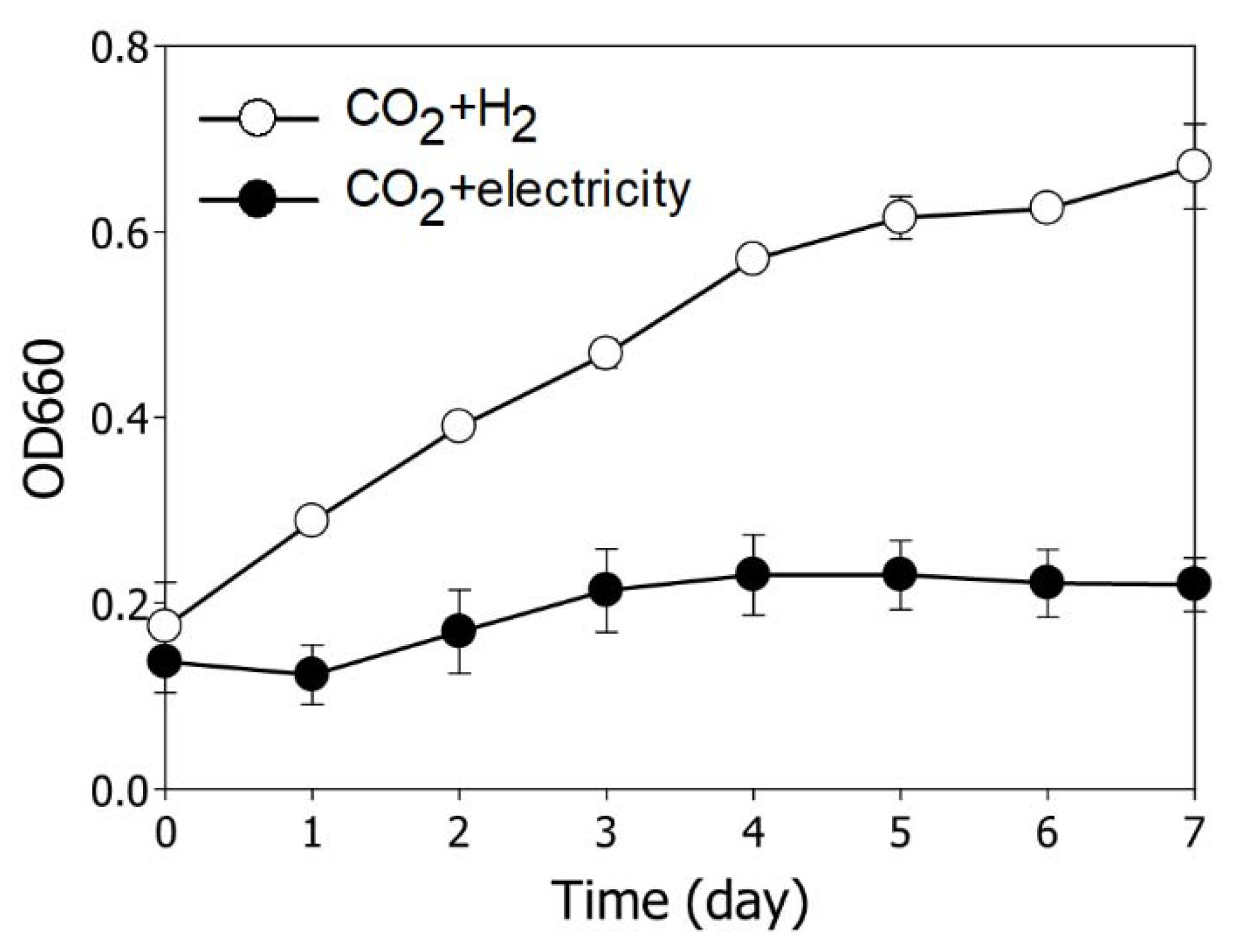
3.1. Comparison of Growth Behavior According to Reducing Source in R. sphaeroides

For CO2 biological fixation using electroactive microorganisms in MES, an appropriate reducing power, such as organic compounds, hydrogen or electricity, is required. Although several studies on MES have been reported, there has been no direct comparison of growth behavior according to reducing source, such as hydrogen or electricity [17,18,19]. Thus, we first examined the cell growth of R. sphaeroides according to the supply of different electron donors in the presence of constant light and CO2 (Figure 1). As a result, it was observed that cell growth was significantly slower when electricity was used as an electron donor than when hydrogen was used. These results indicate that bacterial cell growth is strongly influenced by the type of electron source. Hydrogen is generally one of the most preferred electron donors for autotrophic microorganisms. It readily generates intracellular proton motive force (PMF) to induce sufficient ATP synthesis and promote CO2 transport. This proton gradient caused by hydrogen ion also promotes the generation of reducing cofactor, such as NADPH2 [7,20,21]. NADPH2 is an important biochemical redox cofactor responsible for the transfer of electrons and protons. It is used for reductive biosynthesis, such as biosynthesis of fatty acids and some amino acids. Moreover, it functions as an oxidative stress defense because NADPH2 is essential for the functions of several antioxidant enzymes, including glutathione peroxidase, glutathione S-transferase, and catalase [22,23,24]. When the hydrogen is used as a reducing source, the supply of NADPH2 and ATP are sufficient for CO2 fixation. However, when electricity is used as a reducing source, the supply of NADPH2 and ATP is insufficient due to only electrons are provided and appropriate proton gradient is not formed. Thus, we suspected that the deficiency of ATP and NADPH2 is one of the important factors limiting the bacterial cell growth in MES. For this reason, the direct supply of electrons or the indirect supply of permeated hydrogen ions after water decomposition at the anode is estimated to have lower utilization efficiency in R. sphaeroides compared to the direct supply of hydrogen.
We next performed a transcriptomic analysis to understand the variation of the molecular mechanism in MES systems compared to normal photoautotrophic conditions (Table 1). Consistent with slower cell growth in MES, the expression of hydrogenase (hupSL) gene for uptake of H2 and the Calvin-Benson-Bassham (CBB) cycle (cbbSL, cfxA, fbp1, and cbbR) genes for the uptake of CO2 were downregulated in MES [25,26]. PHB is a representative carbon storage material that accumulates in cells and the expression of PHB biosynthesis (phaA, phaB, phaC1, and phaC2) genes reported to be proportional to cell growth also decreased overall in MES [16]. It is speculated that this is due to the upregulation of the PHB synthesis repressor (PhaR), encoded by RSP_0380 [27]. The transcript levels of genes involved in flagellar biosynthesis (flhAB, flgALK1I1HE, fliEF1KPRD, and motAB) were significantly increased when electricity was used as an electron donor [28]. In MES systems, electron transfer occurs either directly or indirectly through an extracellular electron uptake mechanism. The flagellar mediate direct electron transfer by adhesion to the electrode, and this mechanism has been identified in Desulfovibrio [29,30]. Based on this, it is hypothesized that the upregulation of flagellar biosynthetic gene expression in MES is closely associated with direct electron uptake in R. sphaeroides.
In particular, the genes related to ROS signaling (oxyR, rpoE, chrR, and phrA), redoxins (grxC, RSP_2953, trxA, RSP_0725), and superoxide dismutase (sodC) were highly expressed in MES conditions [31]. In contrast, the expression of catalase gene (katC) involved in hydrogen peroxide scavenging and the carotenoid biosynthetic genes (crtBCDEFI), which were reported to be involved in antioxidant activity, was notably decreased. Overall, the genes related to ROS signaling were upregulated, while the genes related to ROS-scavenging mechanisms were downregulated except for the sodC gene. It is confirmed that expression of the genes related to ROS and antioxidant activity were critically influenced by the direct supply of electricity. These transcriptome analysis results suggest that when CO2 is used as a carbon source, cellular metabolisms are greatly changed depending on the type of reducing power. In particular, it causes a large difference in antioxidant activity, and the mechanism regulating it is very complex, and thus, further studies are needed to elucidate the change in MES.
Many studies have reported that ROS metabolism is very sensitive to changes of environmental growth conditions. In our transcriptome analysis data, it was confirmed that the ROS signaling was strictly associated with the reducing power under photoautotrophic growth conditions. To identify whether changes in transcript levels affected the generation of ROS, we next evaluated the endogenous levels of ROS in MES compared to those under normal photoautotrophic conditions (Figure 2a,b). The levels of endogenous hydrogen peroxide were relatively higher under MES systems than under photoautotrophic conditions. Consistent with this, higher fluorescence values were also observed in MES when the levels of ROS were measured by the detection of fluorescence caused by oxidation of H2DCFDA, an indicator of general oxidative stress. We subsequently measured the activities of peroxidase, which is related to scavenging of hydrogen peroxide (Figure 2c). The peroxidase activities were 4.9-fold lower when electricity was utilized as the electron source than when H2 was utilized. Generally, when the level of endogenous ROS exceeds the detoxification capabilities of microorganisms, cell damage and bacterial cell death occur. It has been reported that the production of endogenous ROS predicted by an ensemble approach of genome-scale increased the susceptibility of E. coli to oxidative attack, quickly leading to cell death [32]. Furthermore, cytotoxic ROS are generated in electrochemical systems through water splitting side reactions. It was also reported that the growth of C. necator in the hybrid microbial-water-splitting catalyst system is inhibited by ROS generated from the electrode in MES when the voltage is less than 2.3 V or higher than 4.0 V. In this system, carbon dioxide converts into biomass and isopropanol along with hydrogen and oxygen produced from water splitting. Since the minimum thermodynamic potential for water splitting is 1.23 V, the relatively high potential for microorganisms was applied in this system [33]. Afterward, to avoid the toxic effect of ROS against microbes and to decrease the potential for water splitting, the biocompatible catalyst system which used an ROS-resistant cobalt-phosphorus alloy cathodic electrode was reported [34]. It was also reported that ROS generated in the inorganic electrocatalytic system, such as O2 and NO, has a toxicity to C. necator, resulting in the inhibition of cell growth and yield of the products [35]. To evade the toxicity of ROS to bacterial cell growth, it has been reported that the two-chamber MES reactor is efficient. This configuration, which separates the cathodic and anodic chamber by a proton exchange membrane (PEM) and has an applied potential at −0.6 V, is similar to our MES reactor [36]. Both the present and the previous observations support that ROS generated in MES systems unfavorably influence cell growth, indicating that the regulation of endogenous ROS is a key element for the improvement of growth in MES.

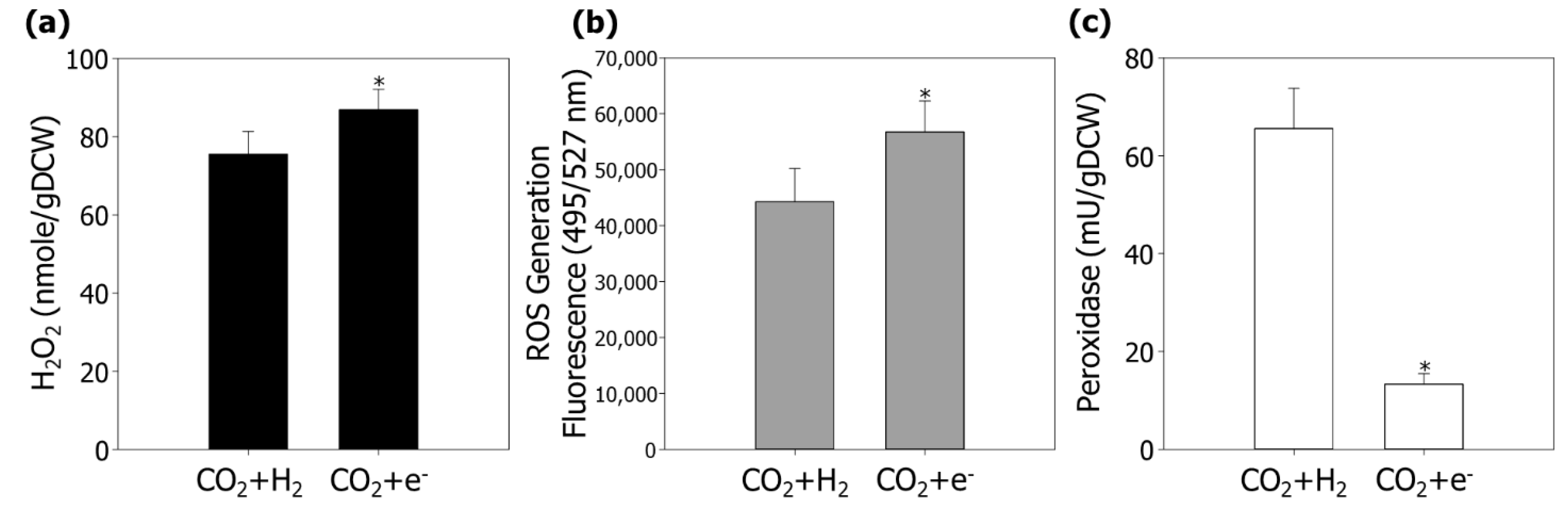
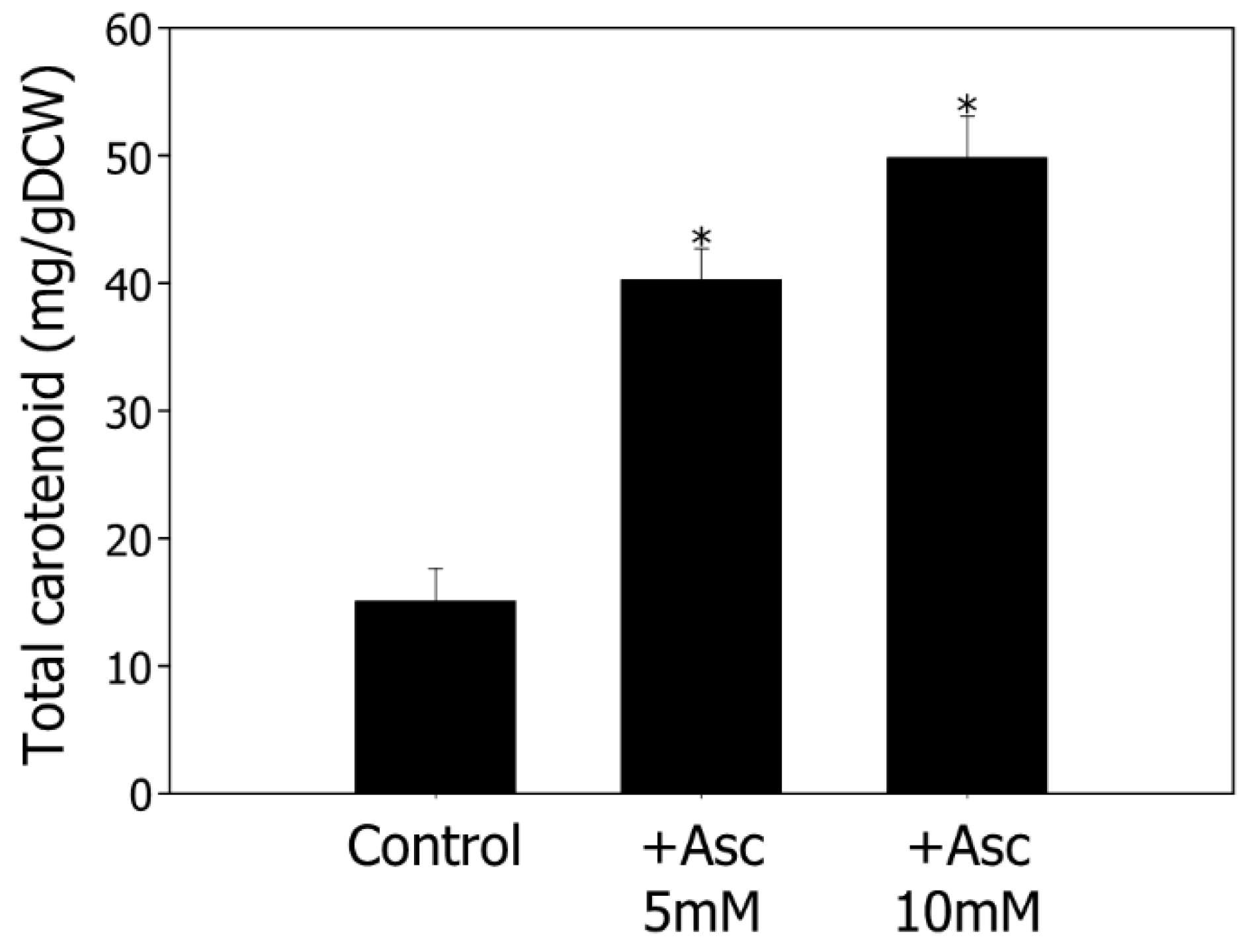
3.2. Enhancement of Bacterial Cell Growth and Carotenoid Production by Modulating ROS in MES

Based on our experimental results so far, when electricity was used as an electron source, we confirmed that the cell growth is slower and the endogenous ROS content is higher compared to when hydrogen is used as an electron source (Figure 1 and Figure 2). Through this, we hypothesized that cell growth is poor in MES due to increased levels of ROS. Furthermore, the growth of microbial cells, including attached cells and planktonic cells, is an important factor in increasing the production of high-value added materials in MES. To improve the cell growth by modulation of ROS while confirming our hypothesis, we introduced ascorbic acid, which is known as a powerful biological antioxidant. The cell growth was observed in MES with the addition of 5 mM and 10 mM ascorbic acid, respectively (Figure 3a). During 10 days of cultivation, the bacterial cell growth was significantly improved by the addition of ascorbic acid. When 5 mM or 10 mM of ascorbic acid was added, the cell density increased by 161% and 131%, respectively. Dosing of more than 10 mM ascorbic acid appears undesirable in MES because excess ascorbic acid can act as an oxidizing agent and inhibit cell growth. To confirm the effect of ascorbic acid on endogenous ROS, we subsequently analyzed the contents of H2O2 and the levels of endogenous ROS (Figure 3b,c). Compared to the control, the levels of H2O2 were reduced proportionally to the concentration of treated ascorbic acid. When ascorbic acid was supplied at 5 mM and 10 mM, the fluorescence value indicating the endogenous contents of ROS was also decreased by 60% and 75%, respectively. In addition, activities of peroxidase were improved 2.9-fold and 2.1-fold in the presence of 5 mM or 10 mM ascorbic acid, respectively (Figure 3d). These observations suggest that supplementation of exogenous antioxidants successfully reduced endogenous levels of ROS, which have a significant impact on the bacterial cell growth.
Ascorbic acid is one of the most important antioxidants present in cells. It prevents oxidative damage caused by ROS and mediates diverse cell metabolism. In plants, ascorbate serves as a cofactor of multiple enzymatic reactions and supports photosynthesis and flowering [37]. Ascorbic acid is able to scavenge hydrogen peroxide, which occurs through the photosynthetic electron transport chain, and thereby contributes to the control of photosynthetic efficiency in plants [38]. Furthermore, the addition of ascorbic acid also has various effects on microbial metabolism. In R. sphaeroides, the activity of light-dependent ATPase involved in ATP synthesis is usually observed to be lower under aerobic conditions than under anaerobic conditions. However, additional supply of ascorbic acid reduced ROS toxicity and recovered ATPase activity to anaerobic levels [39]. The photosynthesis of R. sphaeroides is mediated by the reaction centers (RCs). As the concentration of ascorbate increases, the generation of singlet oxygen (1O2) decreases in the reaction centers [40]. These findings indicate that the supplement of ascorbic acid modulates the production of ROS during photosynthesis, affecting the microbial CO2 fixation efficiency. According to both the present and previous results, ROS regulation by the supply of antioxidants meaningfully improves bacterial cell growth in MES. In order to better understand the effect of regulating antioxidant activity on microorganisms, it will be of great help if we further analyze changes of gene expression patterns and related mechanisms in the future.
The valorization of CO2 into high value-added products is an important aspect of expanding the application of MES. In the context, we next investigated the production of carotenoids, which are naturally accumulated in R. sphaeroides and possess antioxidant activity, under MES systems (Figure 4). When 5 mM and 10 mM of ascorbic acid was added, the contents of total carotenoids were 2.7-fold and 3.3-fold elevated in cells, respectively. These results indicate that the production of total carotenoids in R. sphaeroides is increased in proportion to the concentration of ascorbic acid. Consistent with this, it was reported that a moderate positive correlation was observed between the levels of ascorbate and the contents of total carotenoid in spinach [41]. However, since plants and microorganisms have very different metabolic processes, it is necessary to conduct further research to elucidate the molecular mechanism between ascorbic acid and carotenoid production in R. sphaeroides. Moreover, the antioxidant activity of carotenoids is expected to be beneficial in utilization of CO2 under MES conditions. Previous studies reported that the carotenoid pigments assist the modulation of photosynthetic electron transport and protect against the damage induced by light during photosynthesis [38]. If a synergistic effect between carotenoid biosynthesis and its antioxidant activity could be achieved, then a vigorous electricity-driven cell factory could be established for sustainable carotenoid production in MES [4]. Taken together, the present results show that modulation of ROS by an exogenous antioxidant promoted the production of carotenoids as well as cell growth. These observations suggest that the enhancement of antioxidant activity can be used as a novel strategy to increase the bacterial cell growth and the yield and productivity of valuable products under MES systems.

4. Conclusions

Improving bacterial cell growth is a crucial factor in converting CO2 into valuable materials under MES systems. However, the cell growth in MES has been much slower than that under normal photoautotrophic conditions due to the generation of large amounts of endogenous ROS. To overcome this, we applied on exogenous antioxidant to MES. When ascorbic acid was treated, the cell growth and carotenoid accumulation were significantly increased. Furthermore, while the levels of endogenous ROS were decreased, the activity of ROS scavenging enzyme was increased. Altogether, our results suggest that modulation of ROS is very important to promote bacterial cell growth and carotenoid production under MES conditions.

Author Contributions

Conceptualization, W.-H.L. and S.L.; formal analysis, Y.R.L., S.Y.L., J.L. and H.S.K.; data cura-tion, Y.R.L., S.Y.L., W.-H.L. and S.L.; writing—original draft preparation, Y.R.L., S.Y.L. and S.L.; writing—review and editing, W.-H.L., J.-S.L. and S.L.; funding acquisition, J.-S.L. and S.L. All authors have read and agreed to the published version of the manuscript.

Funding

This research was conducted under the framework of the Research and Development Program of the Korea Institute of Energy Research (KIER-C1-2432); research and development program (C1-5506), which is granted financial resources from Gwangju metropolitan city, Republic of Korea. This work was supported by the Korea Institute of Planning and Evaluation for Technology in Food, Agriculture and Forestry (IPET) through the Useful Agricultural Life Resources Industry Technology Development Program, funded by the Ministry of Agriculture, Food and Rural Affairs (MAFRA) (121048-2).

Institutional Review Board Statement

Not applicable.

Informed Consent Statement

Not applicable.

Conflicts of Interest

The authors declare no conflict of interest. The funders had no role in the design of the study; in the collection, analyses, or interpretation of data; in the writing of the manuscript, or in the decision to publish the results.

References

  1. Mac Dowell, N.; Fennell, P.S.; Shah, N.; Maitland, G.C. The role of CO2 capture and utilization in mitigating climate change. Nat. Clim. Chang. 2017, 7, 243–249. [Google Scholar] [CrossRef] [Green Version]
  2. Lee, S.Y.; Oh, Y.K.; Lee, S.; Fitriana, H.N.; Moon, M.; Kim, M.S.; Lee, J.; Min, K.; Park, G.W.; Lee, J.P.; et al. Recent developments and key barriers to microbial CO2 electrobiorefinery. Bioresour. Technol. 2021, 320, 124350. [Google Scholar] [CrossRef] [PubMed]
  3. Zhang, K.; Zhou, Y.; Song, T.; Xie, J. Bioplastic production from the microbial electrosynthesis of acetate through CO2 reduction. Energy Fuels 2021, 35, 15978–15986. [Google Scholar] [CrossRef]
  4. Wu, H.; Pan, H.; Li, Z.; Liu, T.; Liu, F.; Xiu, S.; Wang, J.; Wang, H.; Hou, Y.; Yang, B.; et al. Efficient production of lycopene from CO2 via microbial electrosynthesis. Chem. Eng. J. 2022, 430, 132943. [Google Scholar] [CrossRef]
  5. Krieg, T.; Sydow, A.; Faust, S.; Huth, I.; Holtmann, D. CO2 to Terpenes: Autotrophic and electroautotrophic α-humulene production with Cupriavidus necator. Angew. Chem. Int. Ed. 2018, 57, 1879–1882. [Google Scholar] [CrossRef]
  6. Batlle-Vilanova, P.; Puig, S.; Gonzalez-Olmos, R.; Balaguer, M.D.; Colprim, J. Continuous acetate production through microbial electrosynthesis from CO2 with microbial mixed culture. J. Chem. Technol. Biotechnol. 2016, 91, 921–927. [Google Scholar] [CrossRef]
  7. Schmid, F.; Novion Ducassou, J.; Couté, Y.; Gescher, J. Developing Rhodobacter sphaeroides for cathodic biopolymer production. Bioresour. Technol. 2021, 336, 125340. [Google Scholar] [CrossRef]
  8. Orsi, E.; Folch, P.L.; Monje-López, V.T.; Fernhout, B.M.; Turcato, A.; Kengen, S.W.M.; Eggink, G.; Weusthuis, R.A. Characterization of heterotrophic growth and sesquiterpene production by Rhodobacter sphaeroides on a defined medium. J. Ind. Microbiol. Biotechnol. 2019, 46, 1179–1190. [Google Scholar] [CrossRef] [Green Version]
  9. Su, A.; Chi, S.; Li, Y.; Tan, S.; Qiang, S.; Chen, Z.; Meng, Y. Metabolic redesign of Rhodobacter sphaeroides for lycopene production. J. Agric. Food Chem. 2018, 66, 5879–5885. [Google Scholar] [CrossRef]
  10. Staerck, C.; Gastebois, A.; Vandeputte, P.; Calenda, A.; Larcher, G.; Gillmann, L.; Papon, N.; Bouchara, J.P.; Fleury, M.J.J. Microbial antioxidant defense enzymes. Microb. Pathog. 2017, 110, 56–65. [Google Scholar] [CrossRef]
  11. Kars, G.; Demirel Kars, M.; Obali, İ.; Emsen, A.; Gündüz, U. Investigation of antioxidant and cytotoxic effects of biotechnologically produced carotenoids from Rhodobacter sphaeroides O.U. 001. Gümüşhane Üniversitesi Fen Bilimleri Dergisi 2020, 10, 559–568. [Google Scholar] [CrossRef]
  12. Tian, B.; Xu, Z.; Sun, Z.; Lin, J.; Hua, Y. Evaluation of the antioxidant effects of carotenoids from Deinococcus radiodurans through targeted mutagenesis, chemiluminescence, and DNA damage analyses. Biochim. Biophys. Acta Gen. Subj. 2007, 1770, 902–911. [Google Scholar] [CrossRef] [PubMed]
  13. Azizi, F.; Farsaraei, S.; Moghaddam, M. Application of exogenous ascorbic acid modifies growth and pigment content of Calendula officinalis L. flower heads of plants exposed to NaCl Stress. J. Soil Sci. Plant Nutr. 2021, 21, 2803–2814. [Google Scholar] [CrossRef]
  14. Sistrom, W.R. The kinetics of the synthesis of photopigments in Rhodopseudomonas spheroides. J. Gen. Microbiol. 1962, 28, 607–616. [Google Scholar] [CrossRef] [Green Version]
  15. Fitriana, H.N.; Lee, J.; Lee, S.; Moon, M.; Lee, Y.R.; Oh, Y.K.; Park, M.; Lee, J.S.; Song, J.; Lee, S.Y. Surface modification of a graphite felt cathode with amide-coupling enhances the electron uptake of Rhodobacter sphaeroides. Appl. Sci. 2021, 11, 7585. [Google Scholar] [CrossRef]
  16. Lee, Y.R.; Nur Fitriana, H.; Lee, S.Y.; Kim, M.S.; Moon, M.; Lee, W.H.; Lee, J.S.; Lee, S. Molecular profiling and optimization studies for growth and PHB production conditions in Rhodobacter sphaeroides. Energies 2020, 13, 6471. [Google Scholar] [CrossRef]
  17. Li, S.; Sakuntala, M.; Song, Y.E.; Heo, J.O.; Kim, M.; Lee, S.Y.; Kim, M.S.; Oh, Y.K.; Kim, J.R. Photoautotrophic hydrogen production of Rhodobacter sphaeroides in a microbial electrosynthesis cell. Bioresour. Technol. 2021, 320, 124333. [Google Scholar] [CrossRef]
  18. Wu, Z.; Wang, J.; Liu, J.; Wang, Y.; Bi, C.; Zhang, X. Engineering an electroactive Escherichia coli for the microbial electrosynthesis of succinate from glucose and CO2. Microb. Cell Fact. 2019, 18, 15. [Google Scholar] [CrossRef]
  19. Chen, J.; Wei, J.; Ma, C.; Yang, Z.; Li, Z.; Yang, X.; Wang, M.; Zhang, H.; Hu, J.; Zhang, C. Photosynthetic bacteria-based technology is a potential alternative to meet sustainable wastewater treatment requirement? Environ. Int. 2020, 137, 105417. [Google Scholar] [CrossRef]
  20. Assil-Companioni, L.; Büchsenschütz, H.C.; Solymosi, D.; Dyczmons-Nowaczyk, N.G.; Bauer, K.K.F.; Wallner, S.; MacHeroux, P.; Allahverdiyeva, Y.; Nowaczyk, M.M.; Kourist, R. Engineering of NADPH Supply Boosts Photosynthesis-Driven Biotransformations. ACS Catal. 2020, 10, 11864–11877. [Google Scholar] [CrossRef]
  21. Nikkanen, L.; Solymosi, D.; Jokel, M.; Allahverdiyeva, Y. Regulatory electron transport pathways of photosynthesis in cyanobacteria and microalgae: Recent advances and biotechnological prospects. Physiol. Plant. 2021, 173, 514–525. [Google Scholar] [CrossRef] [PubMed]
  22. Agledal, L.; Niere, M.; Ziegler, M. The phosphate makes a difference: Cellular functions of NADP. Redox Rep. 2010, 15, 2–10. [Google Scholar] [CrossRef] [PubMed] [Green Version]
  23. Morrison, C.S.; Armiger, W.B.; Dodds, D.R.; Dordick, J.S.; Koffas, M.A.G. Improved strategies for electrochemical 1,4-NAD(P)H2 regeneration: A new era of bioreactors for industrial biocatalysis. Biotechnol. Adv. 2018, 36, 120–131. [Google Scholar] [CrossRef] [PubMed]
  24. Spaans, S.K.; Weusthuis, R.A.; van der Oost, J.; Kengen, S.W.M. NADPH-generating systems in bacteria and archaea. Front. Microbiol. 2015, 6, 742. [Google Scholar] [CrossRef]
  25. Ghirardi, M.L.; Posewitz, M.C.; Maness, P.C.; Dubini, A.; Yu, J.; Seibert, M. Hydrogenases and hydrogen photoproduction in oxygenic photosynthetic organisms. Annu. Rev. Plant Biol. 2007, 58, 71–91. [Google Scholar] [CrossRef]
  26. Lee, S.Y.; Kim, Y.S.; Shin, W.R.; Yu, J.; Lee, J.; Lee, S.; Kim, Y.H.; Min, J. Non-photosynthetic CO2 bio-mitigation by: Escherichia coli harbouring CBB genes. Green Chem. 2020, 22, 6889–6896. [Google Scholar] [CrossRef]
  27. Chou, M.E.; Chang, W.T.; Chang, Y.C.; Yang, M.K. Expression of four pha genes involved in poly-β-hydroxybutyrate production and accumulation in Rhodobacter sphaeroides FJ1. Mol. Genet. Genom. 2009, 282, 97–106. [Google Scholar] [CrossRef]
  28. Baker, A.E.; O’Toole, G.A. Bacteria, rev your engines: Stator dynamics regulate flagellar motility. J. Bacteriol. 2017, 199, e00088-17. [Google Scholar] [CrossRef] [Green Version]
  29. del Pilar Anzola Rojas, M.; Mateos, R.; Sotres, A.; Zaiat, M.; Gonzalez, E.R.; Escapa, A.; De Wever, H.; Pant, D. Microbial electrosynthesis (MES) from CO2 is resilient to fluctuations in renewable energy supply. Energy Convers. Manag. 2018, 177, 272–279. [Google Scholar] [CrossRef]
  30. Tremblay, P.L.; Angenent, L.T.; Zhang, T. Extracellular electron uptake: Among autotrophs and mediated by surfaces. Trends Biotechnol. 2017, 35, 360–371. [Google Scholar] [CrossRef]
  31. Ziegelhoffer, E.C.; Donohue, T.J. Bacterial responses to photo-oxidative stress. Nat. Rev. Microbiol. 2009, 7, 856–863. [Google Scholar] [CrossRef] [PubMed]
  32. Brynildsen, M.P.; Winkler, J.A.; Spina, C.S.; MacDonald, I.C.; Collins, J.J. Potentiating antibacterial activity by predictably enhancing endogenous microbial ROS production. Nat. Biotechnol. 2013, 31, 160–165. [Google Scholar] [CrossRef] [PubMed] [Green Version]
  33. Torella, J.P.; Gagliardi, C.J.; Chen, J.S.; Bediako, D.K.; Colón, B.; Way, J.C.; Silver, P.A.; Nocera, D.G. Efficient solar-to-fuels production from a hybrid microbial-water-splitting catalyst system Proc. Natl. Acad. Sci. USA 2015, 112, E1507. [Google Scholar] [CrossRef] [Green Version]
  34. Liu, C.; Colón, B.C.; Ziesack, M.; Silver, P.A.; Nocera, D.G. Water splitting–biosynthetic system with CO2 reduction efficiencies exceeding photosynthesis. Science 2016, 352, 602–692. [Google Scholar] [CrossRef] [PubMed]
  35. Li, H.; Opgenorth, P.H.; Wernick, D.G.; Rogers, S.; Wu, T.Y.; Higashide, W.; Malati, P.; Huo, Y.X.; Cho, K.M.; Liao, J.C. Integrated electromicrobial conversion of CO2 to higher alcohols. Science 2012, 335, 1596. [Google Scholar] [CrossRef] [PubMed]
  36. Chen, X.; Cao, Y.; Li, F.; Tian, Y.; Song, H. Enzyme-assisted microbial electrosynthesis of poly(3-hydroxybutyrate) via CO2 bioreduction by engineered Ralstonia eutropha. ACS Catal. 2018, 8, 4429–4437. [Google Scholar] [CrossRef]
  37. Gallie, D.R. L-Ascorbic acid: A multifunctional molecule supporting plant growth and development. Sientifica 2013, 2013, 795964. [Google Scholar] [CrossRef] [PubMed] [Green Version]
  38. Foyer, C.H.; Harbinson, J. Relationships between antioxidant metabolism and carotenoids in the regulation of photosynthesis. In The Photochemistry of Carotenoids; Springer: Dordrecht, The Netherlands, 2006; pp. 305–325. [Google Scholar] [CrossRef]
  39. Kim, H.; Tong, X.; Choi, S.; Lee, J.K. Characterization of ATPase activity of free and immobilized chromatophore membrane vesicles of Rhodobacter sphaeroides. J. Microbiol. Biotechnol. 2017, 27, 2173–2179. [Google Scholar] [CrossRef] [Green Version]
  40. Uchoa, A.F.; Knox, P.P.; Turchielle, R.; Seifullina, N.K.; Baptista, M.S. Singlet oxygen generation in the reaction centers of Rhodobacter sphaeroides. Eur. Biophys. J. 2008, 37, 843–850. [Google Scholar] [CrossRef]
  41. Wang, X.; Cai, X.; Xu, C.; Zhao, Q.; Ge, C.; Dai, S.; Wang, Q.H. Diversity of nitrate, oxalate, vitamin C and carotenoid contents in different spinach accessions and their correlation with various morphological traits. J. Hortic. Sci. Biotechnol. 2018, 93, 409–415. [Google Scholar] [CrossRef]
Figure 1. Comparison of growth characteristics according to different reducing power sources in R. sphaeroides. The cells were cultured using CO2 and light, and only the reducing power source was varied. The white circle line denotes CO2 + H2, which represents normal photoautotrophic cultivation. The black circle line denotes CO2 + electricity, which represents MES cultivation. The experiments performed in triplicate and error bars indicate the standard deviation of the mean.
Energies 15 00935 g001
Figure 2. Endogenous levels of ROS in R. sphaeroides under photoautotrophic (CO2 + H2) and MES (CO2 + e) conditions. (a) Endogenous contents of H2O2. (b) Generation of endogenous ROS. The levels of ROS were measured using CM-H2DCFDA. The fluorescence is represented in arbitrary units and normalized by the optical density of the samples. (c) The measurement of peroxidase activity. Experiments were conducted in triplicate and error bars indicate standard deviation of mean. Asterisk represent statistically significant difference, as determined by a Student’s t-test (* p < 0.05).
Energies 15 00935 g002
Figure 3. Effects of ascorbic acid on MES in R. sphaeroides. (a) Cell growth in the presence of 5 mM or 10 mM ascorbic acid. The cultivations were conducted in MES reactors using CO2 as a carbon source and electricity as a reducing source under –0.6 V (vs. Ag/AgCl). Circle lines indicate control (untreated ascorbic acid). Upward triangle lines indicate the addition of 5 mM ascorbic acid. The square line represent the addition of 10 mM ascorbic acid. (b) Measurement of endogenous H2O2. (c) Generation of endogenous ROS. The levels of ROS were measured using CM-H2DCFDA. The fluorescence is represented in arbitrary units and normalized by the optical density of the samples. (d) The analysis of peroxidase activity. Asc, ascorbic acid. Every experiment was conducted in triplicate and error bars indicate standard deviation of mean. Asterisk represents statistically significant difference, as determined by a Student’s t-test (* p < 0.05).
Energies 15 00935 g003
Figure 4. Measurement of total carotenoid contents in the presence of ascorbic acid under MES conditions. Asc, ascorbic acid. Experiments were performed in triplicate and error bars indicate standard deviation of mean. Asterisk represent statistically significant difference, as determined by a Student’s t-test (* p < 0.05).
Energies 15 00935 g004
Table 1. Comparison of transcript levels in MES conditions versus normal photoautotrophic conditions.
Gene NumberGene NameFunctionDescriptionLog2(FC)
RSP_0495hupSHydrogenase protein small subunitEnergy production and conversion−7.6
RSP_0496hupLHydrogenase protein large subunitEnergy production and conversion−9.7
RSP_1281cbbSRibulose 1,5-bisphosphate carboxylase small subunitCarbohydrate transport and metabolism−6.0
RSP_1282cbbLRibulose 1,5-bisphosphate carboxylase large subunitEnergy production and conversion−6.3
RSP_1283cfxAFructose-1,6-bisphosphate aldolaseCarbohydrate transport and metabolism−6.5
RSP_1284prkAPhosphoribulokinaseEnergy production and conversion−6.7
RSP_1285fbp1Fructose-1,6-bisphosphataseCarbohydrate transport and metabolism−6.4
RSP_1286cbbRRuBisCO operon transcriptional regulator, CbbRTranscription−2.2
RSP_0382phaC1Poly-beta-hydroxybutyrate polymeraseLipid transport and metabolism−2.6
RSP_0745phaAAcetyl-CoA acetyltransferaseLipid transport and metabolism−1.8
RSP_0747phaB3-oxoacyl-(Acyl-carrier-protein) reductaseFunction unknown−3.7
RSP_1257phaC2Putative polyhydroxyalkanoic synthaseLipid transport and metabolism−3.6
RSP_0380 Polyhydroxyalkanoate synthesis repressor, PhaRFunction unknown2.9
RSP_0034flhAFlagellar biosynthesis proteinCell motility8.4
RSP_0036flgAFlagella basal body P-ring formation proteinCell motility5.9
RSP_0052fliEFlagellar hook-basal body complex proteinCell motility7.0
RSP_0053fliF1Flagellar M-ring proteinCell motility8.2
RSP_0058fliKFliK, flagellar hook-length control proteinCell motility6.4
RSP_0063fliPFlagellar biosynthetic proteinCell motility9.2
RSP_0065fliRFlagellar biosynthetic proteinCell motility5.8
RSP_0066flhBFlagellar biosynthetic proteinCell motility7.3
RSP_0070fliDFlagellar hook-associated protein 2Cell motility4.0
RSP_0073flgLFlagellar hook-associated protein 3Cell motility5.9
RSP_0074flgK1Flagellar hook-associated protein 1Cell motility6.7
RSP_0076flgI1Flagellar P-ring proteinCell motility8.5
RSP_0077flgHFlagellar L-ring proteinCell motility6.0
RSP_0080flgEFlagellar hook proteinCell motility6.6
RSP_0231motBFlagellar MotB proteinCell motility5.9
RSP_0233motAFlagellar MotA proteinCell motility7.7
RSP_2380katCCatalaseInorganic ion transport and metabolism−1.3
RSP_2780oxyR2Transcriptional regulator, LysR familyTranscription2.4
RSP_1796sodCSuperoxide dismutase [Cu-Zn]Inorganic ion transport and metabolism1.1
RSP_1092rpoEECF RNA polymerase sigma factor RpoETranscription2.8
RSP_1093chrRAnti-sigma-E factor ChrRTranscription2.9
RSP_2143phrADNA photolyase, Cryptochrome 1 apoprotein (Blue light photoreceptor)Replication, recombination and repair4.0
RSP_1194grxCGlutaredoxinPosttranslational modification, protein turnover1.1
RSP_2953 GlutaredoxinPosttranslational modification, protein turnover1.6
RSP_1529trxAThioredoxinPosttranslational modification, protein turnover2.0
RSP_0725 ThioredoxinPosttranslational modification, protein turnover2.6
RSP_0264crtFDemethylspheroidene O-methyltransferaseFunction unknown−4.7
RSP_0265crtEGeranylgeranyl diphosphate synthaseCoenzyme transport and metabolism−6.2
RSP_0266crtDHydroxyneurosporene desaturaseSecondary metabolites biosynthesis−3.9
RSP_0267crtCAcyclic carotenoid 1,2-hydrataseFunction unknown−3.2
RSP_0270crtBPhytoene synthaseLipid transport and metabolism−3.2
RSP_0271crtIPhytoene desaturaseSecondary metabolites biosynthesis−3.8
Publisher’s Note: MDPI stays neutral with regard to jurisdictional claims in published maps and institutional affiliations.

