Article

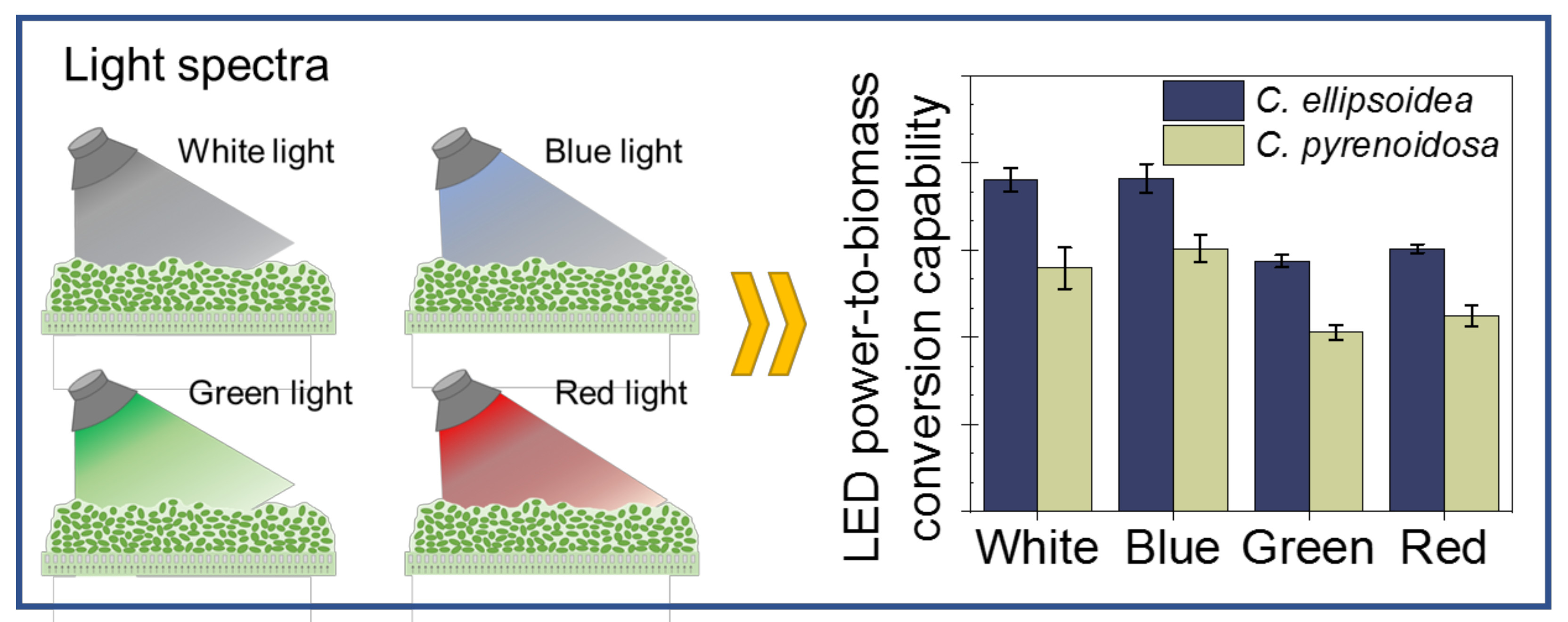
Light-Emitting Diode Power Conversion Capability and CO2 Fixation Rate of Microalgae Biofilm Cultured Under Different Light Spectra

1
School of Energy and Environmental Engineering, University of Science and Technology Beijing, Beijing 100083, China
2
Beijing Key Laboratory for Energy Saving and Emission Reduction of Metallurgical Industry, University of Science and Technology Beijing, Beijing 100083, China
3
Beijing Engineering Research Center of Energy Saving and Environmental Protection, University of Science and Technology Beijing, Beijing 100083, China
*
Author to whom correspondence should be addressed.
Energies 2020, 13(7), 1536; https://doi.org/10.3390/en13071536
Submission received: 30 January 2020 / Revised: 6 March 2020 / Accepted: 19 March 2020 / Published: 25 March 2020
(This article belongs to the Section A4: Bio-Energy)

Abstract

:
Microalgae biofilm-based culture has attracted much interest due to its high harvest efficiency and low energy requirements. Using light-emitting diodes (LEDs) as light source for microalgae culture has been considered as a promising choice to enhance the economic feasibility of microalgae-based commodities. In this work, the LED power conversion capability and CO2 fixation rate of microalgae biofilms (Chlorella ellipsoidea and Chlorella pyrenoidosa) cultured under different light spectra (white, blue, green and red) were studied. The results indicated that the power-to-biomass conversion capabilities of these two microalgae biofilms cultured under blue and white LEDs were much higher than those under green and red LEDs (C. ellipsoidea: 32%–33% higher, C. pyrenoidosa: 34%–46% higher), and their power-to-lipid conversion capabilities cultured under blue LEDs were 61%–66% higher than those under green LEDs. The CO2 fixation rates of these two biofilms cultured under blue LEDs were 13% and 31% higher, respectively, than those under green LEDs. The results of this study have important implications for selecting the optimal energy-efficient LEDs using in microalgae biofilm-based culture systems.

Graphical Abstract

1. Introduction

Microalgae are photosynthetic microorganisms that can convert light, CO2 and nutrients into biomass [1,2,3]. Due to the high growth rates, high lipid contents and ability to mitigate CO2 emissions, microalgae have potential to be promising biological sources to produce high-value biomolecules and biofuels [4,5,6]. However, the success of microalgae-based commodities is dependent on the biomass productivity and production cost [7,8,9]. Recently, some researchers reported that cultivating microalgae as biofilm (i.e., cells are attached to solid surface) can enhance the economic feasibility of microalgae-based commodities, due to its lower water consumption, higher volumetric productivity, higher harvest efficiency and reduced energy requirements compared with suspended systems [10,11,12]. Therefore, developing more efficient biofilm-based microalgae culture system has attracted much attention recently [13,14,15].
To date, various microalgae biofilm-based reactors have been developed to enhance their performance, such as horizontal [16], flow lane [17], twin-layer [18], and rotating biofilm reactors [19]. Extensive studies have reported that many factors can affect the performance of microalgae biofilm-based systems, such as the light condition, the amount of CO2 supplementation, nutrient type and concentration [12,20,21]. Among these factors, light directly affects the photosynthesis of microalgae, thus their growth and cellular composition [22,23,24,25]. Generally, sunlight and artificial illumination are often used as light sources for microalgae culture. The sunlight is cost-effective, but has the disadvantages of changes in weather and season, and day and night cycles. Artificial illuminations, such as fluorescent lamps and light-emitting diode (LED) lights, are more manageable and adjustable, and are of growing interest.
Recently, using LEDs in microalgae culture has been considered as a promising choice due to its advantages of small size, low power consumption, narrow wavelength band and reduced heat release [26,27,28,29]. Some researchers have studied the LED power conversion efficiency of microalgae cultured in suspended systems [30,31]. For example, Ma et al. studied LED power-to-biomass efficiency for Nannochloropsis cultured in suspended systems, and reported that using LED as light source in suspended microalgae culture was more energy efficient [30]. Ajayan et al. found that LEDs provided more light penetration in a column photobioreactor compared with the fluorescent lights, and improved the cell concentration, specific growth rate, total pigments and lipid contents of C. reinhardtii [31]. Maroneze et al. evaluated the role of LED photoperiods on the growth and lipid content of S. obliquus cultured in a bubble column photobioreactor, and found that the photoperiods was an effective strategy to reduce the production cost of microalgae biomass [32]. These above studies indicated that the LEDs might be promising using in suspended microalgae culture systems. It should be noted that, for biofilm-based culture systems, the transmission and refraction of light are totally different from that in suspended systems. This difference may affect the light conversion capability of microalgae [33]. However, to date, little research has studied the LED power conversion capability of microalgae cultured in biofilm-based systems.
To address this gap, this study explored the LED power conversion capability and CO2 fixation rate of two widely used microalgae biofilms cultured under different light spectra (white, blue, green and red). The conversion capabilities of LED power-to-biomass, power-to-lipid, power-to-protein, and power-to-carbohydrate, as well as the CO2 fixation rate for two widely used microalgae biofilms cultured under different LEDs were determined. This study would provide guidance in selecting suitable light spectra and reducing energy consumption for developing more efficient microalgae biofilm-based culture systems.

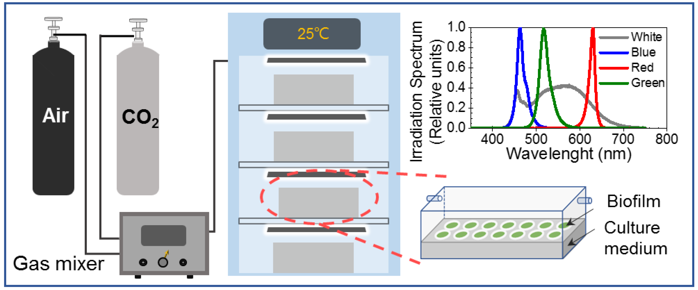
2. Materials and Methods

2.1. Microalgae Biofilm Culture

The microalgae used in this study were Chlorella ellipsoidea (FACHB–40) and Chlorella pyrenoidosa (FACHB–9). Before inoculation of biofilm culture, both microalgae strains were pre-cultivated in 500 mL flasks with 100 mL inoculum at 25 ± 1 °C under continuous irradiance of 100 μmol photons m−2 s−1 for 6 days. Afterwards, these preprepared cell suspensions were evenly vacuum filtered onto some filtration membranes to form microalgae biofilm with an initial inoculum density of 4.0 ± 0.1 g m−2. Then, these filtration membranes were put into the biofilm culture bioreactors (polymethyl methacrylate chambers: 400 × 200 × 200 mm), containing the BG–11 culture medium solidified with 1% agar (see Supplementary Materials Tables S1 and S2 for the details) [34]. As shown in Figure 1, these bioreactors were placed into an incubator to maintain a proper temperature (25 ± 1 °C) for microalgae growth [35]. The inside of these bioreactors was aerated with compressed air enriched with 1% CO2 (vol/vol) at a rate of 0.1 VVM (volume of air per volume of culture per minute).
During cultivation, four kinds of LEDs (J&K Photoelectric Technology, Shanghai, China), including white (JK–W300200, 400–750 nm), blue (JK–B300200, 440–500 nm), green (JK–G300200, 500–550 nm), and red (JK–R300200, 610–650 nm) LEDs were fixed above the bioreactors as the light sources for microalgae cultivation. All the LEDs in the biofilm cultivation were continuous lighting. The photon flux densities of these LEDs at the biofilm surfaces were set to be approximately 100 μmol photons m−2 s−1, which were measured with a 4π quantum scalar sensor (QSL 2100, Biospherical Instruments Inc., San Diego, CA, USA). The light spectra of these four LEDs were characterized with a fiber spectrometer (USB4000, Ocean Optics Inc., Dunedin, FL, USA) between 350 and 750 nm, as shown in Figure 2a. Additionally, the absorption spectra of the C. ellipsoidea and C. pyrenoidosa were characterized by a microplate spectrophotometer (Epoch, BioTek, Winooski, VT, USA), as shown in Figure 2b.

2.2. Determining the Biomass and Chlorophyll Contents

To evaluate microalgae growth, microalgae biofilm were harvested after 6 days culture. The collected biofilm were resuspended with deionized water to remove the soluble nutrients, followed by centrifuging and drying to a constant weight at 105 °C. After cooling in a desiccator, the microalgae biomass was weighed by an analytical balance (XS105, METTLER TOLEDO, Switzerland).
The chlorophyll contents of microalgae cells cultured under white, blue, green and red LEDs were determined according to the methods described by Wellburn [36]. In detail, the chlorophyll was extracted with 80 vol% acetone. The absorbance of chlorophyll solvent was measured at 646 and 663 nm with a visible spectrophotometer (721, INESA, Shanghai, China). The chlorophyll a (chl–a), chlorophyll b (chl–b) concentrations (mg L−1) were calculated by:
chl–a = 12.21 × OD663nm − 2.81 × OD646nm
chl–b = 20.13 × OD663nm − 5.03 × OD646nm
The measurements for biomass and chlorophyll contents were repeated three times. The results were shown as mean ± standard deviation.

2.3. Determining Cellular Composition

The cellular compositions of microalgae are critical evaluation parameters in various microalgae-based commodities. Generally, the main organic components of microalgae are lipids, proteins, and carbohydrates, which represent approximately 80% of the microalgal dry biomass. To investigate the cellular compositions of microalgae biofilm cultured under different LEDs, the harvested cells were frozen at −70 °C and lyophilized. The total lipids were measured according to the methods described by Bligh and Dyer [37], in which chloroform was used to extract the lipid and evaporated at 60 °C. The protein content was measured by a colorimetric method [38,39], in which microalgae biomass was pretreated with thermal alkaline and the standard sample was bovine serum albumin (see Figure S1). The carbohydrate content was determined by the phenol-sulfuric method [40,41], the standard sample was glucose (see Figure S2). All the experiments were repeated in triplicate and the results are shown in mean ± standard deviation.

2.4. LED Power Conversion Capability

We determined the conversion capability of LED power-to-biomass, power-to-lipid, power-to-protein, and power-to-carbohydrate for these microalgae biofilms cultured under different LEDs, which were calculated by [30]:
LED   power   conversion   capability = C t C 0 t × P
where Ct is the accumulation of biomass, lipid, protein and carbohydrate (g m−2) at time t, C0 is the accumulation of biomass, lipid, protein and carbohydrate (g m−2) at time t0 (at the beginning of inoculation), P is the power consumption of different LED units.

2.5. Determining CO2 Fixation Rate

The CO2 fixation rate of microalgae biofilms cultured under white, blue, green and red lights were determined by [42]:
CO 2   fixation   rate = X t X 0 t × C % × 44 12
where Xt is microalgae biomass (g m−2) at time t, X0 is microalgae biomass (g m−2) at time t0 (at the beginning of inoculation), C% is the carbon content of the biomass, which was determined by an elemental analysis (vario EL cube, Elementar, Germany). The experiments were repeated at least three times. The results were shown as mean ± standard deviation.

3. Results and Discussion

3.1. Growth of Microalgae Biofilms Cultured Under Different LEDs

The dry biomass yields and chlorophyll contents (including chl–a and chl–b) of C. ellipsoidea and C. pyrenoidosa biofilms were determined to evaluate their growth. Figure 3 indicates that the microalgae biomasses cultivated under different light spectra were obviously different. In general, for both microalgae, the biomass cultured under the blue LED was much higher than those under the white, green and red LEDs. The total chlorophyll contents of microalgae cultured under the blue and white LEDs were much higher than those under the green and red LEDs.
Previously, many studies reported that both the blue and red light can promote the growth of green algae in suspended culture system, because the green algae can absorb the blue light and red light more efficiently [43]. In this work, Figure 3a indicates that the biomass of C. ellipsoidea was ~14% higher under the blue light than that under the green light. Similarly, Figure 3b shows that the biomass of C. pyrenoidosa was ~26% higher under the blue light than that under the green light. Evidently, the above results suggested that the blue LED was efficient in enhancing cell growth, whereas, we found that the red LED had little influence on cell growth. We think that it may be related to the characteristics of microalgae pigments. On one hand, as shown in Figure 2b, the irradiation spectra of blue LEDs match with the absorption peaks of C. ellipsoidea and C. pyrenoidosa at the light spectra of 420–480 nm (blue), whereas, the irradiation spectra of red LEDs do not match well with the microalgal absorption peaks at the light spectra of 620–680 nm (red). On the other hand, we found that the chlorophyll contents of these two microalgae cultivated under red LEDs were much lower than those cultured under blue LEDs.

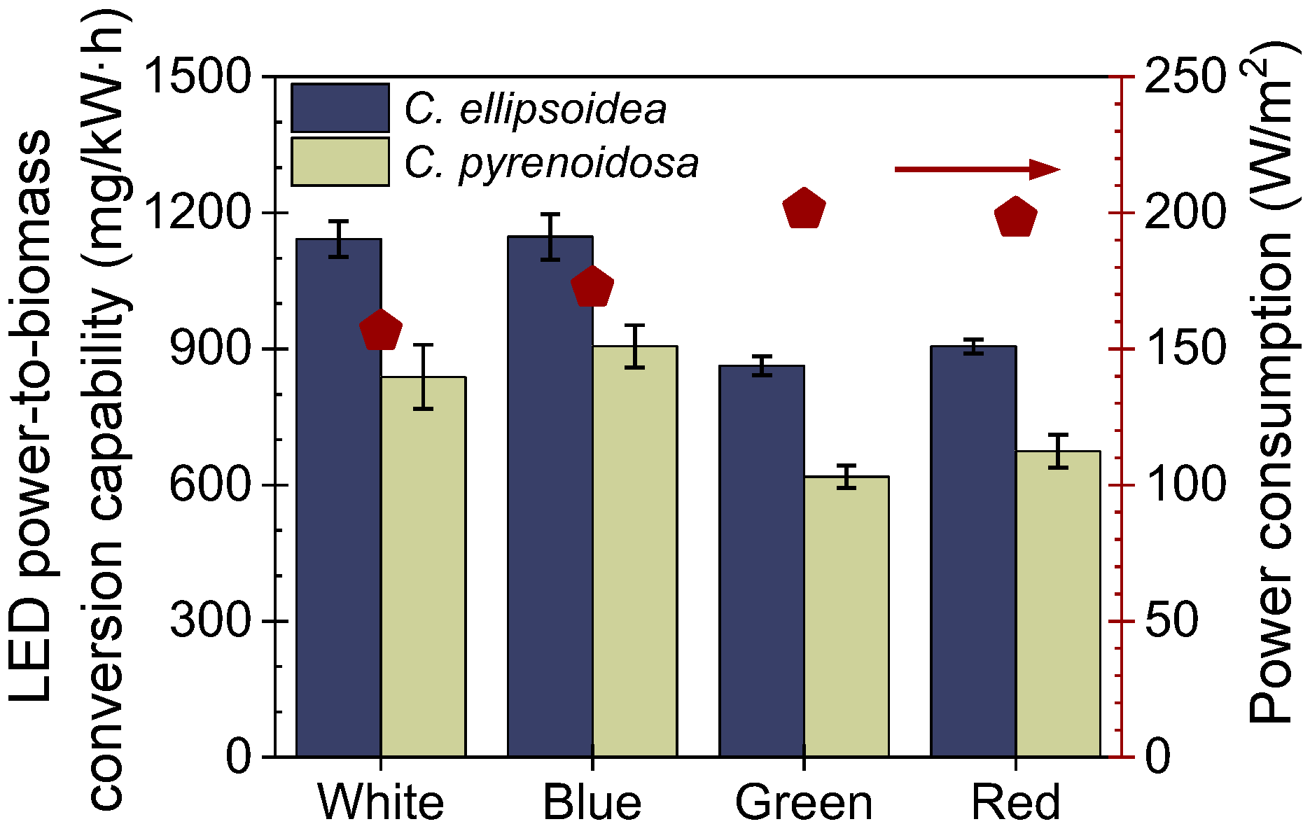
3.2. The LED Power Conversion Capability of Microalgae Biofilm

The LED power conversion capability is an important factor in the assessment of microalgae biofilm cultivation. In this work, we determined the conversion capabilities of LED power-to-biomass, power-to-lipid, power-to-protein, and power-to-carbohydrate. Figure 4 shows the LED power-to-biomass conversion capability for these two microalgae biofilms cultivated under different light spectra, which were calculated based on the biomass accumulation and power consumption of different LEDs (right axis in Figure 4). The results indicate that the power-to-biomass conversion capability of C. ellipsoidea and C. pyrenoidosa cultured under different LEDs ranged from 862 to 1147 mg/kW·h, and from 618 to 905 mg/kW·h, respectively. The LED power-to-biomass conversion capabilities for these two microalgae cultured under white and blue lights were much higher than those cultured under green and red lights, indicating that these microalgae cells utilized blue and white lights more effectively. Particularly, for C. ellipsoidea, the power-to-biomass conversion capability of cells cultured under blue and white lights increased by 32%–33% compared with those under green light. Similarly, for C. pyrenoidosa, the power-to-biomass conversion capability of cells cultured under blue and white lights increased by 34%–46% compared with those under green light.
Furthermore, cellular compositions of microalgae are generally critical evaluation parameters in various applications. Figure S3 shows the lipids, proteins and carbohydrates contents for the C. ellipsoidea and C. pyrenoidosa cultured under different LEDs. It was found that the main organic components in the microalgae represented approximately 80% of microalgae dry biomass, which were consistent with the literature [35]. Based on the cellular composition and power consumption of different LEDs, we determined the conversion capabilities of LED power-to-lipid, power-to-protein, and power-to-carbohydrate. Figure 5 indicates that the power-to-carbohydrate conversion capabilities for both microalgae under white LEDs were the highest. The power-to-protein conversion capabilities for both microalgae cultured under white and blue LEDs were higher than those under green and red lights. Moreover, we found that both microalgae cultured under blue LED showed the highest power-to-lipid conversion capability. In particular, the power-to-lipid conversion capability for C. ellipsoidea and C. pyrenoidosa were 12.6% and 14.8% higher, respectively, under blue light than under white light. The power-to-lipid conversion capability for C. ellipsoidea and C. pyrenoidosa were 60.7% and 66.3% higher, respectively, under blue light than under green light. Similar results have been reported for the suspended culture system. Ra et al. evaluated the effects of LED wavelength on the lipid production of Picochlorum atomus with two-phase suspended cultivation, and found that the lipid accumulation of P. atomus was 329% higher under blue light than that under green light [44]. Kang et al. determined the effect of using wastewater and wavelength filters on microalgal productivity and lipid accumulation with open raceway ponds, and found that the lipid productivity was highest under blue wavelength, at least 46.8% higher than that under white wavelength [45].This may be because blue light can promote the synthesis of lipids [46,47] and has low energy consumption.
Overall, these results suggested that these two microalgae biofilms cultured under blue and white LEDs showed higher conversion capabilities of power-to-biomass and power-to-protein compared with those under green and red LEDs. These two microalgae cultured under blue LEDs possessed the highest power-to-lipid conversion capabilities. The results revealed that it is feasible to induce the synthesis of different chemical components in cells by adjusting the light spectrum in microalgae biofilm-based culture systems.

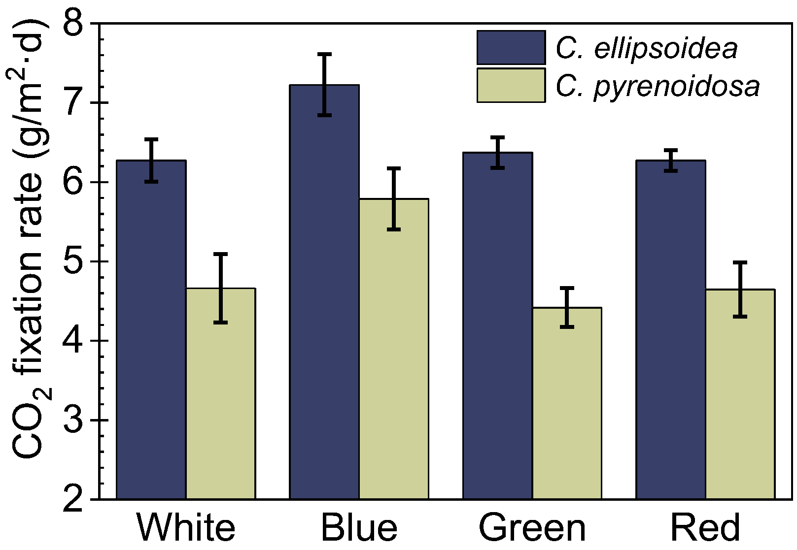
3.3. The CO2 Fixation Rate of Microalgae Biofilm

The CO2 fixation rates of microalgae biofilms cultivated under different LEDs were determined by evaluating the difference in the carbon content (C%) of microalgae between the inoculation and harvest (see Table S3). Figure 6 indicates that the CO2 fixation rates for the C. ellipsoidea and C. pyrenoidosa cultured under blue LEDs were ~13% and ~31% higher, respectively, than those under green light. This may be attributed to the higher photosynthetic performance of microalgae. Additionally, previous study reported that, in suspended culture system, white light was the most effective light for CO2 fixation compared with the blue, red and yellow lights [48]. Evidently, this study indicated that the influence of the light spectra on the CO2 fixation rate of microalgae would be different between the biofilm cultured system and the suspended culture system. This may be due to the different characteristics of light transmission and refraction in these two microalgae culture systems. Further studies should be conducted to understand the light transfer phenomena in microalgae biofilm.

3.4. Implications of LED Selection in Biofilm-Based Microalgae Cultivation

Previous studies reported that using LEDs with specific narrow bands in suspended microalgae culture systems may be more economical than the fluorescent lamp and filament lamp [29,49]. The results of this study suggest that the LEDs are also promising in biofilm-based microalgae culture systems. Furthermore, the study also indicates that the light spectra of LEDs can significantly affect the power conversion capability and CO2 fixation rate of microalgae biofilms. For the C. ellipsoidea and C. pyrenoidosa biofilms, the power-to-biomass conversion capabilities and power-to-protein conversion capabilities were much higher for cells cultured under blue and white LEDs than those under green and red LEDs. Meanwhile, the power-to-lipid conversion capabilities and CO2 fixation rate were the highest for these two microalgae biofilms cultured under blue LEDs. Moreover, considering the simultaneous improvement of power-to-biomass conversion capabilities, power-to-lipid conversion capabilities and CO2 fixation rate, blue LEDs may have great potential using in microalgae biofilm cultivation for the biofuel production and CO2 mitigation. The results of this study will have important implications for selecting the optimal energy-efficient LEDs to use in microalgae biofilm-based culture systems.

4. Conclusions

The results of this study indicated that the LED power conversion capability and CO2 fixation rate of C. ellipsoidea and C. pyrenoidosa biofilms cultured under white, blue, green and red LEDs were significantly different. These two microalgae biofilms cultured under white and blue LEDs showed higher power-to-biomass conversion capabilities and power-to-protein conversion capabilities than those under green and red LEDs. The power-to-lipid conversion capabilities and CO2 fixation rate were the highest for these two microalgae biofilms cultured under blue LEDs. This study would provide guidance in selection of suitable LEDs and reducing energy consumption for developing more efficient microalgae biofilm-based culture systems.

Supplementary Materials

The following are available online at https://www.mdpi.com/1996-1073/13/7/1536/s1: Tables S1 and S2. The culture medium for microalgae. Figure S1. The relationship between the content of bovine serum albumin and the optical density of solution. Figure S2. The relationship between the content of glucose and the optical density of solution. Figure S3. Biochemical composition for the C. ellipsoidea (a) and C. pyrenoidosa (b) under different LEDs. Table S3. The contents of carbon, nitrogen, hydrogen, and oxygen in microalgae determined with an elemental analyzer.

Author Contributions

H.Y. and X.Z. (Xinru Zhang) carried out the experiments, analyzed the data, and drafted the manuscript. Y.W., Y.X., and X.W. carried out part of experiments. X.Z. (Xinru Zhang), Z.J. and X.Z. (Xinxin Zhang) designed the study, and revised the manuscript. All authors have read and agreed to the published version of the manuscript.

Funding

This work is supported by the National Science Foundation of China (No. 51706019), and the Fundamental Research Fund for the Central Universities (No. FRF–AS–17–001 and No. FRF–BD–18–015A).

Conflicts of Interest

The authors declare no conflicts of interest.

References

  1. Maeda, Y.; Yoshino, T.; Matsunaga, T.; Matsumoto, M.; Tanaka, T. Marine microalgae for production of biofuels and chemicals. Curr. Opin. Biotechnol. 2018, 50, 111–120. [Google Scholar] [CrossRef] [PubMed]
  2. Chisti, Y. Biodiesel from microalgae. Biotechnol. Adv. 2007, 25, 294–306. [Google Scholar] [CrossRef] [PubMed]
  3. Saad, M.G.; Dosoky, N.S.; Zoromba, M.S.; Shafik, H.M. Algal biofuels: Current status and key challenges. Energies 2019, 12, 1920. [Google Scholar] [CrossRef] [Green Version]
  4. Chia, S.R.; Ong, H.C.; Chew, K.W.; Show, P.L.; Phang, S.M.; Ling, T.C.; Nagarajan, D.; Lee, D.J.; Chang, J.S. Sustainable approaches for algae utilisation in bioenergy production. Renew. Energy 2018, 129, 838–852. [Google Scholar] [CrossRef]
  5. Guo, M.; Song, W.; Buhain, J. Bioenergy and biofuels: History, status, and perspective. Renew. Sustain. Energy Rev. 2015, 42, 712–725. [Google Scholar] [CrossRef]
  6. Zhang, W.; Zhao, Y.; Cui, B.; Wang, H.; Liu, T. Evaluation of filamentous green algae as feedstocks for biofuel production. Bioresour. Technol. 2016, 220, 407–413. [Google Scholar] [CrossRef] [PubMed] [Green Version]
  7. Clippinger, J.N.; Davis, R.E. Techno-Economic Analysis for the Production of Algal Biomass via Closed Photobioreactors: Future Cost Potential Evaluated Across a Range of Cultivation System Designs; National Renewable Energy Laboratory (NREL): Golden, CO, USA, 2019.
  8. Acién, F.G.; Fernández, J.M.; Magán, J.J.; Molina, E. Production cost of a real microalgae production plant and strategies to reduce it. Biotechnol. Adv. 2012, 30, 1344–1353. [Google Scholar] [CrossRef]
  9. Milledge, J.J.; Nielsen, B.V.; Maneein, S.; Harvey, P.J. A brief review of anaerobic digestion of algae for bioenergy. Energies 2019, 12, 1166. [Google Scholar] [CrossRef] [Green Version]
  10. Berner, F.; Heimann, K.; Sheehan, M. Microalgal biofilms for biomass production. J. Appl. Psychol. 2015, 27, 1793–1804. [Google Scholar] [CrossRef]
  11. Uduman, N.; Qi, Y.; Danquah, M.K.; Forde, G.M.; Hoadley, A. Dewatering of microalgal cultures: A major bottleneck to algae-based fuels. J. Renew. Sustain. Energy 2010, 2, 23–571. [Google Scholar] [CrossRef]
  12. Zhuang, L.L.; Yu, D.; Zhang, J.; Liu, F.F.; Wu, Y.H.; Zhang, T.Y.; Dao, G.H.; Hu, H.Y. The characteristics and influencing factors of the attached microalgae cultivation: A review. Renew. Sustain. Energy Rev. 2018, 94, 1110–1119. [Google Scholar] [CrossRef]
  13. Chang, H.; Quan, X.; Zhong, N.; Zhang, Z.; Lu, C.; Li, G.; Cheng, Z.; Yang, L. High-efficiency nutrients reclamation from landfill leachate by microalgae Chlorella vulgaris in membrane photobioreactor for bio-lipid production. Bioresour. Technol. 2018, 266, 374–381. [Google Scholar] [CrossRef] [PubMed]
  14. Mantzorou, A.; Ververidis, F. Microalgal biofilms: A further step over current microalgal cultivation techniques. Sci. Total Environ. 2019, 651, 3187–3201. [Google Scholar] [CrossRef] [PubMed]
  15. Li, T.; Podola, B.; Schultze, L.K.P.; Melkonian, M. Design scenario analysis for porous substrate photobioreactor assemblies. J. Appl. Psychol. 2019, 31, 1623–1636. [Google Scholar] [CrossRef]
  16. Posadas, E.; García-Encina, P.A.; Soltau, A.; Domínguez, A.; Díaz, I.; Muñoz, R. Carbon and nutrient removal from centrates and domestic wastewater using algal–bacterial biofilm bioreactors. Bioresour. Technol. 2013, 139, 50–58. [Google Scholar] [CrossRef]
  17. Guzzon, A.; Bohn, A.; Diociaiuti, M.; Albertano, P. Cultured phototrophic biofilms for phosphorus removal in wastewater treatment. Water Res. 2008, 42, 4357. [Google Scholar] [CrossRef]
  18. Shi, J.; Podola, B.; Melkonian, M. Application of a prototype-scale Twin-Layer photobioreactor for effective N and P removal from different process stages of municipal wastewater by immobilized microalgae. Bioresour. Technol. 2014, 154, 260–266. [Google Scholar] [CrossRef]
  19. Blanken, W.; Janssen, M.; Cuaresma, M.; Libor, Z.; Bhaiji, T.; Wijffels, R.H. Biofilm growth of Chlorella sorokiniana in a rotating biological contactor based photobioreactor. Biotechnol. Bioeng. 2014, 111, 2436–2445. [Google Scholar] [CrossRef]
  20. Schnurr, P.J.; Allen, D.G. Factors affecting algae biofilm growth and lipid production: A review. Renew. Sustain. Energy Rev. 2015, 52, 418–429. [Google Scholar] [CrossRef]
  21. Wang, J.; Liu, W.; Liu, T. Biofilm based attached cultivation technology for microalgal biorefineries—A review. Bioresour. Technol. 2017, 244, 1245–1253. [Google Scholar] [CrossRef]
  22. Nwoba, E.G.; Parlevliet, D.A.; Laird, D.W.; Alameh, K.; Moheimani, N.R. Light management technologies for increasing algal photobioreactor efficiency. Algal Res. 2019, 39, 101433. [Google Scholar] [CrossRef]
  23. Liao, Q.; Chang, H.X.; Fu, Q.; Huang, Y.; Xia, A.; Zhu, X.; Zhong, N. Physiological-phased kinetic characteristics of microalgae Chlorella vulgaris growth and lipid synthesis considering synergistic effects of light, carbon and nutrients. Bioresour. Technol. 2018, 250, 583–590. [Google Scholar] [CrossRef] [PubMed]
  24. Wang, S.K.; Stiles, A.R.; Guo, C.; Liu, C.Z. Microalgae cultivation in photobioreactors: An overview of light characteristics. Eng. Life Sci. 2014, 14, 550–559. [Google Scholar] [CrossRef] [Green Version]
  25. Ramanna, L.; Rawat, I.; Bux, F. Light enhancement strategies improve microalgal biomass productivity. Renew. Sustain. Energy Rev. 2017, 80, 765–773. [Google Scholar] [CrossRef]
  26. Yang, L.; Li, H.; Liu, T.; Zhong, Y.; Ji, C.; Lu, Q.; Fan, L.; Li, J.; Leng, L.; Li, K.; et al. Microalgae biotechnology as an attempt for bioregenerative life support systems: Problems and prospects. J. Chem. Technol. Biotechnol. 2019, 94, 3039–3048. [Google Scholar] [CrossRef]
  27. Abomohra, A.E.F.; Shang, H.; El-Sheekh, M.; Eladel, H.; Ebaid, R.; Wang, S.; Wang, Q. Night illumination using monochromatic light-emitting diodes for enhanced microalgal growth and biodiesel production. Bioresour. Technol. 2019, 288, 121514. [Google Scholar] [CrossRef]
  28. Anto, S.; Mukherjee, S.S.; Muthappa, R.; Mathimani, T.; Deviram, G.; Kumar, S.S.; Verma, T.N.; Pugazhendhi, A. Algae as green energy reserve: Technological outlook on biofuel production. Chemosphere 2020, 242, 125079. [Google Scholar] [CrossRef]
  29. Glemser, M.; Heining, M.; Schmidt, J.; Becker, A.; Garbe, D.; Buchholz, R.; Brück, T. Application of light-emitting diodes (LEDs) in cultivation of phototrophic microalgae: Current state and perspectives. Appl. Microbiol. Biotechnol. 2016, 100, 1077–1088. [Google Scholar] [CrossRef]
  30. Ma, R.; Thomas-Hall, S.R.; Chua, E.T.; Eltanahy, E.; Netzel, M.E.; Netzel, G.; Lu, Y.; Schenk, P.M. LED power efficiency of biomass, fatty acid, and carotenoid production in Nannochloropsis microalgae. Bioresour. Technol. 2018, 252, 118–126. [Google Scholar] [CrossRef]
  31. Ajayan, K.V.; Anjula, K.; Syamasuruya, A.P.; Harilal, C.C. Energy efficient technology for enhanced growth and lipid production in Chlamydomonas reinhardtii through additional reflector coated LED photo-bioreactor. Biochem. Eng. J. 2019, 144, 81–88. [Google Scholar] [CrossRef]
  32. Maroneze, M.M.; Siqueira, S.F.; Vendruscolo, R.G.; Wagner, R.; De Menezes, C.R.; Zepka, L.Q.; Jacob-Lopes, E. The role of photoperiods on photobioreactors—A potential strategy to reduce costs. Bioresour. Technol. 2016, 219, 493–499. [Google Scholar] [CrossRef] [PubMed]
  33. Huang, Y.; Xiong, W.; Liao, Q.; Fu, Q.; Xia, A.; Zhu, X.; Sun, Y. Comparison of Chlorella vulgaris biomass productivity cultivated in biofilm and suspension from the aspect of light transmission and microalgae affinity to carbon dioxide. Bioresour. Technol. 2016, 222, 367–373. [Google Scholar] [CrossRef] [Green Version]
  34. Yuan, H.; Zhang, X.; Jiang, Z.; Chen, X.; Zhang, X. Quantitative criterion to predict cell adhesion by identifying dominant interaction between microorganisms and abiotic surfaces. Langmuir 2019, 35, 3524–3533. [Google Scholar] [CrossRef]
  35. Zhang, X.; Yuan, H.; Guan, L.; Wang, X.; Wang, Y.; Jiang, Z.; Cao, L.; Zhang, X. Influence of photoperiods on microalgae biofilm: Photosynthetic performance, biomass yield, and cellular composition. Energies 2019, 12, 3724. [Google Scholar] [CrossRef] [Green Version]
  36. Wellburn, A.R. The spectral determination of chlorophylls a and b, as well as total carotenoids, using various solvents with spectrophotometers of different resolution. J. Plant Physiol. 1994, 144, 307–313. [Google Scholar] [CrossRef]
  37. Bligh, E.G.; Dyer, W.J. A rapid method of total lipid extraction and purification. Can. J. Biochem. Physiol. 1959, 37, 911–917. [Google Scholar] [CrossRef] [Green Version]
  38. Lowry, O.H.; Rosebrough, N.J.; Farr, A.L.; Randall, R.J. Protein measurement with the Folin phenol reagent. J. Biol. Chem. 1951, 193, 265–275. [Google Scholar] [PubMed]
  39. Dorsey, T.E.; McDonald, P.; Roels, O.A. Measurements of phytoplankton-protein content with the heated biuret-folin assay. J. Phycol. 1978, 14, 167–171. [Google Scholar] [CrossRef]
  40. Hellebust, J.A.; Stein, J.R.; Craigie, J. Handbook of Phycological Methods: Physiological and Biochemical Methods; Cambridge University Press: Cambridge, UK, 1978; Volume 2. [Google Scholar]
  41. Mercz, T.I. A Study of High Lipid Yielding Microalgae with Potential for Large-Scale Production of Lipids and Polyunsaturated Fatty Acids. Ph.D. Thesis, Murdoch University, Perth, Australia, 1994. [Google Scholar]
  42. Zhou, W.; Wang, J.; Chen, P.; Ji, C.; Kang, Q.; Lu, B.; Li, K.; Liu, J.; Ruan, R. Bio-mitigation of carbon dioxide using microalgal systems: Advances and perspectives. Renew. Sustain. Energy Rev. 2017, 76, 1163–1175. [Google Scholar] [CrossRef]
  43. Szabó, M.; Parker, K.; Guruprasad, S.; Kuzhiumparambil, U.; Lilley, R.M.; Tamburic, B.; Schliep, M.; Larkum, A.W.; Schreiber, U.; Raven, J.A. Photosynthetic acclimation of Nannochloropsis oculata investigated by multi-wavelength chlorophyll fluorescence analysis. Bioresour. Technol. 2014, 167, 521–529. [Google Scholar] [CrossRef] [Green Version]
  44. Ra, C.H.; Kang, C.H.; Jung, J.H.; Jeong, G.T.; Kim, S.K. Enhanced biomass production and lipid accumulation of Picochlorum atomus using light-emitting diodes (LEDs). Bioresour. Technol. 2016, 218, 1279–1283. [Google Scholar] [CrossRef] [PubMed]
  45. Kang, Z.; Kim, B.H.; Ramanan, R.; Choi, J.E.; Yang, J.W.; Oh, H.M.; Kim, H.S. A cost analysis of microalgal biomass and biodiesel production in open raceways treating municipal wastewater and under optimum light wavelength. J. Microbiol. Biotechnol. 2015, 25, 109–118. [Google Scholar] [CrossRef] [PubMed] [Green Version]
  46. Vadiveloo, A.; Moheimani, N.R.; Cosgrove, J.J.; Bahri, P.A.; Parlevliet, D. Effect of different light spectra on the growth and productivity of acclimated Nannochloropsis sp. (Eustigmatophyceae). Algal Res. 2015, 8, 121–127. [Google Scholar] [CrossRef]
  47. Roscher, E.; Zetsche, K. The effects of light quality and intensity on the synthesis of ribulose-1,5-bisphosphate carboxylase and its mRNAs in the green alga Chlorogonium elongatum. Planta 1986, 167, 582–586. [Google Scholar] [CrossRef] [PubMed]
  48. Zhao, X.; Chai, X.; Liu, G.; Hao, Y.; Zhao, Y. Characteristics of light regime on biofixation of carbon dioxide and growth of Scenedesmus obliquus with light-emitting diodes. J. Renew. Sustain. Energy 2014, 6, 033104. [Google Scholar] [CrossRef]
  49. Ra, C.H.; Sirisuk, P.; Jung, J.-H.; Jeong, G.-T.; Kim, S.K. Effects of light-emitting diode (LED) with a mixture of wavelengths on the growth and lipid content of microalgae. Bioprocess Biosyst. Eng. 2018, 41, 457–465. [Google Scholar] [CrossRef]
Figure 1. Schematic diagram of the microalgae biofilm culture system.
Energies 13 01536 g001
Figure 2. Irradiation spectra of different light-emitting diodes (LEDs) (a) and absorption spectra of the C. ellipsoidea and C. pyrenoidosa (b).
Energies 13 01536 g002
Figure 3. Dry biomass yields and chlorophyll contents (including chl–a and chl–b) in C. ellipsoidea (a) and C. pyrenoidosa (b) cultured under different LEDs.
Energies 13 01536 g003
Figure 4. The LED power-to-biomass conversion capability of microalgae cultured under different LEDs (blue and drab column), and power consumption of different LEDs (red pentagon Energies 13 01536 i001).
Energies 13 01536 g004
Figure 5. The conversion capabilities of power-to-carbohydrates, power-to-protein and power-to-lipid for C. ellipsoidea (a) and C. pyrenoidosa (b).
Energies 13 01536 g005
Figure 6. The CO2 fixation rate for C. ellipsoidea and C. pyrenoidosa cultured under different LEDs.
Energies 13 01536 g006

