Next Article in Journal
Eye Inside Out: Endonasal Endoscopic Reposition of Eye from Nose with Complete Vision Regainment
Previous Article in Journal
Reconstruction of a Combined Maxillectomy and Segmental Mandibulectomy Defect in a Seven-Year-Old with a Single Free Fibula Osteocutaneous Flap
 
 
Craniomaxillofacial Trauma & Reconstruction is published by MDPI from Volume 18 Issue 1 (2025). Previous articles were published by another publisher in Open Access under a CC-BY (or CC-BY-NC-ND) licence, and they are hosted by MDPI on mdpi.com as a courtesy and upon agreement with Sage.
Case Report

Orbital Adherence Syndrome Following the Use of Titanium Precontoured Orbital Mesh for the Reconstruction of Posttraumatic Orbital Floor Defects

by
Geraldine Hwee Ping Lee
* and
Samuel Yew Ming Ho
Section of Plastic, Reconstructive and Aesthetic Surgery, Department of General Surgery, Tan Tock Seng Hospital, 11 Jalan Tan Tock Seng, Singapore 308433, Singapore
*
Author to whom correspondence should be addressed.
Craniomaxillofac. Trauma Reconstr. 2017, 10(1), 77-83; https://doi.org/10.1055/s-0036-1584398
Submission received: 18 August 2015 / Revised: 1 November 2015 / Accepted: 14 February 2016 / Published: 23 June 2016

Abstract

:
Orbital blowout fractures are a common occurrence following orbital trauma. Depending on the size of the defect and the contents that have herniated or incarcerated, possible sequelae include enophthalmos, diplopia, dystopia, and entrapment. Surgical intervention aims to prevent or alleviate this through the use of a bone graft or an alloplastic implant to reconstitute the continuity of the orbit. However, in doing so, the implant itself may result in the unexpected adherence of the periorbita, resulting in orbital adherence syndrome. We present two cases of orbital adherence syndrome following the use of titanium mesh for orbital floor reconstruction. In both cases, we also delineate the management of this syndrome. Our first patient reported good recovery after surgical intervention to relieve the tethering to the titanium mesh and subsequent placement of a smooth interface implant. The other patient was managed nonsurgically with resolution of symptoms. We highlight possible signs that might suggest the need for early surgical intervention. Orbital adherence syndrome is a poorly described and understood phenomenon and appears to occur after the use of large-pored titanium mesh for orbital reconstruction. Prevention is possible through careful patient selection and the placement of a smooth interface medium in the initial surgery.

Orbital blowout fractures involving the medial wall and floor are a common occurrence following orbital trauma. [1,2] “Pure” blowout injuries occur in cases where the orbital floor, or less commonly one of the orbital walls, is exclusively involved, and “impure” injuries occur when there is a concomitant fracture of the orbital rim. [1]
Orbital floor fractures result in disruption of bony continuity, permitting herniation of orbital contents into the maxillary sinus, and subsequent incarceration or entrapment. Known sequelae of these fractures include enophthalmos, diplopia, dystopia, infraorbital paresthesia, and entrapment. [3]
Patients with large orbital floor defects, defined in our institution as larger than 2 cm2 on a computed tomographic (CT) scan, or who have symptoms of enophthalmos or entrapment, will require surgical intervention to reposition the herniated tissue, restore orbital volume, and reconstruct the orbital floor defect. [3] This includes the use of autogenous materials such as bone grafts, or more frequently alloplastic materials that are either resorbable (polyglactin based) or nonresorbable (titanium mesh). [4] Proper restoration of orbital contents, the orbital volume, and the bone orbit continuity usually results in an excellent surgical outcome for the patient regardless of the material chosen for reconstruction. However, we present two cases of orbital adherence syndrome after the use of titanium mesh implants, resulting in significant restriction of upward gaze despite anatomical placement of the implants on the postoperative CT scan. We also delineate the management of these patients and the resultant treatment outcomes.

Materials and Methods

The plastic surgery team at our institution evaluates all patients presenting with orbital fractures, with or without the presence of other facial fractures. These patients are also assessed by our ophthalmology colleagues for any ocular injury. Patients with orbital floor defects larger than 2 cm2 on a CT scan, or who have symptoms of enophthalmos or entrapment, are advised for surgical intervention. A postoperative CT scan is performed for all operative cases with titanium implants.
In this study, all orbital fracture cases over 1 year (May 2014 to May 2015) managed operatively by five surgeons from our institution were reviewed. Most patients were operated on within 2 weeks from the time of injury. While most cases were uneventful, we present two cases of orbital adherence syndrome after the use of titanium mesh implants.

Surgical Technique

Approximately 3 mL of 1% lidocaine with 1:80,000 epinephrine was injected into the lateral canthus and inferior fornix. A transconjunctival incision and preseptal approach were used to approach the fracture. A subperiosteal dissection of the orbit was performed to identify the medial, lateral, and posterior boundaries of the defect. The herniated contents are then freed from the edges of the floor defect with blunt dissection using a periosteal elevator. Any orbital rim fractures are reduced and fixation performed with 0.4-mm titanium curved plates. All herniated orbital contents are replaced into the orbit and temporarily held in position with malleable retractors, and the precontoured titanium implant is then placed over the defect in a subperiosteal plane and anchored with screws to the rim and/or the intact floor 1 to 2 mm posterior to the rim. A forced-duction test is performed to ensure there are no residual entrapped orbital contents. The periosteum is then closed with 4–0 polydioxanone suture and the conjunctiva tagged in three locations with 6–0 polyglactin sutures.

Case Reports

Case 1

A 40-year-old man underwent an open reduction internal fixation (ORIF) of his right zygomaticomaxillary complex (ZMC) fracture and orbital floor reconstruction for a right orbital floor fracture, sustained after a road traffic accident (Figure 1). He had no preoperative diplopia.
The surgery took place 10 days following the accident due to neurosurgical concerns regarding his brain injury. Intraoperatively, the periorbita surrounding the herniated contents was noted to be disrupted. A 0.4-mm titanium curved plate was used to reconstruct the orbital rim, and the medium 0.4-mm precontoured titanium orbital plate (Figure 2) was used for reconstruction of the orbital floor defect (DePuy Synthes, Johnson & Johnson, Solothurn, Switzerland). Intraoperatively, he had a negative forced-duction test and postoperatively, he was noted to have full range of extraocular movements of his right eye. The postoperative CT scan demonstrated anatomical placement of the orbital floor plate with reduction of all periorbita (Figure 3).
Five days later, he complained of new-onset diplopia, and was noted to have restriction on right superior gaze. This did not resolve with the resolution of orbital swelling in the following 3 weeks. Conversely, the patient now complained of global diplopia and was noted to have worsening of upward gaze restriction with a positive forced-duction test. Surgical reexploration was undertaken due to the gaze restriction on superior gaze. Intraoperatively, he was noted to have multiple dense fibrous bands growing through the pores of the orbital reconstruction plate causing tethering and a positive forced-duction test (Figure 4).
Meticulous adhesiolysis was performed, and a Suprafoil sheet (S. Jackson Inc., Alexandria, VA) was sized and interposed between the titanium mesh and the periorbita. A negative forced-duction test was obtained intraoperatively. The titanium mesh was left in situ as the second operation was performed 1 month after the primary repair, with the orbital floor defect still present. Postoperatively, the patient reports significant improvement of his right eye symptoms with near full extraocular movements and diplopia isolated to extreme superior and lateral gaze at the 8-week follow-up.

Case 2

A 66-year-old woman sustained a left ZMC fracture and orbital floor fracture following a fall (Figure 5), resulting in left enophthalmos and diplopia on upward gaze. She underwent ORIF and orbital floor reconstruction 12 days after the injury. Both the 0.4-mm titanium orbital mesh and orbital rim plate were used (Figure 6). Postoperatively, she complained of progressive worsening of left upward gaze diplopia, starting around 2 weeks postoperatively and worsening over 4 weeks. There was also dimpling noted over the left lower lid due to lid retraction; however, forced-duction test was negative. She was given lower eyelid scar massage, which was performed in an upward oblique fashion from medial to lateral, and steroid injections (8 IU of triamcinolone) into the lower eyelid. On the 9th week postoperative follow-up, she reported improvement of diplopia but still had lower eyelid retraction and a tugging sensation (Figure 7). She was then given 0.3 mL of hyaluronic acid (HA) filler (Restylane; Galderma, Lausanne, Switzerland) into the posterior lamella space to correct the lower eyelid retraction. She experienced immediate and sustained (at 1 month) relief of symptoms of tugging and eyelid retraction.

Discussion

Orbital floor reconstruction is a commonly undertaken surgical procedure in our trauma center, whether in isolation in pure orbital blowouts or in conjunction with the fixation of accompanying facial fractures. The objectives for orbital floor reconstruction are to replace the herniated orbital contents, reconstruct the orbital floor defect, and to reconstitute the anatomically correct orbital volume. This serves to relieve symptoms of entrapment or incarceration of periorbita, as well as prevent sequelae such as enophthalmos and dystopia. The transconjunctival approach was used, as it causes less cicatricial ectropion and lid retraction. [5]
Reconstruction of the orbital floor may be performed using a variety of materials, broadly divided into biological and alloplastic materials. Autologous bone has been the “gold standard” biomaterial for reconstruction of bony defects in the craniofacial region; however, it suffers from unpredictable resorption and delayed enophthalmos; others include autologous cartilage, fascia, allografts, and xenografts. [2,4,6] Increasingly, there has been a shift toward using alloplastic materials for orbital floor reconstruction; it has the advantage of easy availability without donor site morbidity and decreased operative time. These materials take the form of metals, biological ceramics, resorbable and nonresorbable polymers, and composite implants. [4,6,7]
In the selection of the appropriate alloplast, several factors are pertinent: size of the orbital defect, the ability of the implant to recreate and maintain the normal anatomy, its biologic behavior, and radiopacity. [6,7] Dubois et al. have suggested an algorithm based on defect size. [6] Most materials including resorbable polymers are suitable for smaller defects, although polydioxanone degradation has been shown to be associated with thick scar formation and granulomas, and poly-L-lactic acid with persistence of the implant. Larger, high-complexity defects require more rigid materials that provide support to orbital contents and are capable of being contoured. Titanium and porous polyethylene (PE) are two preferred alloplastic materials.[6,7] The main disadvantage of PE lies in its radiolucency, which precludes postoperative evaluation of the implant position.
In our center, the titanium mesh is the alloplastic implant of choice if a permanent implant is desired; if an absorbable implant is required, the use of Osteomesh (polycaprolactone; Osteopore International, Singapore) or Rapidsorb (DePuy Synthes, Johnson & Johnson) is usually chosen. Titanium is preferred, despite its known propensity for inciting fibrosis, [8] as it possesses sufficient rigidity for the support of orbital contents, easy deformability to allow for adaptation to each individual’s anatomy, [5] is highly biocompatible, has a low extrusion rate, and displays good osseointegration. [7,8]
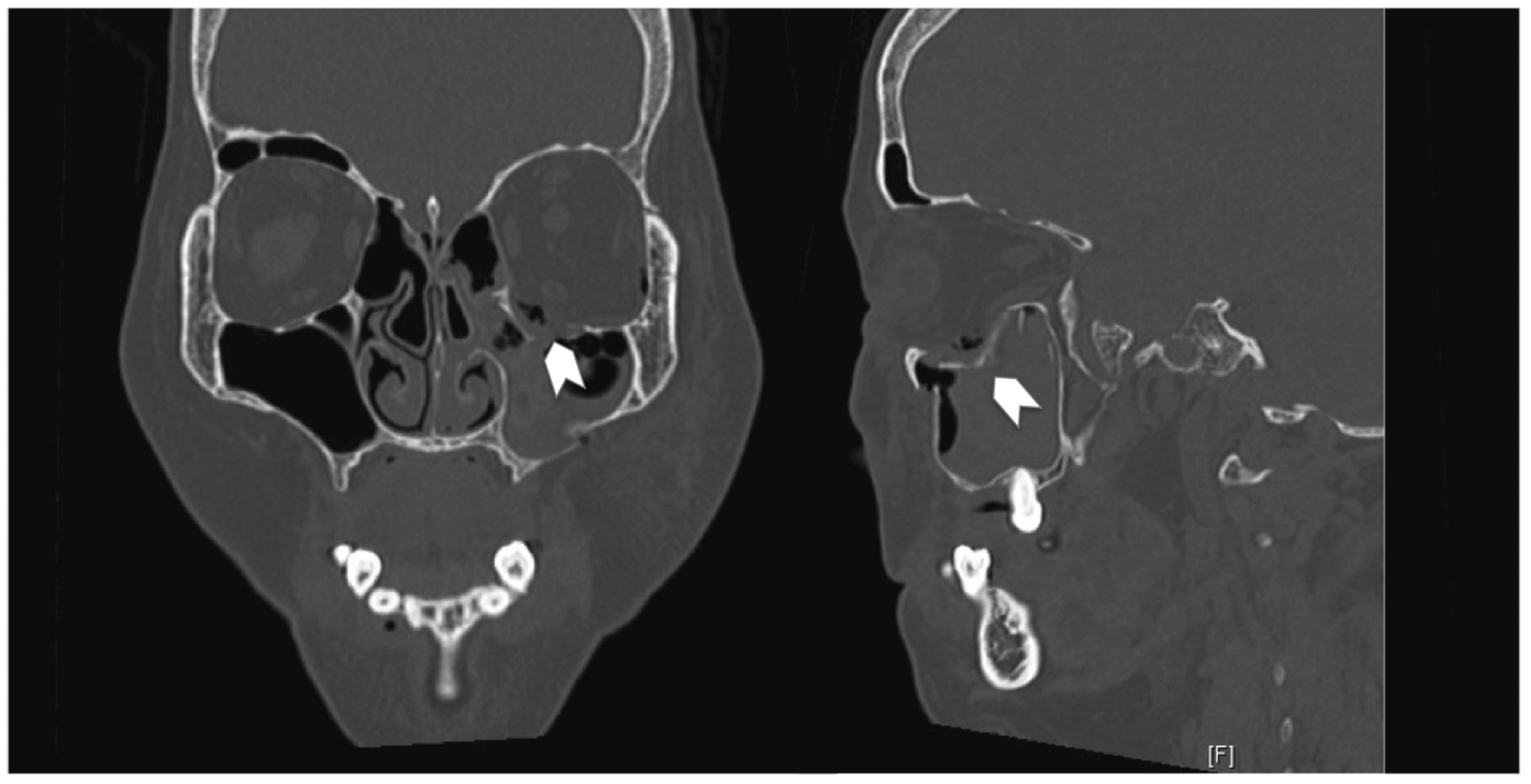
Titanium may be more useful in comminuted fractures where it can secure and/or bridge across multiple bone fragments. [6] The fibrogenic response of titanium that results in orbital adherence syndrome manifests itself in the form of fibrous band ingrowth through the pores in the mesh (Case 1). This then tethers down the periorbita, hindering globe movement in the upward direction. It may also be accompanied by cicatricial eyelid retraction (Case 2).
Le Guéhennec demonstrated that increasing surface irregularities and roughness of the titanium implant resulted in greater bone-to-implant contact, leading to faster osseointegration and stronger adhesion between implant and host. [9] As osseointegration is regarded as immune-modulated inflammatory process, the study of Lee and Nunery on orbital adherence syndrome attributed the extensive fibrotic reaction surrounding the mesh implants to the titanium implant itself. [10] They also theorized that the concomitant use of titanium orbital floor implants anchored to the infraorbital rim and titanium orbital rim plates resulted in greater surface irregularities and exposure of the orbit and eyelid to titanium, resulting in greater fibrosis.
In the 30 cases of orbital fractures performed in our institution over the past year (May 2014 to May 2015) with simultaneous use of titanium mesh and orbital rim implants, only 2 cases were complicated by orbital adherence syndrome (6.67%). Due to the early manifestations of symptoms (<1week) in Case 1, we postulate that another mechanism apart from fibrotic adhesions incited by the titanium implant was responsible for the tethering, as the cicatricial bands would not have been well formed within this time frame to result in significant orbital tethering. As orbital adherence syndrome does not occur in the vast majority of our patients who undergo orbital floor reconstruction with a titanium mesh implant, we postulate that there are certain predisposing risk factors, the significant disruption of periorbital fat after a comminuted orbital floor fracture and the nonmeticulous dissection resulting in periorbital fat disruption.
Pretrauma, the intact periorbita, namely, the periosteum, Lockwood ligament, and the periorbital fat, acts in tandem as an intervening layer between the globe, extraocular muscles, and the bony orbit. This allows for a smooth gliding interface. The periorbital fat is further held intact by the ligaments of Koornneef, which extend throughout the extraocular fat as thin bands dividing the fat into smaller compartments. These ligaments run from the bony orbit to the extraocular muscle fascia and Tenon capsule, maintaining the shape of the soft tissue. The disruption of the periorbita in an orbital floor fracture exposes these small fat components to the orbital mesh. These small fat strands then fall through the pores in the titanium mesh and become incarcerated (similar to an inguinal hernia) due to swelling. Owing to the attachments of the periorbita and fat strands to the muscle fascia via the Koornneef ligaments, this results in tethering of the muscle, manifesting clinically as gaze restriction in the early postoperative period. Subsequently, fat necrosis occurs which incites an inflammatory response; this is exacerbated by the titanium implant material, which results in fibrous ingrowth through the mesh. This leads to persistence or even worsening of symptoms such as diplopia as time passes.
The cases presented by Lee and Nunery [10] and Kersey et al [
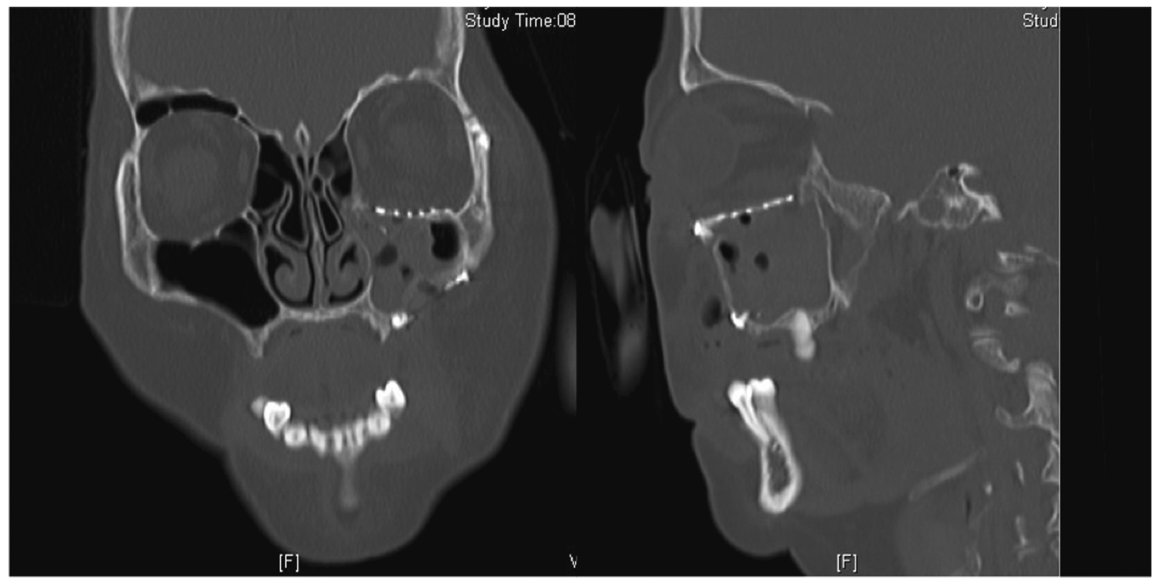
11] were referral cases from other surgeons, and as such, they may not have had the benefit of witnessing the early manifestations of this syndrome. In both case series, a majority of the cases managed operatively underwent removal of the implant, with isolated eyelid retraction repair for two of the eight cases in the study of Lee and Nunery. The mean time to reoperation was 5.8 and 7.1 months, respectively. In this patient (Case 1), the revision operation was performed early, where the orbital floor defect had yet to be sufficiently bridged with bony ingrowth; hence the titanium implant was left in situ. Instead, a smooth Suprafoil (S. Jackson Inc., Alexandria, VA) sheet was placed on top of the titanium implant, thereby creating a smooth gliding plane on the mesh, reducing traction of the mesh on the periorbita, and minimizing the contact of the orbital fat with the titanium implant, yet maintaining orbital support. Suprafoil alone lacks sufficient rigidity to adequately support the orbital contents in their anatomical position. This also prevents small pockets of periorbital fat from herniating through the mesh pores. As can be seen from the negative forced-duction test intraoperatively and the improvement of gaze restriction postoperation, this interposing layer was sufficient to prevent further adhesions.
Thus, the authors propose that in the primary operation, should there be severe disruption of the periorbita with the presence of multiple small fat lobules, we would consider a preemptive onlay alloplastic sheet on the titanium mesh. In cases with generally intact periorbita, the titanium mesh alone would suffice. Composite implants such as a titanium-PE composite implant (Medpor Titan, Stryker, Kalamazoo, MI) have been used to exploit the advantages of each material while minimizing its detrimental side effects [12]; however, this is not available to us locally.
Conversely, the patient in Case 2 was managed successfully with nonsurgical methods. In the study of Kersey et al., all operatively managed cases were noted to have an improvement in globe movement on forced-duction test. [11] There was no mention of the forced-duction test for the conservatively managed patients, or in the study of Lee and Nunery. [10] We hypothesize that patients with a negative forced-duction test (Case 2) are likely to have cicatricial scarring and fibrous ingrowth into the orbital mesh to a lesser degree as compared with patients with a positive forced-duction test. This could indicate greater success with conservative measures, which would involve massage, steroid, or HA injections. In this subset of patients, a higher threshold for operative management might be prudent while awaiting temporal improvement. The authors acknowledge that while scar massage therapy is widely used especially by physical therapists, [13] there are little randomized control trials to determine its efficacy, with even less evidence available on eyelid cicatricial scarring. [14,15] Although vigorous prolonged lid massage might cause a loose lower lid resulting in late ectropion or entropion, it has been shown to improve the outcome of eyelid scars and was initiated in Case 2 as part of the initial management for lid retraction from scarring. [16,17]
In case 2, the lower eyelid retraction and tugging sensation was likely due to a volume contraction in the posterior lamella rather than actual shortening. [18,19] The HA filler acts to restore the volume of the posterior lamella and resulted in relief of lower eyelid retraction and the tugging sensation. A temporary filler is adequate, as HA fillers last at least 9 months or more in the eyelids. Goldberg et al. found that there was still residual effect of the HA filler even at 6 months. [19] While this necessitates repeated treatments, this method is acceptable in patients who may be more inclined toward a less invasive approach.
On occasion, there can be persistent diplopia following orbital floor surgery. Apart from incomplete reduction of herniated orbital contents and entrapment of the periorbita by the implant, an important consideration is the existing trauma to the extraocular muscles, in particular the inferior rectus. This can occur due to the original trauma or surgery, resulting in fibrotic restriction of the muscle. [20] This is not mutually exclusive with orbital adherence syndrome, and may often only be diagnosed after all the other causes have been excluded.
Inherent limitations of this report include its retrospective nature and the small numbers, which would inevitably lead to a degree of bias. Unfortunately, due to the infrequent presentation of orbital adherence syndrome, all published studies on this topic are retrospective, with the exact pathogenesis poorly understood. The two largest case series available are by Lee and Nunery10 and Kersey et al.11; as they recruited cases that were operated on by other surgeons, we believe that they saw a delayed spectrum of this condition. While more cases would lend credence to this study, it may not be possible to add to these case numbers in the near future. Certainly surgical technique could play a causative role; however, blunt dissection was always performed in an attempt to reduce damage to the periorbita. There have been no further cases of orbital adherence syndrome, as the senior surgeon (H.S.) has modified his technique. Certainly further research is required to establish whether the degree of periorbital soft-tissue disruption with the use of titanium implants has a higher complication rate compared with other materials.
Despite the rare complication of orbital adherence syndrome, the authors believe that titanium implants remain a useful option especially in the reconstruction of large, comminuted orbital floor defects. In select cases where there is greater disruption of the periorbita and exposure of the orbital fat, it may be prudent to insert a smooth interposing alloplastic sheet concomitantly in the initial surgery as a preventive measure. In smaller floor defects requiring less rigid support, the authors have moved toward using porous polymer implants alone. Treatment for orbital adherence syndrome is surgical adhesiolysis, with removal of the implants, or placement of a smooth permanent interface sheet in the early recovery period. Patients with a negative forced-duction test would benefit from a trial of conservative management before considering surgical adhesiolysis.

References

  1. Gart, M.S.; Gosain, A.K. Evidence-based medicine: orbital floor fractures. Plast Reconstr Surg 2014, 134, 1345–1355. [Google Scholar] [CrossRef] [PubMed]
  2. Boffano, P.; Roccia, F.; Zavattero, E.; et al. European Maxillofacial Trauma (EURMAT) project: a multicentre and prospective study. J Craniomaxillofac Surg 2015, 43, 62–70. [Google Scholar] [CrossRef] [PubMed]
  3. Burnstine, M.A. Clinical recommendations for repair of orbital facial fractures. Curr Opin Ophthalmol 2003, 14, 236–240. [Google Scholar] [CrossRef] [PubMed]
  4. Gunarajah, D.R.; Samman, N. Biomaterials for repair of orbital floor blowout fractures: a systematic review. J Oral Maxillofac Surg 2013, 71, 550–570. [Google Scholar] [CrossRef] [PubMed]
  5. Ridgway, E.B.; Chen, C.; Colakoglu, S.; Gautam, S.; Lee, B.T. The incidence of lower eyelid malposition after facial fracture repair: a retrospective study and meta-analysis comparing subtarsal, subciliary, and transconjunctival incisions. Plast Reconstr Surg 2009, 124, 1578–1586. [Google Scholar] [CrossRef] [PubMed]
  6. Dubois, L.; Steenen, S.A.; Gooris, P.J.; Bos, R.R.; Becking, A.G. Controversies in orbital reconstruction-III. Biomaterials for orbital reconstruction: a review with clinical recommendations. Int J Oral Maxillofac Surg 2016, 45, 41–50. [Google Scholar] [CrossRef] [PubMed]
  7. Ellis, E., III; Messo, E. Use of nonresorbable alloplastic implants for internal orbital reconstruction. J Oral Maxillofac Surg 2004, 62, 873–881. [Google Scholar] [CrossRef] [PubMed]
  8. Gear, A.J.; Lokeh, A.; Aldridge, J.H.; Migliori, M.R.; Benjamin, C.I.; Schubert, W. Safety of titanium mesh for orbital reconstruction. Ann Plast Surg 2002, 48, 1–7, discussion 7–9. [Google Scholar] [CrossRef] [PubMed]
  9. Le Guéhennec, L.; Soueidan, A.; Layrolle, P.; Amouriq, Y. Surface treatments of titanium dental implants for rapid osseointegration. Dent Mater 2007, 23, 844–854. [Google Scholar] [CrossRef] [PubMed]
  10. Lee, H.B.; Nunery, W.R. Orbital adherence syndrome secondary to titanium implant material. Ophthal Plast Reconstr Surg 2009, 25, 33–36. [Google Scholar] [CrossRef] [PubMed]
  11. Kersey, T.L.; Ng, S.G.; Rosser, P.; Sloan, B.; Hart, R. Orbital adherence with titanium mesh floor implants: a review of 10 cases. Orbit 2013, 32, 8–11. [Google Scholar] [CrossRef] [PubMed]
  12. Tabrizi, R.; Ozkan, T.B.; Mohammadinejad, C.; Minaee, N. Orbital floor reconstruction. J Craniofac Surg 2010, 21, 1142–1146. [Google Scholar] [CrossRef] [PubMed]
  13. Roques, C. Massage applied to scars. Wound Repair Regen 2002, 10, 126–128. [Google Scholar] [CrossRef] [PubMed]
  14. Mustoe, T.A.; Cooter, R.D.; Gold, M.H.; InternationalAdvisory Panel on Scar Management; et al. International clinical recommendations on scar management. Plast Reconstr Surg 2002, 110, 560–571. [Google Scholar] [CrossRef] [PubMed]
  15. Shin, T.M.; Bordeaux, J.S. The role of massage in scar management: a literature review. Dermatol Surg 2012, 38, 414–423. [Google Scholar] [CrossRef] [PubMed]
  16. Bodian, M. Use of massage following lid surgery. Eye Ear Nose Throat Mon 1969, 48, 542–547. [Google Scholar] [PubMed]
  17. Rathore, D.S.; Chickadasarahilli, S.; Crossman, R.; Mehta, P.; Ahluwalia, H.S. Full thickness skin grafts in periocular reconstructions: longterm outcomes. Ophthal Plast Reconstr Surg 2014, 30, 517–520. [Google Scholar] [CrossRef] [PubMed]
  18. Chang, H.S.; Lee, D.; Taban, M.; Douglas, R.S.; Goldberg, R.A. “En-glove” lysis of lower eyelid retractors with AlloDerm and dermis-fat grafts in lower eyelid retraction surgery. Ophthal Plast Reconstr Surg 2011, 27, 137–141. [Google Scholar] [CrossRef] [PubMed]
  19. Goldberg, R.A.; Lee, S.; Jayasundera, T.; Tsirbas, A.; Douglas, R.S.; McCann, J.D. Treatment of lower eyelid retraction by expansion of the lower eyelid with hyaluronic acid gel. Ophthal Plast Reconstr Surg 2007, 23, 343–348. [Google Scholar] [CrossRef] [PubMed]
  20. Silbert, D.I.; Matta, N.S.; Singman, E.L. Diplopia secondary to orbital surgery. Am Orthopt J 2012, 62, 22–28. [Google Scholar] [CrossRef] [PubMed]
Figure 1. Coronal and sagittal cuts of the CT scan of the face (Case 1) demonstrating the presence of a large right orbital floor defect (arrows).
Cmtr 10 00013 g001
Figure 2. Precontoured titanium implants composed of commercially pure titanium with a variable pore size ranging from 1 × 3 mm to 3 × 5 mm.
Cmtr 10 00013 g002
Figure 3. Coronal and sagittal cuts of the postoperative CT scan of the face (Case 1) demonstrating reduction of all periorbita.
Cmtr 10 00013 g003
Figure 4. Intraoperative photo demonstrating fibrous ingrowth through the orbital mesh plate (arrows).
Cmtr 10 00013 g004
Figure 5. Coronal and sagittal cuts of the CT scan of the face (Case 2) demonstrating the presence of a large left orbital floor defect (arrows).
Cmtr 10 00013 g005
Figure 6. Coronal and sagittal cuts of the postoperative CT scan of the face (Case 2) demonstrating reduction of all periorbita. Although the posterior edge of the plate is slightly depressed, there is no tethering of the inferior rectus muscle.
Cmtr 10 00013 g006
Figure 7. At 9-week postoperation. Left—left lower lid dimpling on primary gaze; right—no tethering of inferior rectus and inferior oblique muscles.
Cmtr 10 00013 g007

