Next Article in Journal
Capillary Hemangioma of the Foot
Previous Article in Journal
Pedal Gangrene Secondary to Disseminated Intravascular Coagulation with Gastric Carcinoma
 
 
Journal of the American Podiatric Medical Association is published by MDPI from Volume 116 Issue 1 (2026). Previous articles were published by another publisher in Open Access under a CC-BY (or CC-BY-NC-ND) licence, and they are hosted by MDPI on mdpi.com as a courtesy and upon agreement with American Podiatric Medical Association.
Article

Congenital Absence of the Tibial Sesamoid

by
Fred Day
1,
P. Clint Jones
2 and
Christi L. Gilbert
2
1
American College of Foot and Ankle Surgeons
2
Doctors Hospital, New Boston, TX, USA
J. Am. Podiatr. Med. Assoc. 2002, 92(3), 153-154; https://doi.org/10.7547/87507315-92-3-153
Published: 1 March 2002

Abstract

Congenital absence of the tibial sesamoid of the hallux is an infrequent occurrence. The authors present a case of congenital absence of the tibial sesamoid and a review of the literature regarding the clinical significance of this anomaly.

The sesamoids provide a variety of functions. They protect the flexor hallucis brevis and longus tendons, increase the mechanical advantage of the flexor hallucis brevis tendon, absorb a portion of the weightbearing stresses on the medial forefoot, elevate the first metatarsal head off the ground, and diminish the friction between the tendons and the metatarsophalangeal joint [1] Root et al [2] stated that congenital absence or surgical excision of sesamoids significantly reduces the ability of the flexor tendons to maintain propulsive stability of the hallux. Jeng et al [3] proposed that congenital absence of the sesamoid may represent merely a delay or failure of ossification.
There is little information and few case studies of congenital absence of the tibial sesamoid. Sarrafian [4] described the tibial and fibular sesamoids of the hallux as being present 100% of the time. This has been confirmed by other studies as well. In 1936, Inge [5] and Kewenter [6] were the first to report cases of tibial sesamoid absence. Subsequently, Lapidus [7] in 1939 and Hubay [8] in 1949 reported cases. In 1976, Vranes [9] reported one case and Zinsmeister and Edelman [10] and Goez and DeLauro [11] found a cartilaginous precursor upon surgical exploration in one case. After reviewing the medical literature, the authors found that only nine cases have been reported [5,6,7,8,9], [12,13,14] Two patients have been reported with congenital absence of the fibular sesamoid. [3], [13], [15]
The propensity for these anomalies to be symptomatic, painful, or deforming is uncertain. In the Inge [5] study, two patients were both asymptomatic and had normal examinations. DuVries [16] reported an associated hammertoe of the hallux with painful callus under the first metatarsal head and hypothesized that the absence of the sesamoid weakened the flexor hallucis brevis tendon, resulting in clawing of the metatarsophalangeal joint. Mann and Coughlin [1] stated that most of these patients were asymptomatic and absence of the sesamoid could lead to clawing of the hallux and possibly hallux abducto valgus deformity. Zinsmeister and Edleman [10] found that the absence of the tibial sesamoid reduces the ability of the medial head of the short flexor to stabilize the hallux, which contributes to an imbalance at the first metatarsophalangeal joint. This allows the lateral head of the flexor hallucis brevis to gain a mechanical advantage, resulting in the formation of a hallux abductus or hallux abducto valgus deformity. In one of the cases presented in their study, Zinsmeister and Edelman [10] surgically explored the first metatarsophalangeal joint and a cartilaginous precursor of the tibial sesamoid was visualized.
Congenital absence of the sesamoid may represent merely a delay or failure of ossification. Inge and Ferguson [17] found that the hallucal sesamoids first appear as undifferentiated islands of connective tissue beneath the metatarsophalangeal joint in an 8-week-old fetus. This tissue is continuous with the periosteum of the metatarsal proximally and the phalanx distally. By 5 months, the sesamoids have already assumed their normal adult shape but will continue to grow in size. At 8 years of age, the sesamoids begin to ossify and are completed by age 11. Congenital anomalies may occur from an arrest in maturation of the bone.

Case Report

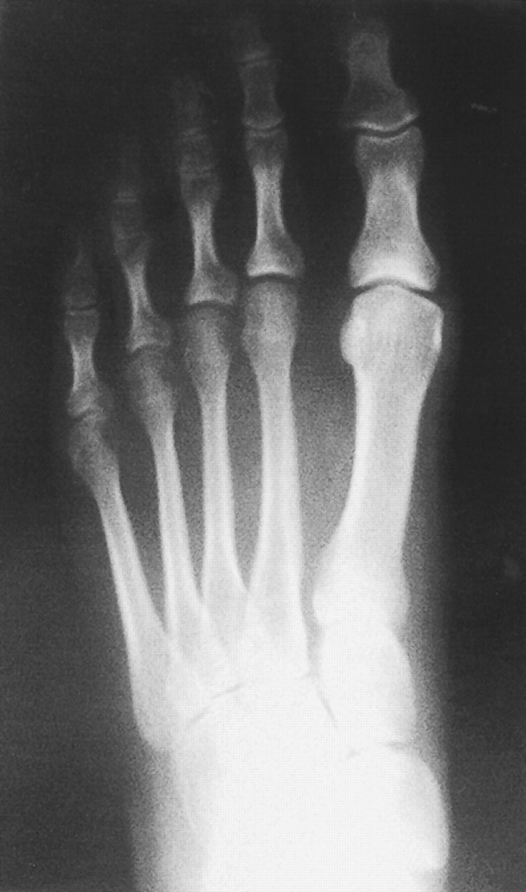
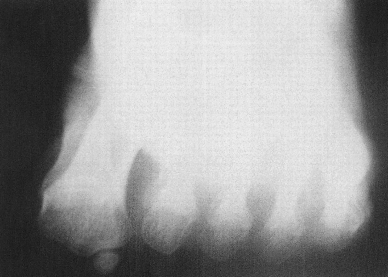
A 57-year-old woman presented to the office of the senior author (F.D.) for evaluation and treatment of a stress fracture of the second metatarsal of the right foot. Absence of the tibial sesamoid was an incidental radiographic finding (Figs. 1 and 2). A contralateral view confirmed the absence of the tibial sesamoid. Clinically and radiographically, the patient was asymptomatic at the first metatarsophalangeal joint and only the stress fracture warranted treatment.

Conclusion

This relatively rare and infrequently reported anomaly has been presented, along with a case report and a review of the literature. The delay or failure of ossification is discussed as a possible cause of a congenital absence of the tibial sesamoid.
  1. Mann RA, Coughlin MJ (eds): Surgery of the Foot and Ankle, 6th Ed, p 474, CV Mosby, St Louis, 1993..
  2. Root ML, Orien WP, Weed JH: Normal and Abnormal Function of the Foot, p 285, Clinical Biomechanics Corp, Los Angeles, 1977..
  3. Jeng CL, Maurer A, Mizel MS: Congenital absence of the hallux fibular sesamoid: a case report and review of the literature. .Foot Ankle Int19::329. ,1998. .
  4. Sarrafian SK: Anatomy of the Foot and Ankle: Descriptive, Topographic, Functional, p 227, JB Lippincott, Philadelphia, 1983..
  5. Inge GA: Congenital absence of the medial sesamoids bone of the great toe. .J Bone Joint Surg18::188. ,1936. .
  6. Kewenter Y: Die sesambeine des I: metatarsophalangealgelenks des menschen. .Acta Orthop Scand2 (suppl)::1. ,1936. .
  7. Lapidus PW: Congenital unilateral absence of the medial sesamoids of the great toe. .J Bone Joint Surg21::208. ,1939. .
  8. Hubay CA: Sesamoid bone of the hands and feet. .Am J Roentgenol61::493. ,1949. .
  9. Vranes R: Hallux sesamoids: a divided issue. .JAPA66::687. ,1976. .
  10. Zinsmeister BJ, Edelman R: Congenital absence of the tibial sesamoid: a report of two cases. .J Foot Surg24::266. ,1985. .
  11. Goez J, DeLauro T: Congenital absence of the tibial sesamoid. .JAPMA85::509. ,1995. .
  12. Berquist TH (ed): Radiology of the Foot and Ankle, p 21, Raven, New York, 1989..
  13. Jahss MH (ed): Disorders of the Foot and Ankle: Medical and Surgical Management, 2nd Ed, p 1064, WB Saunders, Philadelphia, 1991..
  14. Wright SM: Congenital hallux varus deformity with bilateral absence of the hallucal sesamoids. .JAPMA88::47. ,1998. .
  15. Jahss MH: The sesamoids of the hallux. .Clin Orthop157::88. ,1981. .
  16. DuVries HL: Surgery of the Foot, CV Mosby, St Louis, 1959..
  17. Inge G, Ferguson H: A surgery of the sesamoid bones of the great toe. .Arch Surg27::466. ,1933. .
Figure 1. Anteroposterior view; note healing of the stress fracture.
Japma 92 00153 g001
Figure 2. Sesamoidal view of the stress fracture.
Japma 92 00153 g002