Mailing address: Podiatry Associates of Belleville, 155 Belleville Ave, Belleville, NJ 07109.
Hemangiomas are benign, rarely malignant neoplasms composed of vascular channels that affect any living tissue. Capillary hemangiomas are a common vascular lesion of skin and mucous membranes in infancy and childhood. Hemangioma neoplasms are caused by vasoformative tissue proliferation. The lesion grows rapidly and can erupt through the skin, resulting in a stalk or pedicle formation. The majority of these lesions are located in the head and neck area [
1] In a review of 178 cases, Patrice et al[
2] found that lesions were most commonly located in the head and neck area (62.4%), followed by the trunk (19.7%), upper extremity (12.9%), and lower extremity (5.0%).
The etiology of capillary hemangioma is unknown. In their study of 256 cases, Jenkins and Delaney [
3] found that 47% were of congenital origin and trauma was the primary factor in 17% of cases. Hemangiomas can affect any living tissue with a vascular supply. According to various studies, capillary hemangiomas are among the most common orbital tumors in infants [
4], [
5] One-third of all orbital capillary hemangiomas are noted at birth, and virtually all are diagnosed by 6 months of age. In a study of 600 hemangiomas, Lampe and Latouretter[
6] found that 61% were present at birth and 86% appeared within the first month of life.
Capillary hemangiomas begin as an erythematous macula that evolves into a papule. The tumor may first be seen as a flat, circumscribed lesion with telangiectatic vessels in the superficial layers of the skin. The lesion grows rapidly and frequently becomes pedunculated. Skin discoloration may be present when the soft-tissue mass is in close proximity to the epidermis, varying from red to brown or blue to purple in color. Capillary hemangiomas may vary in consistency from soft and spongy to a hard mass and may be fixed or movable beneath the epidermis. Capillary hemangiomas often ulcerate and protrude through the epidermis. Pulsations are rare in hemangiomas, but if found they are usually from adjacent tumor-free vessels and are felt distal to the lesion [
7]
A history of spontaneous ulceration and bleeding is common in capillary hemangioma. The first visit to a physician is usually initiated by epidermal breakdown, crusting, and bleeding. The most common symptom is tenderness due to the hemangioma compressing a nearby nerve trunk or a nerve directly. Impairment of function depends on the location of the tumor.
The typical low-power microscopic appearance of a hemangioma is a well circumscribed, exophytic mass attached to a narrow stalk, consisting of aggregates of proliferating capillaries located within an edematous matrix. The vascular tumor is composed of closely aggregated, thin-walled vessels arranged in lobules [
8] The epidermal surface often shows focal areas of atrophy or ulceration.
Clinical diagnosis of capillary hemangioma is difficult to make prior to pathologic evaluation. The differential diagnosis of soft-tissue tumors may include a cyst, arteriovenous fistulas, eruptive senile angiomas, granuloma pyogenicum, angioblastomas, gemma angiomas, verrucous hemangiomas, tufted hemangiomas, and angiosarcoma.
The usual course of treatment is cauterization with silver nitrate, electrodessication, curettage, or tangential excision and cautery. Argon and pulsed dye lasers have been used for treatment of hemangiomas, but surgical excision with ligation of the surrounding vessels remains the treatment of choice [
9]
Case Report
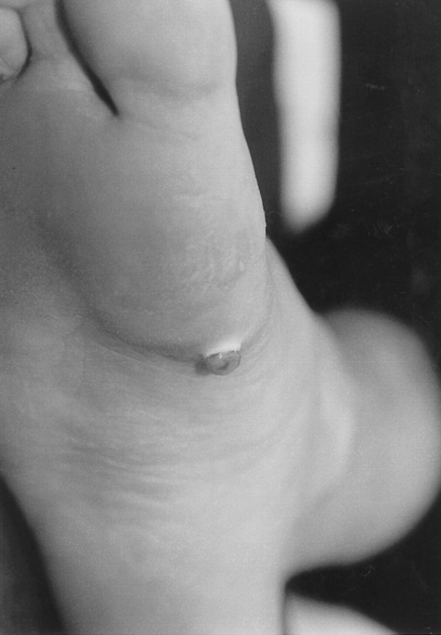
A 43-year-old male presented to the authors’ office with a chief complaint of a painful, growing mass on the bottom of his right foot. The patient reported that he felt a lump underneath the skin with an overlying blister formation. The overlying skin was discolored from red to purple. The patient stated that he had popped the blister and over the course of a week the mass had protruded through the skin (
Fig. 1). The patient said that the mass bled actively, especially upon weightbearing. The patient had pain upon ambulation and denied any history of trauma to the foot.
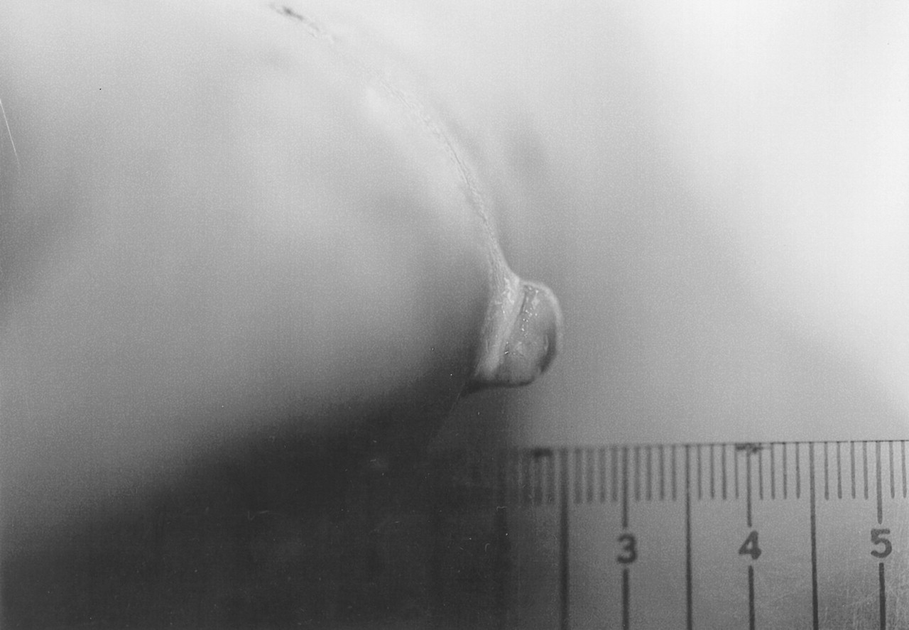
An examination of the plantar aspect of the right foot found a protruding mass on the plantar arch, just proximal to the first metatarsal head. The cylindrical mass protruded 10 mm through the epidermis and was approximately 10 mm in diameter (
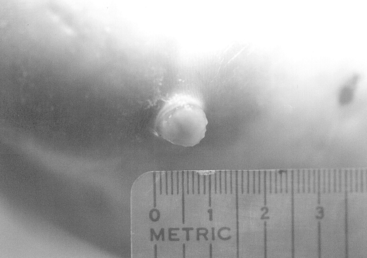
Fig. 2). The mass was erythematous, suggestive of a neoplasm of vascular origin. The surrounding skin at the base of the lesion was erythematous and had a purplish discoloration (
Fig. 3). No drainage, purulence, active bleeding, or local signs of infection were noted. The mass was of a soft, spongy consistency upon palpation of the lesion. There was no tenderness upon palpation of the lesion or surrounding area. No pulsations were noted and active bleeding was elicited upon compression of the lesion.
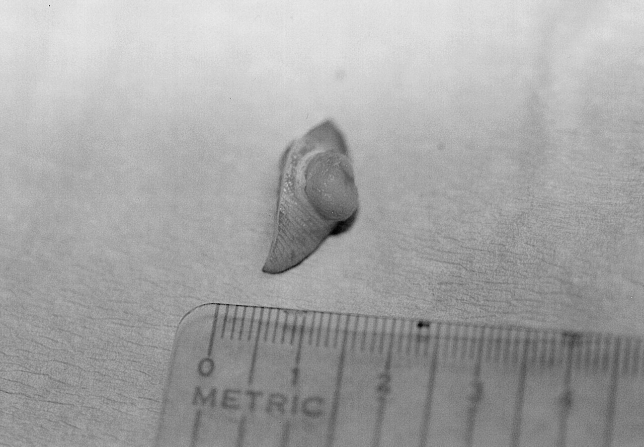
Proposed treatment was surgical resection of the mass, including an excisional biopsy of the lesion. The patient was placed in a supine position and the right foot was injected just proximal to the mass with local anesthesia consisting of 1% lidocaine. Two converging 3-cm semielliptical skin incisions were made over the mass and the involved section of skin was excised. The mass was freed from the surrounding and underlying tissue by sharp, blunt dissection. All vessels were severed and ligated as necessary. The mass was excised along with a border of healthy skin surrounding the neoplasm (
Fig. 4). The deeper structures were closed with 4.0 Vicryl and the skin was closed with 4.0 nylon. A dry, sterile, compressive dressing was placed on the right foot. The patient was instructed to walk with crutches and stay strictly nonweightbearing for 2 weeks. The sutures were removed 2 weeks after surgery. Mild skin sloughing was encountered for the following week, but the skin incision healed uneventfully at 4 weeks postoperative. All specimens were sent to the pathology laboratory for gross and microscopic examination.
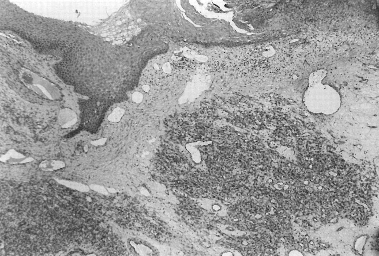
The pathology report revealed a wedge of epithelium exhibiting multiple dilated capillaries situated in the papillary dermis. Multiple small-dilated capillaries were seen with the middermis (
Fig. 5). The capillaries appeared to be completely filled with blood. No signs of inflammation, infection, or malignancy were present. The pathologist confirmed the diagnosis of capillary hemangioma.
Conclusion
Hemangiomas are benign vascular tumors. They are congenital and rarely hereditary, though trauma may be a predisposing factor. Common symptoms include pain, swelling, skin discoloration, and an associated mass. Definitive diagnosis is made through gross and microscopic examination of the mass and surgical excision is the treatment of choice.