Disseminated intravascular coagulation is a consumptive coagulopathy that involves both abnormal coagulation and hemorrhage resulting from a clotting cascade initiated by injury to tissues or vascular endothelium.[
1] Although seen in a wide array of disorders, disseminated intravascular coagulation occurs most commonly in infectious disease processes, such as septicemia, and in 30% to 50% of patients with gram negative sepsis.[
2] Tissue injury occurring in malignancy may also cause disseminated intravascular coagulation. While observed in essentially all hematologic malignancies, disseminated intravascular coagulation is most commonly encountered in acute promyelocytic leukemia,[
3] and in 10% to 15% of solid tumors.[
2] The thrombotic episodes resulting from the disrupted clotting cascade may lead to organ failure, peripheral tissue damage, and necrosis. Disseminated intravascular coagulation resulting in gangrene of the lower extremities has been observed in a newborn infant in which the etiology was unknown.[
4] It has also been observed in a splenectomized Hodgkin’s disease patient who presented with bacteremia,[
5] in patients with measles,[
6,
7] and in patients after various systemic infections.[
8–
10] Recently, two cases of lower-extremity gangrene were described, one involving a human immunodeficiency virus-positive patient presenting with
S Aureus endocarditis,[
11] and another presenting with disseminated intravascular coagulation of unknown origin.[
12] However, few cases of peripheral gangrene in malignancy are reported in the medical literature, and even fewer involve the lower extremity. One case of pedal gangrene in malignancy has been reported,[
13] but it did not directly identify disseminated intravascular coagulation as a consequence of the malignancy. In a prospective evaluation of cases of digital ischemia in malignancy,[
14] 23 previously published cases of digital gangrene associated with malignancy were identified; however, the typical presentation was gangrene of the finger. In another review of 71 cases of symmetrical peripheral gangrene,[
15] the most common underlying condition (82%) was infection. Of the remaining cases, only one involved malignancy, specifically Hodgkin’s disease. The case presented in this article identifies gastric carcinoma as the mechanism initiating a clotting cascade and resulting in pedal gangrene.
Case Report
A 57-year-old male presented to the emergency room at the Edward Hines, Jr Hospital in Hines, Illinois, complaining of pain in his right foot for 3 days that had begun with redness and pain upon ambulation. The foot then turned purple and the intensity of the pain increased. Upon presentation, the patient could only stand upright with excruciating pain, and he felt pain even at rest. The patient denied any episodes of tingling or pins and needles in the extremity, nausea, or vomiting. He had chills the morning of presentation. The patient reported a history of epigastric pain that radiated to the back and ribs bilaterally and a history of esophageal reflux for 1 year prior to the onset of symptoms. A diagnosis of peptic ulcer disease was made and the patient had been treated with medication that failed to alleviate his symptoms. The patient history was also significant for increased fatigue, decreased appetite, an unintentional weight loss of 25 pounds, and darkening of the urine for 3 months.
Past medical and surgical histories were unremarkable and the patient denied medications or allergies. The patient reported that he had smoked two packs of cigarettes daily for 30 years. Alcohol consumption of two six packs of beer or hard liquor daily had stopped 6 years earlier. The patient denied illicit drug use.
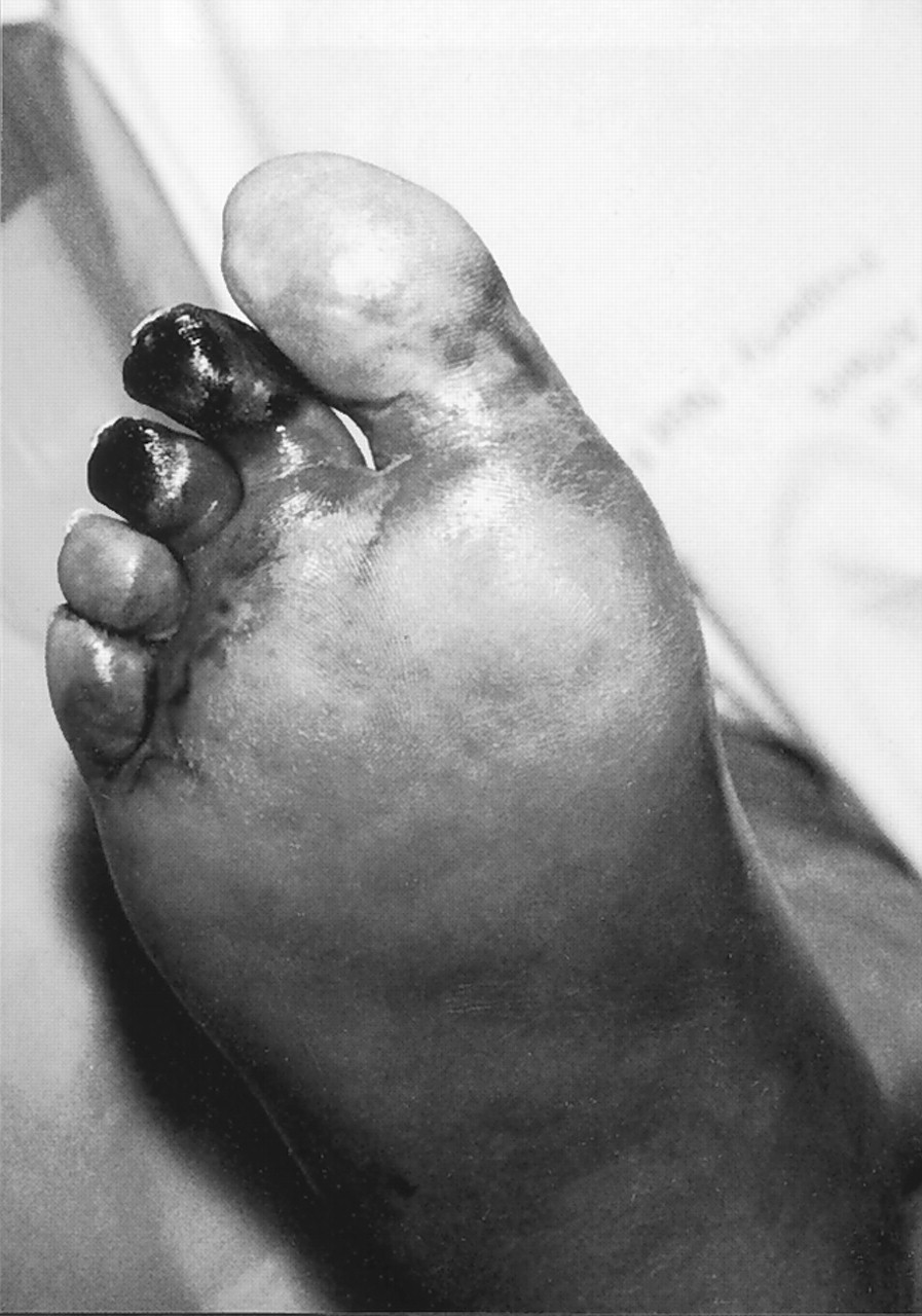
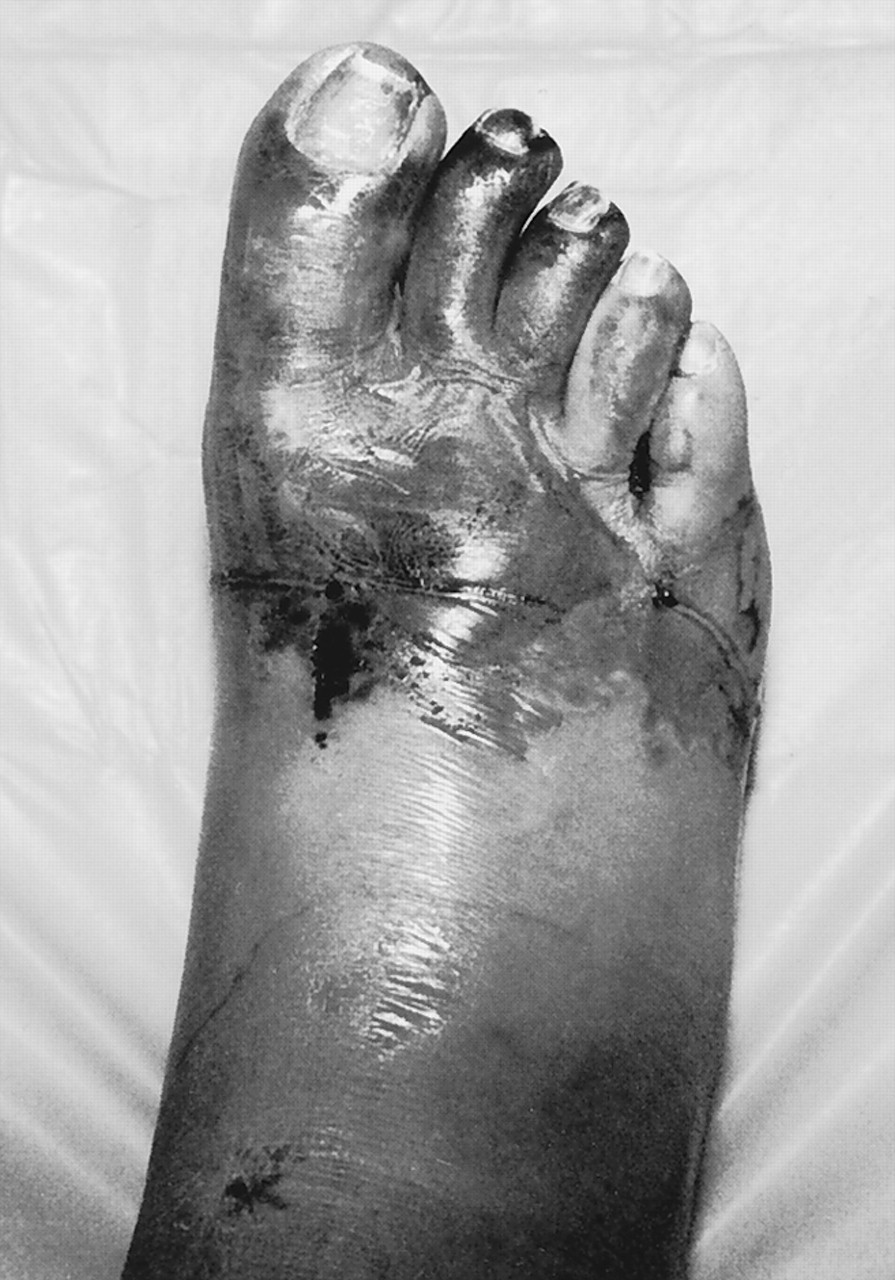
Upon physical examination, the patient’s oral temperature was 100.3°; pulse, 62; respiration, 20/min; blood pressure, 169/86 mm Hg. The general appearance of the patient was that of a cachectic, apprehensive male. The skin appeared slightly jaundiced, and the sclera was icteric. Examination of the abdomen showed marked epigastric tenderness along with a palpable mass in the epigastric area that was hard, tender, and not mobile. Voluntary guarding was noted. The liver was enlarged with unevenness to palpation. Lower-extremity examination revealed a right foot with tight, shiny skin and bluish discoloration up to the level of the midfoot; necrosis of the distal aspect of the second and third digits (
Fig. 1); and ecchymosis medial aspect of the left hallux. Erythema which did not reduce upon elevation was observed bilaterally, and the erythematous areas were cool to touch. A 4-cm hemorrhagic bulla was present on the dorsal aspect of the right foot extending from the second to fourth metatarsophalangeal joints (
Fig. 2). No open lesions or signs of infection were noted. Vascular examination revealed palpable pedal pulses and 2/4 pitting edema bilaterally. Capillary fill time was absent to the right foot. Radiographs of the right foot failed to demonstrate evidence of fracture or osteomyelitis.
Figure 1.
Plantar view of the right foot. Note the taut skin and discoloration with necrosis of digits two and three.
Figure 1.
Plantar view of the right foot. Note the taut skin and discoloration with necrosis of digits two and three.
Figure 2.
Note the large hemmorrhagic bulla on the dorsal aspect of the right foot.
Figure 2.
Note the large hemmorrhagic bulla on the dorsal aspect of the right foot.
Blood tests performed upon admission revealed fibrin split product level of 5-20 μg/mL, which peaked at 80-160 μg/mL the next day; partial thromboplastin time, 28.3; promthombin time, 14.3; and international normalized ratio, 1.36. The white blood cell count was elevated at 16,000/mm3; platelet count, 83,000; fibrinogen value, 117 mg/dL. Liver enzymes were also abnormal: alkaline phosphatase, 850 μ/L; alanine aminotranferase, 141 μ/L; asparate aminotransferase, 143 μ/L.
A working diagnosis of blue toe syndrome secondary to disseminated intravascular coagulation and obstructive jaundice with epigastric mass was made. The patient was started on heparin therapy for the embolic complications. Pain management was initiated to mollify the gangrene symptoms.
A computed tomography examination of the abdomen revealed thickening of the mucosa in the antrum of the stomach, which was consistent with a primary neoplastic process, and multiple hypodense areas in the liver that was suggestive of metastasis. Subsequent biopsies of the stomach and liver confirmed the diagnosis of gastric carcinoma and poorly differentiated large cell carcinoma of the liver. In addition, Doppler scans of the lower extremity revealed bilateral deep vein thrombosis.
As the patient was afebrile and there was no clinical infection, local wound care was initiated. The patient’s condition never stabilized and he died approximately 6 weeks after admission.
Discussion
The pathophysiologic mechanisms of disseminated intravascular coagulation are complex. The two major mechanisms are tissue injury, which is believed to be the major inciting factor in malignancy, and endothelial cell alteration, which plays a significant role in the initiation of disseminated intravascular coagulation in infection and trauma. Tissue injury that occurs in malignancy causes tissue factor, which is expressed on the cell surface, to circulate through the vascular tree.[
3] Tissue factor then complexes with factor VII and generates the prothrombin complexes necessary for generation of thrombin.[
16] As these factors circulate, the coagulation cascade is initiated in organ systems and the extremities, leading to the sequelae often observed, such as symmetrical peripheral gangrene and acute renal failure. Other inflammatory mediators such as interleukin-6 and tumor necrosis factor lead to the deregulation of the coagulation and the fibrinolytic pathways, and defects in inhibitors of coagulation also occur.[
2] The combination of defects in coagulation and fibrinolysis leads to the syndrome of disseminated intravascular coagulation, which includes simultaneous pathologic hemorrhage and coagulation.
The diagnosis of gangrene due to disseminated intravascular coagulation can be made from physical signs and confirmed by laboratory tests. The first sign is often erythema of the extremity, which progresses to gangrene within 12 to 24 hours of presentation, and hemorrhagic bullae often develop. Areas of purpura may also develop in the region but may not progress to frank gangrene.[
15] The presence of an underlying disease associated with disseminated intravascular coagulation is the most important initial indicator. However, the only reliable diagnostic test is the presence of fibrin split products.[
17] In addition, a rapid decline in platelet count or a platelet count of less than 100,000 mm
3, prolonged promthombin and partial thromboplastin time, an elevated international normalized ratio, and low plasma levels of coagulation inhibitors are all specific signs of disseminated intravascular coagulation.[
2] Measurement of fibrinogen levels has been indicated; however, these levels often remain within normal limits since fibrinogen is manufactured very rapidly by the liver under stressful situations.[
16]
Treatment of disseminated intravascular coagulation and symmetrical peripheral gangrene is first directed at the underlying disorder. The paradoxical cascade of thrombus generation and hemorrhage must be halted initially. This is accomplished through the use of anticoagulants such as heparin, low molecular weight heparin, and antithrombin III inhibitors.[
2] Novel treatments such as the use of plasminogen activator have also been utilized and preliminary results appear promising.[
17]
It is important to note that the initial chief complaint of the patient in this case report was a black and blue foot. The epigastric pain and weight loss apparently were not significant to the patient until they were noted to be part of a larger syndrome. Although the patient had sought treatment for his gastrointestinal complaints, the symptoms were believed to be a component of peptic ulcer disease or gastroesophageal reflux disease. At the time the patient presented to the emergency room, the malignancy had metastasized to the liver and obstructive jaundice was apparent.
Patients who present with gangrene of the extremities must be carefully considered for the presence of underlying disorders. The diagnosis of disseminated intravascular coagulation resulting in symmetrical peripheral gangrene was made quickly after admission, and if the gastric carcinoma had been in a less advanced stage, treatment would have been administered to the affected extremities. For the right foot, this probably would have involved amputation to restore function. In addition, the patient was diagnosed with bilateral deep vein thrombus. In a hypercoagulable state, the physician must be suspicious of other thrombotic events that may present an acute threat to the patient’s life.
It is also important to differentiate the case of symmetrical peripheral gangrene from cellulitis. Although the patient presented with an elevated white blood cell count and was febrile, and the feet were edematous and erythematous, no other clinical signs of infection were present. The extremities were cool to the touch and no open lesions or purulence were noted. The clinical appearance and the laboratory examinations were clear diagnostic indicators of underlying disease.
Conclusion
Although rare, disseminated intravascular coagulation may present as lower-extremity gangrene. The underlying disorder must be recognized and treated quickly, and the phenomenon of disseminated intravascular coagulation should be quickly halted. If treatment of the underlying disorder is successful, the extremities may be salvaged in a manner that will restore function.