Several topical prescription and nonprescription products have become widely used for the treatment of xerosis. Xerosis is defined as dehydrated skin showing redness, dry scaling, and fine crackling that may resemble crackled porcelain.[
1] The hallmarks of xerosis are scaliness and loss of elasticity, which are frequently seen on the extremities, especially the legs and feet.[
1,
2] In 1986, more than 1.5 million Americans visited podiatric physicians seeking relief of painful, excessive, hyperkeratotic pedal skin.[
3]
Xerotic skin is a reaction pattern to a variety of disorders, such as winter xerosis and hereditary ichthyosis, which have abnormalities of desquamation in common. In xerosis, the desquamation process is impaired, and desmosomes accumulate in the outer layers of the stratum corneum, leading to an increase in the surface corneocytes. The corneocytes and their intercellular cohesive structures are prerequisites for the function of the stratum corneum as a physical-chemical barrier. In winter xerosis and ultraviolet-damaged skin, there is a significant increase in desmoglein in the superficial layers, which is not found in normal stratum corneum. Desmoglein is believed to be a major component of the intercorneocyte cohesion in the stratum corneum. A decrease in stratum corneum lipids may explain the high incidence of dry, winter skin in older people.[
4–
6]
Patients with xeroxis present with a chief complaint of skin tightness, and a rough, scaly, and sensitive skin surface. The treatment of xerosis involves replacing water content, maintaining hydration, alleviating the symptomatology, and controlling keratinization to reduce scaling. The relief and prevention of dry skin focuses on maintaining the proper hydration of the epidermis, particularly the stratum corneum. In the past, therapy has been limited to topical applications of hydrating emollients designed to soften the stratum corneum and alleviate dry scaliness. Emollients are used to maintain enough water in the stratum corneum to reduce the possibility of cracking and flaking. Emollients, such as lanolin and glycerin, are hydrophilic, acting primarily on the skin’s surface to form an occlusive barrier that decreases evaporation.
In vitro studies have shown that glycerol facilitates the digestion of desmosomes, thereby possibly alleviating skin xerosis
in vivo.[
1,
7–
9]
Humectants, which are skin protectants that contain hygroscopic substances, increase skin moisture and reduce water loss.[
10] In addition to having a humectant effect on xerosis, lactic acid may also reduce the thickness of hyperkeratotic stratum corneum.[
8] Multiple studies have demonstrated the clinical effectiveness of lactic acid and ammonium lactate in the treatment of xerosis.[
11–
15] Lactinol (Pedinol Pharmaceuticals, Farmingdale, New York), which is 10% lactic acid, is used for the treatment of xerosis and is designed to produce humectant effects that reduce the xerotic disease state. According to the
Physicians’ Desk Reference,[
16] Lac-Hydrin 12% lotion (Bristol-Myers Squibb, Princeton, New Jersey) is a formulation of 12% lactic acid neutralized with ammonium hydroxide to produce a humectant effect. The
Physicians’ Desk Reference cautions fair-skinned individuals and individuals with sensitive skin on using the product since irritation and mild transient stinging may occur upon application to abraded or inflamed areas.[
16]
Materials and Methods
This double-blind, randomized comparison clinical trial investigated the effect of Lactinol versus Lac-Hydrin 12% lotion in mild to moderate foot xerosis. The study was conducted at the Foot Clinics of New York, the clinical arm of the New York College of Podiatric Medicine.
Criteria for enrollment included mild to moderate bilateral foot xerosis. The patient had to be able to apply (or have applied) Lactinol on one foot and Lac-Hydrin 12% lotion on the other foot. Pregnant women, nursing mothers, or patients with known hypersensitivity to Lactinol, Lac-Hydrin 12%, lactic acid, or ammonium lactate were excluded. Patients with peripheral vascular disease or known dermatologic diseases, such as psoriasis, lichen planus or eczema, or who were currently on immunosuppressants, were also excluded.
After meeting the inclusion criteria and signing an Institutional Review Board-approved informed consent form, patients were randomly assigned by means of a computer-generated random code. If necessary, a potassium hydroxide test was taken to rule out possible fungal infection. The patient was discontinued from participation in the study if the culture was positive. Since the inclusion criteria specifically stated bilateral mild to moderate xerosis, each patient served as his or her own control. One test medication was applied to each foot. The lotion bottles were exactly the same and clearly labeled with directions to apply the right foot medication with the left hand and the left foot medication with the right hand. This procedure reduced patient variability in the assessment of therapeutic change.
Clinical assessment of xerosis was performed at the baseline visit, and the designated sites were evaluated 2 and 4 weeks after treatment began. The assessments were based on the xerosis severity scale as presented by Rogers et al[
12] (
Table 1).
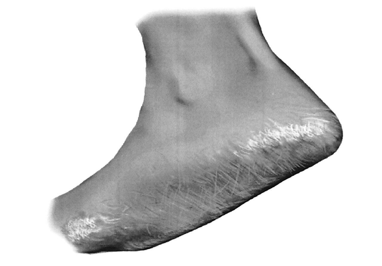
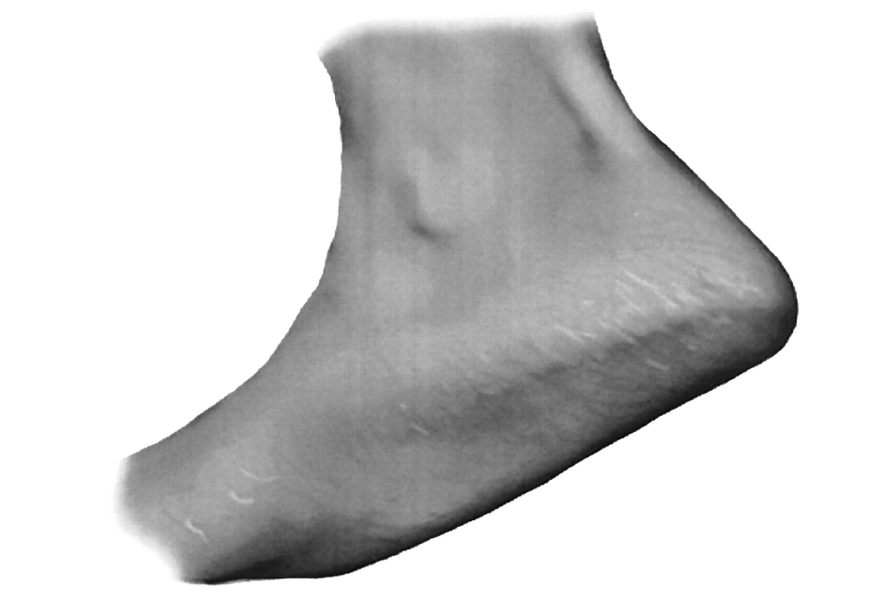
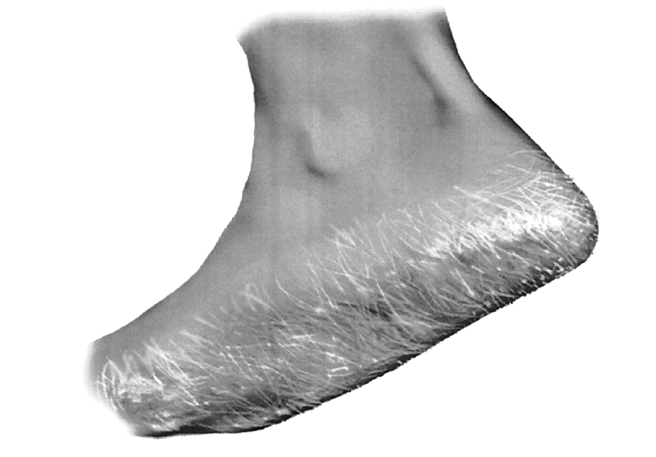
Figure 1 shows xerosis severity score 1, with occasional minute skin flakes. Xerosis severity score 2 shows many minute skin flakes (
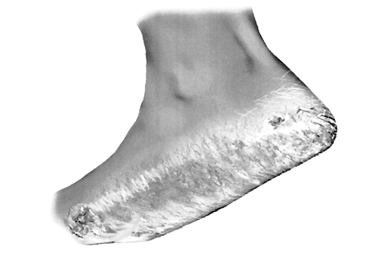
Fig. 2).
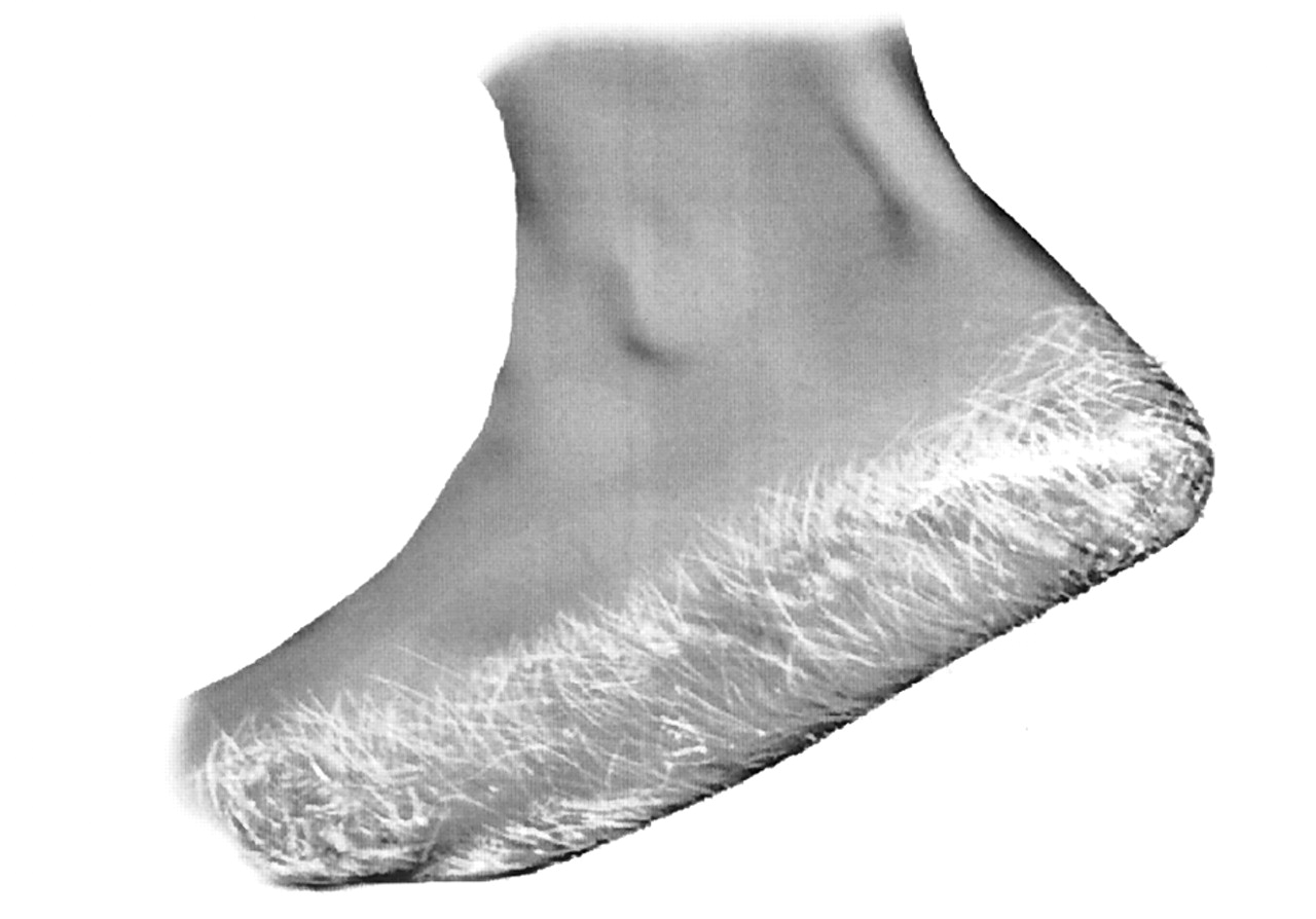
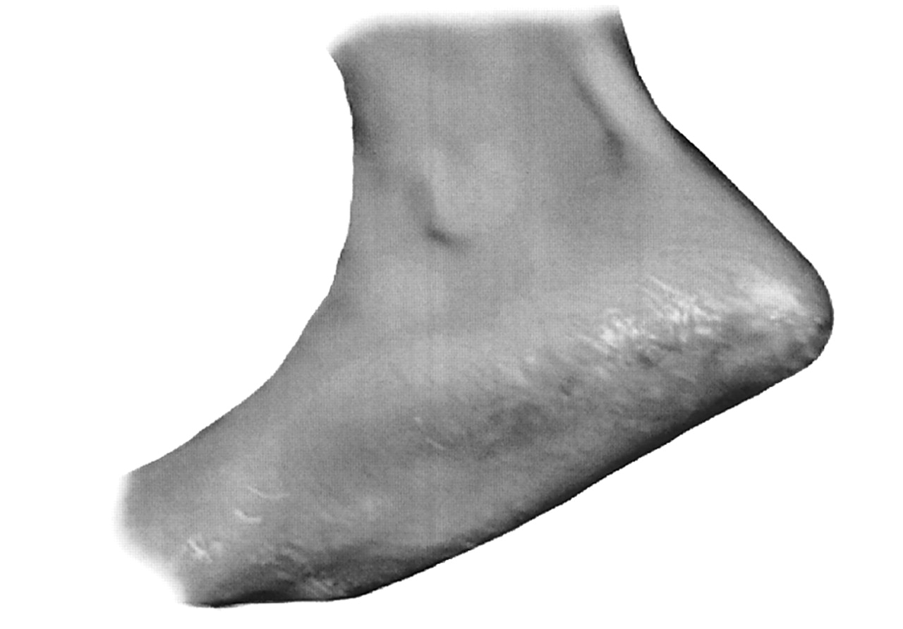
Figures 3 and
4 show more defined scaling, with severity score 4 having raised borders. The most severe xerosis is shown in
Figures 5 and
6, with large-scale plates and deep fissures for xerosis severity score 6.
Tenderness was assessed by the patient and the investigator at each visit. The following 4-point scale, based on the level of discomfort experienced by the patient with palpation of the feet, was used: 0 = none; 1 = mild (discomfort upon deep palpation); 2 = moderate (discomfort upon moderate palpation); and 3 = severe (discomfort upon slight palpation). At each visit, the patient was asked to assess any pruritus as none, mild (slight), moderate (somewhat), or severe (very).
Table 1.
Xerosis Severity Scale
Table 1.
Xerosis Severity Scale
Figure 1.
Xerosis severity score 1: Dusty appearance, occasional minute skin flakes.
Figure 1.
Xerosis severity score 1: Dusty appearance, occasional minute skin flakes.
Figure 2.
Xerosis severity score 2: Generalized dusty appearance, many minute skin flakes.
Figure 2.
Xerosis severity score 2: Generalized dusty appearance, many minute skin flakes.
Figure 3.
Xerosis severity score 3: Defined scaling with flat borders.
Figure 3.
Xerosis severity score 3: Defined scaling with flat borders.
Figure 4.
Xerosis severity score 4: Well defined, heavy scaling with raised borders, shallow fissures.
Figure 4.
Xerosis severity score 4: Well defined, heavy scaling with raised borders, shallow fissures.
Figure 5.
Xerosis severity score 5: Large scale plates, fissures.
Figure 5.
Xerosis severity score 5: Large scale plates, fissures.
Figure 6.
Xerosis severity score 6: Large scale plates, deep erythematous fissures.
Figure 6.
Xerosis severity score 6: Large scale plates, deep erythematous fissures.
After the fourth week of treatment, the patient and clinician independently rated the overall result on the following 6-point scale: 5 = worse; 4 = no improvement; 3 = slight improvement; 2 = moderate improvement; 1 = good improvement; and 0 = clear.
To ensure patient compliance, both tubes of medication were weighed at all three visits. Adverse events occurring during the trial were documented at each evaluation visit.
Demographic data, including age, ethnicity, and gender were tabulated, and statistical analyses were conducted for all tabulated efficacy variables. Mean xerosis severity and overall treatment evaluation scores were analyzed by means of the Wilcoxon signed-ranks test. Mean differences in medication use were analyzed by means of the paired t-test.
Results
Patient demographic data are shown in
Table 2. Of the 53 patients enrolled, 66% were female and 34% male. These data coincide with the overall gender distribution of patients at the Foot Clinics of New York, which is 62% female and 38% male. The age range of the patients was from 26 to 83 years, with an average of 50 years.
Table 2.
Characteristics of Study Population at Baseline, 2 Weeks, and 4 Weeks
Table 2.
Characteristics of Study Population at Baseline, 2 Weeks, and 4 Weeks
After the baseline visit, nine patients were lost to follow-up and were excluded from the analysis. Final analysis excluded data from five patients because of noncompliance after 4 weeks of therapy. No patients were discharged for positive fungus culture. Four patients were discharged from the study at 2 weeks due to an adverse event.
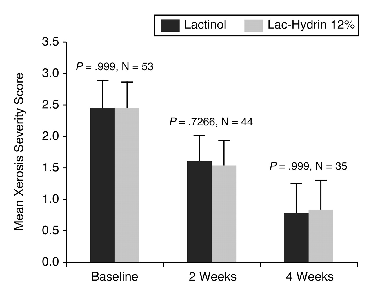
At baseline, there was no statistically significant difference between mean xerosis severity scores (
P = .999) (
Fig. 7). Although there was significant improvement in xerosis scores after 2 and 4 weeks of treatment, no statistically significant difference was observed between treatment groups at either 2 weeks (
P = .7266) or 4 weeks (
P = .999) (
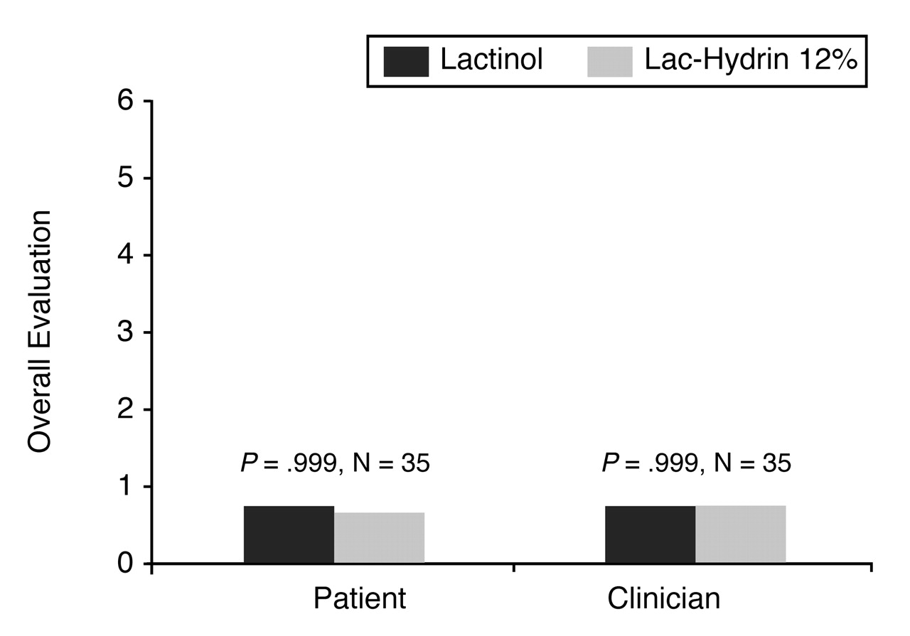
Fig. 7). The mean overall treatment evaluation scores of the patients and investigators after 4 weeks of treatment with Lactinol
versus Lac-Hydrin 12% lotion is shown in
Figure 8. There was no statistically significant difference in treatment evaluation scores for patients or investigators for Lactinol
versus Lac-Hydrin 12% treatment.
Table 3 lists the number of patients with a complete cure (return to normal skin) and with no change in condition throughout the treatment regimen. Of the 35 patients who were compliant for the study, two patients were completely healed (both feet) by 2 weeks of therapy. By 4 weeks of therapy, 11 patients were completely cured. One patient was completely cured with the Lactinol foot by week 2; this patient’s Lac-Hydrin foot had a xeroxis score of 1 by week 4. By the fourth week of therapy, four patients experienced a cure in the Lactinol foot whereas the Lac-Hydrin foot still had a xerosis score of 1. In two patients, the Lac-Hydrin foot was completely cured by 4 weeks of therapy, whereas the Lactinol xerosis score was 1. Four of the patients who completed the study showed no improvement but no worsening of their condition. Eight patients who completed 2 weeks of therapy showed no improvement without worsening of their condition.
Table 4 lists the data for the Lactinol and Lac-Hydrin feet by change in xerosis score, with the decrease measured by 1, 2, or 3 scores. There was no statistically significant difference in change of xerosis scores from baseline to 2 weeks of therapy, or from 2 weeks to 4 weeks of therapy, in either of the treatment groups.
Figure 7.
Mean xerosis severity scores at baseline and after 2 and 4 weeks of treatment.
Figure 7.
Mean xerosis severity scores at baseline and after 2 and 4 weeks of treatment.
Figure 8.
Comparison of overall evaluation of treatment by patients and clinician after 4 weeks of treatment.
Figure 8.
Comparison of overall evaluation of treatment by patients and clinician after 4 weeks of treatment.
Table 3.
Complete Cure versus No Change in Condition
Table 3.
Complete Cure versus No Change in Condition
Table 4.
Data for Lactinol and Lac-Hydrin Feet by Decrease in Xerosis Scores
Table 4.
Data for Lactinol and Lac-Hydrin Feet by Decrease in Xerosis Scores
Table 5 lists the number of adverse events. There were 14 reported cases of adverse events. Four of these patients were discontinued from the treatment plan. Two had mild to moderate itching as a result of the Lactinol and discontinued treatment. One had severe burning with Lac-Hydrin 12% lotion and moderate burning with Lactinol. One patient had a severe allergic reaction to both medications, which required immediate discontinuation of research medication and treatment with 50 mg oral benadryl at bedtime and application of 0.25% topicort cream twice daily. The patient improved within 1 week, and the condition was completely resolved by the third week after treatment.
Table 5.
Number of Adverse Events
Table 5.
Number of Adverse Events
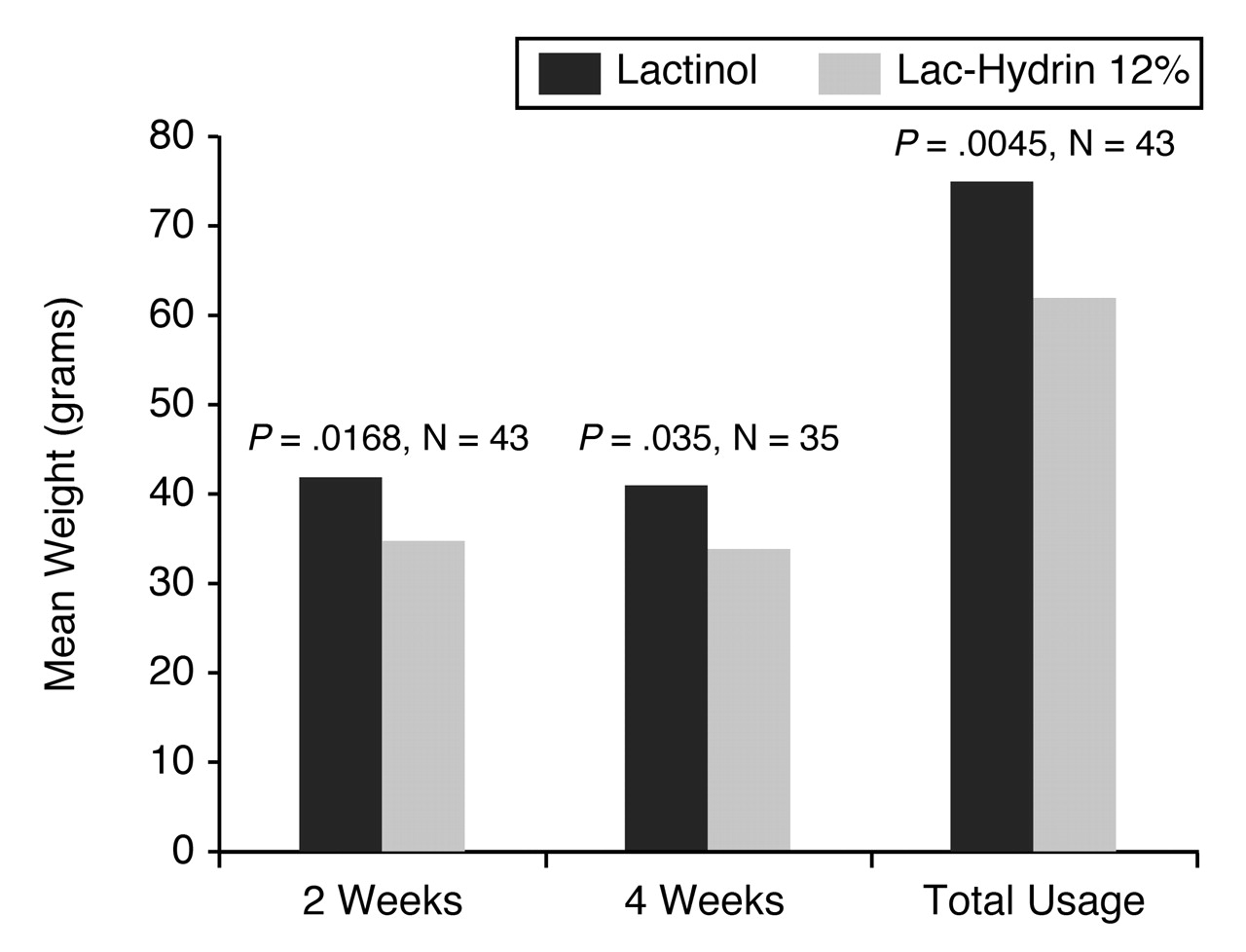
Figure 9 shows the mean patient medication use by week for the 4-week treatment plan. A statistically significant difference between the use of Lactinol
versus Lac-Hydrin 12% was observed throughout the study. On average, subjects used approximately 20% more Lactinol than Lac-Hydrin 12% lotion.
Figure 9.
Mean weight of Lactinol and Lac-Hydrin 12% lotion used after 2 and 4 weeks of treatment.
Figure 9.
Mean weight of Lactinol and Lac-Hydrin 12% lotion used after 2 and 4 weeks of treatment.
Of the 44 patients queried on whether they had a preference for either medication, 22 responded affirmatively. Further discussion revealed that the patients’ preference was based on the texture and the perceived efficacy of the two medications. Of the 22 patients with a preference, 72% preferred Lactinol. Patients were inclined to use more Lactinol since they were more likely to believe it was efficacious.
Discussion
Xerosis is a skin condition characterized by dehydration, which manifests as redness, scaling, and crackling. In this clinical trial, the use of both Lactinol and Lac-Hydrin 12% lotion resulted in a significant improvement in the severity of the xerotic skin. Lactinol and Lac-Hydrin 12% were both effective in treating xerosis when used for 4 weeks. Of the 44% of patients who expressed a preference, a highly significant percentage (72%) preferred Lactinol. This preference may account for the greater than 20% increase in the overall use of Lactinol in the study.
The two most commonly reported adverse reactions, burning and pruritus, were resolved in all cases. The allergic reaction experienced by one patient is something that cannot be predicted but can be resolved with antihistamine and topical steroid application.
Conclusion
Lactinol and Lac-Hydrin 12% both resulted in a significant reduction in the severity of skin dryness after 4 weeks of therapy, with no statistically significant difference observed between treatments. As with all dermatologic agents for the treatment of xerosis, subject preference for Lactinol may positively affect patient outcome by affecting compliance with the treatment regimen.