Diabetic peripheral neuropathy is partly a consequence of diabetes-mediated impairment in blood flow to, and resultant hypoxia of, nerves [
1]. There is no treatment to reverse the neurologic deficit of this disease manifestation, although capsaicin cream, tricyclic antidepressants, and valproic acid are efficacious in diminishing pain [
2]. Studies have demonstrated some increase in conduction velocity with the use of aldose reductase inhibitors [
3]. Due to the notable problems in feasibility, logistics, and efficacy that accompany each of these approaches, additional research into preventing and treating diabetic neuropathy has become a major research focus of the American Diabetes Association, the Juvenile Diabetes Foundation, and the National Institutes of Health.
Diabetic peripheral neuropathy is considered to be a progressive disease. Impaired sensation in the feet becomes evident several years after the onset of diabetes [
4]. Ultimately, the loss of feeling can result in one or more ulcerations of the foot. If the degree of sensory impairment reaches a level in which the subject is insensate to the Semmes-Weinstein 5.07 monofilament, there is a very high likelihood of ulceration, followed by amputation [
5]. Therefore, improving blood flow in the feet of patients with diabetes could help restore sensation; furthermore, restoration of adequate circulation may reverse neuropathy and thus delay the onset of ulcerations that often lead to amputation.
In the authors’ practices, many patients have been treated with monochromatic near-infrared photo energy (MIRE) in a protocol designed to heal otherwise recalcitrant ulcers, including venous stasis and diabetic ulcers of the lower leg [
6]. MIRE is also used to facilitate the progress of patients treated with physical therapy after musculoskeletal and soft-tissue injuries, as it has been cleared by the US Food and Drug Administration under 510k for increasing circulation and reducing pain. In many instances, patients being treated for a variety of problems at the outpatient Physical Therapy Department of The Medical Center of Aurora, Colorado, have told their therapists that they could feel warmth during MIRE application, although they had been unable to discern differences in temperature prior to MIRE treatment. These reports usually occurred several days after treatment began.
To investigate whether sensation returned in the lower extremities during MIRE treatment, a prospective study was performed on subjects with diabetes in whom the loss of sensory perception could be easily documented. The sole complaint of the subjects was neuropathy; none had lower-extremity ulcers. At the end of the 30-day trial, all 49 subjects had partial restoration of feeling in their feet. To the authors’ knowledge, this is the first highly successful, noninvasive, drug-free therapy that restores, at least temporarily, neural sensation in subjects with diabetes.
Materials and Methods
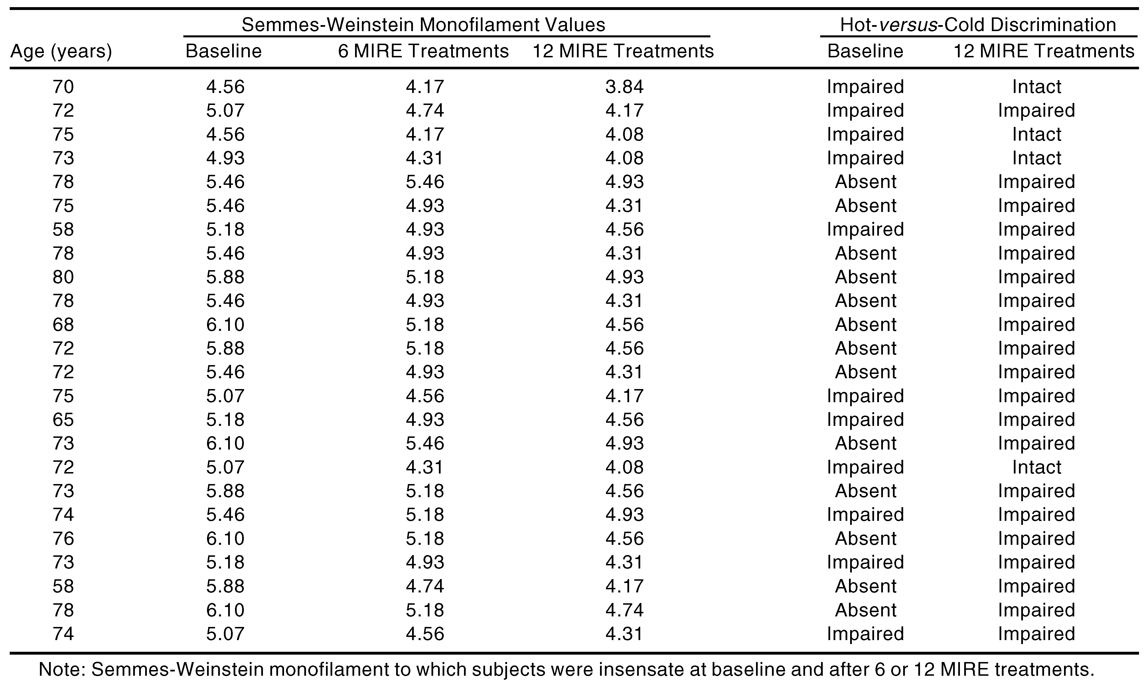
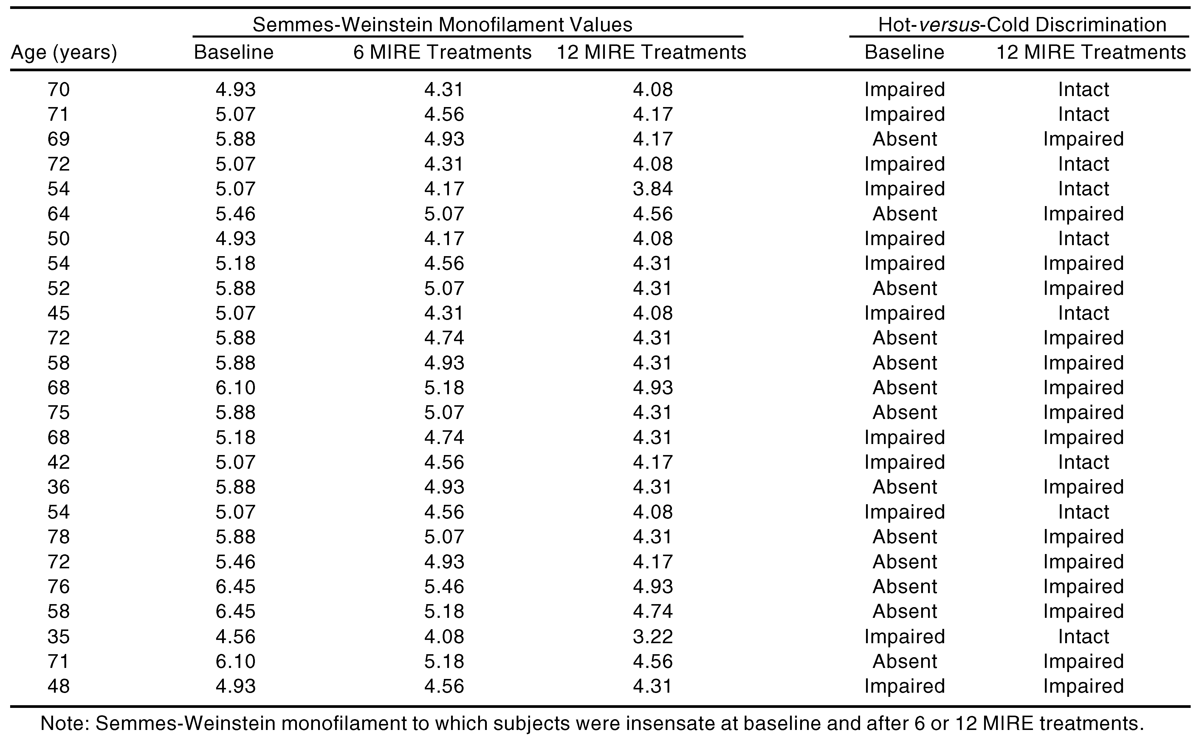
All of the subjects in the study were treated at the Physical Therapy Department of The Medical Center of Aurora, Colorado. The subjects ranged from 35 to 80 years of age; 25 had type 1 diabetes and 24 had type 2 diabetes (
Table 1 and
Table 2). On the basis of the Semmes-Weinstein monofilament test, all had peripheral neuropathy. The ability to detect hot
versus cold was also absent or impaired in all subjects. No novel treatments or pharmaceuticals that would have uniquely modified circulation in the lower extremities were employed for 30 days prior to beginning this study. No changes were made in the standard of medical care associated with diabetes for these subjects, including insulin or oral hypoglycemic agents, diet, blood pressure medications, or exercise. The Semmes-Weinstein test is often used as an adjunct to gait-testing analysis in a physical therapy department and such information can guide the therapist in reeducating the muscles of the lower leg despite ongoing neuropathy [
7]. The present study included patients with diabetes whose Semmes-Weinstein, hot-
versus-cold discrimination, and gait analysis values were abnormal. In an outpatient physical therapy department, the goal is to rehabilitate patients as quickly as possible. None of the following clinical research tools were used: transcutaneous partial pressure of oxygen, vibratory sensation measuring instrumentation, or reflex measuring devices.
MIRE is delivered from a series of 60 gallium aluminum arsenide diodes in a flexible pad (diode array) placed on the foot or lower leg. The device used in this study was the Anodyne Therapy System (Anodyne Therapeutics LLC, Denver, Colorado). Four diode arrays were used and each application lasted 30 minutes. One diode array was placed on the distal posterior aspect of the tibia in an effort to alter circulation in the posterior tibial artery, and a second diode array was placed over the anterior distal tibia in an effort to affect the dorsalis pedis artery. The third and fourth diode arrays were placed on the dorsal and ventral surfaces of the foot. This was done to each foot. If the posterior tibia region was uncomfortable for the subject, two diode arrays were placed on the plantar surface of each foot.
Several sizes of Semmes-Weinstein monofilaments (3.22, 3.84, 4.08, 4.17, 4.31, 4.56, 4.74, 4.93, 5.07, 5.18, 5.46, 5.88, 6.10, and 6.45) were used to determine the absolute level of neurological impairment. The monofilaments were randomly applied to three test sites: the great toe, plantar arch region, and fourth toe. The same locations were tested at each visit. The filament was applied until it began to bend and it was held in place for approximately 1.5 seconds. Each site was tested three times. Care was taken to test areas in which the keratin layer was the least thick. The response to the filament testing was based on the subjective response of the subject, who was asked to say “now” when the filament could be felt. In addition, the subject was queried as to the location on the foot where the monofilament was sensed to assure objectivity of the measurements taken.
Hot-versus-cold testing was done at the same test sites on the feet. Response to the hot-versus-cold testing was determined from the subject’s reports as to whether he or she could correctly identify the hot or cold bar at three sites. The responses were graded as absent (0 of 3 correct answers at all sites); impaired (correct discrimination at one or two sites); or intact (correct discrimination at all three sites). One of the authors (A.B.K.) performed all sensory tests and applied the diode arrays to each subject.
Results
The data for subjects with type 1 and type 2 diabetes were grouped and analyzed by repeated measures analysis. Values were reported as mean ± SD and significance was accepted at P < .05.
The ages of the subjects, type of diabetes (type 1 or 2), Semmes-Weinstein values, and hot-
versus-cold discrimination ability prior to beginning the study and after MIRE treatments are shown in
Table 1 and
Table 2. As indicated in
Table 3, subjects with type 1 diabetes (60.4 ± 12.8 years of age) were approximately 12 years younger than the type 2 subjects (72.5 ± 5.5 years of age).
Baseline Semmes-Weinstein deficits were virtually identical for type 1 (5.49 ± 0.52) and type 2 (5.44 ± 0.47) subjects (
Table 3). There were 13 subjects with type 1 diabetes and 13 type 2 subjects who had no ability to discriminate between hot and cold prior to MIRE treatment (
Table 1,
Table 2, and
Table 4).
Table 1.
Data for Subjects with Type 1 Diabetes (N = 25)
Table 1.
Data for Subjects with Type 1 Diabetes (N = 25)
Table 2.
Data for Subjects with Type 2 Diabetes (N = 24)
Table 2.
Data for Subjects with Type 2 Diabetes (N = 24)
Table 3.
Subject Characteristics and Semmes-Weinstein Monofilament Values (Mean ± SD)
Table 3.
Subject Characteristics and Semmes-Weinstein Monofilament Values (Mean ± SD)
Table 4.
Subject Characteristics and Hot-versus-Cold Sensation Discrimination
Table 4.
Subject Characteristics and Hot-versus-Cold Sensation Discrimination
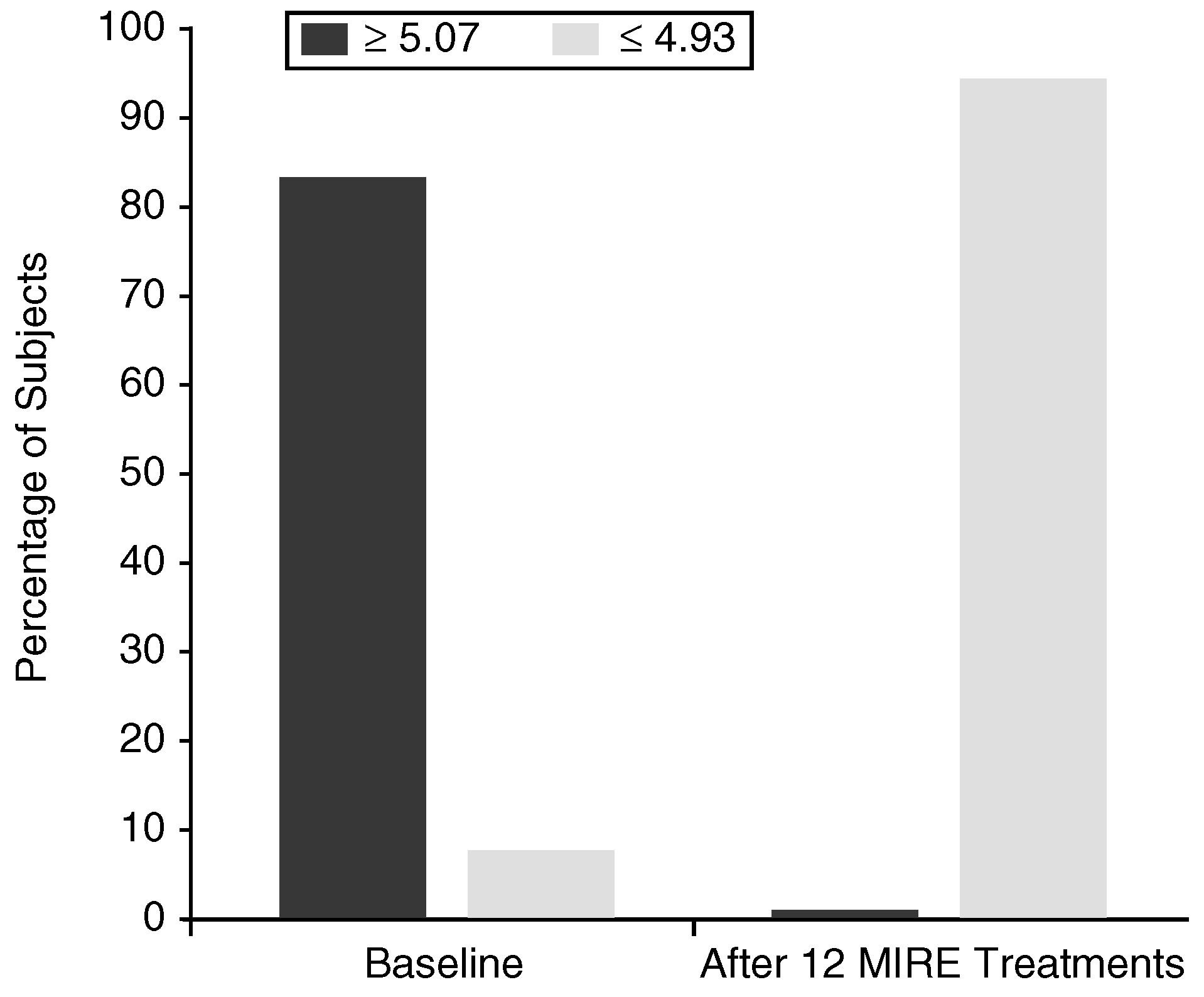
After 12 MIRE treatments, 100% of the type 1 subjects had Semmes-Weinstein monofilament values below 5.07 (
Figure 1). Mean Semmes-Weinstein values for all 25 type 1 subjects were 4.26 ± 0.34 after 12 MIRE treatments (
Table 3). Because the Semmes-Weinstein scale is a log
10 scale, this result demonstrates that subjects who could only detect a force of approximately 20 g prior to MIRE were now able to detect a force of approximately 2 g.
Figure 1.
Percentage distribution of patients with type 1 diabetes (N = 25) with Semmes-Weinstein monofilament values ≥ 5.07 and ≤ 4.93 before (left) and after (right) 12 MIRE treatments.
Figure 1.
Percentage distribution of patients with type 1 diabetes (N = 25) with Semmes-Weinstein monofilament values ≥ 5.07 and ≤ 4.93 before (left) and after (right) 12 MIRE treatments.
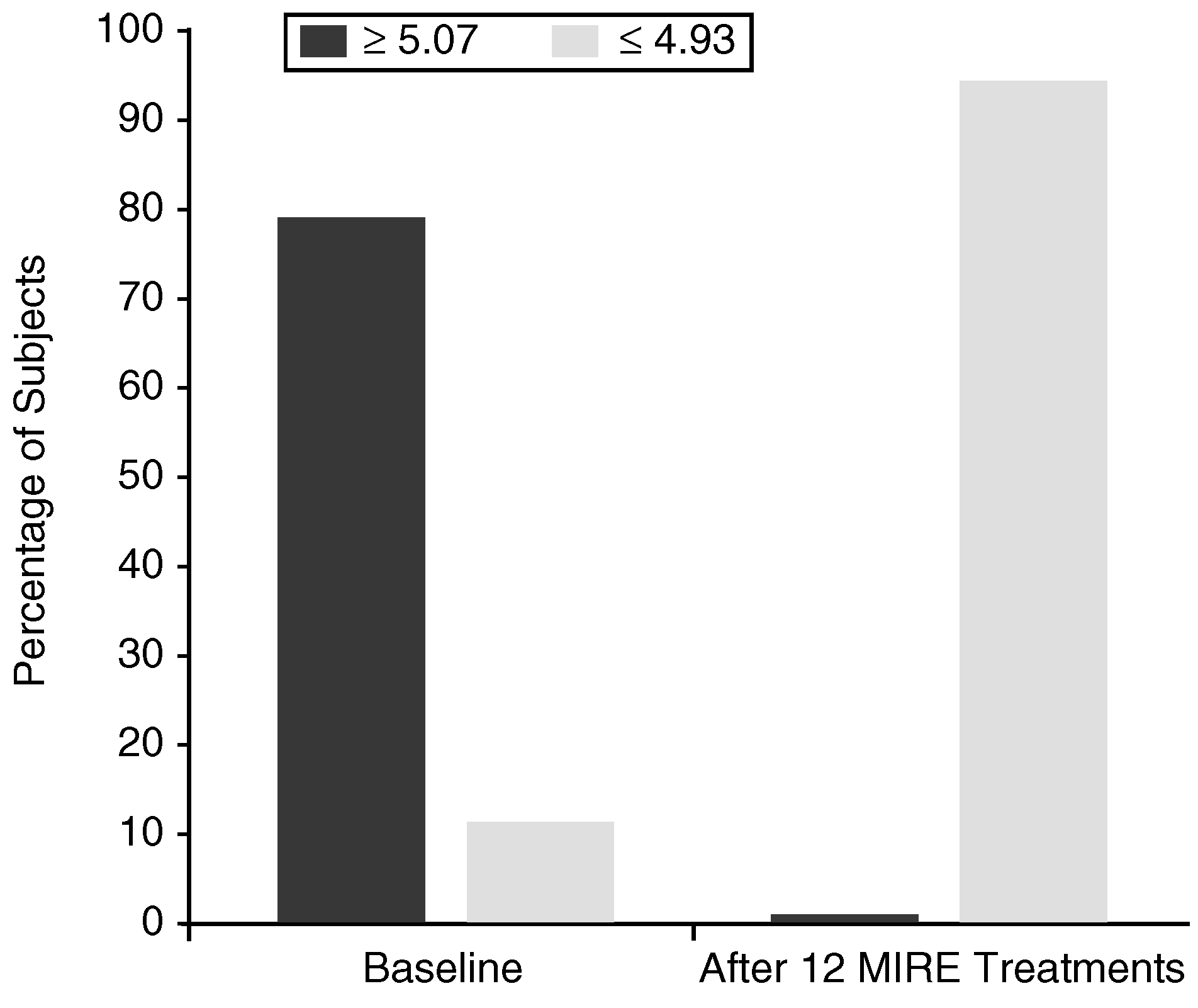
Figure 2 documents a similar response to MIRE treatment in the somewhat older type 2 diabetic subjects. After 12 MIRE treatments, 100% of the type 2 subjects had Semmes-Weinstein values below 5.07, and the mean for all 24 subjects was 4.45 ± 0.32 (
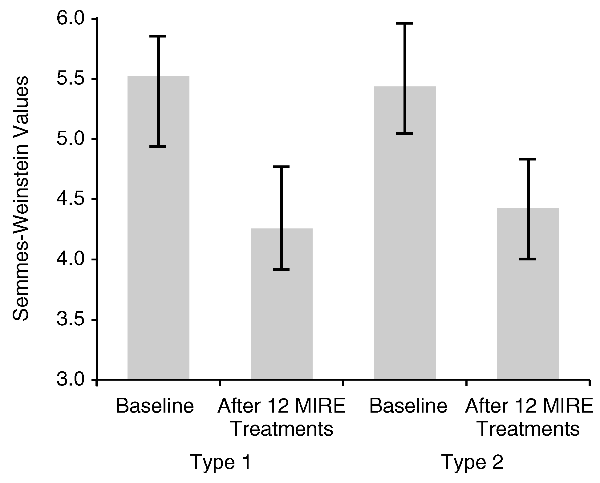
Table 3). These results reflect an average of 85% improvement in sensory perception from approximately 20 g to 3 g following MIRE treatment. The mean Semmes-Weinstein values before and after 12 treatments with MIRE for all of the subjects are shown in
Figure 3. Whereas 42 of 49 subjects (21/25 type 1, 21/24 type 2) had values at or above 5.07 prior to initiating the study, after 12 MIRE treatments none had values higher than 4.93 (
Table 1 and
Table 2).
Figure 2.
Percentage distribution of patients with type 2 diabetes (N = 24) with Semmes-Weinstein monofilament values ≥ 5.07 and ≤ 4.93 before (left) and after (right) 12 MIRE treatments.
Figure 2.
Percentage distribution of patients with type 2 diabetes (N = 24) with Semmes-Weinstein monofilament values ≥ 5.07 and ≤ 4.93 before (left) and after (right) 12 MIRE treatments.
Figure 3.
Semmes-Weinstein monofilament values (Mean ± SD) in all subjects before and after 12 MIRE treatments. P < .0001.
Figure 3.
Semmes-Weinstein monofilament values (Mean ± SD) in all subjects before and after 12 MIRE treatments. P < .0001.
After 12 MIRE treatments, 9 of 12 (75%) subjects with type 1 diabetes converted from impaired hot-
versus-cold sensation to an intact ability to discriminate hot from cold (
Table 1), and 4 of 11 (36%) subjects with type 2 diabetes were able to discriminate hot
versus cold after 12 MIRE treatments (
Table 2).
Discussion
The results of the present study suggest that there is a potentially effective therapy currently available that will, at least temporarily, reverse diabetic peripheral neuropathy in all patients as documented by the Semmes-Weinstein monofilament.
The insensitivity to a 5.07 (10 g) Semmes-Weinstein monofilament is “reliable and may be superior to biothesiometry in screening for patients at risk for foot ulceration since sensitivity is the more important parameter,” as pointed out by Kumar et al [
8]. Recently, Mayfield and Sugarman [
9] noted: “The Semmes-Weinstein monofilament is currently the best choice for screening for clinically significant neuropathy because it is portable, inexpensive, painless, easy to administer, acceptable to the patient, and provides good predictive ability for the risk of ulceration and amputation.” In this study, 42 of 49 subjects had loss of protective sensation (Semmes-Weinstein value of 5.07 or greater) at baseline, yet MIRE treatment was able to reverse neuropathic impairment to below 5.07 in every subject. Improving sensation to this degree reduces the risk of an eventual foot ulceration or amputation.
The present study shows that MIRE treatment in an outpatient setting can reverse, at least temporarily, the sensory deficits in all diabetic subjects treated so far. Admittedly the trial was small and lasted only a month. However, there were no restrictions as to subject selection, and subjects were not required to alter any aspect of their lifestyle, dietary intake, or drug or exercise regimen.
Although no placebo was used, this outpatient clinical study was performed in a setting in which the goal is to expedite patient recovery. Historically, diabetic peripheral neuropathy of long duration does not reverse spontaneously. Moreover, the usefulness of placebo arms in objective studies has recently been questioned [
10].
The physiologic basis of the improvement in neural function may be due, in part, to improved circulation related to the localized release of nitric oxide. However, it is emphasized that in this outpatient study, neither nitric oxide nor any of its surrogates were measured directly. Accordingly, any possible involvement of nitric oxide is purely speculative. However, nitric oxide might be involved for the following reasons:
1) It is well recognized that photo energy can modulate circulation, as evidenced by the early work of Nobel laureate Robert Furchgott [
11], although the precise biological effects of MIRE are less well understood. Recently, experimental studies in rats have demonstrated that 890 nm near-infrared photo energy, virtually identical to MIRE, increases blood flow partly through an effect mediated by endothelial nitric oxide synthase or nitric oxide; the vasodilation was sustained for several hours even after the photo energy was removed [
12].
2) Red blood cells are able to store large amounts of nitric oxide [
13], partly in the form of nitrosothiols [
14], and the absorption of this wavelength of photo energy by hemoglobin is well documented [
15]. Thus, vasodilation mediated by photo energy may be due, in part, to the localized release of nitric oxide from the red blood cells continuously passing through vessels exposed to the MIRE [
12,
16].
3) Glycosylated hemoglobin, characteristic of diabetes, avidly binds nitric oxide [
17]. This suggests that even the smaller-than-normal amounts of nitric oxide produced by patients with diabetes [
18,
19] may not be easily released from red blood cell hemoglobin at microcirculatory sites. Perhaps MIRE enables the nitric oxide to be released from glycosylated hemoglobin more easily.
Conclusion
The results of this study suggest that in an outpatient setting MIRE consistently has the effect of improving neural function in patients with diabetes. Future studies should be directed at assessing whether nitric oxide may be involved in these outcomes and at the long-term duration of the improvement in sensory deficits that were observed with this 1-month treatment protocol.