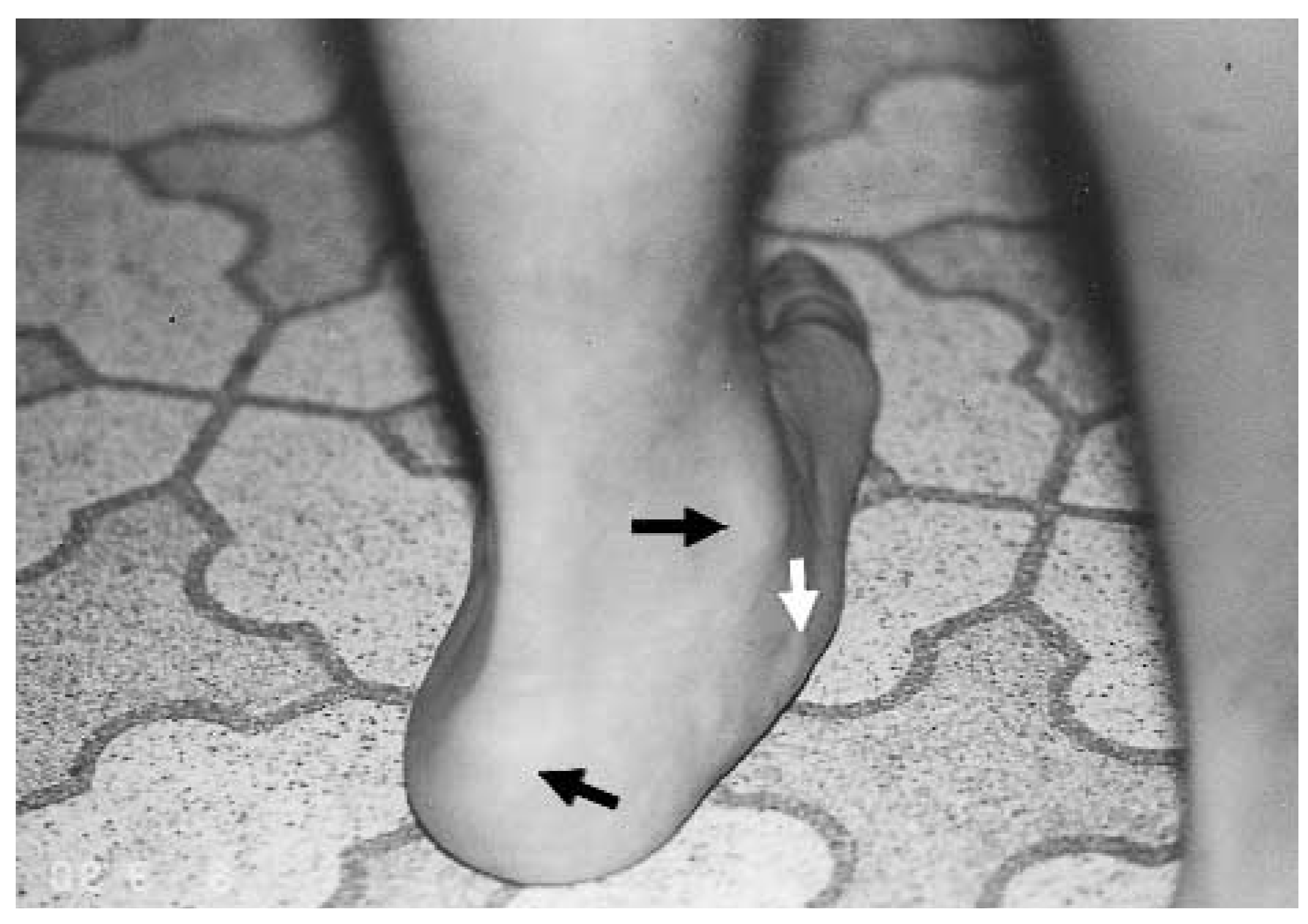
The medical literature includes many articles on the flexible flatfoot. The terminology is often confusing and excessive. Perhaps the most descriptive term used is “collapsing pes valgo planus,” in which collapsing refers to the flexibility of the deformity, pes refers to the foot, planus refers to the flattened arch, and valgus refers to the everted calcaneus (
Fig. 1).
Figure 1.
Posterior view of the left foot–everted heel (inferior black arrow), flattened arch (white arrow), and internally rotated leg (superior black arrow), resulting in a medially located ankle relative to the heel.
Figure 1.
Posterior view of the left foot–everted heel (inferior black arrow), flattened arch (white arrow), and internally rotated leg (superior black arrow), resulting in a medially located ankle relative to the heel.
The biomechanics of the maximally pronated pes valgo planus foot type consists of plantarflexion and medial rotation of the talus and eversion of the calcaneus during weightbearing. The maximally pronated subtalar joint unlocks the midtarsal joint, which becomes hypermobile and unstable. The medial and lateral column instability that results eventually leads muscles to fatigue. The muscles are inadequate to compensate for the structural instability or to resist the strong pronatory forces. A vast majority of these flexible flatfeet can be controlled with functional orthoses, but the worst deformities may require surgical intervention.
The purpose of this study was to evaluate the subjective and radiographic results of the STA-peg arthroereisis procedure (Flake-Austin modification) for the correction of collapsing pes valgo planus in children. Two previous studies have looked at the STA-peg arthroereisis procedure.[
1,
2] Their data included radiographic and clinical measurements, but they both failed to adequately include the subjective results.
Many procedures have been advocated over the years to correct collapsing pes valgo planus. The procedures vary from soft-tissue to bony correction and can include combinations of both. The most common soft-tissue medial column procedures are the Kidner[
3] and Young[
4] procedures, in which the advancement of the tibialis posterior tendon and the redirecting of the tibialis anterior tendon occur, respectively.
Bony procedures include calcaneal osteotomies as described by Evans,[
5] Dwyer,[
6] Silver et al,[
7] and Koutsogiannis.[
8] Arthrodesis procedures include talonavicular fusion, navicular-cuneiform fusion as described by Hoke,[
9] first metatarsal-cuneiform-navicular fusion as described by Miller,[
10] subtalar joint arthrodesis, and triple arthrodesis as described by Ryerson.[
11]
In many cases, soft-tissue and bony procedures are done concomitantly, depending on the degree of deformity and age of the patients. The Kidner/Young/Koutsogiannis combination is one such example.
Many bony procedures attempt to block the sinus tarsi and prevent excessive pronation. The Green-Grice[
12] procedure uses a bone graft in the sinus tarsi to create an extra-articular arthrodesis of the talus and the calcaneus. When successful, not only is excessive pronation eliminated but all motion between the calcaneus and the talus also is eliminated. However, other procedures must be performed to eliminate excessive pronation but still allow supination. In 1946, Chambers[
13] elevated the calcaneal sulcus to prevent the forward progression of the talus into the sinus tarsi. Baker and Hill[
14] elevated the posterior facet of the calcaneus by using a bone graft. Selakovich[
15] elevated the sustentaculum tali with bone graft in an attempt to prevent the talus from plantarflexing and adducting. LeLievre[
16] and Haraldsson[
17] placed a bone graft in the sinus tarsi to block excessive motion while attempting to avoid an arthrodesis.
The subtalar joint arthroereisis procedure was developed to block the sinus tarsi and prevent the pes valgo planus. The block is placed in the sinus tarsi to prevent end-range pronation by limiting plantarflexion and medial rotation of the talus, which still allows for some pronation and full supination during gait.
Various materials have been used to block the sinus tarsi. Many studies have used Silastic material with various designs[
18-
20] while others have used silicone.[
21,
22] Two component systems of stainless steel and polyethylene also have been used.[
23] Valenti employed a polyethylene screw[
24] and Pisani used a stainless steel screw with a polyethylene crown on the head.[
25]
In 1976, the Smith subtalar arthroereisis implant (STA-peg) (Wright Pharmaceutical, Arlington, Tennessee) was released. This implant is made of an ultra-high molecular weight polyethylene disk and stem. The STA-peg comes in two sizes, small and medium.[
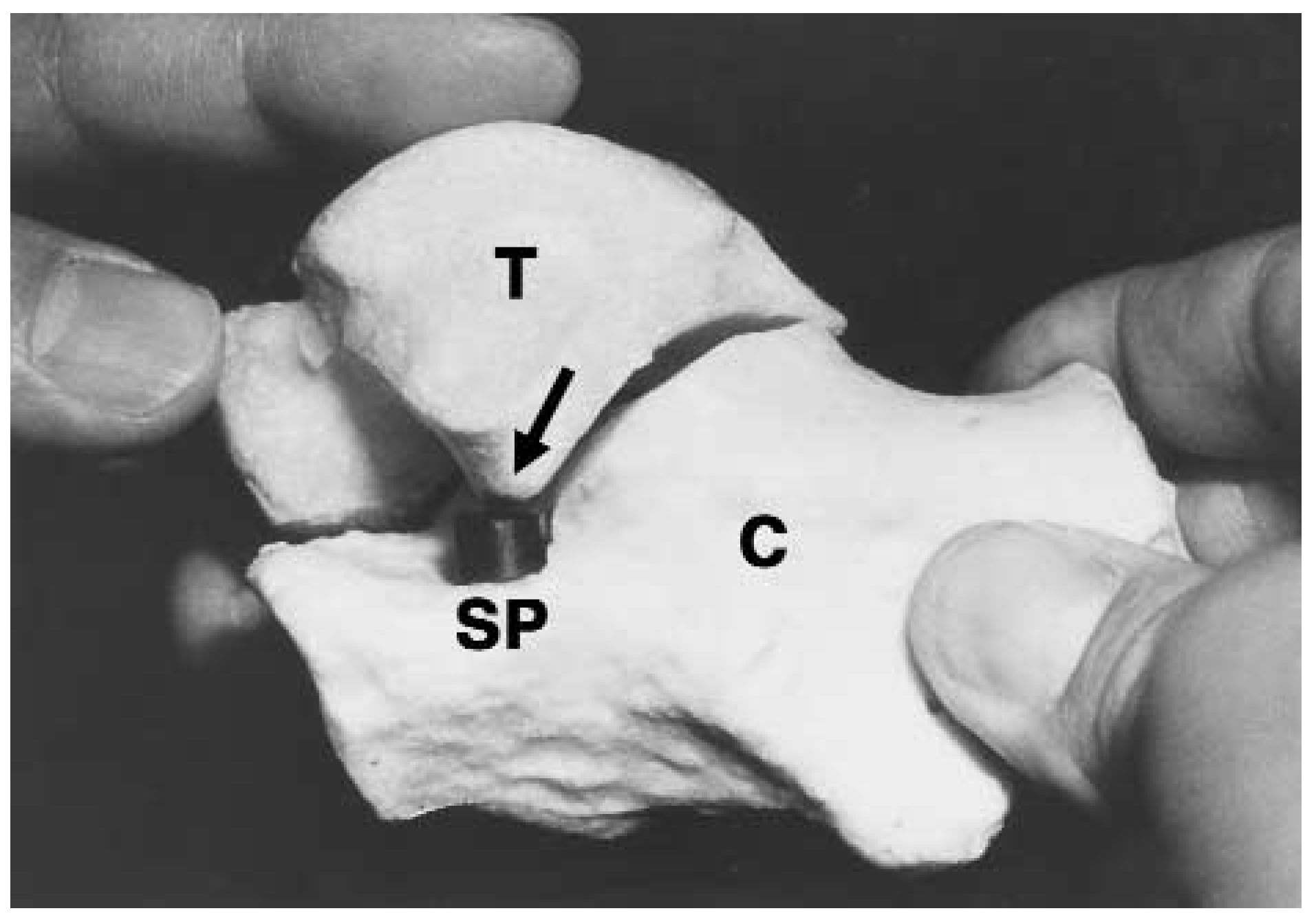
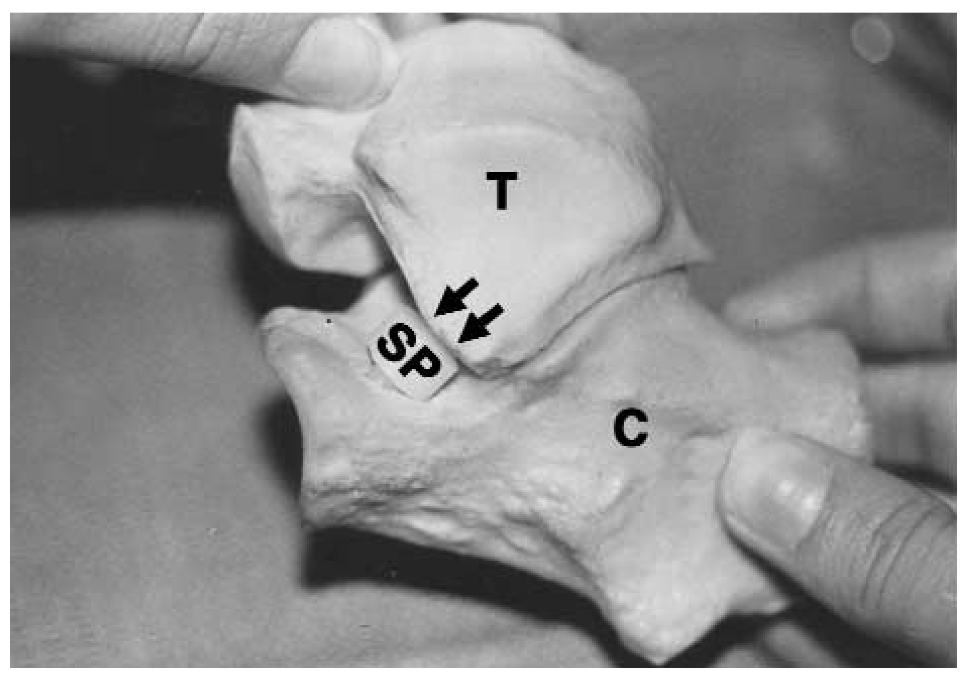
1] The implant is placed in the floor of the sinus tarsi so that the posterior facet of the talus can glide onto the dorsal surface of the implant, preventing plantarflexion and medial rotation of the talus in the sinus tarsi (
Fig. 2). The implant later was modified with an anterior incline to further increase the blockade and decrease the medial rotation of the talus. This inclined or angled implant comes in three sizes: small, medium, and large.[
2] Sgarlatto Labs (Los Gatos, California) offers an implant that has a similar angled design, the LSI, which has a longer stem and comes in extra-small and extra-large sizes, as well as small, medium, and large.
Figure 2.
Model of the talus (T) and calcaneus (C) with the STA-peg (SP) perpendicular in the sinus tarsi, which blocks the forward progression of the lateral talar facet as it rides up on the STA-peg in pronation.
Figure 2.
Model of the talus (T) and calcaneus (C) with the STA-peg (SP) perpendicular in the sinus tarsi, which blocks the forward progression of the lateral talar facet as it rides up on the STA-peg in pronation.
Flake et al[
26] modified the placement of these implants, and advocated placing the implant at approximately a 45° angle so that the dorsal aspect of the disk comes in complete contact with the lateral leading wall of the talus (
Fig. 3). There is no gliding of the talus on the implant but an actual blockage of motion. This utilizes the entire surface of the implant against the lateral leading wall of the talus and further decreases the medial rotation of the talus, which is especially helpful in individuals with a high subtalar joint axis (a large transverse planar dominant foot type).
Figure 3.
Model of the left talus (T) and calcaneus (C) with the STA-peg (SP) angled in the calcaneus so that the surface of the peg abuts the leading wall of the posterior facet of the talus in a flush manner in pronation.
Figure 3.
Model of the left talus (T) and calcaneus (C) with the STA-peg (SP) angled in the calcaneus so that the surface of the peg abuts the leading wall of the posterior facet of the talus in a flush manner in pronation.
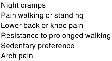
The subjective criterion for surgery, in general, is often pain. However, with pes valgo planus, the child often does not present the symptoms as actual pain. It is not until after careful questioning that symptoms consistent with postural fatigue are identified, including night cramps, pain walking or standing, lower back or knee pain, resistance to prolonged walking, sedentary preference, or arch pain (
Table 1). More commonly, parents seek medical attention because of a child’s abnormal shoe wear, fallen arches, or abnormal foot appearance. The parent may have noticed that the child is clumsy and has trouble keeping up with other children of the same age.
Table 1.
Postural Fatigue Symptoms.
Table 1.
Postural Fatigue Symptoms.
The expectations of the patient, doctor, and parent of the STA-peg arthroereisis procedure also are important. Overcorrection can be worse than the original deformity; undercorrection with some capability of pronation is desirable. Therefore, it must be emphasized that postoperatively the child’s foot is not expected to look perfectly normal and that some mild degree of pronation (flatfoot) appearance will remain. There will be radiographic and clinical evidence of a pronated foot although to a much less severe degree than preoperatively. Surgery generally is required only in more significantly pronated feet. The patient also will still require orthotic treatment until the age of maturity, at 18 to 22 years. However, subjective symptoms should improve or even may be eliminated. It is, therefore, imperative that data be collected on subjective symptoms both quantitatively and qualitatively in the preoperative and postoperative periods.
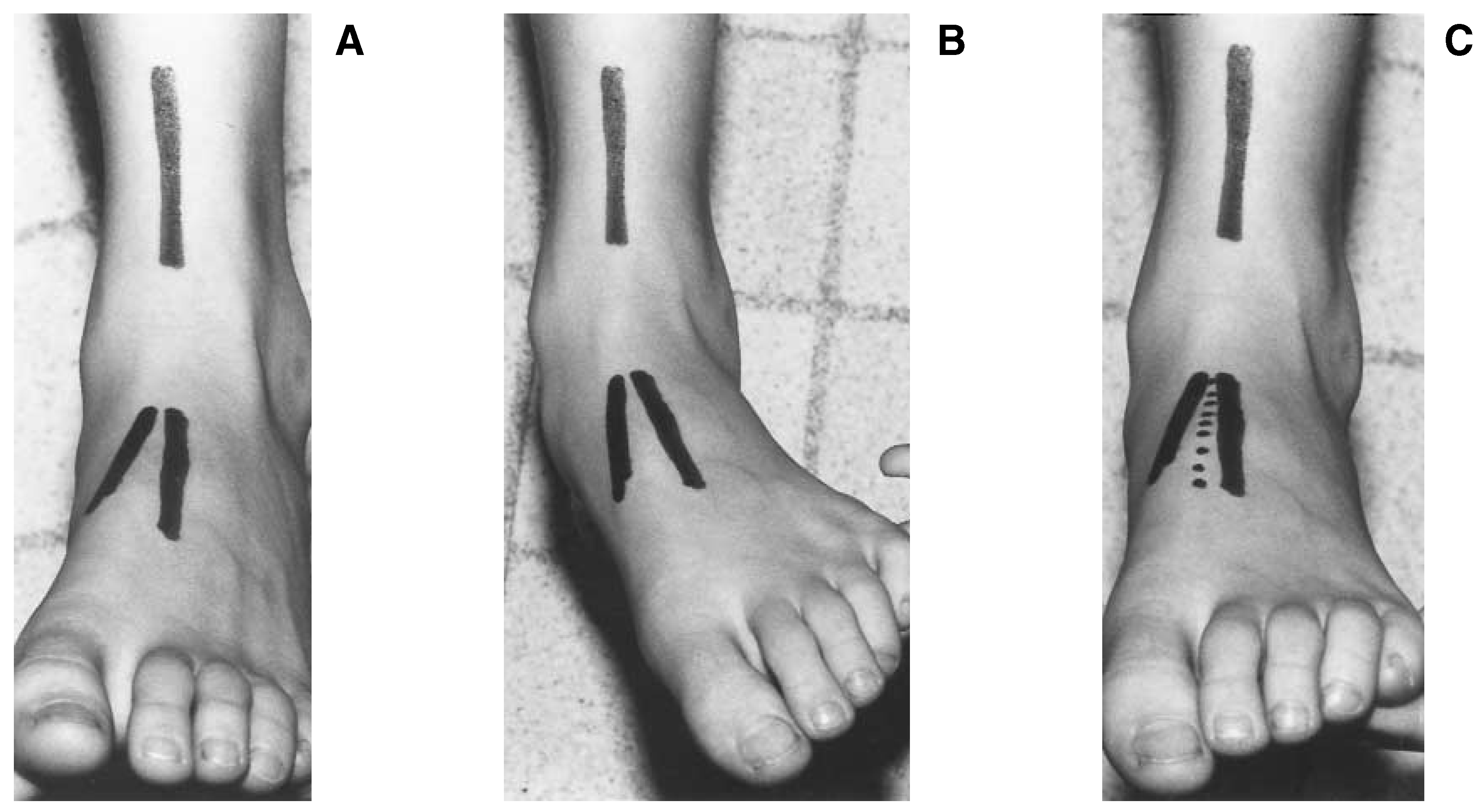
During the surgical procedure, alignment markers are mapped out by drawing a straight line on the anterior aspect of the leg. The line is extended onto the dorsum of the foot with the foot in neutral position. A second line is extended onto the dorsum of the foot with the foot in a maximally pronated position. When evaluating the effectiveness of the STA-peg, the amount of correction should fall between these two lines (
Fig. 4), allowing some pronation but eliminating excessive pronation. Overcorrection should be avoided or postural symptoms and fatigue will ensue.
Figure 4.
A, Foot in neutral subtalar joint position. The leg line is drawn on the distal anterior aspect of the leg. The foot line is an extension of the leg line in the neutral position. B, Foot in maximally pronated subtalar joint position. The foot line is an extension of the leg line in the maximally pronated position. C, The broken line is an extension of the leg line with the foot mildly pronated. This is the desired position to hold the foot with the STA-peg.
Figure 4.
A, Foot in neutral subtalar joint position. The leg line is drawn on the distal anterior aspect of the leg. The foot line is an extension of the leg line in the neutral position. B, Foot in maximally pronated subtalar joint position. The foot line is an extension of the leg line in the maximally pronated position. C, The broken line is an extension of the leg line with the foot mildly pronated. This is the desired position to hold the foot with the STA-peg.
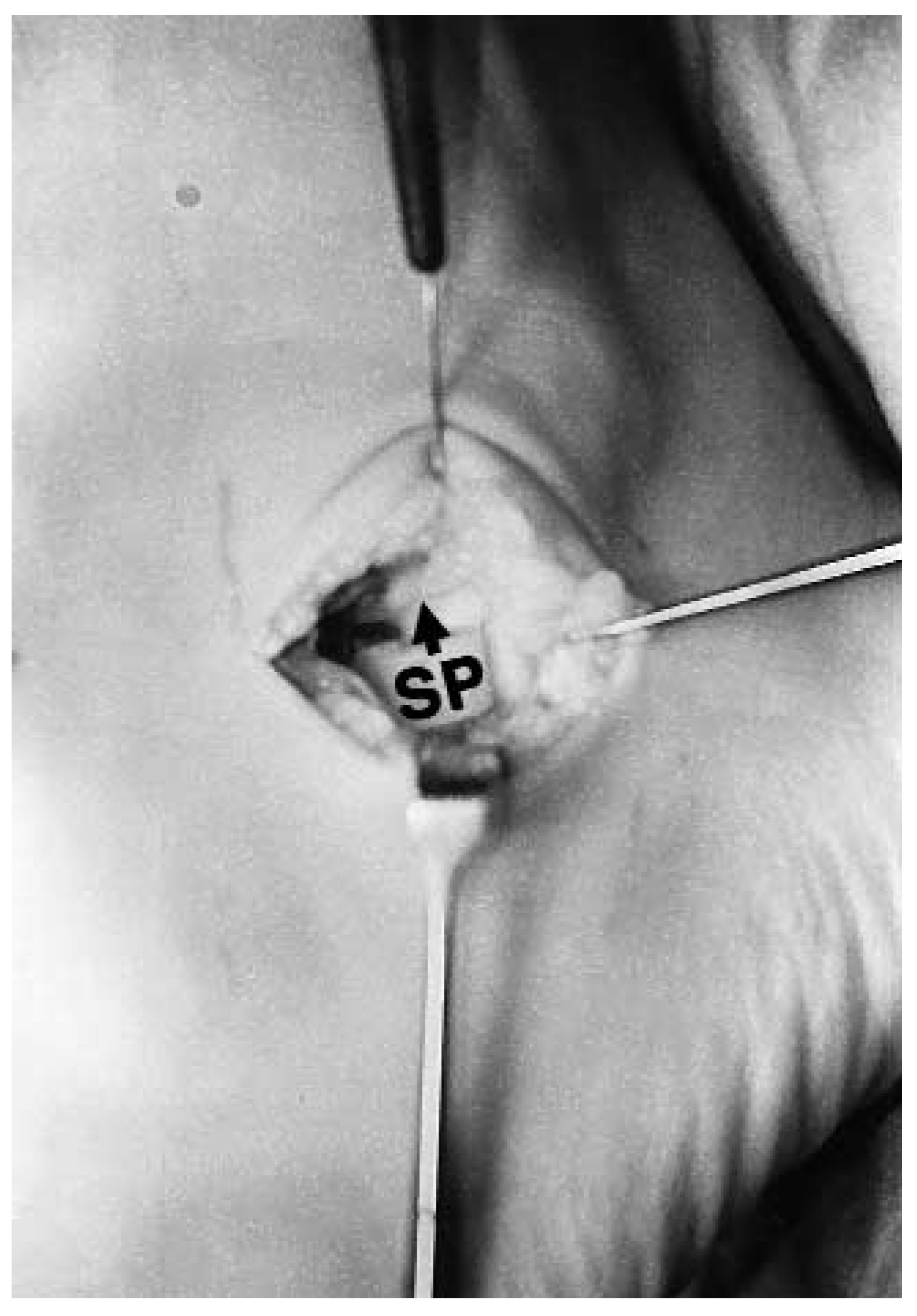
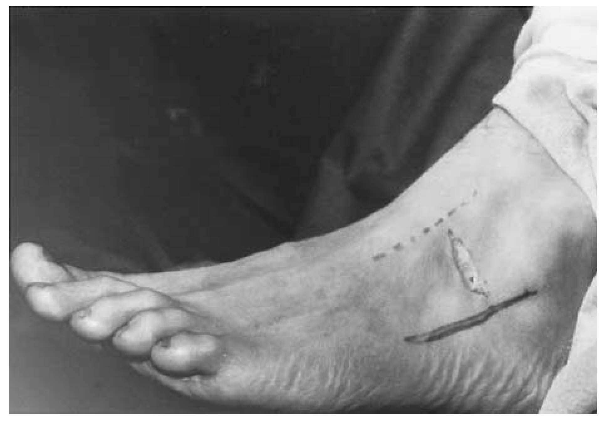
Attention is then directed to the lateral aspect of the foot, and the intermediate dorsal cutaneous nerve and peroneal tendons are identified and marked. The sinus tarsi is then identified and palpated and a modified Ollier incision following the skin lines is made, staying between the anatomical markers (
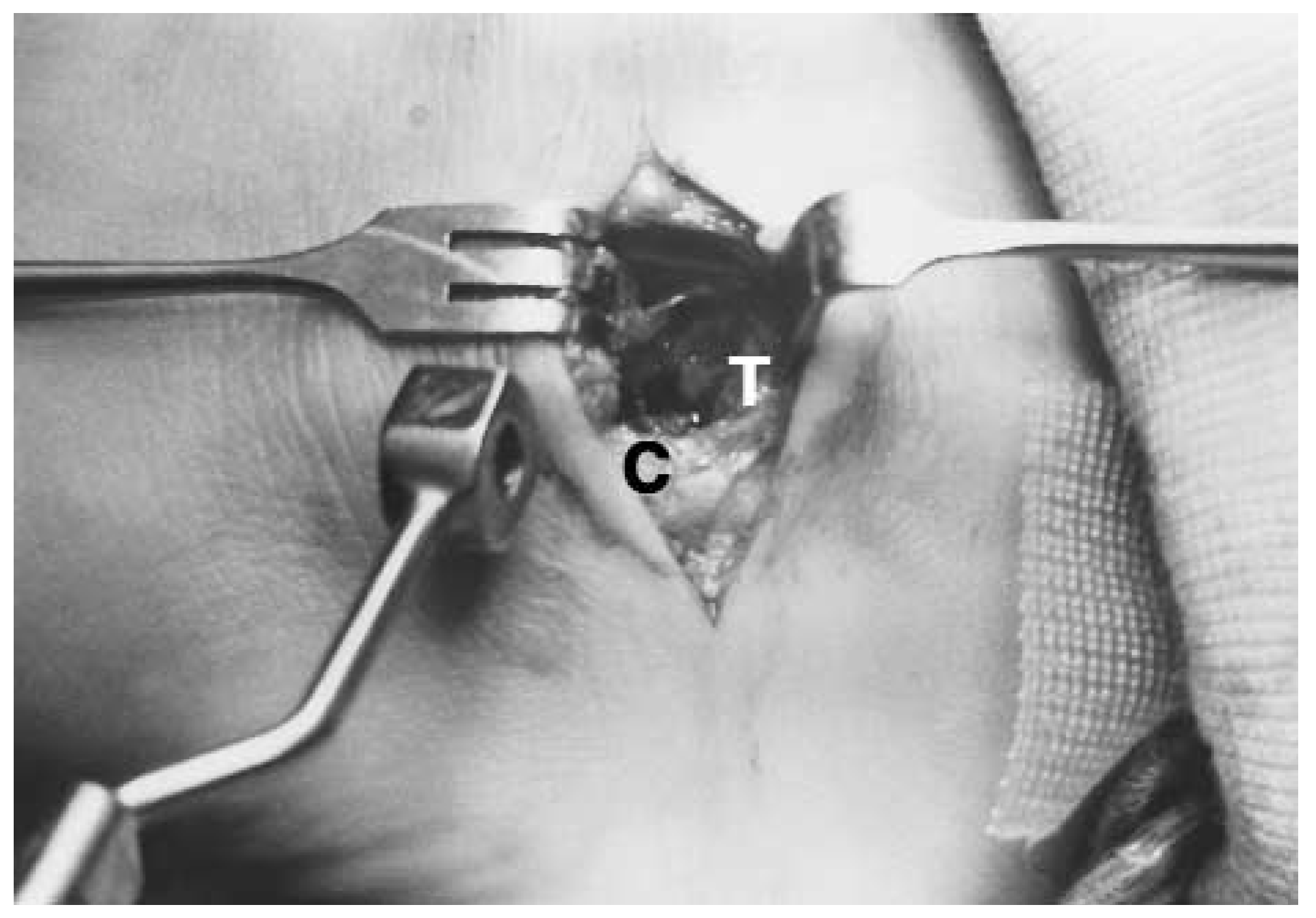
Fig. 5). Dissection is performed in layers. The deep fascia is identified and an L-shaped incision is made through it. The vertical incision follows the lateral leading wall of the talus and the more horizontal incision follows the floor of the sinus tarsi. The deep fascia is then reflected and the fat plug is identified and removed. The lateral leading wall of the talus, the floor of the calcaneus, and the posterior facet of the subtalar joint should be identifiable (
Fig. 6). The sinus tarsi ligament deep in the wound is preserved as much as possible.
Figure 5.
Ollier incision made along the skin lines over the lateral sinus tarsi between the intermediate dorsal cutaneous nerve and the peroneal tendons.
Figure 5.
Ollier incision made along the skin lines over the lateral sinus tarsi between the intermediate dorsal cutaneous nerve and the peroneal tendons.
Figure 6.
Sinus tarsi cleared off exposing the floor of the sinus tarsi (C) and the leading wall of the talus (T). The template with its central hole is poised to be inserted into the sinus tarsi.
Figure 6.
Sinus tarsi cleared off exposing the floor of the sinus tarsi (C) and the leading wall of the talus (T). The template with its central hole is poised to be inserted into the sinus tarsi.
A template with a medium- and a small-size metal head duplicating the shape of the disk portion of the straight STA-peg is then used to determine the proper size of the implant. Sgarlatto Labs has developed a template that duplicates the shape of the various angled-size widths of the implants to determine more exactly the proper size of the implant. The template is placed flush against the lateral leading wall of the talus and the foot is maximally pronated (
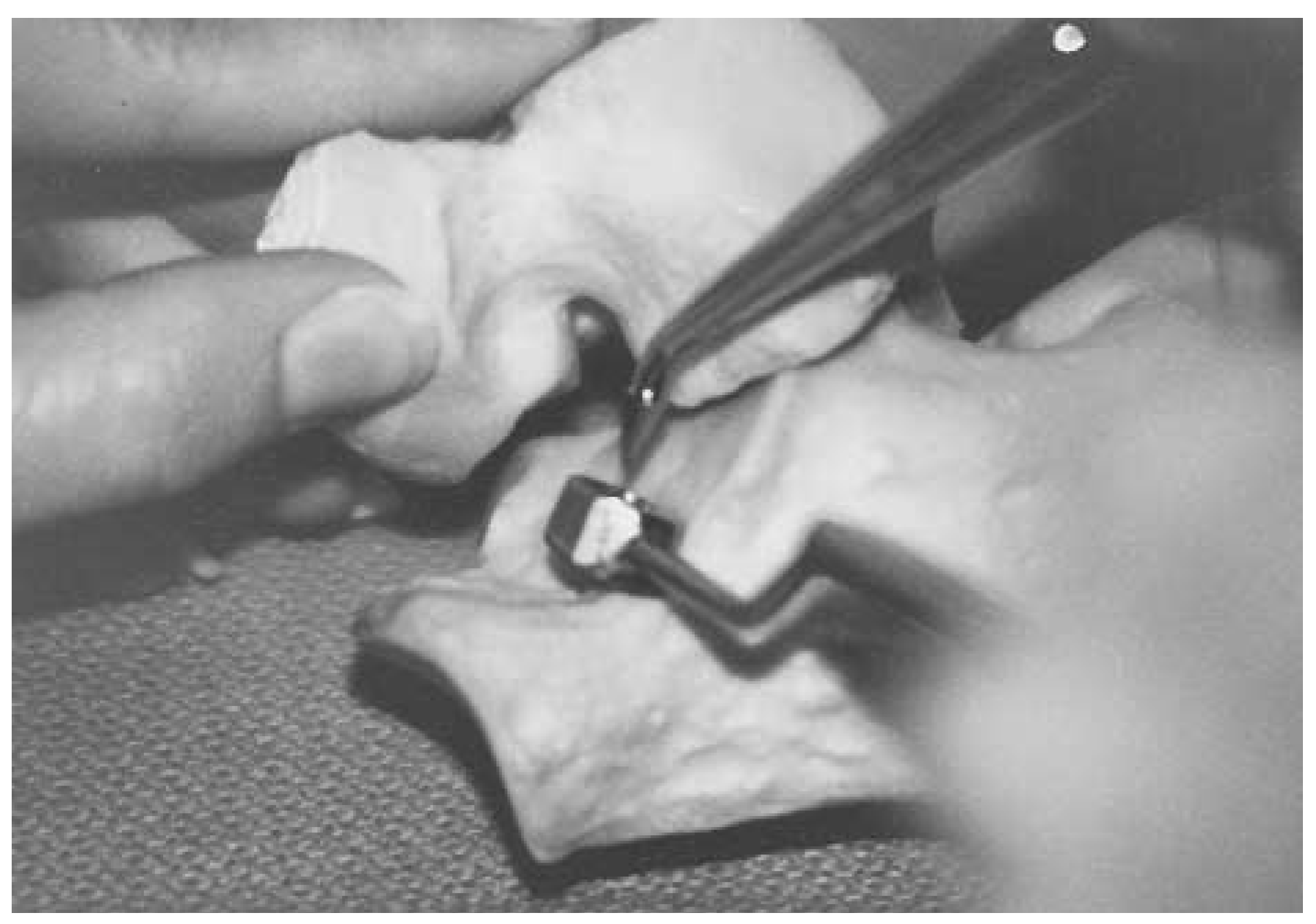
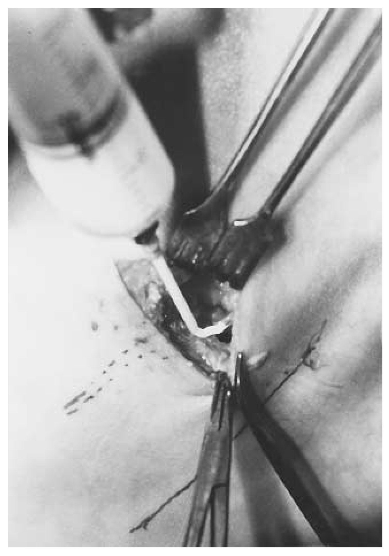
Fig. 7). The alignment markers are then checked to ensure that the appropriate amount of correction has been achieved. The foot is then supinated, maintaining the template on the floor of the calcaneus, and a sharp awl is placed through the opening in the template to mark the position of the stem of the implant (
Fig. 8). The template is then removed and the hole is widened and deepened with a curette and a curved hemostat. A spacer is then seated into the calcaneus for a trial fitting.
Figure 7.
The template is inserted in place so that it lies flush to the leading wall of the posterior facet of the talus (T) in the pronated position of the bone models. C, calcaneus.
Figure 7.
The template is inserted in place so that it lies flush to the leading wall of the posterior facet of the talus (T) in the pronated position of the bone models. C, calcaneus.
Figure 8.
With the subtalar joint supinated, the template is held against the floor of the sinus tarsi. An awl is placed in the center of the template to mark the position and direction of the peg hole.
Figure 8.
With the subtalar joint supinated, the template is held against the floor of the sinus tarsi. An awl is placed in the center of the template to mark the position and direction of the peg hole.
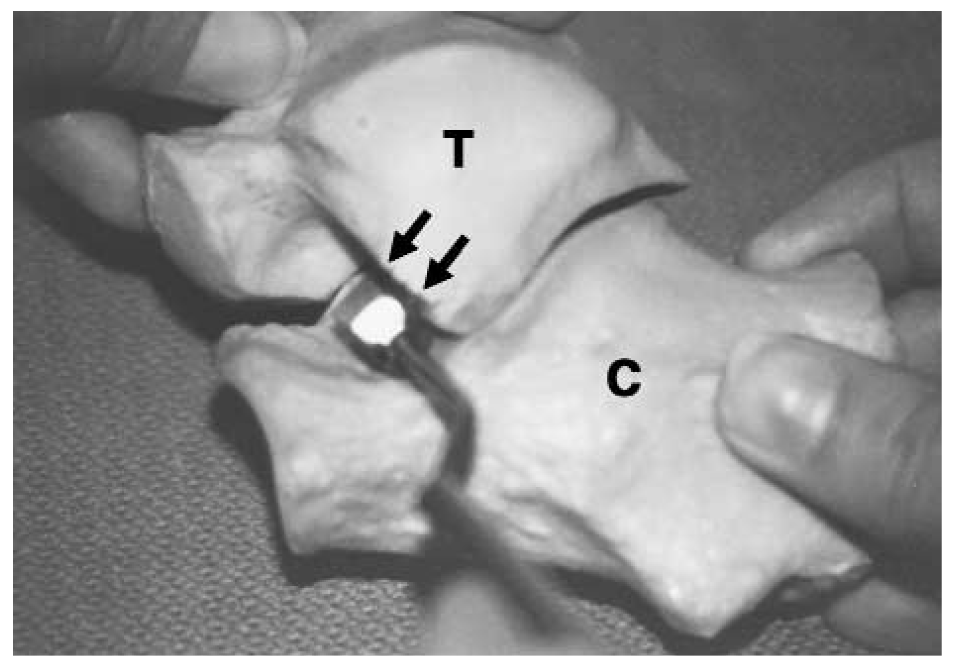
The lateral leading wall of the talus must be flush with the dorsal aspect of the implant when the foot is maximally pronated (
Fig. 9). The angled implant is often used to achieve this flush fit. Once the appropriate size implant has been determined, the sizer is removed and the implant is seated. The amount of maximum pronation is reduced, but supination still is available (
Fig. 10).
Figure 9.
The STA-peg implant (SP) remains in place while the subtalar joint is supinated, demonstrating the distance between the STA-peg implant and the leading lateral wall of the talus in supination.
Figure 9.
The STA-peg implant (SP) remains in place while the subtalar joint is supinated, demonstrating the distance between the STA-peg implant and the leading lateral wall of the talus in supination.
Figure 10.
In a minority of cases, a small amount of methyl methacolate may be necessary to help secure appropriate positioning of the STA-peg.
Figure 10.
In a minority of cases, a small amount of methyl methacolate may be necessary to help secure appropriate positioning of the STA-peg.
If the implant is not secure and moves in the calcaneus, or if the dorsal aspect of the implant is not flush with the lateral leading wall of the talus, methyl methacolate may be necessary to secure this position, although this usually is not required. Care should be taken not to have excessive methyl methacolate in the sinus tarsi. The wound is then flushed with normal saline and closed in layers. There should be no soft tissue interposed between the seating of the implant or between the talus and the dorsal aspect of the implant.
In many cases, ancillary procedures are required. Children with a gastrocnemius soleus complex require a tendo Achillis lengthening. Children with significant gastrocnemius soleus equinus require a gastrocnemius recession. Older children with severe pronating forces leading to subluxation may require reconstruction of the medial arch. Depending on the age of the patient and the amount of deformity, additional soft-tissue or bony auxiliary procedures may be performed.
Postoperative care consists of immediate weightbearing in a surgical shoe and compressive dressing for 2 weeks if the STA-peg procedure is done as an isolated procedure. Early range of motion is started and the patient may return to a soft-soled shoe in 2 weeks. Peroneal longus strengthening exercises are also initiated. The patient is advised not to run or jump for 3 months; normal vigorous activity can begin at 6 months. The authors recommend that the STA-peg be removed at the age of maturity, at 18 to 22 years of age.
Materials and Methods
A subjective questionnaire was sent to all patients of two of the authors (E.F., D.G.) who had an arthroereisis with at least 1 year of follow-up. Patients completed the questionnaire with the help of their parents when necessary. Charts were reviewed to aid in identifying preoperative signs and symptoms. A group of 21 patients representing 40 STA-peg procedures responded. The ages of the patients at the time of surgery ranged from 4 to 16 years with an average age of 9.7 years. The follow-up period ranged from 12 to 90 months with an average of 36 months. Six patients had ancillary procedures, three patients had Kidner procedures, one patient had bilateral tendo Achillis lengthening, and two patients had three McBride bunionectomies and epiphyseal staplings. Patients completed the questionnaire both preoperatively and postoperatively. Each child was interviewed about the results of the questionnaire. Charts were reviewed to confirm preoperative signs and symptoms. All of the results were collected by the lead author (P.F.)
Questionnaire Results
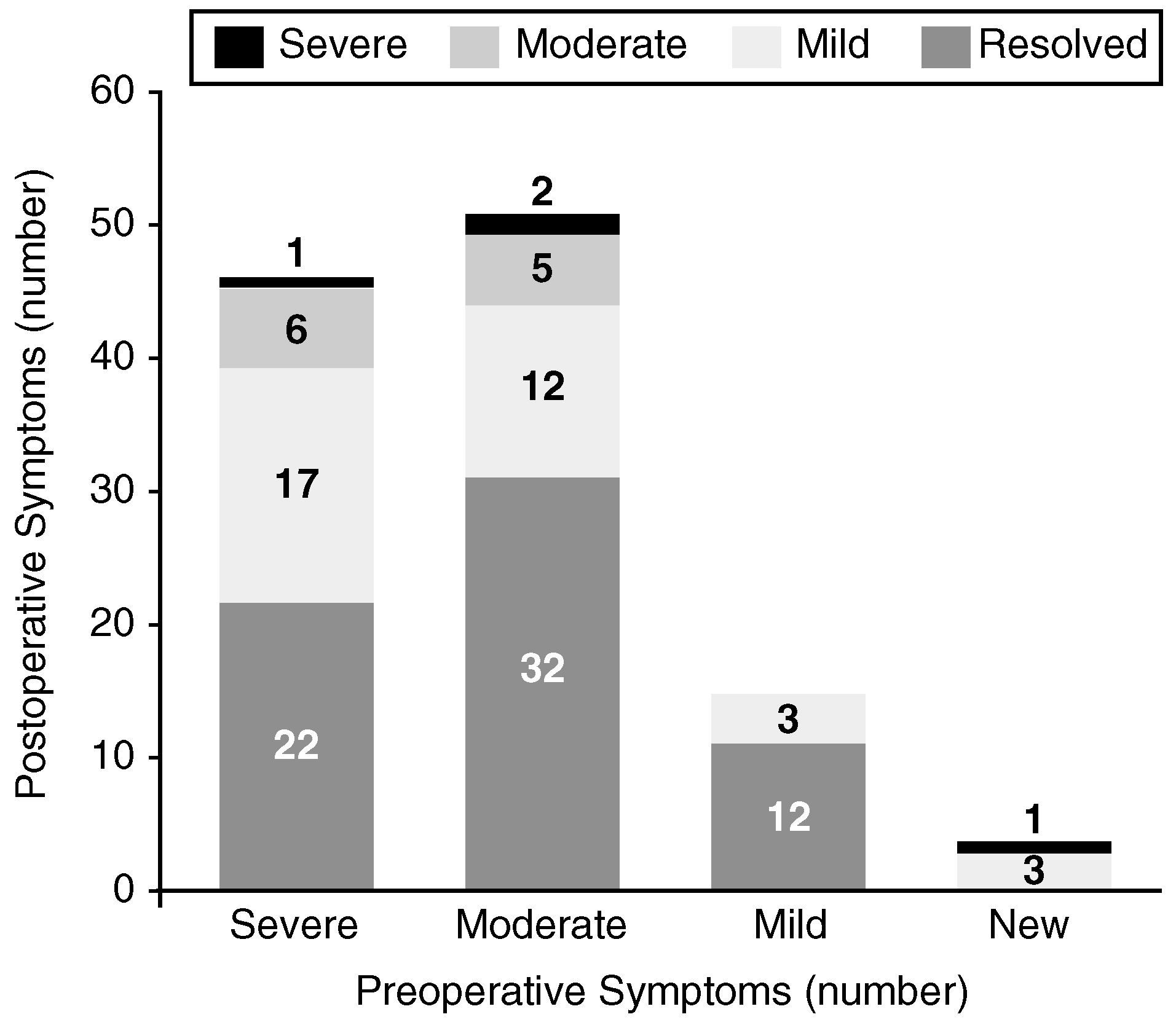
In the subjective questionnaire, patients indicated what signs and symptoms were present preoperatively and postoperatively. These data are provided in
Table 2. The symptoms included night cramps, arch pain, pain walking or standing, resistance to prolonged walking, and lower back or knee pain. The signs included fallen arches, inability or lack of desire to participate in sports, abnormal shoe wear, and clumsiness. These signs and symptoms were then rated as severe, moderate, or mild. In the present study, the percentage of complaints was drastically decreased postoperatively, except in the category of night cramps, which increased 33%. Of the 112 preoperative subjective symptoms reported, 46 were rated as severe, 51 as moderate, and 15 as mild.
Table 2.
Patient Questionnaire.
Table 2.
Patient Questionnaire.
An analysis of the patients’ symptoms preoperatively and postoperatively is found in
Figure 11. Postoperatively, 50 subjective symptoms were reported: 4 severe, 11 moderate, and 35 mild. Dramatic improvements in subjective symptoms postoperatively were recorded. Of the 46 severe symptoms reported preoperatively, 45 had reduced postoperatively, with 17 reduced to mild, 6 to moderate, and 22 to no symptoms whatsoever. One patient continued with symptoms severe enough to interfere with her ability to participate in sports.
Figure 11.
Analysis of subjective symptoms based on questionnaire results.
Figure 11.
Analysis of subjective symptoms based on questionnaire results.
Fifty-one subjective symptoms were initially reported as moderate; of these, 49 were reduced or remained the same. Two moderate symptoms, representing one patient, increased to severe. Of the remaining 49 symptoms, 32 were eliminated, 12 were reduced to mild, and 5 remained unchanged.
The mild subjective symptoms preoperatively also showed dramatic improvement. Of the 15 mild symptoms reported preoperatively, 12 resolved and 3 remained unchanged postoperatively.
Four new symptoms were reported postoperatively: three patients reported mild night cramps, which were attributed to increased weightbearing activities, such as sports involvement. One patient developed symptoms that were not present preoperatively; these symptoms were identified as severe lower back or knee pain.
The subjective questionnaire also included two questions on the patient’s overall satisfaction with the STA-peg arthroereisis procedure (
Tables 3 and
4). Fifteen of the 21 children responding felt they had at least 90% improvement and would highly recommend the procedure. Three of the 21 children felt that they had at least 70% improvement of signs and symptoms and would recommend the procedure. One child reported worse symptoms postoperatively. Two children did not answer the question but did express their satisfaction with the procedure elsewhere on the questionnaire.
Table 3.
Chief Complaints Satisfactorily Resolved.
Table 3.
Chief Complaints Satisfactorily Resolved.
Table 4.
Overall Satisfaction.
Table 4.
Overall Satisfaction.
Interviews with the children revealed that all but two are now involved in normal sports activity. One patient plays basketball and tennis and another patient races BMX bicycles. One patient who stated preoperatively that he was unable to participate in sports or do any activity that caused him to be on his feet now plays basketball and works as a busboy in a restaurant. Eleven patients felt that they could now keep up with their peers and one is involved with a gymnastics team. One mother stated that her daughter’s lower-extremity muscle tone has drastically improved, enabling her to wear regular shoes. Three patients reported that they had mild pain in the sinus tarsi region with increasing activity, but they all stated that this was not significant when compared with their preoperative limitations. One patient stated that her pain was decreasing as her stamina improved.
Of the two patients who were not involved in sports activity, one indicated that she could walk and stand more readily postoperatively, but she could not participate in sports that required running. The other patient’s symptoms had worsened postoperatively and she stated that she was unable to participate in any sports activities.
Radiographic Results
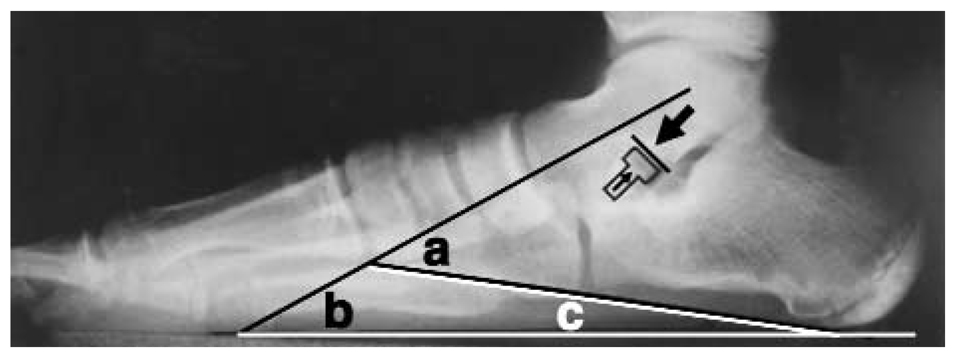
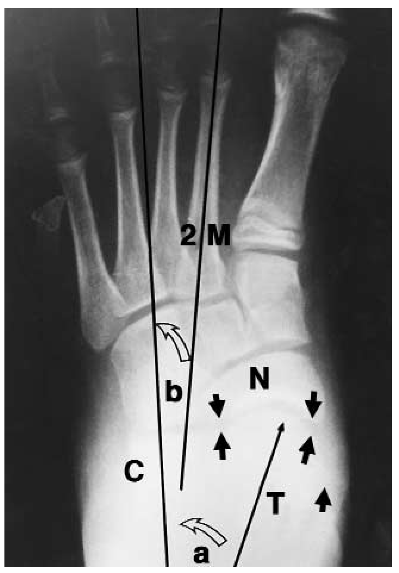
Radiographic evaluation in the present study included preoperative and postoperative evaluation of the talar declination angle, calcaneal inclination angle, lateral talocalcaneal angle, sinus tarsi occlusion, dorsoplantar talocalcaneal angle (Kite’s angle), and talonavicular articulation (
Figs. 12–
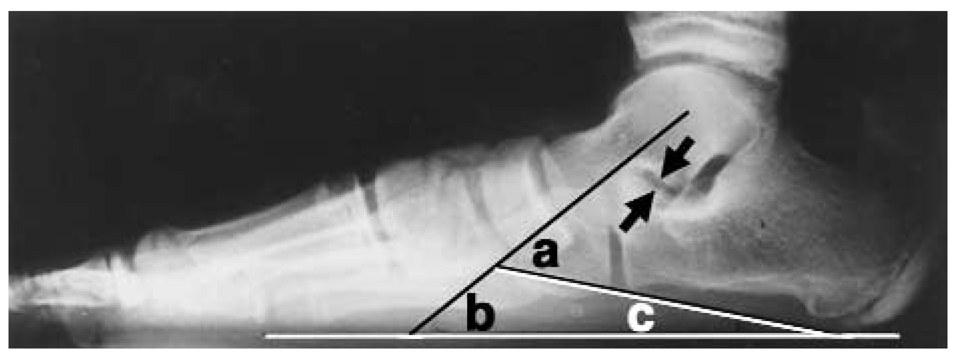
15). These angles were measured to the 10th degree by one physician (P.F.). Sinus tarsi occlusion (Kirby’s sign) is a radiographic change that can indicate abnormal pronation. In this study, sinus tarsi occlusion was measured by the distance between the leading edge of the posterior facet of the talus and the superior aspect of the floor of the calcaneus in the resting calcaneal stance position.
Figure 12.
Preoperative lateral x-ray: a, talocalcaneal angle; b, talar declination angle (pitch of the bisection of the head and neck of the talus); c, calcaneal inclination angle. The arrows indicate a positive Kirby’s sign or occlusion of the sinus tarsi (close approximation of the lateral leading wall of the talus and the calcaneal floor of the sinus tarsi in the pronated relaxed position).
Figure 12.
Preoperative lateral x-ray: a, talocalcaneal angle; b, talar declination angle (pitch of the bisection of the head and neck of the talus); c, calcaneal inclination angle. The arrows indicate a positive Kirby’s sign or occlusion of the sinus tarsi (close approximation of the lateral leading wall of the talus and the calcaneal floor of the sinus tarsi in the pronated relaxed position).
Figure 13.
Pronated postoperative lateral x-ray with the STA-peg implant in place: a, lateral talocalcaneal angle decreased from the preoperative position; b, talar declination angle decreased from the preoperative position; c, calcaneal inclination angle was unchanged from the preoperative position. The arrows show that the STA-peg implant prevents occlusion of the sinus tarsi.
Figure 13.
Pronated postoperative lateral x-ray with the STA-peg implant in place: a, lateral talocalcaneal angle decreased from the preoperative position; b, talar declination angle decreased from the preoperative position; c, calcaneal inclination angle was unchanged from the preoperative position. The arrows show that the STA-peg implant prevents occlusion of the sinus tarsi.
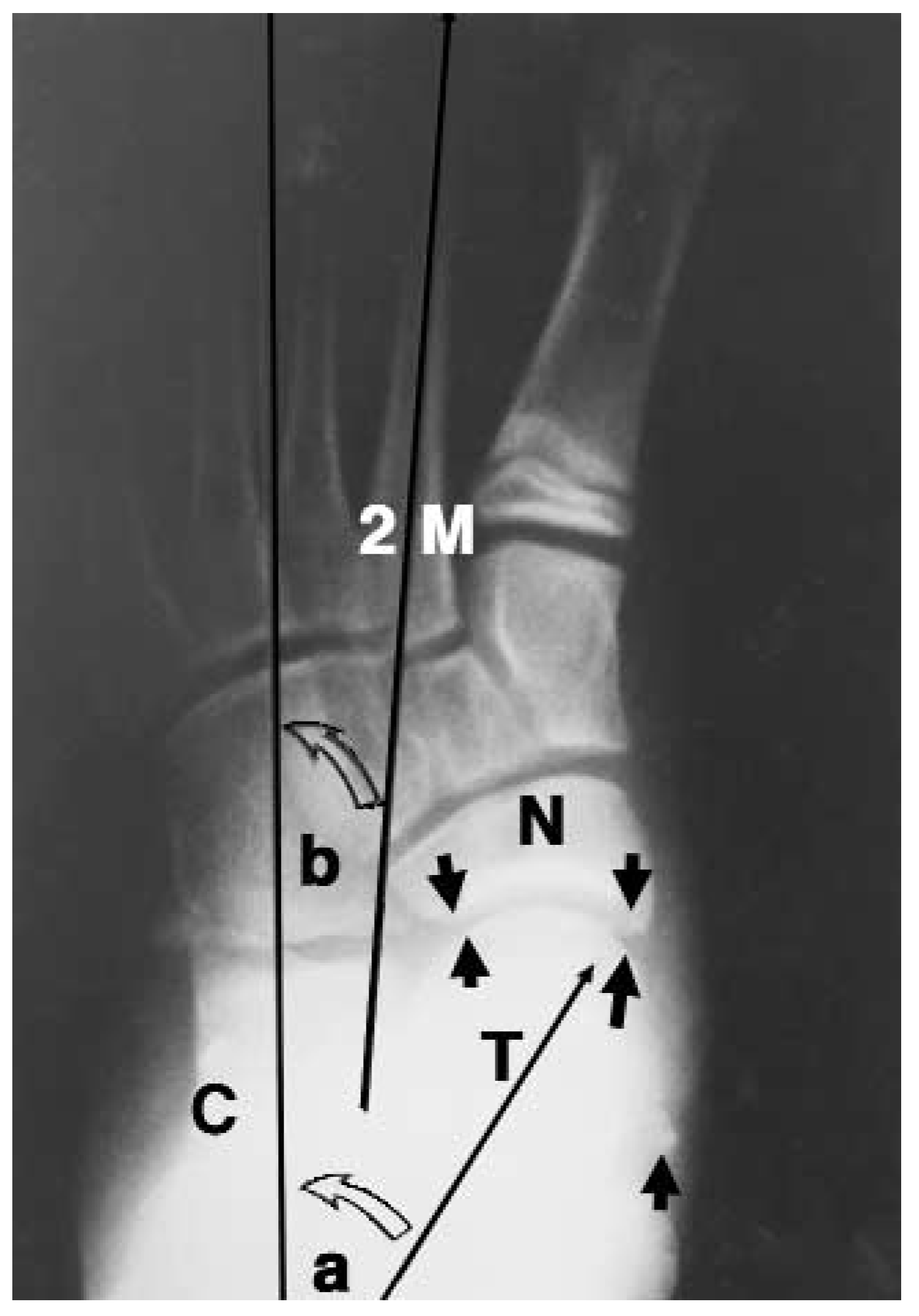
Figure 14.
Preoperative dorsoplantar x-ray in pronated relaxed stance position: a, Kite’s angle (talo [T] calcaneal [C] angle); b, forefoot adductus angle (second metatarsal [2M] versus rearfoot–calcaneus [C]). The arrows indicate the percent of articulation of the talus (T) and the navicular (N).
Figure 14.
Preoperative dorsoplantar x-ray in pronated relaxed stance position: a, Kite’s angle (talo [T] calcaneal [C] angle); b, forefoot adductus angle (second metatarsal [2M] versus rearfoot–calcaneus [C]). The arrows indicate the percent of articulation of the talus (T) and the navicular (N).
Figure 15.
Postoperative dorsoplantar x-ray with the STA-peg in place: a, Kite’s angle decreased from the preoperative position; b, forefoot adductus angle increased from the preoperative position. The arrows indicate that the percent of articulation of the talus (T) and the navicular (N) increased from the preoperative position. 2M, second metatarsal.
Figure 15.
Postoperative dorsoplantar x-ray with the STA-peg in place: a, Kite’s angle decreased from the preoperative position; b, forefoot adductus angle increased from the preoperative position. The arrows indicate that the percent of articulation of the talus (T) and the navicular (N) increased from the preoperative position. 2M, second metatarsal.
The talonavicular articulation indicates how much of the talus is articulating with the navicular. This articulation decreases with pronation as the talus rotates medially and plantarly. It is measured by the distance of the articulating surface of the talus in contact with the navicular that is then divided by the total articular surface of the talus.
Sixteen children with a total of 31 feet had preoperative, postoperative, and long-term postoperative x-rays (at least 1.5 years) available for review. The patients’ ages ranged from 4 to 14 years with a mean age of 8.5 years. The follow-up period ranged from 18 to 57 months with a mean of 34 months. Five children had undergone ancillary procedures, two children had two Kidner procedures, one child had bilateral tendo Achillis lengthening, one child had bilateral McBride bunionectomies and epiphyseal staplings, and one child had a unilateral McBride bunionectomy with epiphyseal stapling.
The radiographic results are reported in
Table 5. The talar declination angle showed a mean decrease of 3.6°, decreasing from 25.1° preoperatively to 21.5° postoperatively. The calcaneal inclination angle measured 16.8° preoperatively and 15.2° postoperatively for a mean decrease of 1.6°. The lateral talocalcaneal angle preoperatively was 41.9° and postoperatively was 35.7°, decreasing 6.2°. The sinus tarsi occlusion measured 4.5 mm preoperatively and 9.2 mm postoperatively for a change of 4.7 mm (
Figs. 12 and
13). The Kite’s angle was 29.1° preoperatively and 19.8° postoperatively, resulting in a decrease of 9.3°, while the navicular coverage of the talar head increased from 48.1% preoperatively to 71.5% postoperatively (
Figs. 14 and
15;
Table 5) for a change of 23.4%. The forefoot adduction angle increased, in general, from preoperatively to postoperatively, although no specific figures were tabulated.
Table 5.
Radiographic Results.
Table 5.
Radiographic Results.
Smith and Millar[
1] stated that the talar declination angle was the most reliable angle to measure because the talus adducts, plantarflexes, and moves anteriorly during pronation. They argued that this movement would manifest itself radiographically by the talar declination angle. Flake et al[
26] indicated, however, that the talar declination angle was not adequate by itself and that the lateral talocalcaneal angle also must be evaluated. They indicated that the variability of the structural calcaneal inclination angle would affect the talar declination angle. The calcaneal inclination angle is a structural angle measuring the pitch of the calcaneus, which changes very little with supination and pronation (
Figs. 12 and
13).[
27,
28]
In patients who have had instability due to the pronation for a long enough period of time to allow for subluxation of the midfoot, plantar subluxation of the rearfoot (talus and calcaneus) can lead to a structurally low calcaneal pitch. Some view this same action as the dorsal subluxation of the forefoot on the rearfoot.[
29] It is simply a matter of perspective, however, since the result is the same. In any event, if this foot supinates, the calcaneal inclination changes little and remains low.
The calcaneal inclination angle can vary significantly from very low in a pes planus foot to very high in a cavus foot. The normal angle ranges from 18° to 30° with an average of 24.5°.[
28] This is grossly independent of a supinated and pronated position. In a cavus foot type with a high calcaneal inclination angle (a high pitch to the whole rearfoot), a lower than normal talar declination angle would be expected in a neutral subtalar joint position. The lateral talocalcaneal angle may be normal. If this same foot were pronated, the talar declination angle may increase to what would be considered a normal range. But when considering both the calcaneal inclination angle and the talar declination angle, an increased lateral talocalcaneal angle would demonstrate a significant increase in pronation.
The talar declination angle is a positional angle that measures the general direction of the head and neck of the talus on a lateral x-ray view (
Figs. 12 and
13). The normal talar declination angle is 21°.[
28] The head and neck of the talus plantarflex with pronation, thus increasing the talar declination angle. Dorsiflexion of the head and neck of the talus occurs in supination, thereby decreasing the talar declination angle.
The lateral talocalcaneal angle is the relationship of the head and neck of the talus to the pitch of the calcaneus (
Figs. 12 and
13). This angle equals the sum of the talar declination angle and the calcaneal inclination angle. Thus, the normal angle is 45.5° (21° plus 24.5°). This angle increases with pronation and decreases with supination. However, the new talar position average angle is variable depending on the structural calcaneal inclination angle.
Occlusion of the sinus tarsi (Kirby’s sign) in a severely pronated foot is the result of the leading edge of the posterior facet of the talus coming into contact with the floor of the calcaneus and occluding this space. This is clearly demonstrated by Green[
27] in his radiocinematography of the foot. When the STA-peg implant is placed in the sinus tarsi, the leading wall of the posterior facet of the talus is blocked from continuing onto the floor of the calcaneus, preventing excess pronation and occlusion of the sinus tarsi (
Figs. 12 and
13). In the present study, the space measured is a direct result of the size of the STA-peg implant. In this study, the children had approximately 5 mm of space improvement postoperatively.
In a pes planus foot type with a low calcaneal inclination angle (low pitch to the rearfoot), a higher than normal talar declination angle might be expected in a neutral subtalar joint position. However, in order for the bisection of the head and neck of the talus to pass through the first metatarsal head, a lower than normal talar declination angle would be expected. In any event, a lower than normal lateral talocalcaneal angle would be expected in the neutral subtalar joint position.
In the present study, the average calcaneal inclination angle of 16.8° was far below normal. The normal angle ranges from 18° to 30° with an average of 24.5°.[
28] Since the calcaneal inclination angle is a structural angle, it changed very little with pronation and supination. In fact, this study demonstrated a small decrease in the calcaneal pitch postoperatively in a less pronated position, averaging a 1.6° decrease to 15.2°.
The talar declination angle was higher than normal at 25.07° preoperatively in this series. The normal angle is 21°. As a result of the low calcaneal inclination angle, the talar declination angle in a neutral position would be expected to be even lower than normal. This is primarily a positional angle, which increases in pronation and decreases with supination. The talar declination angle decreased postoperatively on average 3.6° to a relatively normal 21.5°. However, this postoperative relaxed stance position is expected still to be mildly pronated.
The lateral talocalcaneal angle was 41.9° (calcaneal inclination angle of 16.8° plus talar declination angle of 25.1°). The normal value would be expected to be 24.5° plus 21° degrees, or 45.5°. The measured lateral talocalcaneal angle in this pronated group was lower than normal. With such a low calcaneal inclination angle, any lateral talocalcaneal angle that would approach a more normal range actually would demonstrate significant pronation. The lateral talocalcaneal angle was reduced by an average of 6.2° postoperatively to a low of 35.7°, although this postoperative relaxed stance position ideally is still mildly pronated.
The greatest amount of correction noted in the present study was seen in the transverse plane
versus the sagittal plane (
Figs. 14 and
15). The dorsoplantar talocalcaneal angle, or Kite’s angle, is a positional angle measuring the transverse plane relationship of the head and neck of the talus to the remaining rearfoot (the calcaneus). This angle increases with pronation and decreases with supination. A normal Kite’s angle is 18°.[
28]
The percentage that the navicular covers the head of the talus is a positional relationship that also reflects the transverse relationship of the midfoot and the talus. The talar head becomes more uncovered with pronation and more completely covered with supination. Normally, 75% of the talar head should be covered by the navicular.[
30]
The forefoot adductus angle is a positional angle that measures the transverse plane relationship of the forefoot (second metatarsal) to the rearfoot (calcaneus). This angle decreases with pronation and increases with supination. Note, however, that the metatarsus adductus angle is a structural angle measuring the transverse plane relationship of the forefoot (second metatarsal) to the midfoot or lesser tarsus and changes very little with pronation and supination.
The Kite’s angle was 29.1° preoperatively in this study, which is high and demonstrates a pronated foot. This figure reduced 9.3° to 19.8° postoperatively. Furthermore, 48.1% of the talar head was covered by the navicular preoperatively. This figure increased 23.4% to 71.5% postoperatively. Although detailed figures of the forefoot adductus angle were not compiled, this angle increased from the preoperative value to the postoperative value.
The Kite’s angle, the coverage of the talus by the navicular, and the forefoot adductus angle all demonstrate good correction in the transverse plane and are probably due to the Flake-Austin modification used in the placement of the STA-peg implant in this study. The placement of the implant at a 45° angle more efficiently prevents excessive medial rotation of the talus. Therefore, the transverse planar dominant foot is more corrected than with the traditional method of placement of the STA-peg.
Pronation of the subtalar joint has been defined as adduction and plantarflexion of the talus and eversion of the calcaneus, which encompasses all three body planes. Root et al[
31] described the average subtalar joint axis to be located 42° from the transverse plane and 16° from the sagittal plane, allowing equal amounts of abduction/adduction to eversion/inversion. However, since this is only an average it can be assumed that some individuals deviate from this axis, making planar dominance an important clinical entity.[
32] Planar dominance can be identified easily in the clinical examination by determining the subtalar joint axis. If an individual has a high subtalar joint axis, the majority of motion will be in the transverse plane. During pronation, this manifests clinically as abduction of the forefoot relative to the rearfoot. Radiographically, an increase in the Kite’s angle, a decrease in the percentage of navicular coverage of the talus, and less predominance of the talar declination angle would be noted with pronation.
If the individual had a low subtalar joint axis, the plane of dominance would be the frontal plane. Clinically, a larger calcaneal eversion would be identified during pronation. Radiographically, there would be a decrease in the first metatarsal declination angle and an increase in the lesser tarsal superimposition and midtarsal breech due to the unlocking of the midtarsal joint by excessive calcaneal eversion. This unlocking of the midtarsal joint eventually leads to subluxation.
The complexity of radiographic interpretation indicates that no single radiographic angle can assess all patients with pes planovalgus. The biomechanical evaluation must be considered first before evaluating the radiographs.
Complications
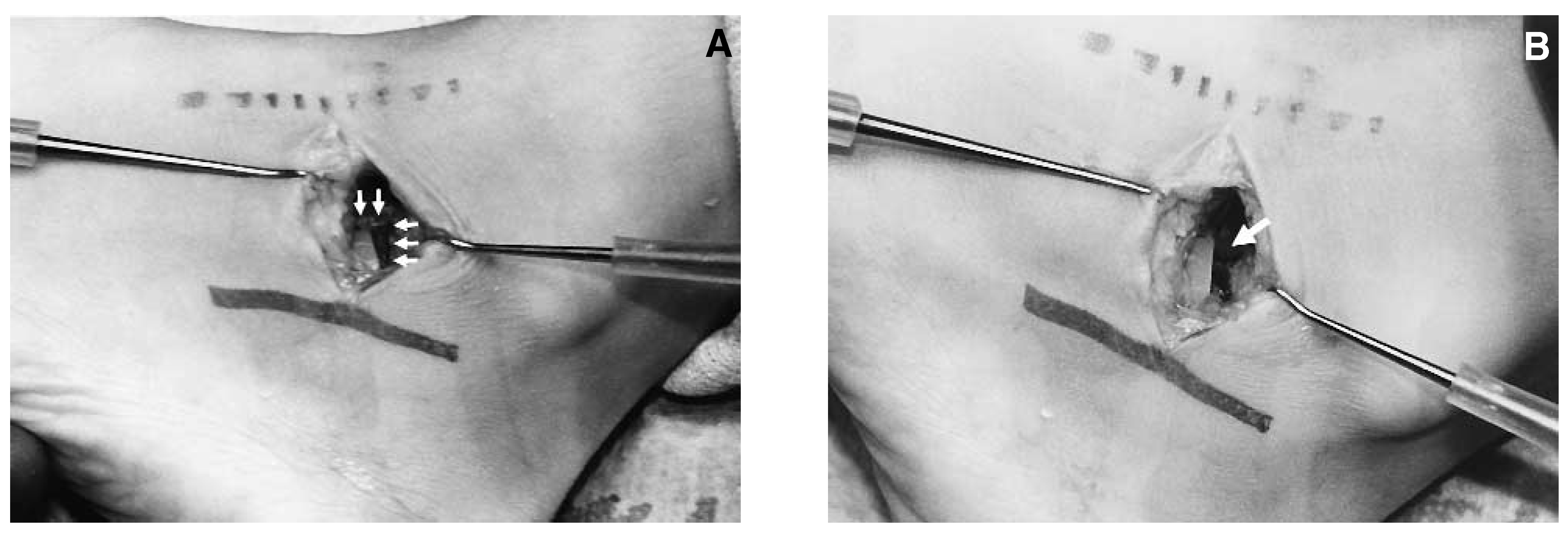
Three complications occurred in the patients in this study. One patient developed peroneal spasm and pain after a ball was forcibly smacked into the dorsolateral aspect of his right foot 5 months postoperatively. The patient’s symptoms gradually worsened, although the left foot, which also had the STA-peg arthroereisis procedure, remained asymptomatic. The patient underwent conservative therapy, which failed, and exploratory surgery was done at 1 year postoperative. During surgery, a 2-mm ledge of talar bone was noted contouring to the deep border of the STA-peg implant (
Fig. 16A). The ledge of bone was removed and the implant was inspected and found to be seated well without loosening (
Fig. 16B). The implant was left in place. The patient has been followed for approximately 4 years without any symptoms.
Figure 16.
A, Sinus tarsi with STA-peg in place. The deep talar exostosis is contoured to the dorsal deep aspect of the STA-peg that is marked with two arrows. B, Sinus tarsi with STA-peg still in place. As indicated by the single arrow, the talar exostosis has been removed.
Figure 16.
A, Sinus tarsi with STA-peg in place. The deep talar exostosis is contoured to the dorsal deep aspect of the STA-peg that is marked with two arrows. B, Sinus tarsi with STA-peg still in place. As indicated by the single arrow, the talar exostosis has been removed.
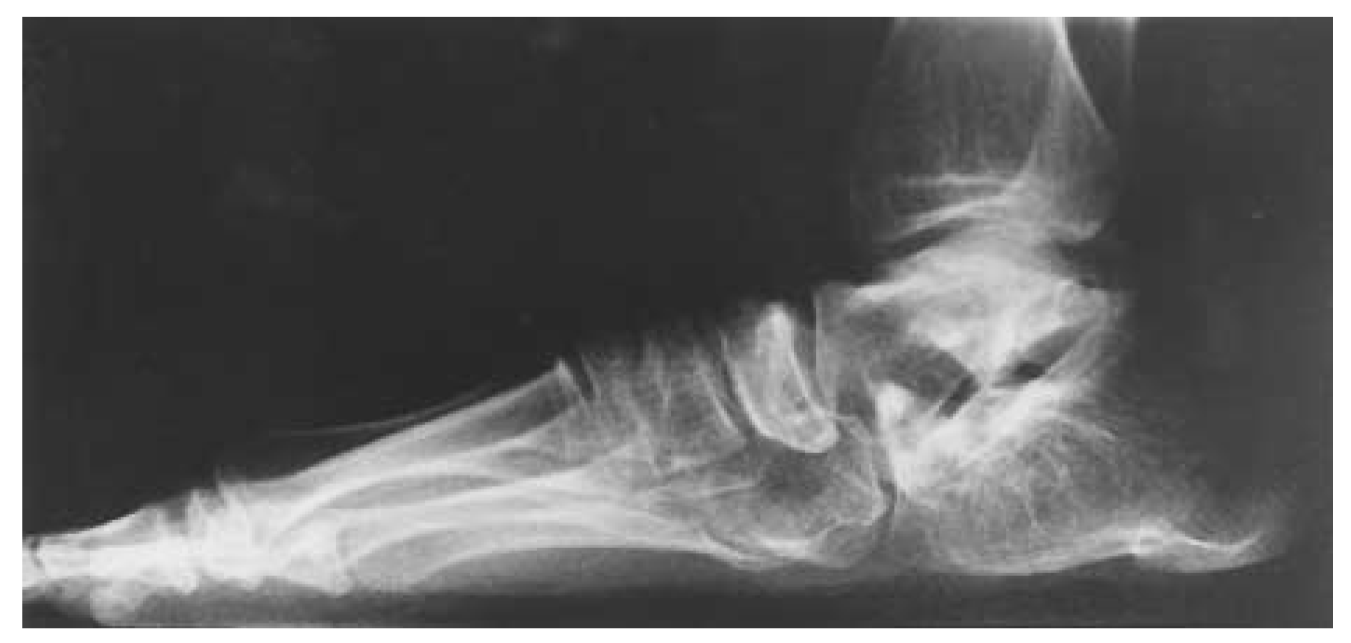
A second patient has had occasional, but recurring, sinus tarsi pain that has required several injections postoperatively. However, the patient’s overall symptoms have decreased significantly and she is very pleased with the results. This patient has a congenital arthropathy with some malformation in the pedal bones (
Fig. 17). The patient’s mother had a similar arthritic involvement and has limited extended ambulatory capacity. This patient was unable to participate in regular physical education classes and therefore this is included as a complication, but she has significantly improved her normal walking patterns.
Figure 17.
STA-peg in a patient with familial arthropathy at 3 years postoperative. Note the irregular contour of the pedal bones, especially the talus.
Figure 17.
STA-peg in a patient with familial arthropathy at 3 years postoperative. Note the irregular contour of the pedal bones, especially the talus.
A third patient experienced increased symptoms postoperatively and required removal of both STA-pegs. The patient had excessive range of motion and methyl methacolate was used with the STA-peg to build up the sinus tarsi. There was some question as to excessive amounts of cement used. Some viscous material, which appeared to be synovial or ganglionic in nature, was drained from the sinus tarsi areas bilaterally on several occasions. The implants were removed bilaterally. However, even after the STA-pegs and the cement were removed, the patient still experienced pain. This is the only true long-term complication in this study group. This patient is now more than 9 years postoperative from the original surgery and 8 years postoperative from the removal of the implants. Her mother reports that in spite of the complications and the additional surgery required, her daughter is functioning slightly better than she did preoperatively.
Conclusion
This study’s review of the STA-peg arthroereisis procedure has shown many rewarding results and supports favorable findings that also have been published by Smith and Millar[
1] and Lundeen.[
2] In addition to objective results, the authors also quantitatively and qualitatively recorded very positive subjective results. There are many advantages to using the STA-peg arthroereisis in children who experience excessive pronation. The growing child’s osseous structure potentially is able to adapt to a more corrected position if severe pronatory forces can be neutralized.
An important advantage of the Flake-Austin placement of the STA-peg arthroereisis procedure is that it does not involve placing the peg into an articulating joint. If complications do arise, the implant is easily removed, as in the most serious complication in this study.
A further advantage with the modification of the STA-peg placement is that the transverse planar dominant deformities are controlled more readily. It is a relatively benign procedure for the growing child with more severe forms of flexible pes valgo planus deformities and it also allows the bones to adapt to a more normal structure rather than be condemned to continue to grow into the deformed compensated position.