Elective foot and ankle surgery in patients with peripheral neuropathy, whether due to diabetes mellitus or some other etiology, is commonly performed without major concern. Often, diabetic patients have no history or radiographic signs of Charcot’s arthropathy and, in fact, have a subclinical case of peripheral neuropathy. In such cases, these patients, similar to non-neuropathic patients, suffer from painful bone and joint pathologies that include hallux valgus, hallux limitus, hammer toes or claw toes, and exostoses. An important issue is whether the procedure or the postoperative course in patients with the potential to develop Charcot’s arthropathy should be modified. This article presents multiple reports of cases in which podiatric surgery resulted in Charcot-type degeneration. Four cases are presented in detail and an additional five cases are reviewed in table format.
Unfortunately, very little information on this topic exists in the medical literature. Multiple cases of iatrogenic causation of Charcot’s disease following nerve ablation have been reported. For example, in 1970, Falzi and Falzi[
1] reported on two cases of Charcot’s arthropathy following traumatic lesions of peripheral nerve trunks. In 1993, Smith and Shane[
2] reported on a case of Charcot’s foot following medial and lateral calcaneal nerve excision for recalcitrant heel pain. More recently, a report from Darst et al[
3] described Charcot-type degeneration following a Keller arthroplasty. According to Darst et al, “Charcot’s joint has not previously been reported as a sequela of elective foot surgery.” A review of the medical literature revealed only a few reports on the topic.
It has been reported that trauma can be a precipitating factor in the development of Charcot’s arthropathy. For example, in 1988, Mok et al[
4] described the development of a Charcot joint after an intertrochanteric fracture of the femur. In addition, Berg[
5] reported on three cases of middle-aged patients with diabetes who developed Charcot’s hips following acetabular fractures. Prior to injury and subsequent open reduction and internal fixation, these patients had no radiographic evidence of Charcot’s arthropathy. A debatable question is which trauma caused the Charcot’s degeneration: the original fracture or the surgical procedure?
Another interesting question is whether clinical neuropathy must be present for a patient to develop a Charcot joint. According to Kuur,[
6] “Charcot arthropathy may develop in patients with normal protective joint sensitivity.” Kuur reported on two cases of Charcot’s shoulder joints in two non-neuropathic patients.
Case Reports
Case 1
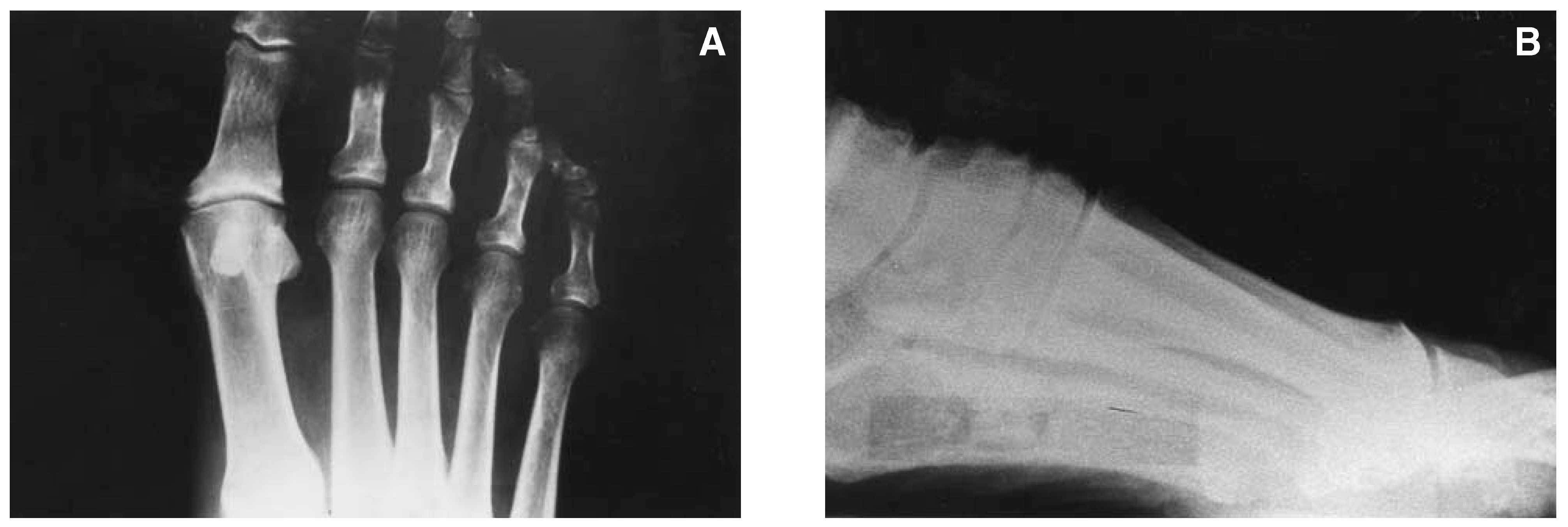
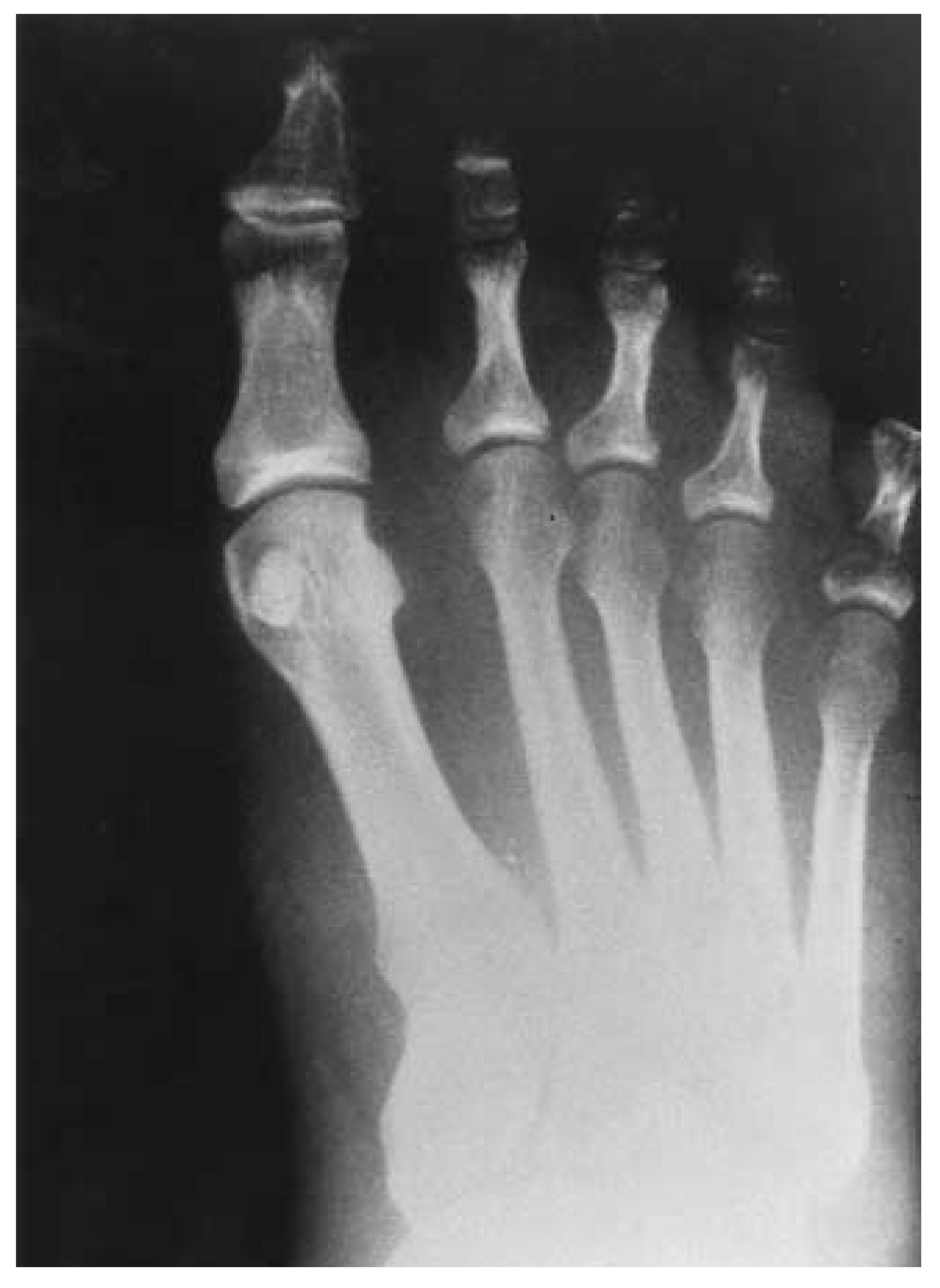
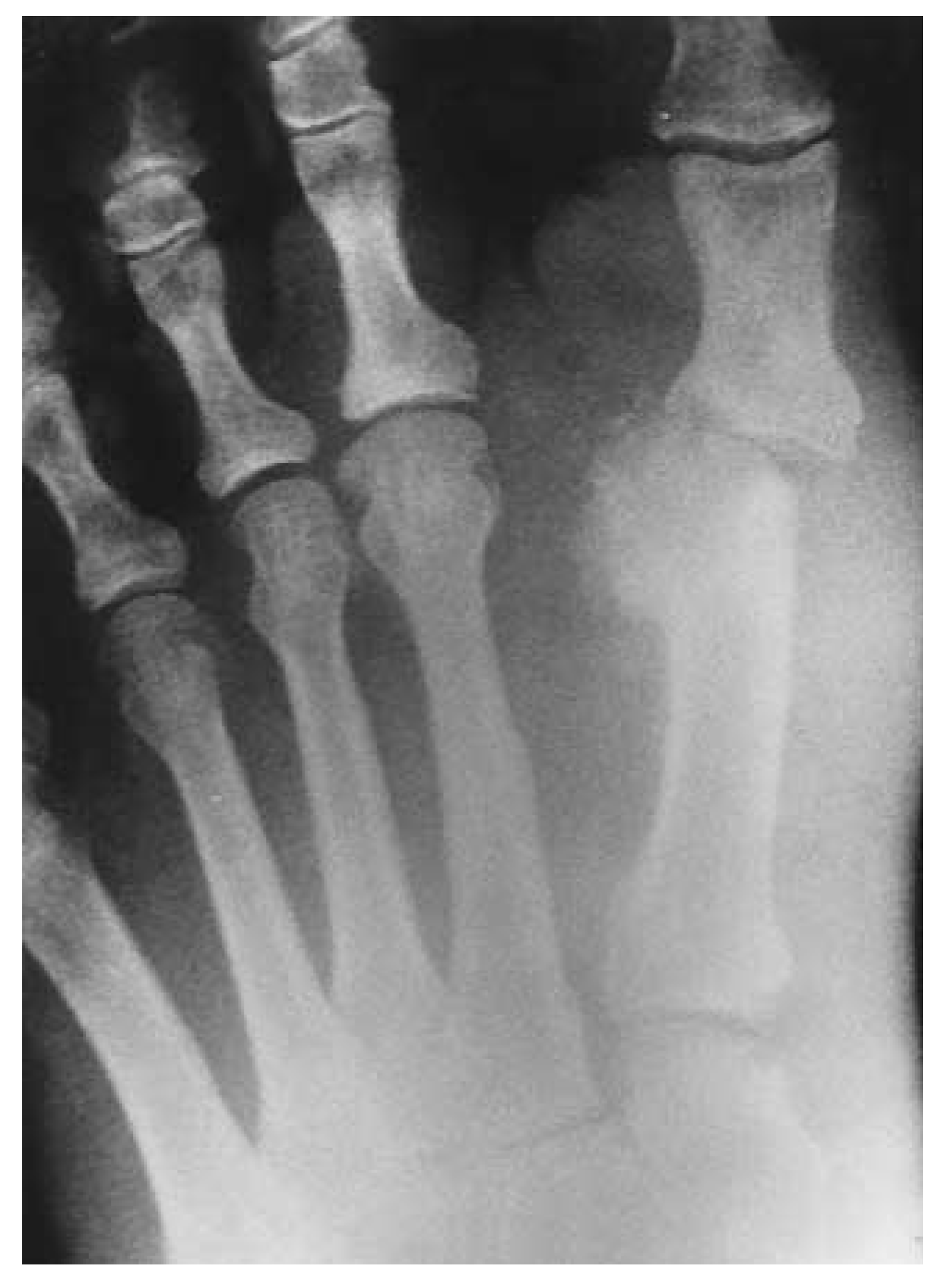
A 38-year-old man was being treated for a neurotrophic ulceration at the plantar medial aspect of the hallux of the right foot. He had had diabetes mellitus for approximately 5 years and was taking an oral hypoglycemic agent. The range of motion of the first metatarsophalangeal joint was significantly reduced, to about 25° of dorsiflexion. There was a “hard” end range of motion of the first metatarsophalangeal joint, which produced pain. Radiographs revealed a mild hallux valgus deformity with nonuniform joint-space narrowing. Also noted was a squared-off metatarsal head with a mild dorsal exostosis (
Fig. 1). A diagnosis of hallux limitus and associated neurotrophic ulcer was made. Conservative treatment (wound care and off-loading) was attempted, but it failed to heal the ulcer. The patient was then scheduled for hallux limitus repair, which consisted of a Waterman-Green subcapital osteotomy. In addition, an exostectomy of the plantar medial condyle of the base of the proximal phalanx was performed.
Figure 1.
A, Anteroposterior and B, lateral preoperative views. Note the squared-off metatarsal head and mild dorsal exostosis.
Figure 1.
A, Anteroposterior and B, lateral preoperative views. Note the squared-off metatarsal head and mild dorsal exostosis.
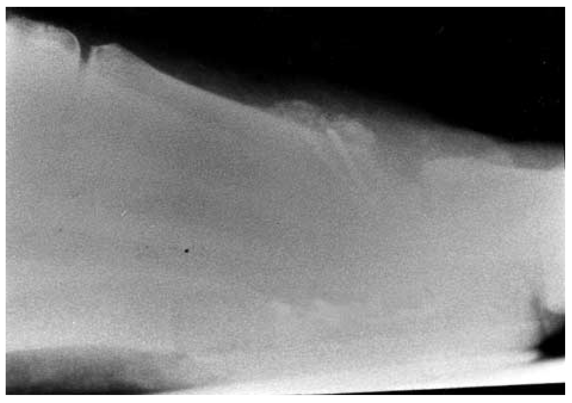
Immediate postoperative radiographs revealed crossing Kirschner-wire fixation. Slight shortening and plantarflexion of the first metatarsal head were achieved as well as relocation of articular cartilage. At the patient’s first postoperative checkup, there were no problems with incision healing, nor were there any signs of infection. At 2 weeks postoperatively, radiographs were taken, which were unremarkable. Fixation was adequate, and there was no rotation or disturbance of the capital fragment. At 2 months postoperatively, the patient complained of redness and swelling at the surgical site. He noted some pain at times; however, it was not debilitating. Clinically, increased temperature and obvious edema about the first metatarsophalangeal joint were noted. There was no pain with range of motion. Radiographs were taken and revealed a significant amount of hypertrophic bone formation, most notably on the lateral view (
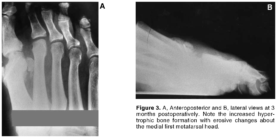
Fig. 2). A tentative diagnosis of Charcot’s arthropathy was made at this point. The patient had been using a postoperative surgical shoe and was instructed to use a removable cast. The patient was seen for follow-up visits regularly and experienced very little pain. The joint became less warm and swollen by the 12th postoperative week. Serial radiographs were taken at 3 months (Fig. 3) and 21 weeks (Fig. 4).
Figure 2.
Lateral view at 2 months postoperatively. Note the hypertrophic bone formation about the osteotomy site.
Figure 2.
Lateral view at 2 months postoperatively. Note the hypertrophic bone formation about the osteotomy site.


Case 2
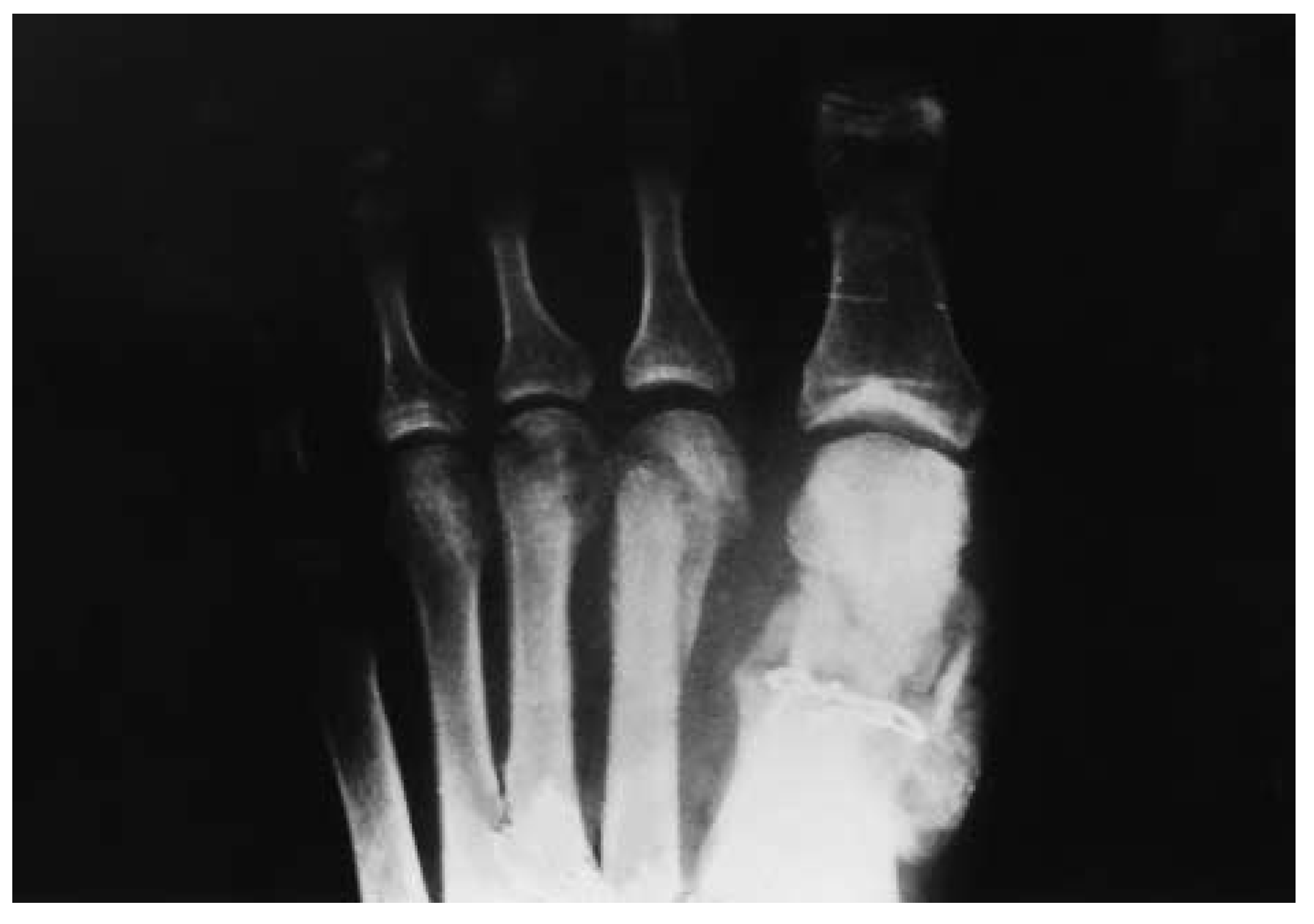
A 32-year-old insulin-dependent diabetic woman presented with a chronic malperforans ulcer under the first metatarsal head of the left foot. After conservative treatment was attempted without success, the patient underwent a dorsiflexory osteotomy of the first metatarsal. Preoperative radiographs failed to reveal any bony pathology. The patient was nonweightbearing for 6 weeks; radiographs taken at this time showed adequate bone healing. The patient was then allowed progressive weightbearing. She returned 1 week later with a hot, swollen foot. An area of induration was felt at the osteotomy site. Immediate casting and nonweightbearing were instituted. Radiographs taken after 3 weeks of cast immobilization revealed Charcot-type degeneration not only at the osteotomy site but also at the second and third metatarsal heads (
Fig. 5). The bones healed after approximately 7 months in a removable cast boot.
Figure 5.
Anteroposterior view 3 weeks after the initial Charcot episode. Note the degeneration at the osteotomy site and at the second and third metatarsal heads.
Figure 5.
Anteroposterior view 3 weeks after the initial Charcot episode. Note the degeneration at the osteotomy site and at the second and third metatarsal heads.
Case 3
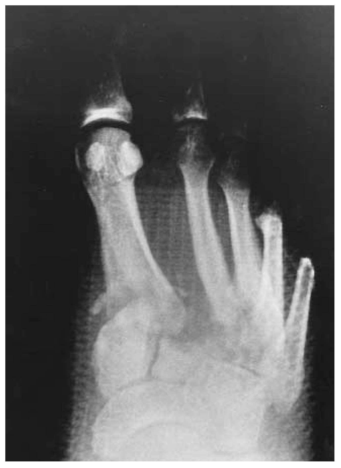
In the case of a progressive acute infection, amputation is often necessary (even when it is biomechanically unsound) to prevent loss of limb or life. The following is an example of such a patient with a severe diabetic foot infection that resulted in incision and drainage with partial resection of the fourth and fifth rays. A preoperative radiograph is shown in
Figure 6. The patient ultimately recovered from the infection and was discharged from the hospital when the wound was free of infection and of adequate healing potential, the white blood cell count was normal, and the patient was afebrile. The patient was closely followed at weekly intervals. At 3 weeks postoperatively, the surgical wound was closed and there was no sign of infection. Protected weightbearing was instituted at that time. At approximately 1 month postoperatively, the foot became red, hot, and swollen. Radiographs showed Charcot-type degeneration across the Lisfranc’s joint (
Fig. 7).
Figure 6.
Preoperative anteroposterior view.
Figure 6.
Preoperative anteroposterior view.
Figure 7.
Anteroposterior view at 1 month postoperatively. Note the degeneration at the Lisfranc’s joint.
Figure 7.
Anteroposterior view at 1 month postoperatively. Note the degeneration at the Lisfranc’s joint.
Case 4
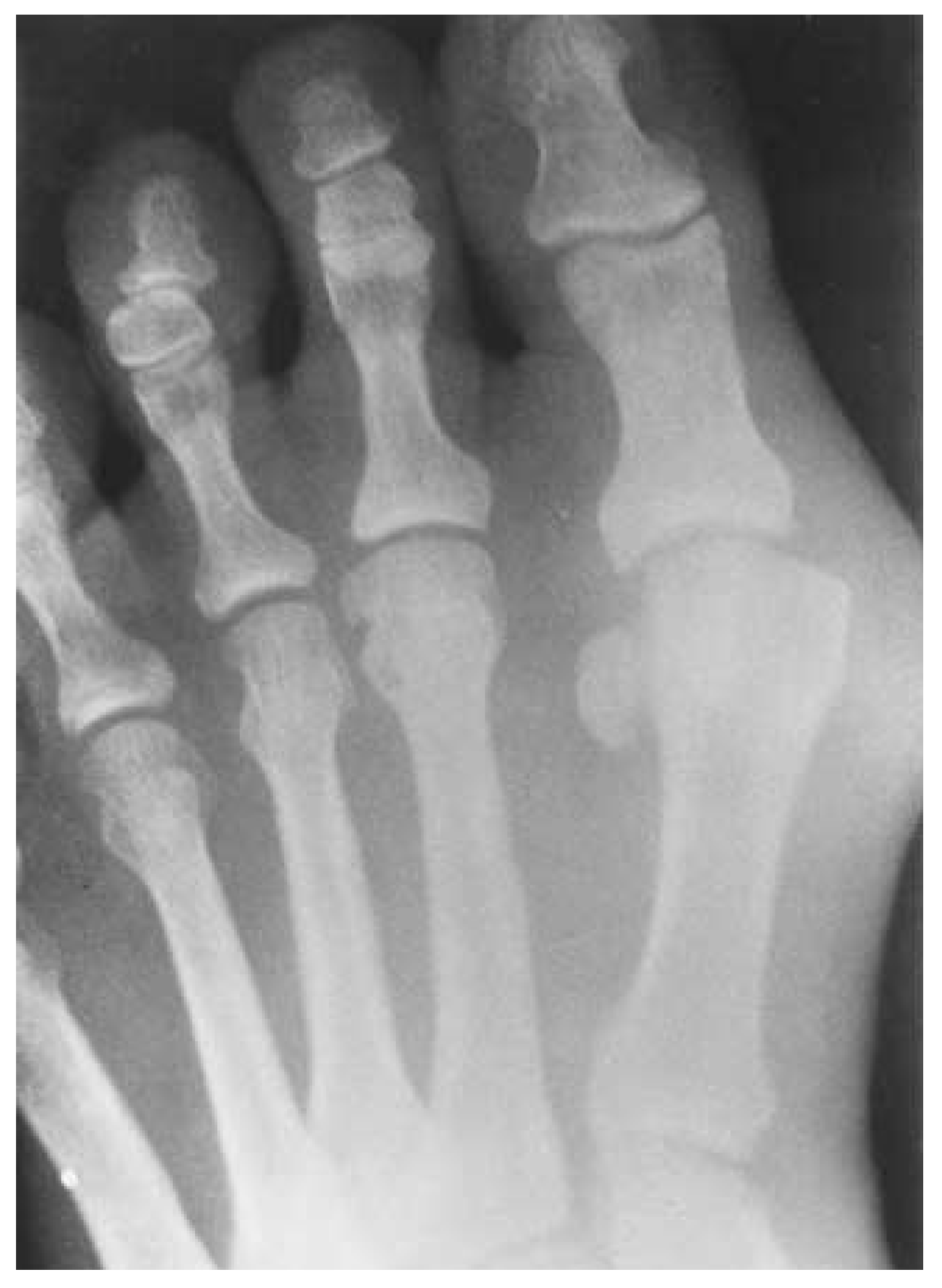
A 60-year-old man presented for correction of hallux valgus (
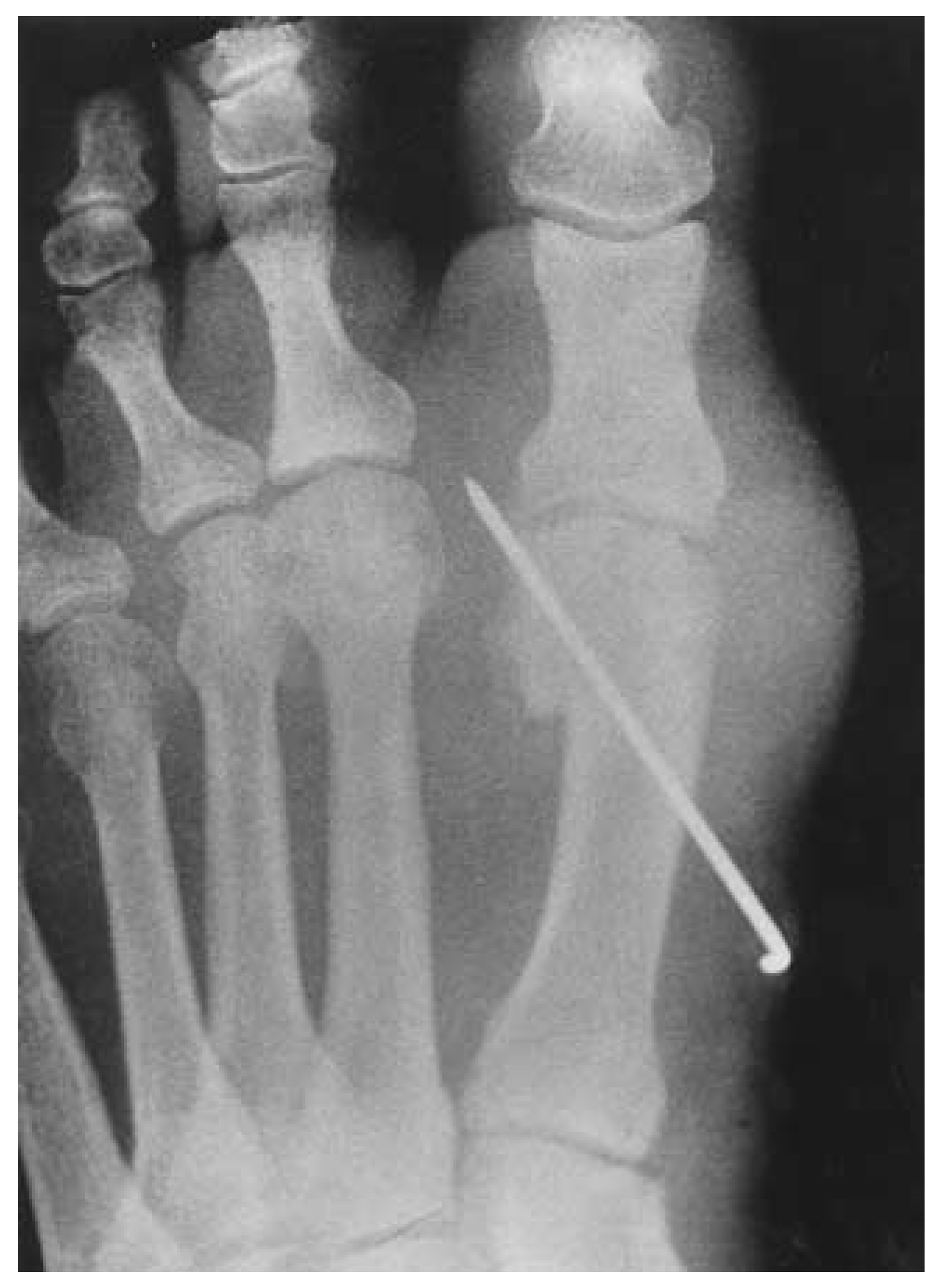
Fig. 8). He had had diabetes for a number of years and was taking an oral hypoglycemic agent. A traditional Austin bunionectomy with percutaneous Kirschner-wire fixation was performed (
Fig. 9). His operation and postoperative course were uneventful until approximately 8 weeks postoperatively, when he presented with a red, hot, swollen foot. Radiographs showed Charcot-type degeneration with development of hallux varus (
Fig. 10). Continued deterioration of the first metatarsophalangeal joint and eventual dorsal dislocation of the second digit were noted at 10 weeks postoperatively.
Figure 8.
Preoperative anteroposterior view revealing a moderate hallux valgus deformity.
Figure 8.
Preoperative anteroposterior view revealing a moderate hallux valgus deformity.
Figure 9.
Anteroposterior view immediately postoperatively.
Figure 9.
Anteroposterior view immediately postoperatively.
Figure 10.
Anteroposterior view 8 weeks postoperatively.
Figure 10.
Anteroposterior view 8 weeks postoperatively.
Table 1 lists five additional cases of surgically induced Charcot’s foot.
Table 1.
Additional Cases of Surgically Induced Charcot’s Foot.
Table 1.
Additional Cases of Surgically Induced Charcot’s Foot.
Discussion
Nine cases of foot surgery in the diabetic population have been presented. Four cases were reviewed in detail, with case histories and supporting radiographs. In each instance, Charcot’s arthropathy developed. Only one patient had a preexisting Charcot episode. In each of these cases, the etiology of Charcot’s arthropathy is attributed to alteration of weightbearing forces. In addition, changes in the motion of bones or joints play a significant role in the development of Charcot’s disease. Alteration of weightbearing can occur with the removal of a sesamoid bone, the shortening or elevating of a metatarsal, increasing or decreasing joint range of motion, and after removal of a metatarsal head.
A red, hot, swollen foot following surgery should make one suspect infection as well. An argument could be made that some of the patients with these symptoms may have had cellulitis or infection. This can be a diagnostic dilemma, as there is no reliable test to differentiate Charcot’s arthropathy from infection, other than recovery of an infectious agent by tissue or blood culture. The unique features in each of these cases include lack of constitutional symptoms, delayed onset of symptoms following surgery, the appearance (often) of symptoms shortly after the start of weightbearing, and, in some cases, the occurrence of bone destruction in areas far from the operative site. Moreover, in each case, the acute episode resolved with immobilization without administration of anti-infective agents. This information is sufficient to make one cautious in the selection of procedures and postoperative protocol in patients who are susceptible to Charcot-type degeneration.
There is conflicting information regarding the most common sites for Charcot’s joints in the foot. Most authors agree that the forefoot (metatarsophalangeal joints and interphalangeal joints) and the midfoot (tarsometatarsal joint) are the most common sites for Charcot’s arthropathy.[
7-
11] Regardless of whether these are the most common sites, certain assumptions can be made when performing surgery in these areas.
Protection against bending forces on the surgery site is critical during postoperative healing. Osteotomies or exostectomies of the midfoot may require extra care, especially if there is an equinus deformity of the ankle. Traditionally, when significant ankle joint equinus is present, Charcot’s arthropathy will occur at the site of maximum bending force (the midfoot) to allow dorsiflexion of the foot, instead of at the ankle.
Conclusion
Surgically induced Charcot’s foot is not as rare as the medical literature suggests. When surgery is performed on patients with diabetes, there is always a serious risk of developing a Charcot foot. Surgery ultimately alters the weightbearing forces and function of the foot. A sudden increase in temperature or swelling in the foot during the postoperative period should raise the index of suspicion for acute Charcot’s arthropathy in this patient population. Strict nonweightbearing immobilization is recommended.
The less-is-more approach may be advisable in patients who are at high risk for Charcot’s disease. For example, a patient who requires hallux valgus correction may be well suited for a modified McBride bunionectomy rather than an osteotomy. Furthermore, distal osteotomies for hallux valgus surgery usually can be followed by full weightbearing in a postoperative shoe. For patients who are at risk of developing Charcot’s disease, protected weightbearing, bracing, or even immobilization may be prudent.