Next Article in Journal
Talar Fractures. Three Case Studies
Previous Article in Journal
Flake-Austin Modification of the STA-Peg Arthroereisis. A Retrospective Study
 
 
Journal of the American Podiatric Medical Association is published by MDPI from Volume 116 Issue 1 (2026). Previous articles were published by another publisher in Open Access under a CC-BY (or CC-BY-NC-ND) licence, and they are hosted by MDPI on mdpi.com as a courtesy and upon agreement with American Podiatric Medical Association.
Article

Hemophilic Arthropathy: Considerations in Management

by
Thomas J. Chang
1,
Shenin Mohamed
2 and
Julie Hambleton
3
1
Clinical Professor and Immediate Past Chairman, California College of Podiatric Medicine; private practice San Francisco and Santa Rosa, CA. Mailing address: 2281 Cleveland Ave, Santa Rosa, CA 95403
2
Submitted during second-year residency, Memorial Hospital of Rhode Island, Brown University, Pawtucket, RI
3
Associate Clinical Professor, University of California, San Francisco School of Medicine, San Francisco, CA
J. Am. Podiatr. Med. Assoc. 2001, 91(8), 406-414; https://doi.org/10.7547/87507315-91-8-406

Published: 1 September 2001

Abstract

The authors evaluate nonsurgical and surgical approaches to treating patients with hemophilic arthropathy and review the functional and economic limitations imposed on treating these patients. Indications for surgery are discussed and a case study that incorporates both conservative and surgical management options is presented. While the advent of factor replacement therapy has dramatically changed the course of treatment and prognosis for patients with hemophilia, the authors argue that the economic burden of treating these patients is still very high. The authors recommend that proper conservative and surgical management options for patients with hemophilia should be based upon a thorough understanding of the disease process.

In severe hemophilia, osteoarthropathy of one or more joints is an expected and common sequela to recurrent episodes of acute, spontaneous hemorrhages into the joints. Due to chronic intra-articular bleeding, degenerative changes such as hypertrophy and inflammation of the synovial tissues result, followed by the subsequent release of proteolytic enzymes from the synovium that serve to further destroy the surrounding articular cartilage.
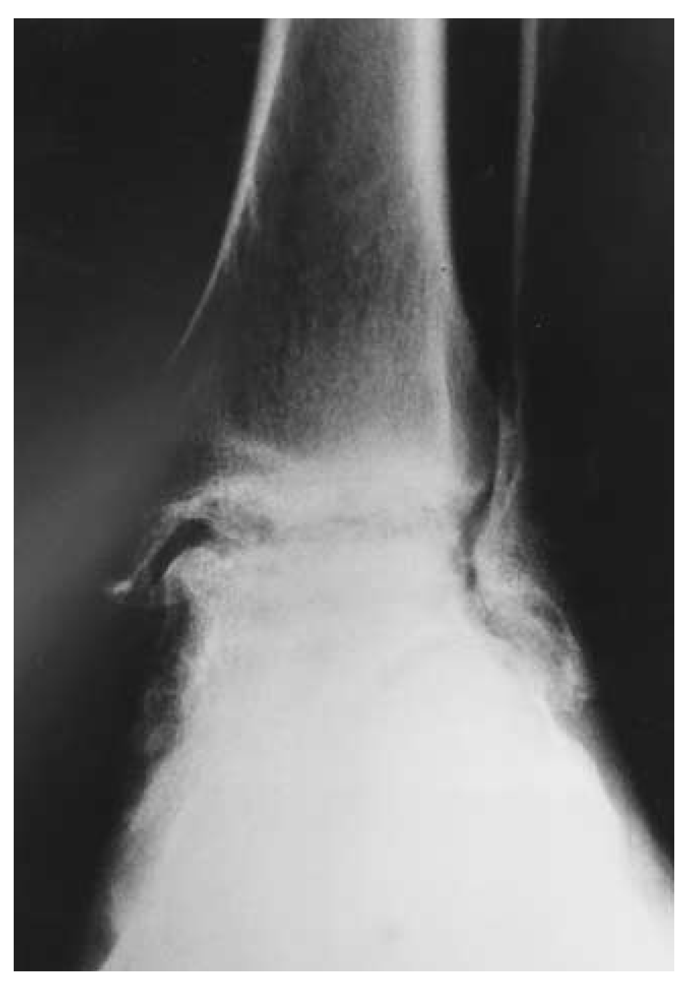
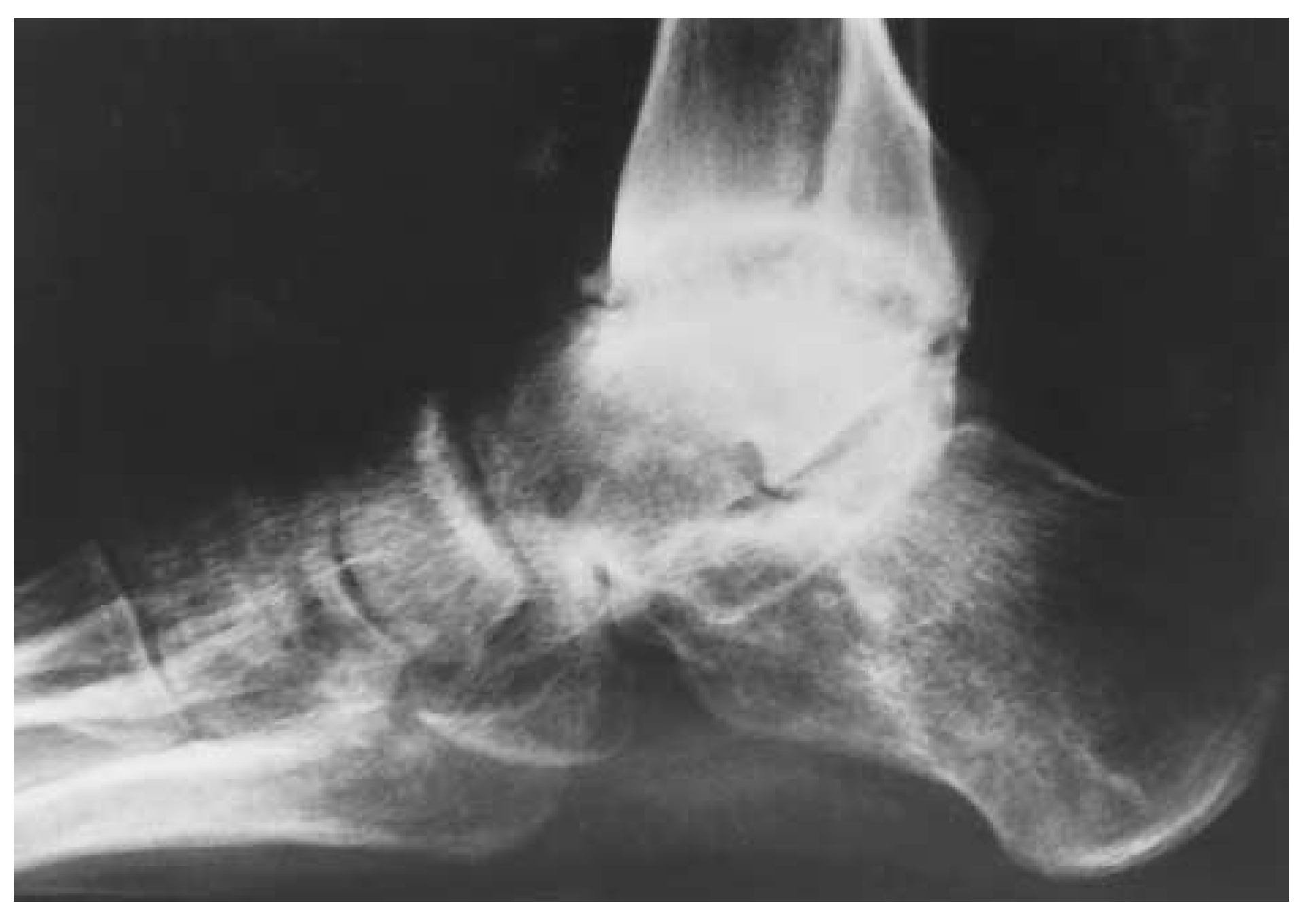
Joints in which there is repeated hemarthrosis will suffer from longer and more intense episodes of intra-articular bleeding and may become chronically affected by synovitis (Figs. 1 and 2). Therefore, early treatment for the prevention of chronic synovitis is very important.[1-8] Arthropathy is often polyarticular, and the lower limbs are affected more often than the upper limbs. In order of frequency, the knee joint is affected most often, followed by the elbow and then the ankle.[3-5,8-14]
Figure 1. Classic example of hemophilic intra-articular bleeding into the ankle joint with subsequent degeneration.
Japma 91 00406 g001
Figure 2. Lateral view of ankle joint.
Japma 91 00406 g002
Before the era of replacement therapy with human clotting factor, there was no effective treatment to control spontaneous episodic hemorrhages. The only treatments available were noninvasive modalities, such as immobilization and rest. However, with the advent of human factor VIII and IX replacement therapy, it has become possible to perform major surgical procedures on patients with hemophilia, such as total hip replacements, provided that hemostatic levels of the clotting factor are secured during the perioperative period.[2,15,16] Ironically, however, the development of prophylactic factor replacement therapy has dramatically decreased the need for surgical intervention to correct lower-extremity pathology during the past 10 years. Progress in the treatment of patients with hemophilia has significantly improved their quality of life, resulting in a population of hemophiliac patients who are living longer and functioning without pain or complications associated with factor deficiency.[17,18]

Nonsurgical Approach to Treatment

Patients with hemophilic arthropathy often experience early joint destruction, which is commonly followed by biomechanical malalignments such as joint contractures and limb angulations of the lower extremity. The most common deformities affecting the ankle and subtalar joints are fixed plantarflexion due to degenerative arthritis of the anterior ankle, varus hindfoot due to malalignment of the subtalar joint, and valgus rotation at the ankle due to differential overgrowth of the distal tibial epiphysis during adolescence or progressive degenerative disease during maturity. These disabilities often result in severe pain and physical limitations, motivating the patient to seek immediate relief.[18-23]
Conservative and prophylactic treatments always should be suggested and attempted prior to surgical intervention. Conservative treatment for ankle arthropathy involves the application of splints, braces, wedge insoles, and calipers. In addition, regular prophylactic transfusions of clotting factor may prevent recurrent intra-articular bleeds and further development of hemarthrosis. Both of these factors will allow inflammation of synovial tissue to be resolved. For control of acute hemorrhages, factor replacement treatment should be administered immediately. For recurrent, chronic episodes, factor replacement is given at frequent prophylactic intervals at an adequate dosage to prevent breakthrough bleeding.[13-26]
Previously, it was thought that nonsteroidal anti-inflammatory agents to treat pain and inflammation associated with acute hemarthrosis, synovitis, and chronic arthritis in patients with hemophilia would result in an increased frequency of gastrointestinal bleeding as an adverse drug reaction. However, this has proven to be incorrect. Patients with chronic arthritis and synovitis may safely benefit from oral treatment with nonsteroidal anti-inflammatory agents. The effect of platelet binding with these agents affects a different pathway for clotting than the intrinsic cascade. If analgesia is still inadequate, opioids should be added to the regimen.[17,27]
Hemarthrosis imposes functional and economic limitations. Even if a transfusion of human clotting factor is given immediately, an acute hemarthrosis is extremely painful, limiting a patient’s ability to walk and participate in physically demanding activities for at least 1 to 10 days.[2-5,8-11] Surgery, as well, can be very stressful for the patient and runs the risk of exposing the patient to the development of antibodies against the high doses of clotting factor that may need to be infused over a short perioperative period of time.[28,29]
There are two forms of hemophilia. Hemophilia A is when human clotting factor VIII is deficient, and hemophilia B is when human clotting factor IX is deficient. Hemophilia A and B are further divided into three categories depending on their level of factor VIII: severe (< 1%), moderate (2%–5%), and mild (> 5%). Patients with severe hemophilia have been receiving human clotting factor VIII replacement therapy for prophylaxis in hemophilia A since 1958. Factor IX replacement therapy for hemophilia B has been available since 1972. Regular prophylactic therapy, although expensive, always should be a therapeutic consideration in the management of all patients with hemophilia, as the efficacy of this treatment is such that, if started early enough, future needs for osseous or soft-tissue surgical interventions become unnecessary. In fact, during the last 20 years, levels of prophylactic replacement therapy have intensified, now generally beginning when patients are 1 to 2 years of age. Replacement therapy is given at dosages high enough to prevent the concentration level of the factor from falling below 1% to 2% of normal, reducing episodes of intra-articular bleeding.[2,11,17,26-28,30-34]
The advent of clotting factor and the increased availability of factor concentrates have led to a trend over the last 10 years of an increasing survival rate for patients with hemophilia, an older average age of patients, reduced incidence of severe joint disability, and, ultimately, a decreased need for classic hemophilic surgery. However, it also should be noted that the availability of coagulation factor replacement therapy has made reconstructive and corrective surgery possible for the relief of pain caused by severe arthropathy. The regimen for the infusion of factor concentrates for surgery is based on specified protocols derived from serum factor studies conducted before surgery, as well as the patient’s weight and the presence of any inhibitor.[11,17,26-28,30,31]
To raise the level of serum clotting factor by 2%, 1 unit/kg of factor VIII must be infused. Thus, to raise the level of serum clotting factor to 80% in a patient with severe hemophilia, 40 units/kg must be infused. For a 70-kg patient, this would mean 2,800 units of factor. As the biological half-life of factor VIII is 12 hours, infusions must be administered every 8 to 12 hours to ensure that steady levels of serum factor are maintained throughout the perioperative period. The half-life for factor IX is longer, so dosing is given over longer intervals. Concentrates may be administered by an intravenous push or a continuous fusion.[17,27,35]
Approximately 30 min before a major operation, the patient usually is given a transfusion to raise the serum level of the deficient clotting factor to levels 80% to 150% of normal. The target factor level is dependent on the type of procedure to be performed. Repeat doses may be given during the surgery if the procedure continues for longer than 90 min, or if the plasma factor concentration falls below 30%.[2-5,8]
Transfusions are continued throughout the duration of the patient’s 10 to 14 days in the hospital, ensuring that factor levels are maintained at 30% to 40% of normal. During the process of wound reorganization, at 3 to 5 days postoperatively, the clot is actually weaker than at the time of surgery. It is important to maintain at least a 50% factor level during this early period of wound healing. The factor is then given at longer intervals after the patient’s discharge, maintaining factor concentrations of 10% to 20% of normal. Physical therapy in the early weeks after surgery also may cause bleeding to occur and factor levels should be considered. An adequate regimen serves to minimize the risk of spontaneous hemarthrosis during the immediate postoperative period.36-38 For minor surgery, the serum levels are kept significantly lower and maintained for shorter periods of time.[26,27,31,33,36,38]
The economic burden of just a single transfusion to treat a minor episode of intra-articular bleeding is very high. A single transfusion of factor VIII concentrate can cost from $1,200 to $1,500. In 1994, one unit of factor VIII concentrate at the University of North Carolina Hospital (Chapel Hill, North Carolina) was priced at approximately $1.01 for factor VIII produced by monoclonal antibody technique, or $1.23 for recombinant DNA factor VIII. The dosage of a transfusion for an uncomplicated acute hemarthrosis is 25 units/kg of body weight. Therefore a 50-kg patient would require 1,250 units of factor at a cost ranging from $1,263 to $1,538 for a single transfusion.[8,36-39] Factor IX is twice as expensive as factor VIII.
For major cases of hemarthrosis or surgery, several additional transfusions would be required. Despite the cost of multiple clotting factor infusions necessary during the perioperative period, the number of transfusions required after a synovectomy or arthroplasty is significantly decreased by the end of the first postoperative year. Therefore, long-term health-care costs are reduced considerably through surgical intervention with the expected reduction in the number of intra-articular hemorrhages.[8,37,38]
During the 1970s and early 1980s, the factor concentrates used to treat patients with hemophilia were of only intermediate purity. Unfortunately, they also were associated with the transmission of hepatitis B, hepatitis C, and the human immunodeficiency virus (HIV). The factor concentrates at that time were pooled from thousands of donors. Since patients with hemophilia are obligate recipients of commercial clotting factor, there was a steady increase in the prevalence of HIV-1 seroconversion among hemophiliac patients from 1981 to 1987. By 1985, when the assay for HIV-1 antibody became available for clinical use, 80% of the HIV-seropositive hemophiliac patients had already seroconverted. By 1995, 75% percent of patients with severe hemophilia A were HIV-1 seropositive, of which more than 1,800 developed clinical acquired immunodeficiency syndrome (AIDS) and more than 1,000 died as a result of AIDS-related complications. As of January 1988, the mortality rate of hemophiliac patients with AIDS was 58%, of which 84% died within the first year of diagnosis.[40-42]
Hepatitis C infection is just as significant as HIV in the hemophilic population. Some patients with chronic liver disease attributed to hepatitis C undergo a liver transplant, which is curative for hepatitis as well as hemophilia since the endothelial cells from the liver have mRNA that can produce factor VIII. Unfortunately, the transplant patient has a host of new concerns dealing with graft rejection and immunosuppression. The selection of candidates for liver transplantation is rare since many of these patients also have an HIV infection.
With the advent of new purification and viral attenuation techniques during the last few years, human clotting factor concentrates of very high purity have been introduced and the transmission of HIV-1 infection virtually has been eliminated. Owing to the relatively recent availability of solvent detergent and pasteurization techniques to reduce the risk of virus transmission, the incidence of AIDS among patients with hemophilia is directly related to the age of the patient and the severity of the factor deficiency. As such, there is a higher incidence of AIDS among patients with hemophilia who are 34 years of age or older versus younger adolescents and children.[40-42]

Indications for Surgery

The three main indications for surgery in patients with hemophilia are intractable pain, disfigurement, and arthritis. Surgery should be contemplated when the patient’s disability is severe and when the patient does not achieve any pain relief from alternative modalities. When episodes of bleeding become extremely frequent, prophylactic factor replacement regimens often become ineffective at controlling the recurrent hemarthrosis. Disability results owing to damage to the synovial tissue, inflammation, and loss of motion followed by degenerative arthritis.[3-7,19-23] As a result, despite replacement therapy, patients with severe hemophilia often require surgical intervention of the affected joint as young as 20 to 30 years of age. The most common surgical approach is synovectomy, in an attempt to control repetitive intra-articular bleeds and joint pain, when the level of the arthropathy does not require reconstructive surgery. The second most common surgical approach is conventional arthroplasty, or arthrodesis, for correction or reconstruction of joint deformities.[2,28,30]
In the initial stages, when recurrent episodes of hemarthrosis lead to hypertrophic synovitis and boggy swelling around the affected joint, surgical synovectomy should be a serious consideration. The hypervascular synovial tissue, in the presence of deficiencies with coagulation, will predispose the patient to relapsing episodes of hemarthrosis, which, in turn, will exacerbate further synovitis.[2,8-10] The end result is a painful arthritis. With synovectomy of the joint, hypertrophied, highly vascular synovium is excised and the source of hemarthrosis therefore is eliminated. Surgical synovectomy is one of the best treatments available to date in the treatment of hemophilic arthropathy. It is a relatively uncomplicated procedure that is successful at significantly reducing the frequency of intra-articular bleeding and chronic joint pain. Unfortunately, joint mobility may not improve postoperatively and joint deterioration is not arrested, although it continues to progress at a much slower pace.[2,13-16,24] Joint debridement is an effective method for restoring joint function with minimal risks and long-lasting benefits that appears to slow the evolution of radiographic pathology. When debridement fails to relieve pain, joint replacement must be considered.[29]
Arthroplasty and arthrodesis are performed in end-stage joint disease and have proven successful at completely abolishing hemarthrosis and symptoms of disabling arthropathy. However, in contrast to patients with rheumatoid arthritis, patients with hemophilia who undergo arthroplasty will continue to have a lack of range of motion postoperatively, owing to the extensive involvement of periarticular soft tissue, residual weakness, and recurrent intramuscular bleeding.[1,2]
For optimal management and care for the surgical candidate, the operating facility should be adequately equipped with a special coagulation laboratory. There must be strong cooperation between the hematologist (proficient in the management of coagulation disorders) and the podiatric surgeon. The success of the surgery also depends on the patient’s full cooperation and compliance with the rehabilitation program.[17,31] Most patients’ activity level on follow-up is normal, and after surgery patients will achieve full unprotected weightbearing and ambulation without the assistance of crutches or orthoses.[3-5]

Case Report

A 31-year-old man with hemophilia presented to the California College of Podiatric Medicine in San Francisco, California, with a painful right ankle that had become intolerable after a series of inversion injuries during the last 10 years. The pain was of a dull, aching nature, localized, nonradiating, and aggravated by weightbearing activities and cold weather (Fig. 3). The patient’s condition only mildly improved with rest, ice, compression, and elevation therapy. When the condition became unbearable, he was forced to walk nonweightbearing on crutches (Figs. 4 and 5). Over the last year, the pathology had severely affected his independence and activity level, resulting in a significantly diminished quality of life.
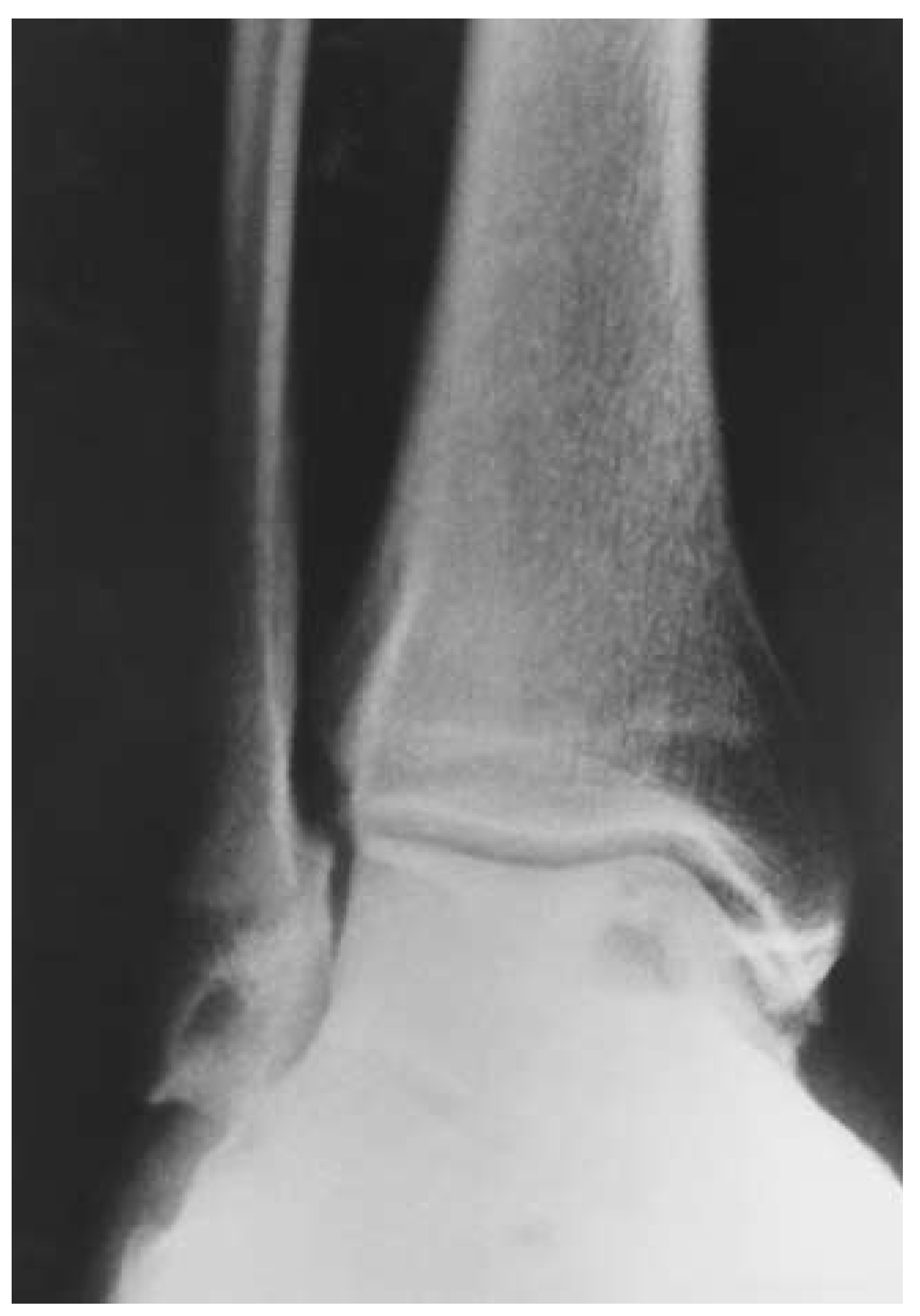
Figure 3. Preoperative anteroposterior radiograph. Note the flattening of the talus and the cyst under the medial shoulder of the talar dome.
Japma 91 00406 g003
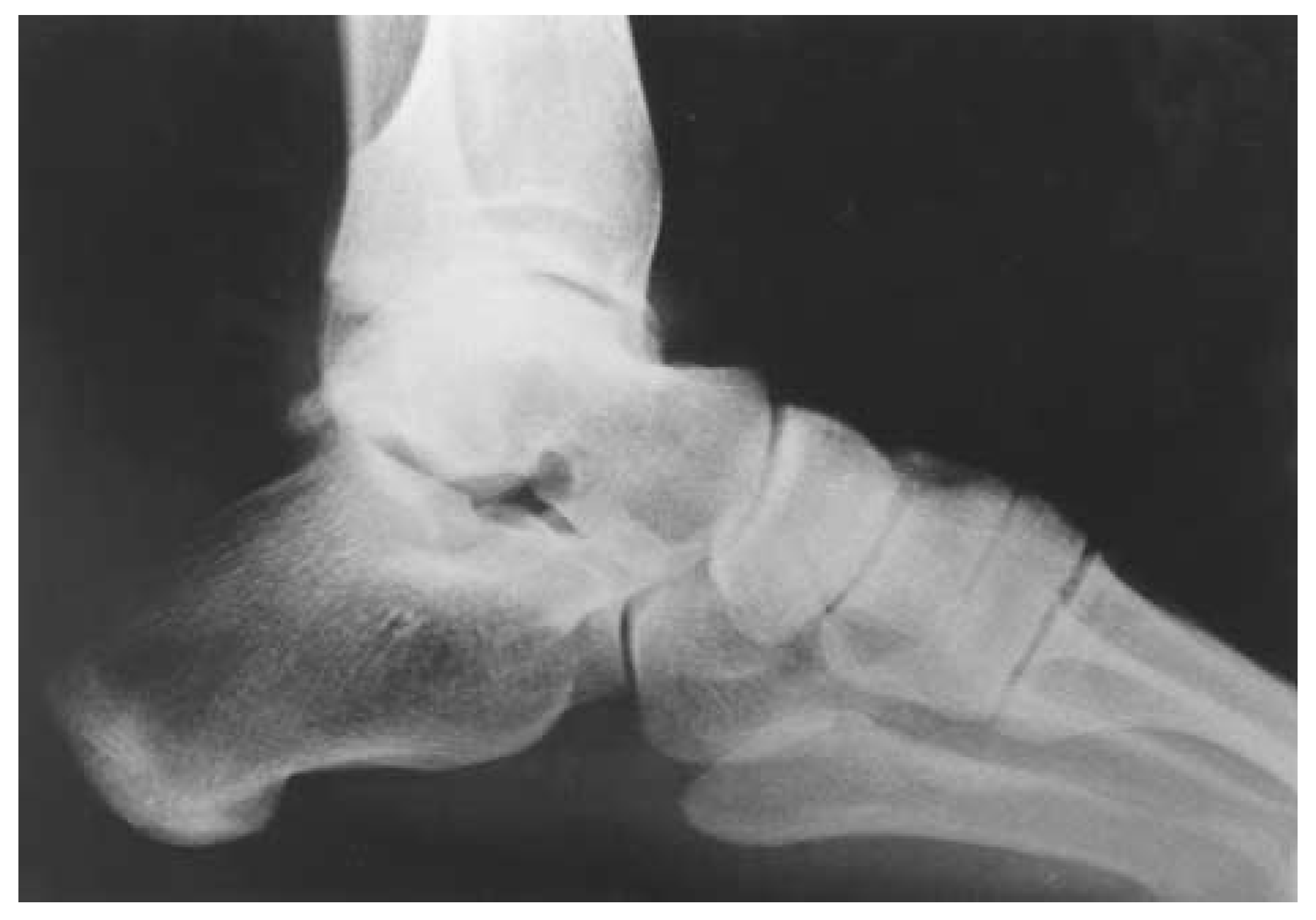
Figure 4. Preoperative lateral radiograph.
Japma 91 00406 g004
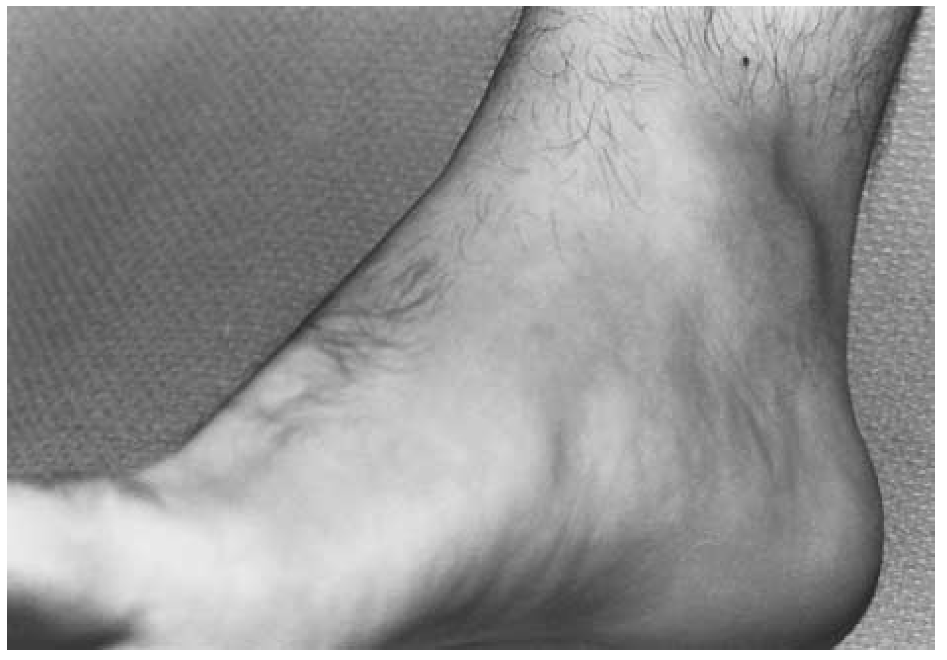
Figure 5. Preoperative clinical photo. Note the fullness in the anterior and medial ankle region consistent with chronic inflammation in the joint.
Japma 91 00406 g005
Surprisingly, the patient was not diagnosed with hemophilia until he was 17 years of age. The diagnosis was made after he developed a hematoma in his hip after a groin injury that occurred during a sporting activity. Owing to his late diagnosis, he had not received a factor replacement transfusion until 1986 and had not contracted HIV as so many other hemophiliac blood recipients did prior to 1982.
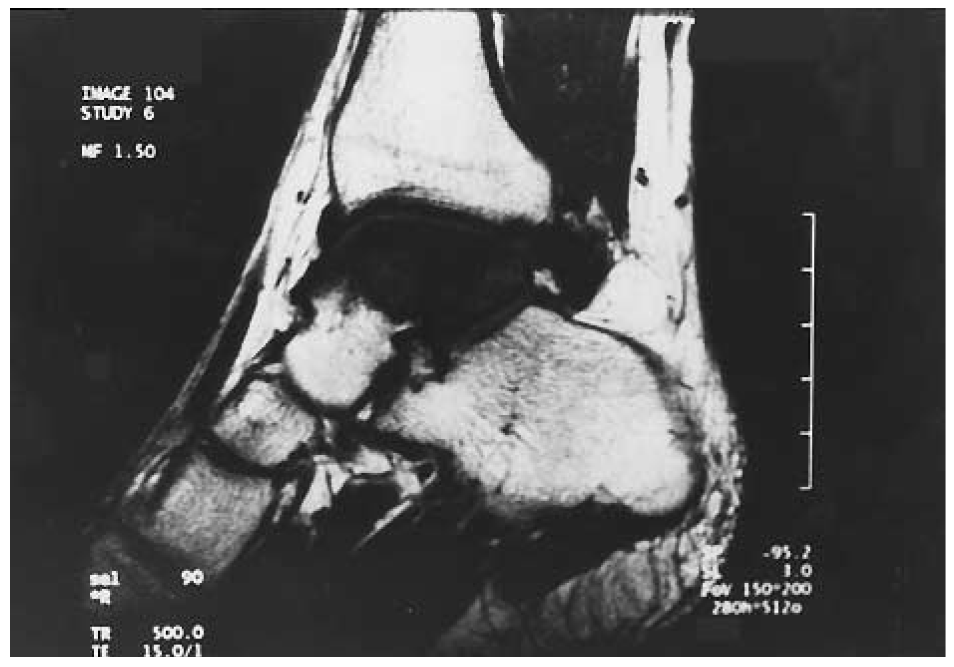
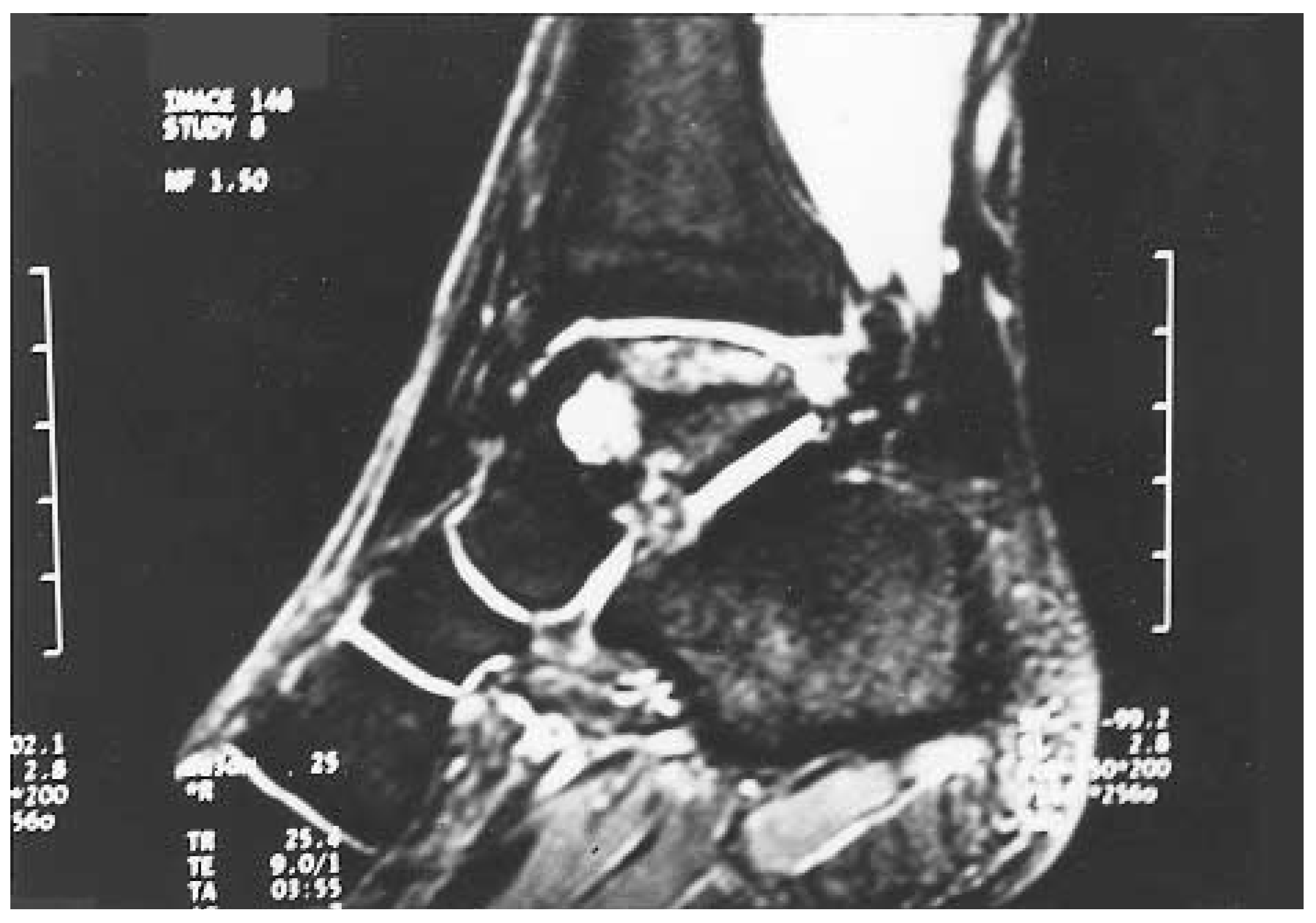
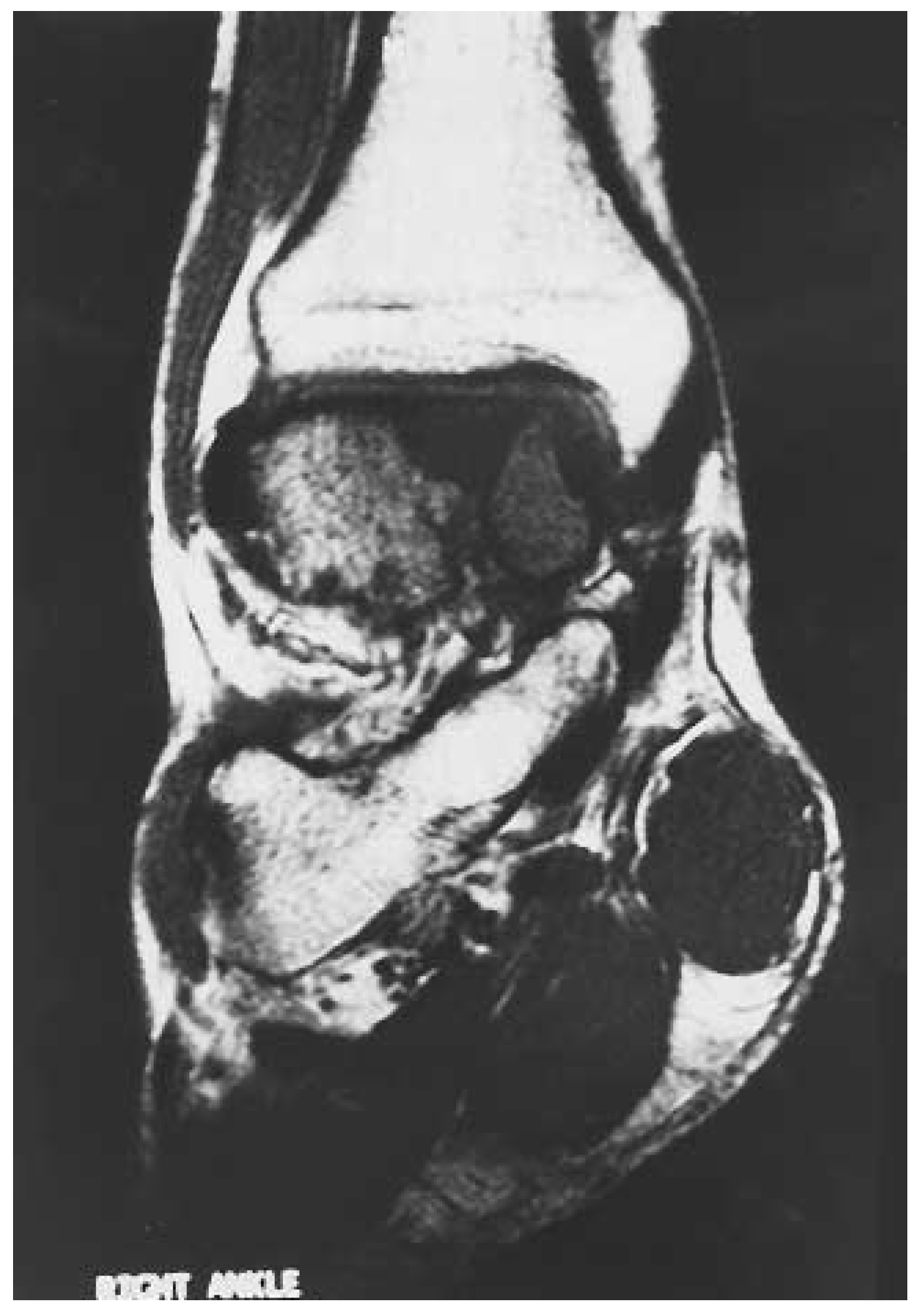
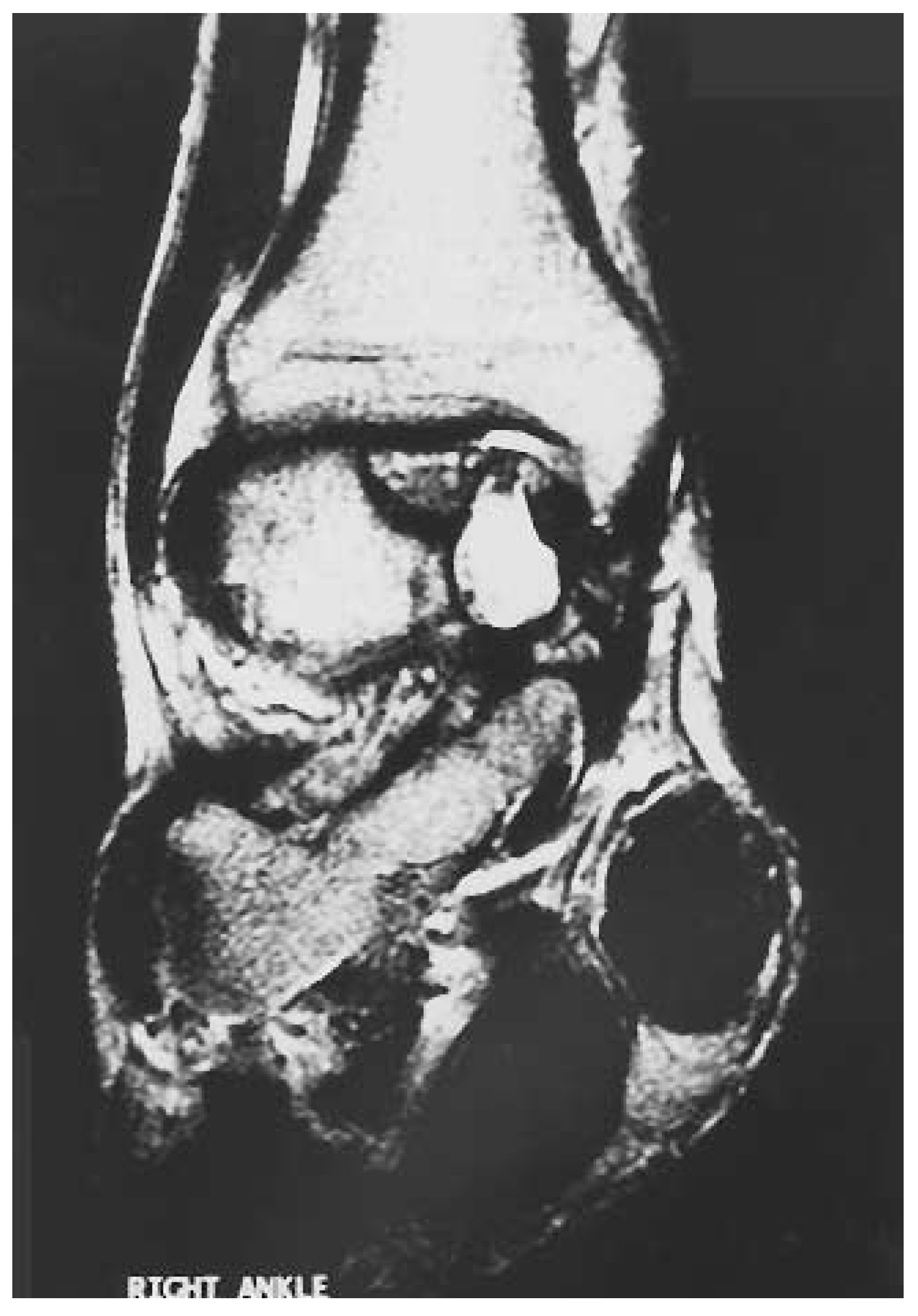
A magnetic resonance image (MRI) revealed the following findings: avascular necrosis of the right talus, ankle-joint degeneration with periarticular osseous fragmentation, a cyst in the medial aspect of the talar dome, and a fracture of the os trigonum with resultant hypertrophy of soft tissue posteriorly (Figs. 69). Prior to surgical intervention, a diagnostic intra-articular injection was recommended to further assess the contributions from the ankle. The cost of prophylactic factor replacement therapy for the injection alone was $3,000. After consultation with the hematologist, it was decided to bypass the injection and proceed with surgery.
Figure 6. Sagittal MRI T-1 scan. Note the avascularity of the talar dome on the images.
Japma 91 00406 g006
Figure 7. Sagittal MRI T-2 scan.
Japma 91 00406 g007
Figure 8. Frontal MRI T-1 scan.
Japma 91 00406 g008
Figure 9. Frontal MRI T-2 scan.
Japma 91 00406 g009
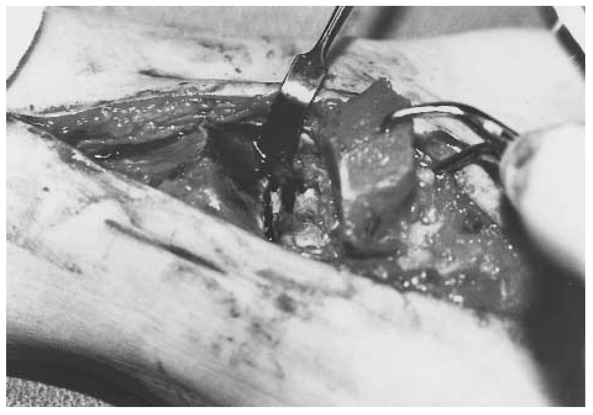
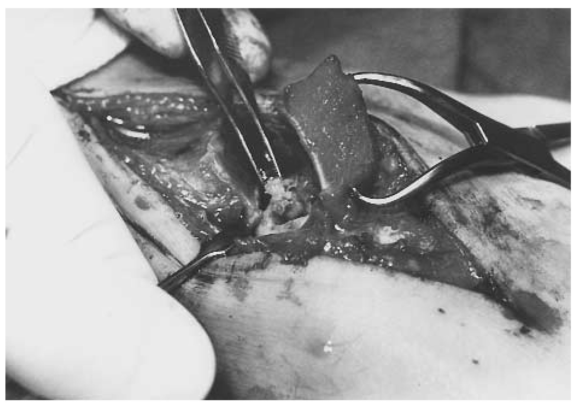
Surgery involved the following procedure: excision of the os trigonum fracture fragment; osteotomy of the medial malleolus to facilitate debridement and removal of the degenerative cartilage on the talar dome with drilling of the talar dome; cutting of a cortical window; curettage of medial talar dome cyst; and packing of the cyst with autogenous calcaneal bone graft (Figs. 10 and 11).
Figure 10. Intraoperative photo showing exposure into the joint and medial face of the talar dome.
Japma 91 00406 g010
Figure 11. Autogenous chips from the calcaneus are being placed through a cortical window in the medial wall.
Japma 91 00406 g011
Factor VIII replacement therapy was monitored and continued postoperatively. The cost of the replacement therapy for the complete perioperative management totaled more than $75,000. At 3½ years postoperative, the patient was doing extremely well, with good function to his ankle (Figs. 12 and 13). The number of intra-articular bleeds had decreased and the patient was pleased with his progress. He also reported that his ankle pain ranged from minimal to none.
Figure 12. Anteroposterior radiograph at 18 months postoperative. Note osseous healing of the medial cyst.
Japma 91 00406 g012
Figure 13. Lateral radiograph at 18 months postoperative.
Japma 91 00406 g013

Conclusion

Proper conservative and surgical management options for patients with hemophilia can be recommended only after a thorough understanding of the disease process. The advent of factor replacement therapy has dramatically changed the course of treatment and prognosis for patients with hemophilia. Although cost is still a significant factor, the long-term benefits, absent the risk of disease transmission, provide these patients with an optimistic outlook.

References

  1. Teigland JC, Tjonnfjord GE, Evensen SA, et al: Knee arthroplasty in hemophilia 5-12 year follow-up of 15 patients. .Acta Orthop Scand64::153. ,1993.
  2. Arnold WD, Hilgartner MW: Hemophilic arthropathy: current concepts of pathogenesis and management. .J Bone Joint Surg Am59::287. ,1977.
  3. Horoszowski H, Heim M, Schulman S, et al: Multiple joint procedures in a single operative session on hemophilic patients. .Clin Orthop328::60. ,1996.
  4. Duthie RB, Dodd CAF: “Chronic Haemophilic Arthropathy,” in The Management of Musculo-skeletal Problems in the Haemophilias, 2nd Ed, ed by RB Duthie, CR Rizza, PLF Giangrande, et al, p 159, Oxford University Press, New York, 1994..
  5. Horoszowski H, Heim M, Seligsohn U, et al: “Use of the Laser Scalpel in Orthopaedic Surgery on Haemophilic Patients,” in Haemophilia, ed by U Seligsohn, A Rimon, H Horoszowski, p 189, Castle House Publishing Ltd, Turnbridge Wells, UK, 1981..
  6. Ragni MV, Crossett LS, Herndon JH: Postoperative infection following orthopaedic surgery in human immunodeficiency virus: infected hemophiliacs with CD4 counts ≤ 200/mm3. .J Arthroplasty10::716. ,1995.
  7. Levine PH: “Clinical Manifestations and Therapy of Hemophilias A and B,” in Hemostasis and Thrombosis: Basic Principles and Clinical Practice, ed by RW Coleman, J Hirsh, VJ Marder, et al, p 67, JB Lippincott, Philadelphia, 1982..
  8. Greene WB: Synovectomy of the ankle for hemophilic arthropathy. .J Bone Joint Surg Am76::812. ,1994.
  9. Fabry G: Early biochemical and histological findings in experimental hemarthrosis in dogs. .Arch Orthop Trauma Surg100::167. ,1982.
  10. Mainardi CL, Levine PH, Werb Z, et al: Proliferative synovitis in hemophilia: biochemical and morphologic observations. .Arthritis Rheum21::137. ,1978.
  11. Canale ST, Dugdale M, Howard BC: Synovectomy of the knee in young patients with hemophilia. .South Med J81::1480. ,1988.
  12. Casscells CD: Commentary: the argument for early arthroscopic synovectomy in patients with severe hemophilia. .Arthroscopy3::78. ,1987.
  13. Clark MW: Knee synovectomy in hemophilia. .Orthopedics1::285. ,1978.
  14. DeGnore LT, Wilson FC: Surgical management of hemophilic arthropathy. .Instr Course Lect38::383. ,1989.
  15. Wiedel JD: Arthroscopic synovectomy of the knee in hemophilia: 10-to-15 year followup. .Clin Orthop328::46. ,1996.
  16. Bussi L, Silvello L, Baudo F, et al: Results of synovectomy of the knee in haemophilia. .Haematolgica59::81. ,1974.
  17. Shopnick RI, Brettler DB: Hemostasis: a clinical review of conservative and operative care. .Clin Orthop328::34. ,1996.
  18. Lofqvist T, Sanzen L, Petersson C, et al: Total hip replacement in patients with hemophilia: 13 hips in 11 patients followed for 1-16 years. .Acta Orthop Scand67::321. ,1996.
  19. Pearce MS, Smith MA, Savidge GF: Supramalleolar tibial osteotomy for haemophilic arthropathy of the ankle. .J Bone and Joint Surg Br76::947. ,1994.
  20. Atkins RM, Henderson NJ, Duthie RB: Joint contractures in the hemophilias. .Clin Orthop219::97. ,1987.
  21. Houghton GR, Dickson RA: Lower limb arthrodeses in haemophilia. .J Bone Joint Surg Br60::387. ,1978.
  22. Luck JV Jr: Surgical management of advanced hemophilic arthropathy. .Prog Clin Biol Res324::241. ,1990.
  23. Smith MA, Urquhart DR, Savidge GF: The surgical management of varus deformity in haemophilic arthropathy of the knee. .J Bone Joint Surg Br63::261. ,1981.
  24. Dyszy-Laube B, Kaminski W, Gizycka I, et al: Synovectomy in the treatment of hemophilic arthropathy. .J Pediatr Surg9::123. ,1974.
  25. Gamba G, Grignani G, Ascari E: Synoviorthesis versus synovectomy in the treatment of recurrent haemophilic haemarthrosis: long-term evaluation. .Thromb Haemost45::127. ,1981.
  26. van den Berg HM, Nieuwenhuis K, Mauser-bunschoten EP, et al: Hemophilia prophylaxis in the Netherlands. .Semin Hematol31::13. ,1994.
  27. Steven MM, Small M, Pinkerton L, et al: Non-steroidal anti-inflammatory drugs in haemophilic arthritis: a clinical and laboratory study. .Haemostasis15::204. ,1985.
  28. Lofqvist T, Nilsson IM, Petersson C: Orthopaedic surgery in hemophilia: 20 years’ experience in Sweden. .Clin Orthop332::232. ,1996.
  29. Rodriguez Merchan EC, Magallon M, Galindo E: Joint debridement for haemophilic arthropathy of the knee. .Int Orthop18::135. ,1994.
  30. Ahlberg A: Treatment and prophylaxis of arthropathy in severe hemophilia. .Clin Orthop53::135. ,1967.
  31. Brettler DB, Levine PH: Factor concentrates for treatment of hemophilia: which one to choose. ?Blood73::2067. ,1989.
  32. Scharf R, Kucharski W, Nowak T: Surgery in hemophilia A patients with factor VIII inhibitor: 10-year experience. .World J Surg20::1171. ,1996.
  33. O’Marcaigh AS, Schmalz BJ, Shaughnessy WJ, et al: Successful hemostasis during a major orthopedic operation by using recombinant activated factor VII in a patient with severe hemophilia A and a potent inhibitor. .Mayo Clin Proc69::641. ,1994.
  34. Teigland JC, Tjonnfjord GE, Evensen SA, et al: Synovectomy for haemophilic arthropathy: 6-21 years of follow-up in 16 patients. .J Intern Med235::239. ,1994.
  35. Nilsson IM, Berntorp E, Ljung R, et al: Prophylactic treatment of severe hemophilia A and B can prevent joint disability. .Semin Hematol31::5. ,1994.
  36. Aronstam A, Browne RS, Wassef M, et al: Clinical features of early haemarthoses in severely affected adolescent haemophiliacs. .Clin Lab Haematol6::9. ,1984.
  37. Greene WB, McMillan CW: Nonsurgical management of hemophilic arthropathy. .Instr Course Lect38::367. ,1989.
  38. Wilson FC, Mahew DE, McMillan CW: Surgical management of musculoskeletal problems in hemophilia. .Instr Course Lect32::233. ,1983.
  39. McMillan CW, Greene WB, Blatt PM, et al: The management of musculoskeletal problems in hemophilia. Part I: principles of medical management of hemophilia. .Instr Course Lect32::210. ,1983.
  40. Unger AS, Kessler CM, Lewis RJ: Total knee arthroplasty in human imunodeficiency virus–infected hemophiliacs. .J Arthroplasty10::448. ,1995.
  41. Goedert JJ, Kessler CM, Aledort LM, et al: A prospective study of human immunodeficiency virus type I infection and the development of AIDS in subjects with hemophilia. .N Engl J Med321::1141. ,1989.
  42. Greene WB, DeGore LT, White GC: Orthopaedic procedures and prognosis in hemophilic patients who are seropositive for human immunodeficiency virus. .J Bone Joint Surg Am72::2. ,1990.

Additional References

  1. Brettler DB, Forsberg AD, Levine PH, et al: The use of porcine factor VIII (Hyate:C) in the treatment of patients with inhibitor antibodies to factor VIII. A multicenter US experience. .Arch Intern Med149::1381. ,1989.
  2. Brown DC: Adverse events reported in association with the use of monoclonal antibody purified factor VIIIC: monoclate. .Semin Hematol27::16. ,1990.
  3. Gatti L, Mannucci PM: Use of porcine factor VIII in the management of seventeen patients with factor VIII antibodies. .Thromb Haemost51::379. ,1984.
  4. Goldsmith JC, Gordon EM: Treatment of hemophilia B: serendipitous use of continuous infusion coagulation factor IX. .Thromb Res70::265. ,1993.
  5. Hedner U: Factor VIIa in the treatment of haemophilia. .Blood Coagul Fibrinolysis1::307. ,1990.
  6. Hedner U, Feldstedt M, Glazer S: “Recombinant FVIIa in Hemophilia Treatment,” in Hemophilia and von Willebrand’s Disease in the 1990s: Proceedings of the XIX Congress of the World Federation of Hemophilia, Washington, DC, August 14–19, 1990, ed by JM Lusher, CM Kessler, p 283, Elsevier Science, New York, 1991..
  7. Kasper CK: Treatment of factor VIII inhibitors. .Prog Hemost Thromb9::57. ,1989.
  8. Kasper CK: Complications of hemophilia A treatment: factor VIII inhibitors. .Ann N Y Acad Sci614::97. ,1991.
  9. Lethagen S, Egervall K, Berntorp E, et al: The administration of desmopressin by nasal spray: a dose-determination study in patients with mild hemophilia A or von Willebrand’s disease. .Haemophilia1::97. ,1995.
  10. Lusher JM, Shapiro SS, Palascak JE, et al: Efficacy of prothrombin-complex concentrates in hemophiliacs with antibodies to factor VIII: a multicenter therapeutic trial. .N Engl J Med303::421. ,1980.
  11. Mannucci PM: Desmopressin (DDAVP) for treatment of disorders of hemostasis. .Prog Hemost Thromb8::19. ,1986.
  12. Mannucci PM, Bettaga D, Cattaneo M: Patterns development of tachyphylaxis in patients with haemophilia and von Willebrand disease after repeated doses of desmopressin (DDAVP). .Br J Haematol82::87. ,1992.
  13. Mariani G, Scheibel E, Nagao T, et al: Immunotolerance as treatment of alloantibodies to factor VIII in hemophilia. .Emin Hematol (suppl)31::62. ,1994.
  14. Nilsson IM: Factor VIII inhibitor treatment: immune tolerance. .Semin Hematol31 (suppl 4)::44. ,1994.
  15. Nilsson IM, Berntorp E, Freiburghaus C: Treatment of patients with factor VIII and IX inhibitors. .Thromb Haemost70::56. ,1993.
  16. Nilsson IM, Berntorp E, Zettervall O: Induction of immune tolerance in patients with hemophilia and antibodies to factor VIII by combined treatment with intravenous IgG, cyclophosphamide, and factor VIII. .N Engl J Med318::947. ,1988.
  17. Petrini P, Lindvall N, Egberg N, et al: Prophylaxis with factor concentrates in preventing hemophilic arthropathy. .Am J Pediatr Hematol Oncol13::280. ,1991.
  18. Ragni MV, Hanley EN: Septic arthritis in hemophilic patients and infection with human immunodeficiency virus (HIV). .Ann Intern Med110::168. ,1989.
  19. Rudowski WJ, Scharf R, Ziemski JM: Is major surgery in hemophiliac patients safe. ?World J Surg11::378. ,1987.
  20. Seremetis SV: The clinical use of factor VIIa in the treatment of factor VIII inhibitor patients. .Semin Haemtol31 (suppl)::53. ,1994.
  21. Soreff J: Joint debridement in the treatment of advanced hemophilic knee arthropathy. .Clin Orthop191::179. ,1984.
  22. Weber DJ, Becherer PR, Rutala WA, et al: Nosocomial infection rate as a function of human immunodeficiency virus type I status in hemophiliacs. .Am J Med (suppl)91::206. ,1991.