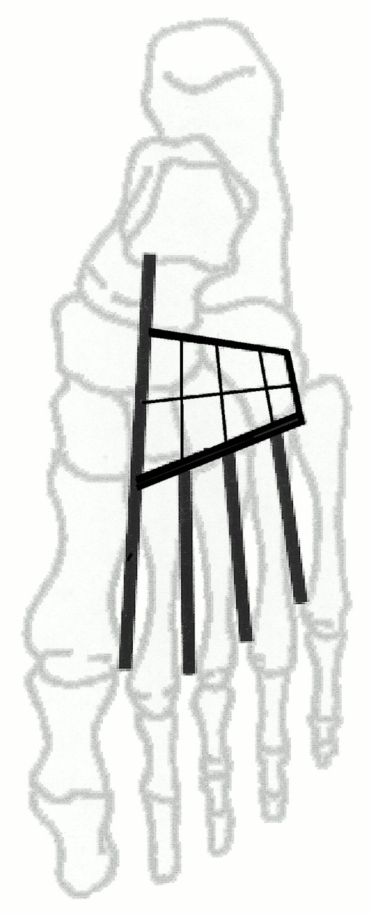
The arcuate artery is described as arising laterally from the dorsalis pedis artery at the level of the first tarsometatarsal joint and continuing laterally across the bases of metatarsals 2 through 4 to supply the second through fourth dorsal metatarsal arteries.[
1-
3] In commonly used textbooks and atlases on the lower extremity, the occurrence of the arcuate artery is cited as approximately 54%.[
2,
3] However, the incidence of the arcuate artery in cadaveric studies has been empirically found to range from 10% to 67% depending on the precise definition of the artery.[
4-
10] Chaney et al[
4] found the arcuate present in roughly 61% of specimens sampled and noted that the artery was of varying diameter. Yamada et al[
5] claimed that the arcuate was absent in 33% of their specimens. Huber[
6] found a significant arcuate artery in 54% of his specimens. His definition of arcuate artery included origin at the naviculocuneiform joint or the tarsometatarsal joint. In contrast, Adachi[
7] favored the name “distal lateral tarsal artery” for the artery with a naviculocuneiform origin off the dorsalis pedis. Consequently, Adachi found a lower occurrence of the arcuate artery, at 30.6%. If Huber’s study is limited to that artery with origin only at the first tarsometatarsal joint, the incidence of the arcuate artery is 35%. Huber further categorized the arcuate artery by the number of dorsal metatarsal arteries it supplied. He found that only 16.5% of the specimens had an arcuate artery at the level of the tarsometatarsal joint supplying the second through fourth dorsal metatarsal arteries. If Adachi is held to the same definition, only 13% of his specimens had a complete arcuate artery. Similarly, in three additional studies, the incidence of the arcuate artery with origin at the tarsometatarsal joint and supplying dorsal metatarsal arteries 2 through 4 was found to be as low as 20%,[
8] 19%,[
9] and 10%.[
10]
The lateral tarsal artery frequently provides blood supply to the dorsal metatarsal arteries in the absence of a complete arcuate artery. Published values indicate an 18% to 33% incidence of a lateral tarsal artery providing blood supply to dorsal metatarsal arteries 2 through 4.[
9,
10] The lateral tarsal artery arises laterally off the dorsalis pedis artery above the naviculocuneiform joint. Its origin off the dorsalis pedis varies widely; at times, it originates off the anterior tibial artery. There may even be two lateral tarsal arteries.[
2] The lateral course of the artery ends in anastomosis with the lateral malleolar or arcuate arteries. Prior to termination, the lateral tarsal artery sends one to three branches to the intermetatarsal spaces to supply the second through fourth dorsal metatarsal arteries.
Dorsal metatarsal arteries 2 through 4 may also arise from proximal perforating arteries. In this situation, the dorsal metatarsal arteries are supplied by the plantar arterial system. Adachi[
7] and Hamada et al[
10] claim values as high as 57% for nondorsal arteries contributing the primary supply to one or all of the dorsal metatarsal arteries. Lippert and Pabst[
8] and Albaret et al[
9] provide lower values at 40% and 15%, respectively; however, both of these studies described solely plantar supply to all three lateral dorsal metatarsal arteries. Huber[
6] described the plantar supply to the second through fourth dorsal metatarsal arteries as the principal supply in 33.5%, 23%, and 37.5%, respectively.
It is the authors’ contention that the frequency of occurrence of the arcuate artery in anatomical course and as supplier of dorsal metatarsal arteries 2 through 4 is significantly lower than is commonly held in many lower-extremity–anatomy teaching atlases and textbooks. The authors sought to determine the occurrence of the arcuate artery as the major supplier of blood to dorsal metatarsal arteries 2 through 4.
Materials and Methods
Ninety-four cadaveric feet were dissected and examined. The specimens were dissected to the level of the arterial network on the dorsum of the foot. An eosin-dye mixture consisting of 7.5 g of eosin dye powder (Fisher Scientific, Pittsburgh, Pennsylvania) in 250 mL distilled water was injected into the anterior tibial artery. If the outflow vessels were obstructed or severed, more dye was injected distal to that site to make sure that all vessels were perfused. The anterior tibial, dorsalis pedis, lateral tarsal, arcuate, and dorsal metatarsal arteries were then identified. The dorsal arterial network was visualized and copied to a scaled-down drawing of the foot bones. The drawings were then classified according to the following criteria: right or left foot; presence of lateral tarsal arteries; presence of true or variant arcuate artery; major blood supply to dorsal metatarsal arteries 2 through 4 as arcuate, lateral tarsal, proximal perforating, or some combination thereof.
The following definitions were used to identify the major dorsal arteries supplying the dorsal metatarsal arteries. The arcuate artery was defined as that artery arising laterally from the dorsalis pedis artery at or below the level of the tarsometatarsal joint and coursing laterally to supply dorsal metatarsal arteries 2 through 4. An arcuate artery variant was defined as an artery arising laterally from the dorsalis pedis artery at or below the tarsometatarsal joint and coursing laterally to supply fewer than the three lateral dorsal metatarsal arteries. The variant may also arise laterally from the dorsalis pedis artery up to the naviculocuneiform joint and then course laterally to supply any of the dorsal metatarsal arteries 2 through 4. The lateral tarsal artery was defined as that artery arising laterally off the dorsalis pedis artery at or above the naviculocuneiform joint, coursing laterally across the midtarsus to anastomose with the lateral malleolar or arcuate arteries. In its course, the lateral tarsal artery supplies blood to the extensor digitorum brevis muscle. Only those arteries that anastomosed laterally were included in this study. The lateral tarsal artery sent zero to three longitudinal branches to intermetatarsal spaces 2 through 4. Proximal perforating arteries were identified via direct anastomosis with the dorsal metatarsal arteries. Once the arterial supply to the dorsal metatarsal arteries was determined and classified, simple percentages were calculated for each scenario against the total number of samples.
Results
Initially, 94 specimens were examined. Twenty-two samples were considered indeterminate, owing either to previous dissection loss or fungus, leaving a total of 72 feet for the final analysis.
Arcuate Artery
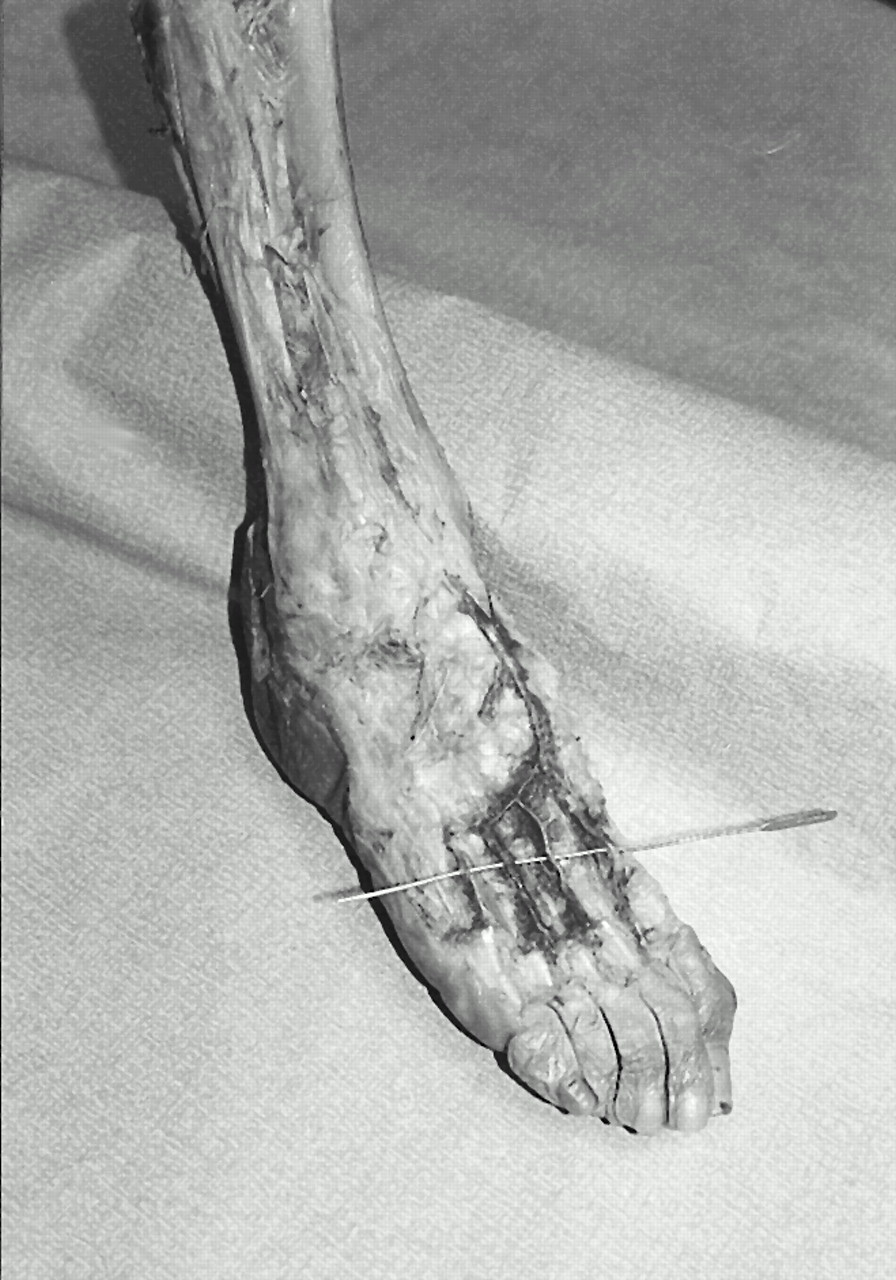
An examination of the dorsal arterial network showed that 12 of 72 feet (16.7%) had an arcuate artery with origin at or below the tarsometatarsal joint and supplying dorsal metatarsal arteries 2 through 4 (
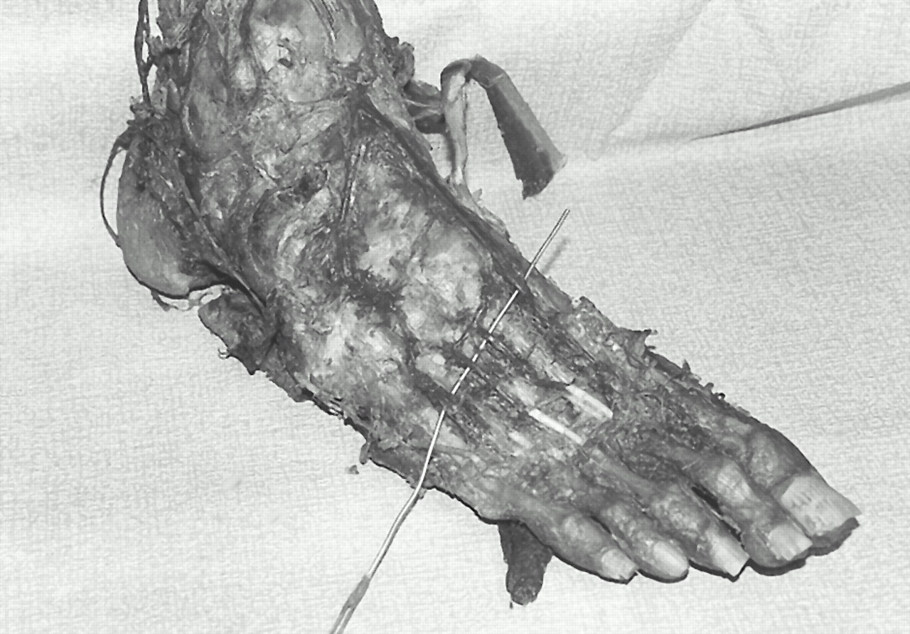
Fig. 1). Fifteen of 72 samples (20.8%) had an arcuate variant, either incomplete in supply to the second through fourth dorsal metatarsal arteries or originating above the tarsometatarsal joint (
Fig. 2). Nine feet (12.5%) had an arcuate-level artery supplying the second dorsal metatarsal artery. Four feet (5.6%) had an arcuate-level artery supplying the second and third dorsal metatarsal arteries. Two feet (2.8%) had an arcuate variant supplying the second through fourth dorsal metatarsal arteries. A total of 27 feet (37.5%) examined had some form of arcuate artery.
Figure 1.
Arcuate artery as defined. The artery branches off the dorsalis pedis at or below the level of the tarsometatarsal joint, continues in course laterally across the bases of metatarsals 2 through 4, and supplies dorsal metatarsal arteries 2 through 4.
Figure 1.
Arcuate artery as defined. The artery branches off the dorsalis pedis at or below the level of the tarsometatarsal joint, continues in course laterally across the bases of metatarsals 2 through 4, and supplies dorsal metatarsal arteries 2 through 4.
Figure 2.
Arcuate artery variant. The artery branches off the dorsalis pedis below the cuneonavicular joint. It supplies dorsal metatarsal arteries 2 through 4 only when branching above the tarsometatarsal joint. When it branches at or below the tarsometatarsal joint, as in this case, it supplies fewer than three of the lateral dorsal metatarsal arteries. In this specimen, the fourth dorsal metatarsal artery is supplied by the lateral tarsal artery.
Figure 2.
Arcuate artery variant. The artery branches off the dorsalis pedis below the cuneonavicular joint. It supplies dorsal metatarsal arteries 2 through 4 only when branching above the tarsometatarsal joint. When it branches at or below the tarsometatarsal joint, as in this case, it supplies fewer than three of the lateral dorsal metatarsal arteries. In this specimen, the fourth dorsal metatarsal artery is supplied by the lateral tarsal artery.
Lateral Tarsal Artery
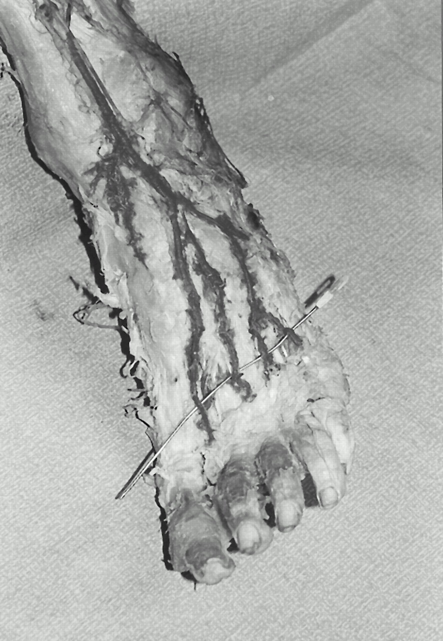
Forty-nine of the 72 feet (68.1%) examined had at least one lateral tarsal artery as defined. When examined further, 34 of 72 feet (47.2%) had a lateral tarsal artery feeding dorsal metatarsal arteries 2 through 4 (
Fig. 3). Of these 34 feet, 6 (17.6%) had dual lateral tarsal arteries off the dorsalis pedis. Twelve of 72 feet (16.7%) had a lateral tarsal artery feeding dorsal metatarsal arteries 3 and 4; and 2 of 72 feet (2.8%) had a lateral tarsal artery that fed the dorsal metatarsal artery to the fourth interspace only.
Figure 3.
Lateral tarsal artery. The artery branches off the dorsalis pedis artery at or above the cuneonavicular joint. It tends laterally in course and provides zero to three branches to intermetatarsal spaces 2 through 4. In this specimen with a laterally tending dorsalis pedis artery, the lateral tarsal artery supplies dorsal metatarsal arteries 2 through 4.
Figure 3.
Lateral tarsal artery. The artery branches off the dorsalis pedis artery at or above the cuneonavicular joint. It tends laterally in course and provides zero to three branches to intermetatarsal spaces 2 through 4. In this specimen with a laterally tending dorsalis pedis artery, the lateral tarsal artery supplies dorsal metatarsal arteries 2 through 4.
Proximal Perforating Artery
Nine of 72 feet (12.5%) exhibited primarily plantar blood supply to the second through fourth dorsal metatarsal arteries via the proximal perforating arteries. One foot (1.4%) had plantar supply to the second and third dorsal metatarsal arteries and one foot (1.4%) had plantar supply to the fourth dorsal metatarsal artery.
Combination Supply
A total of 21 feet (29.2%) had combination blood supply to dorsal metatarsal arteries 2 through 4. Nine (12.5%) had an arcuate-level branch to the second and a lateral tarsal artery branch to the third and fourth dorsal metatarsal arteries. Three feet (4.2%) had an arcuate-level supply to the second and third and a lateral tarsal supply to the third and fourth dorsal metatarsal arteries. In this situation, there was clear anastomosis between the arcuate and lateral tarsal arteries at the third dorsal metatarsal artery. One foot (1.4%) had an arcuate-level artery to the second and third dorsal metatarsal arteries and a lateral tarsal supply to the fourth. One foot (1.4%) had proximal perforating supply to the second and third dorsal metatarsal arteries and lateral tarsal supply to the fourth. One foot (1.4%) had lateral tarsal supply to the second and third dorsal metatarsal arteries and proximal perforating supply to the fourth.
Bilateral Symmetry
Fourteen of 31 (45.2%) usable pairs exhibited bilateral symmetry of dorsal blood supply to the dorsal metatarsal arteries. This value did not include two usable pairs with complete lateral tarsal supply to the second through fourth dorsal metatarsal arteries when one of the feet had dual lateral tarsal arteries.
Discussion
The occurrence of the arcuate artery in the cadaveric specimens in this study was indeed lower than the 54% commonly claimed in current teaching texts. The present value of 16.7% corroborates previous findings by Huber,[
6] Adachi,[
7] Lippert and Pabst,[
8] Albaret et al,[
9] and Hamada et al.
10 It is important to recognize that the definition of the arcuate artery was crucial in the interpretation of the results in each of these studies. The current study maintained a strict definition of the arcuate artery as that artery arising at or below the tarsometatarsal joint and coursing laterally to supply dorsal metatarsal arteries 2 through 4. When the above-mentioned studies were interpreted according to this definition, the occurrence of the arcuate artery was similar to that in the current study. However, as these and other studies referred to the arcuate artery with a broader definition, they found higher occurrences of the arcuate artery.[
4-
7] Consequently, textbooks and atlases of lower-extremity anatomy have combined strict definitions of the arcuate artery with statistics from more general definitions to create a less than accurate picture.[
1-
3] However, it would be inaccurate to say that these textbooks have not included descriptions of the variability of the dorsal arterial network; in fact, they have.
The lateral tarsal artery was found to contribute blood supply to the second through fourth dorsal metatarsal arteries in nearly one-half of the specimens in the present study. Previous studies found the lateral tarsal artery providing the primary dorsal blood supply to the lateral dorsal metatarsal arteries 18% to 33% of the time.[
8,
9] The current value suggests that the lateral tarsal artery provides the majority of the blood to dorsal metatarsal arteries 2 through 4. Six occurrences of lateral tarsal artery blood supply to the dorsal metatarsals involved dual lateral tarsal arteries. These were included in this category because the arteries to the intermetatarsal spaces were originating from arteries above the arcuate artery or its variants and the origin of both of these arteries was the dorsalis pedis artery above the cuneonavicular joint.
Other sources of blood to the dorsal metatarsal arteries include the proximal perforating arteries and a combination supply from the arcuate, lateral tarsal, and proximal perforating arteries. The current study found proximal perforating arteries supplying dorsal metatarsal arteries 2 through 4 in 12.5% of the specimens. This value corroborates the study by Albaret et al,[
9] which found a 15% occurrence of complete plantar blood supply to the lateral dorsal metatarsal arteries.
Combinatory blood supply to the dorsal metatarsal arteries was less common than anticipated. It occurred in only 30% of the specimens. No single combination occurred more frequently than the three individual supplies did as the sole source of blood to the second through fourth dorsal metatarsal arteries. The most common combination consisted of an arcuate-level branch to the second dorsal metatarsal artery and a lateral tarsal branch to the third and fourth dorsal metatarsal arteries. The variation seen in the combination supply specimens and their relationship to the arcuate and lateral tarsal arteries as suppliers of the dorsal metatarsal arteries suggests that the mature arterial structure develops out of a consistent developmental network.
Bilateral symmetry of the blood supply to the dorsal metatarsal arteries occurred in 45.2% of the usable, paired feet. This value compares the feet only on the basis of the origin of supply to the second through fourth dorsal metatarsal arteries. Huber[
6] states that although 65% of paired feet were found to be categorizable to the same regional dorsal arterial supply, only 4% of his specimens had exactly matching bilateral arterial patterns. The present study confirms that bilateral symmetry among feet does occur. However, as descriptive definitions become stricter, the occurrence decreases greatly.
Interpretation of the dorsal forefoot arteries is a difficult task, as evidenced by the numerous studies of the pedal arteries. Consequently, much has been written about the dorsal arterial tree of the foot. However, owing to the variability of the arterial pattern, a simple teachable structure remains elusive.[
2,
6,
11,
12] Toward a more understandable and reliable interpretation, Adachi[
7] contributed the concept, which Huber[
6] and Edwards[
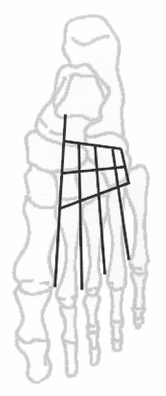
11] espoused, of a “grundform” or very constant dorsal arterial rete. This concept incorporates the frequently encountered anatomical variants into a predictive pattern that accounts for most of the variety witnessed upon dissection. This approach theorizes a constant multifaceted network of dorsal arteries that, through enlargement and stasis during development, adapt to create the variety of dorsal anastomoses (
Fig. 4). The network consists of proximal and distal lateral tarsal arteries and an arcuate artery branching laterally off the dorsalis pedis. These three arteries course laterally over the foot and anastomose or otherwise terminate at the lateral border. During their course, they send distal branches toward the three intermetatarsal spaces that effectively create a web of arteries covering the dorsal foot. Huber6 has theorized that this pattern is present in early development. Subsequently, these vessels increase or decrease in size to create a specific dorsal blood supply that is related to the initial network, but different from other developmental possibilities. Therefore, the occurrence, in percentage terms, of the vessels of the dorsal foot reflects the noticeable enlargement of specific vessels and the absence of enlargement of other potentially contributory vessels to that region. This accounts for the variation in blood supply to the dorsal metatarsal arteries and helps explain the diverse combinations of arteries contributing to the dorsal metatarsal arteries. For example, the arcuate artery, commonly labeled the primary source of dorsal metatarsal arteries 2 through 4, serves in this role only when it is sufficiently large (
Fig. 5). Therefore, a patent but extremely small arcuate artery might be discovered in dissection, but a prominent lateral tarsal artery providing three convincing branches that enter the intermetatarsal spaces dorsally would be described as the source of the dorsal metatarsal arteries. In this case, an arcuate artery, although present, certainly does not serve as the primary source of the dorsal metatarsal arteries.
Figure 4.
Developmental arterial tree of the dorsal foot. Early anastomosis includes two lateral tarsal arteries and an arcuate artery. Three perpendicular branches span these three arteries and extend into the dorsal intermetatarsal spaces.
Figure 4.
Developmental arterial tree of the dorsal foot. Early anastomosis includes two lateral tarsal arteries and an arcuate artery. Three perpendicular branches span these three arteries and extend into the dorsal intermetatarsal spaces.
Figure 5.
Mature arterial tree of the dorsal foot. The arcuate artery has enlarged to become the main supplier of dorsal metatarsal arteries 2 through 4. The proximal lateral tarsal artery has maintained its size, while the distal lateral tarsal has atrophied. The perpendicular branches have atrophied between the lateral tarsal and arcuate arteries.
Figure 5.
Mature arterial tree of the dorsal foot. The arcuate artery has enlarged to become the main supplier of dorsal metatarsal arteries 2 through 4. The proximal lateral tarsal artery has maintained its size, while the distal lateral tarsal has atrophied. The perpendicular branches have atrophied between the lateral tarsal and arcuate arteries.
Most branches of the dorsal arterial network described by Adachi were evidenced in the cadavers. Not all of these vessels were equivalent in size and not all matched the pattern exactly. Most often, a clearly discernible supply was discovered, with much smaller contributory vessels “filling in the blanks” of the developmental dorsal network. These smaller vessels occurred in the course of the arcuate and lateral tarsal arteries and their branches.
Conclusion
The current study found the occurrence of the complete arcuate artery to be 16.7%, a figure similar to previously published data. The lateral tarsal artery was determined to be a more consistent supply of blood to dorsal metatarsal arteries 2 through 4 than the arcuate artery. At 47.2%, the lateral tarsal artery provided complete blood supply to the dorsal metatarsal arteries in nearly one-half of the specimens. Plantar blood supply to the dorsal metatarsal arteries was significant, with complete supply to the second through fourth dorsal metatarsal arteries occurring 12.5% of the time. Bilateral symmetry of the dorsal arterial network was common, occurring in nearly one-half of the usable paired specimens; however, blood supply was assessed only to dorsal metatarsal arteries 2 through 4. The variety of the dorsal arterial network of the foot is best described by an early developmental network, which enlarges and atrophies to form the adult network.