Extraskeletal chondroma, or chondroma of soft parts, is a benign cartilaginous tumor. Although it is relatively rare, when it does occur it is most often found in the soft tissues of the hands and feet. Its variable histologic presentation, including immature-appearing cells, can pose a problem in accurate diagnosis and successful treatment [
1,
2]. Despite its prevalence in the feet, extraskeletal chondroma has not, to the authors’ knowledge, been described in the podiatric medical literature.
There have been two large-scale studies describing extraskeletal chondromas. The first, reported by Dahlin and Salvador [
3] in 1974, consisted of 70 patients treated or seen for diagnostic consultation at the Mayo Clinic. The second study was performed by Chung and Enzinger [
4] in 1978. They reviewed 104 cases of soft-tissue chondroma that were from the files of the Armed Forces Institute of Pathology and covered a 23-year period.
These studies confirmed that, even though these lesions may vary histologically, at times having the cellular appearance of chondrosarcoma, they are benign in nature and seldom recur following simple excision. The studies also found that these lesions occur predominantly in the extremities, particularly in the hands and the feet.
Individual reports of extraskeletal chondroma have been noted in the literature and describe variations of these lesions occurring primarily in the hands [
5,
6,
7,
8,
9].
Extraskeletal chondroma is most common among individuals from 30 to 60 years of age and occurs fairly equally in males and females [
3,
4]. Clinically, the lesion appears as a slow-growing, painless mass of variable duration, usually measuring less than 3 cm in diameter, although cases of lesions over 10 cm have been reported [
3]. Ninety-six percent of cases occur in the extremities, with 72% in the upper extremities and 24% in the lower extremities. Among lower-extremity lesions, 88% occur in the foot and ankle [
4].
Radiographs often show a soft-tissue mass demonstrating areas of calcification. The lesion does not typically involve the underlying osseous or periosteal structures, and cortical erosions are not characteristically present as in periosteal chondroma [
1,
3,
4]. Magnetic resonance imaging has proved to be helpful in localizing and differentiating these tumors [
9].
The tumor is usually a well-circumscribed, nodular mass with a grayish-white appearance; it is usually easily enucleated at the time of surgery. Some tumors may be closely associated with tendon, tendon sheath, or periarticular soft tissues [
1,
2,
3,
4]. The tumor differs from synovial chondromatosis, which occurs primarily in the larger joints and is associated with articular loose bodies [
1,
3,
4].
Most tumors are composed primarily of adult-type hyaline cartilage with a lobular pattern; however, some may exhibit plump, immature-appearing cells within a myxoid matrix closely simulating chondrosarcoma, but without having the characteristic cellular alignment in strands or cords [
1,
4]. Others have focal areas of ossification or diffuse calcification, which often outline the periphery of cartilage cells in a lacelike pattern [
1,
4]. If calcification is extensive, extraskeletal chondroma may mimic tumoral calcinosis, a lesion that does not possess cartilage cells. Extraskeletal chondroma may also resemble calcifying aponeurotic fibroma. Calcifying aponeurotic fibroma, however, usually does not occur distally in the digits and is most common in patients under 25 years old [
1]. Proliferation of multinucleated giant cells may be seen in the periphery of the calcified nodules, resembling that of giant-cell tumor [
4]. However, the cells in extraskeletal chondromas lack the more uniform cellular pattern commonly seen in giant-cell tumors [
1].
The treatment of choice for soft-tissue chondroma is simple excision. The lesion has been shown to have a very low recurrence rate, and there have been no documented cases of metastasis [
3,
4].
Case Report
A 52-year-old man presented for evaluation of a small, painless nodule on the flexor surface of the left second toe. The patient described a gradual onset of the nodule over 2 to 3 years. He denied any history of trauma to the area. His medical history was significant for gout, for which he was taking medication, a heel spur on the right foot, and osteomyelitis of the right foot as a child. The patient was otherwise in good general health.
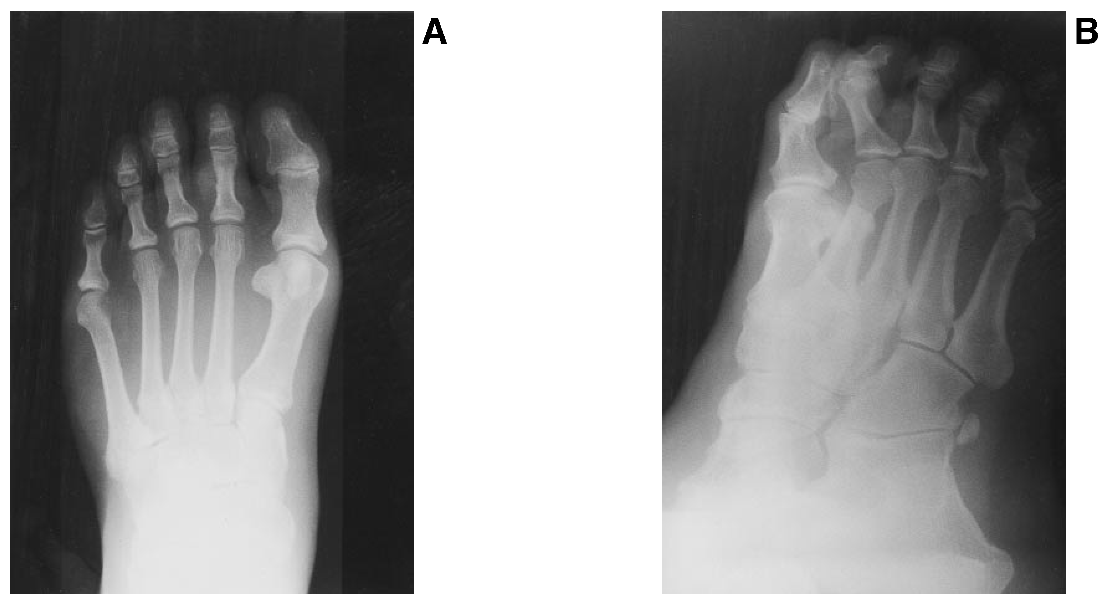
Physical examination of the lower extremities was normal except for the nodule under the left second toe; the nodule was firm and immobile and was not tender upon palpation or upon active or passive motion. Radiographs revealed a soft-tissue mass around the proximal phalanx of the second digit with diffuse calcification (
Figure 1). There was no evidence of periosteal reaction or cortical erosion.
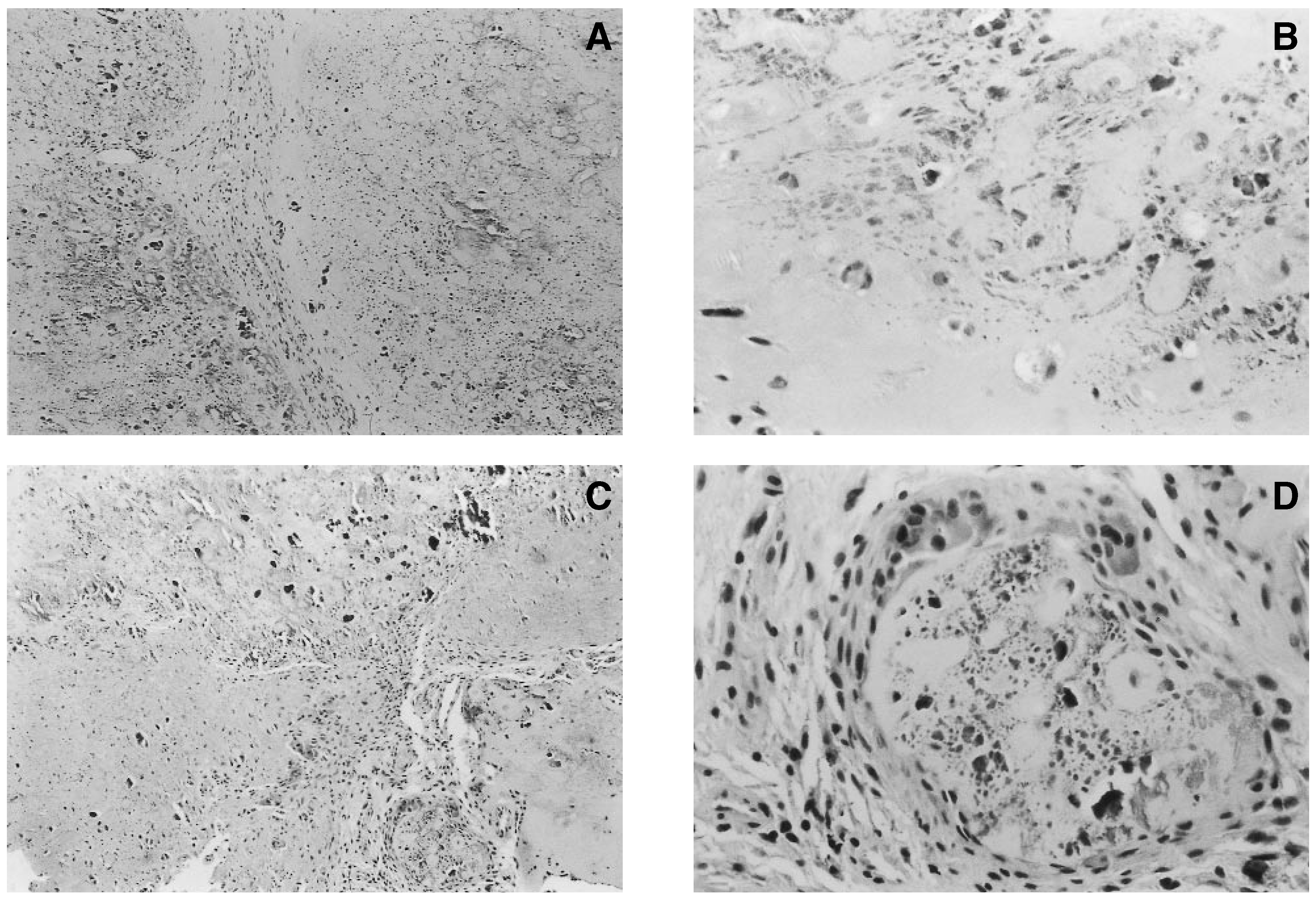
The patient underwent surgical excision of the lesion, which revealed a firm, well-encapsulated, grayishwhite mass approximately 2 cm in diameter, appearing to be closely associated with the flexor tendon sheath. Histologically, the lesion was chondroid with areas of extensive calcification. Hypocellular lobules were noted with cellular areas showing moderate pleomorphism. Giant-cell reaction was noted toward the periphery of some of the lobules (
Figure 2). Pathology reports showed these findings to be consistent with extraskeletal chondroma.
The postoperative course was uneventful. The patient showed no evidence of recurrence or metastasis of the lesion 8 months following surgical excision.
Discussion
The present case of extraskeletal chondroma is similar to those previously described in its clinical presentation as a small, painless mass found in a toe, a common location for soft-tissue chondromas. Radiographic findings in this case were highly suggestive of a soft-tissue tumor with diffuse calcification not associated with underlying osseous structures.
Although this lesion did exhibit the typical lobulated chondroid appearance of extraskeletal chondroma, mature, adult-type hyaline cartilage was scarce. Extensive calcification, as seen in one-third of all lesions, and giant-cell proliferation, which can occur in 15% of cases, were also seen [
1].
Despite the lesion’s variability and occasional atypical cellular characteristics, distinguishing benign extraskeletal chondromas from other lesions is made easier by considering both the clinical and histologic features such as the patient’s age, the location and size of the lesion, and the predominant cell type of the lesion. By so doing, the physician can make a more accurate assessment and avoid unnecessary radical surgery.