The opinions and assertions contained herein are the private views of the authors and are not to be construed as official or as reflecting the views of the Department of Veterans Affairs.
Poroid hidradenoma is a benign neoplasm with eccrine differentiation. It has both solid and cystic components and is contained entirely within the dermis [
1]. Eccrine poromas have been previously reported; however, to the authors’ knowledge, the present article is the first in the podiatric literature to report a case of poroid hidradenoma, a recent subclassification of the poromas [
2,
3].
Poroid hidradenomas are made up of poroid and cuticular cells similar to those in the intradermal eccrine duct and in the lower segment of the intraepidermal eccrine duct. There are now four subtypes of poromas: hidroacanthoma simplex, eccrine poroma, dermal duct tumor, and poroid hidradenoma [
1]. The poromas are classified on the basis of the location of the neoplastic cells. When the cells are entirely in the epidermis and arranged in ovoid nests, the lesion qualifies as hidroacanthoma simplex. When the neoplastic cells are without cysts and have widely spaced nodules located completely in the dermis, the lesion is called a dermal duct tumor, and when the basal layer of the epidermis and the upper part of the dermis contain the neoplastic cells, the tumor is referred to as an eccrine poroma. When both solid and cystic components are found in a tumor that lies entirely in the dermis with no connection to the epidermis, the neoplasm is termed poroid hidradenoma [
4].
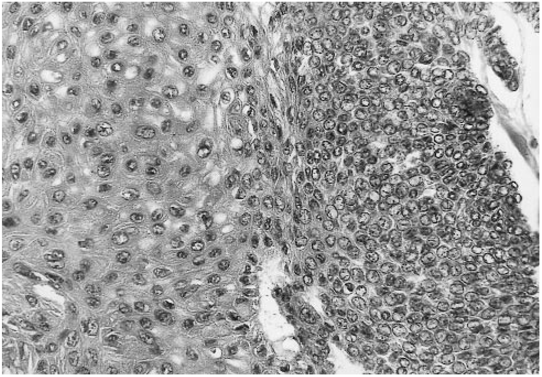
Hidradenomas are divided into two groups. The first, those of eccrine differentiation, constitute 5% of all hidradenomas; they are characterized by single, multilobulated dermal nodules that have no connection to the epidermis. These neoplasms consist of two cell types, poroid and cuticular, that are identical to the cell types found in other poromas. They are known as poroid hidradenomas (
Fig. 1). Ninety-five percent of all hidradenomas fall into the second group, consisting of neoplasms with apocrine differentiation. They are composed of clear, polygonal, and mucinous cells with foci of tubular differentiation [
5]. These are sometimes referred to in the literature as clear cell hidradenomas. Some confusion has resulted from the fact that the tumor described in the present article is classified in two different ways. It has the architectural characteristics of a hidradenoma and the cytologic characteristics of a poroid neoplasm; hence its name.
Although several authors previously reported tumors that closely resembled poroid hidradenomas, Abenoza and Ackerman [
1] in 1990 were the first to use the term “poroid hidradenoma” in describing this tumor. In 1966, Winkelman and McLeod [
6] described a distinct tumor of the dermis that closely fit the description of what Abenoza and Ackerman called a poroid hidradenoma. In 1968, Winkelman and Wolff [
7] described two neoplasms, which they termed “solid-cystic hidradenoma,” that were consistent with the characteristic features of a poroid hidradenoma. Then, in 1976, Ishikawa [
8] described a dermal neoplasm that resembled a nodular hidradenoma with no connection to the dermis.
Clinically, a poroid hidradenoma presents as a solitary neoplasm in 99% of cases. It is generally asymptomatic, but can exhibit slight tenderness. Pain or loss of epicritic sensation may be noted if compression of a neurovascular bundle has occurred. The tumor is typically well circumscribed and is most frequently seen in the sixth decade of life; however, those affected range widely in age, from 3 to 93 years [
7,
9]. No sex or ethnic predilection has been noted. The reported frequencies of tumor locations are as follows: head, 50%; extremities, 21%; trunk, 20%; and neck, 8%. The extremity poromas commonly occur on the palms and soles where the concentration of eccrine glands approaches 620/cm
2; however, poroid hidradenomas are not commonly found on the plantar aspect of the foot [
1,
10]. Most tumors are slow growing, with an average of 1 to 5 years elapsing before the patient presents to a physician. Approximately 15% demonstrate ulceration at presentation due to direct trauma or abrasion to the mass. One-quarter of these tumors have an underlying blue hue that results from secretory or hemorrhagic fluid within the cystic structures [
5]. These neoplasms are considered benign, because the risk of malignant transformation is less than 1% [
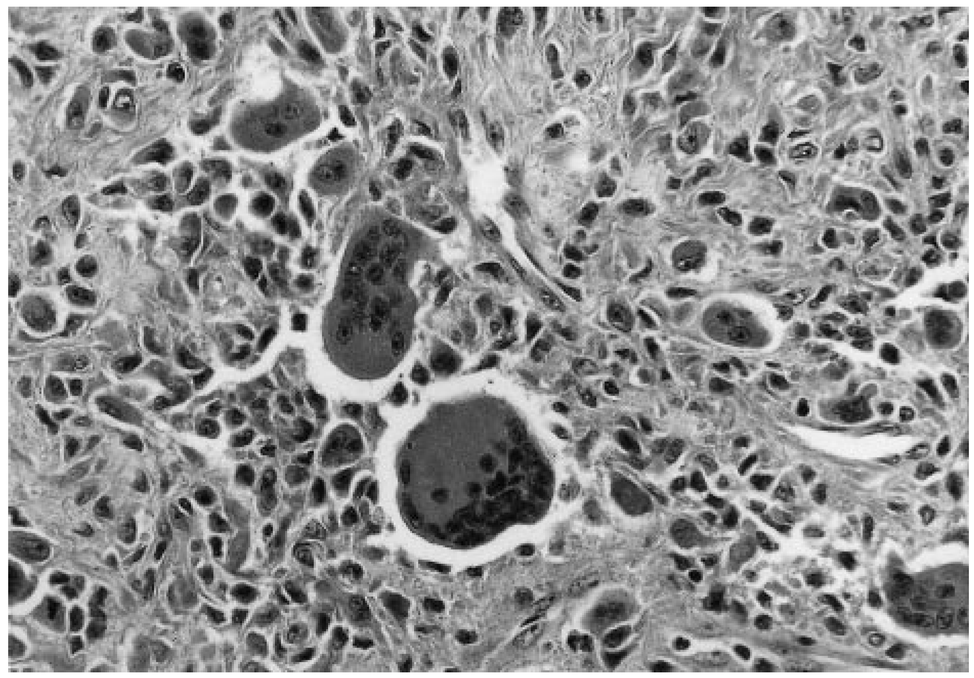
1]. The histologic distinction between a benign poroma and a porocarcinoma can be difficult, as the benign poromas can show nuclear atypia and mitotic figures suggestive of malignancy (
Fig. 2). However, malignant tumors have an irregular growth pattern in addition to atypia. It is therefore important for the pathologist to evaluate the entire intact tumor [
1].
The differential diagnosis of poroid hidradenoma includes the other poromas as well as the apocrine hidradenomas. Other clinical considerations include seborrheic keratosis, actinic keratosis, basal cell carcinoma, squamous cell carcinoma, pyogenic granuloma, fibroma, hemangioma, amelanotic melanoma, dermatofibroma, and epidermal nevus [
1,
10].
The treatment for poroid hidradenoma is total excision, which is usually curative [
10]. However, if the lesion is not removed
in toto, the recurrence rate is high, although no figure is stated in the literature. If potential malignancy is a concern preoperatively, then microscopically controlled excision may be necessary to ensure that the lesion has been completely excised [
10].
Case Report
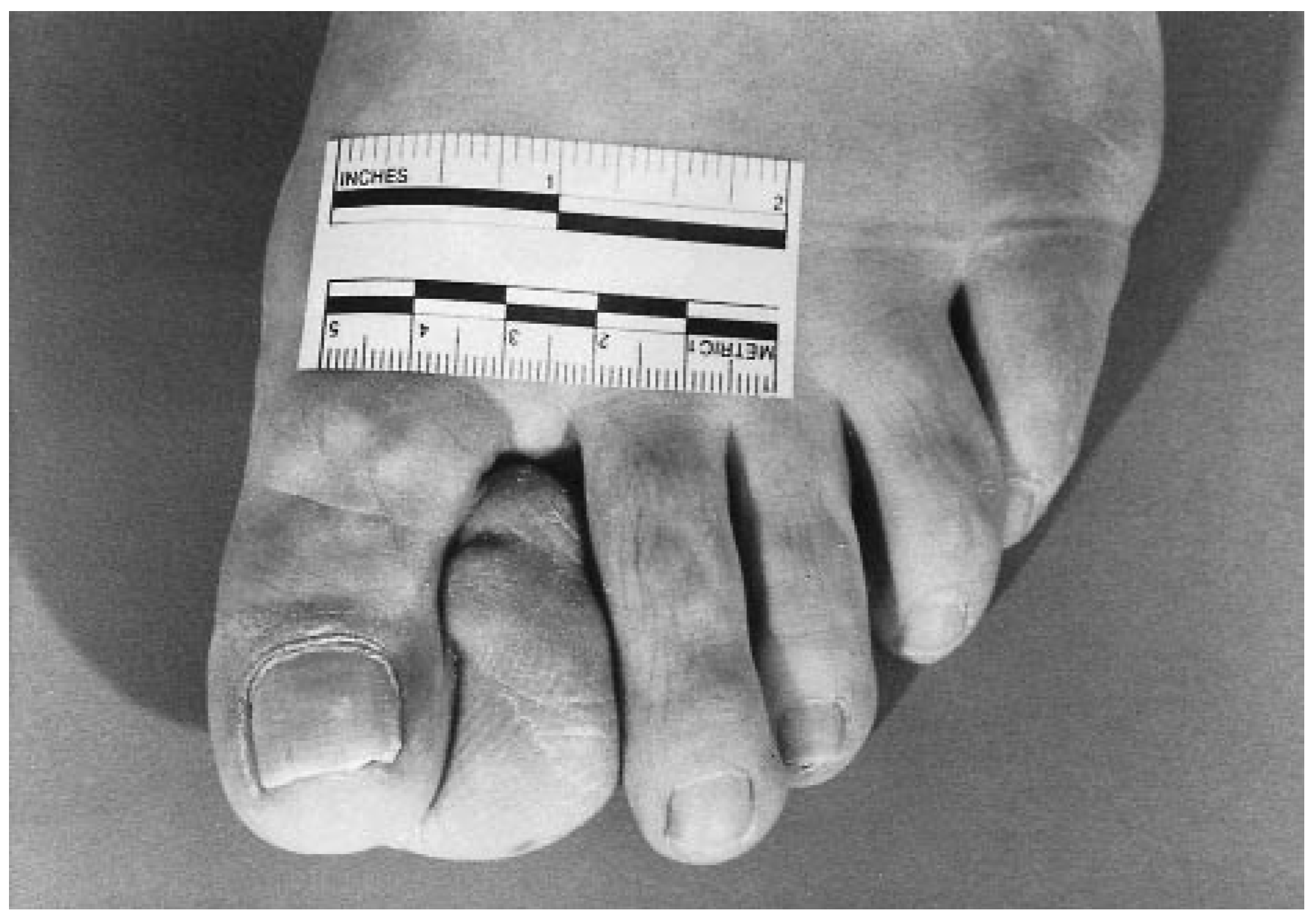
A 56-year-old woman presented to Naval Hospital in Great Lakes, Illinois, complaining of a painful mass on her left great toe. The patient reported that the mass had been present for approximately 12 years and had gradually increased in size. She denied color changes or bleeding. She stated that the mass prevented her from wearing normal shoes and was painful primarily during ambulation (
Fig. 3).
The patient’s medical history was significant for type 2 diabetes mellitus for 10 years and nephrolithiasis at age 6. She denied other lesions and had no other complaints. Medications included glipizide, estrogen, and medroxyprogesterone acetate. Social and family history were noncontributory.
Physical examination revealed a 4.5 × 3 × 2.5-cm lobulated mass arising from the plantar lateral left hallux. The mass was nonpulsatile, exhibited an underlying blue hue, and was not painful upon palpation. The remainder of the lower-extremity examination was unremarkable.
Radiographic evaluation revealed increased soft-tissue density of the first interdigital space, no periosteal reaction, and no osseous erosion. A computed tomographic scan in the coronal projection showed a large soft-tissue mass measuring 4 × 3.5 × 3.5 cm. The mass did not erode into adjacent bony structures, and there was no evidence of significant enhancement on postenhancement scans. The radiologic differential diagnosis included fibrous tumor and nodular tenosynovitis of the joint synovium and tendon sheath.
Ultrasonography was subsequently performed and revealed a solid soft-tissue mass demonstrating multiple internal septations. The lack of through transmission ruled out the possibility of a fluid-filled cyst. Duplex sonography failed to reveal significant arterial or venous flow. The sonographic differential diagnosis included fibroma, lipoma, and unspecified soft-tissue neoplasm.
An ultrasound-guided biopsy was performed using two passes of a 15-gauge ASAP® (Meditech, Natick, MA.) biopsy gun. With the initial pass a small core of tissue was obtained and serous fluid was extruded from the mass. A second pass yielded an additional core of tissue. Microscopic examination revealed fragments of a benign-appearing adnexal tumor most consistent with eccrine poroma.
After discussion of both conservative and surgical treatment options, the patient opted for surgical excision. The standard preoperative evaluation revealed no surgical contraindications, and the patient was taken to the operating room for an excisional biopsy performed under local anesthetic.
A dorsolinear incision was made incorporating the entire length of the mass. During subcutaneous dissection, a large amount of straw-colored serosanguineous fluid was expressed. The mass was separated from the surrounding tissue, taking care to preserve any neurovascular structures encountered, and excised
in toto. The specimen was sent to the pathology department for analysis. After copious lavage and subcutaneous closure, a large amount of redundant skin remained owing to the original size of the lesion. Dermoplasty was performed by means of the excision of two Burow’s triangles [
11,
12] at the distalmost aspect of the incision and the surgical site was converted to a Y-incision.
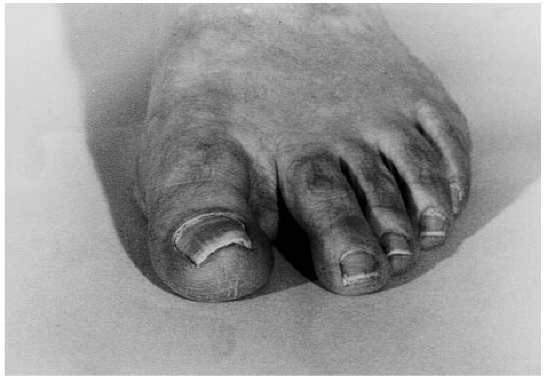
The postoperative course was uneventful. Epicritic sensation was intact with the exception of a 5-mm area of decreased sensation at the distalmost aspect of the incision. The patient remains asymptomatic without evidence of recurrence 4 years after the initial diagnosis (
Fig. 4).
Pathologic examination of the specimen revealed a 3.3 × 2.1 × 1.5-cm portion of yellow-to-pale-tan soft tissue with multiple thin-walled cystic spaces. Microscopically, two cell types were evident: larger cuticular cells with more and paler cytoplasm, and smaller, darker poroid cells. These cell types were contained in the dermis, with no connection to the epidermis. Multinucleated cells were also found in this lesion; these are not uncommon in poroid hidradenomas. The final pathologic diagnosis was poroid hidradenoma, thus highlighting the limitations of the biopsy results and the importance of detailed pathologic examination.
Summary
The clinical, gross, and microscopic characteristics of a poroid hidradenoma have been described, and one case of poroid hidradenoma has been presented.
It is estimated that poroid hidradenoma occurs in the extremities in only 21% of cases. When treating a region of the body with such a high concentration of eccrine sweat glands, however, it is necessary to include this tumor in the differential diagnosis for asymptomatic, slow-growing, solitary skin tumors. Although these neoplasms are benign, they may display some of the microscopic features of a malignant tumor and therefore may be misdiagnosed as such. This underscores the importance of the use of several diagnostic modalities, as in this case.