Kaposi’s sarcoma is the most common malignant lesion in patients who test seropositive for the human immunodeficiency virus (HIV) [
1]. There are no reported cases of trauma as the precipitating event in the development of lesions of Kaposi’s sarcoma in the foot or ankle. In 1872, Dr. Moritz Kaposi [
2] described unusual hyperpigmented, cutaneous nodules on the lower extremities of three men. Appropriately, using the original description set forth by this Hungarianborn dermatologist, the neoplasm came to be known as Kaposi’s sarcoma.
There is considerable epidemiologic evidence suggesting that acquired immunodeficiency syndrome (AIDS)–associated Kaposi’s sarcoma is caused by a transmittable agent. Although the exact etiology of the disease is unknown, increasing evidence suggests that Kaposi’s sarcoma may result from infection with a sexually transmitted organism in addition to HIV [
1]. The primary clinical manifestations of Kaposi’s sarcoma are variable. Lesions often involve the skin and oral mucosa, but may have extramucocutaneous presentations as well, including sites in the head and neck, eyes, vocal cords, heart, lungs, liver, spleen, thyroid, gastrointestinal tract, bone, and lymph nodes [
1]. Typical primary lesions are pink to deep purple or brown in color and are often irregularly shaped and either nodular, macular, patch-like, or plaque-like [
1,
3]. Because of the variability of lesions of Kaposi’s sarcoma, they often mimic other dermatologic pathology. The lesions may or may not be painful, and sometimes follow a bilateral, symmetrical distribution along the lines of skin cleavage [
1,
3]. It is important to note that the lesions of Kaposi’s sarcoma are considered to be autochthonous—ie, not metastatic—with each lesion arising independently of others [
4].
Koebner’s phenomenon has been described as the development of skin disease at a trauma site in a patient who is susceptible to that particular skin disease [
5]. Although Koebner’s phenomenon has previously been reported as the mechanism of onset of Kaposi’s sarcoma, none of the reports describe instances following blunt trauma to the foot or ankle [
5,
6,
7,
8]. Janier et al. [
6] reported lesions developing at the site of venipuncture, following tuberculin skin tests, and as a result of trauma to a patient’s shoulder. Koebner’s phenomenon leading to Kaposi’s lesions has been reported in cases following insect bites, after eczematous skin eruptions, and at sites of surgical scars in HIV-1-ab-positive patients [
5,
6,
7,
8]. French et al. [
5] have suggested that Kaposi’s lesions are not caused by trauma, but rather that the predisposition of an HIV+ patient to Kaposi’s sarcoma results in Koebner’s phenomenon [
5]. Although the exact mechanism is still unclear, the development of lesions following Koebner’s phenomenon is a finding worthy of reporting owing to its rarity.
A definitive diagnosis of Kaposi’s sarcoma is made by biopsy [
4,
6,
9]. Upon histologic confirmation, it is essential that HIV screening and physical examination be performed and a comprehensive history be obtained in order to rule out the existence of opportunistic infection [
9]. Treatment options are numerous and are based on a variety of factors, including the relative immune status of the patient, the presence or absence of systemic opportunistic infections, and the overall nature and location of the tumors or lesions [
10,
11].
The authors report a case of trauma-induced lesions in the foot that was successfully treated with radiotherapy.
Case Report
A 48-year-old HIV+ man presented to the Podiatric Rehabilitation Clinic at Jackson Memorial Hospital in Miami, Florida, with the chief complaint of a nonpainful mass on his left hallux. The patient stated that he dropped a piece of wood on his great toe 1 month earlier and then noticed, within a matter of days, a slowly developing mass. The patient presented with a gauze bandage covering the mass on his left hallux, and reported performing daily dressing changes because of the constant drainage from the lesion. The patient described the drainage as a yellow liquid mixed with blood. The patient denied having prior cutaneous lesions on his lower extremities.
A thorough medical history revealed the presence of HIV for 8 years, with the patient reporting a concentration of CD4+ T lymphocytes of 465 cells per cubic millimeter. Further questioning revealed the presence of type 2 diabetes mellitus, depression, and hypertension. Current medications included glyburide, nifedipine, lisinopril, trazodone hydrochloride, buspirone hydrochloride, and paroxetine hydrochloride. The patient denied having any allergies or relevant surgical history.
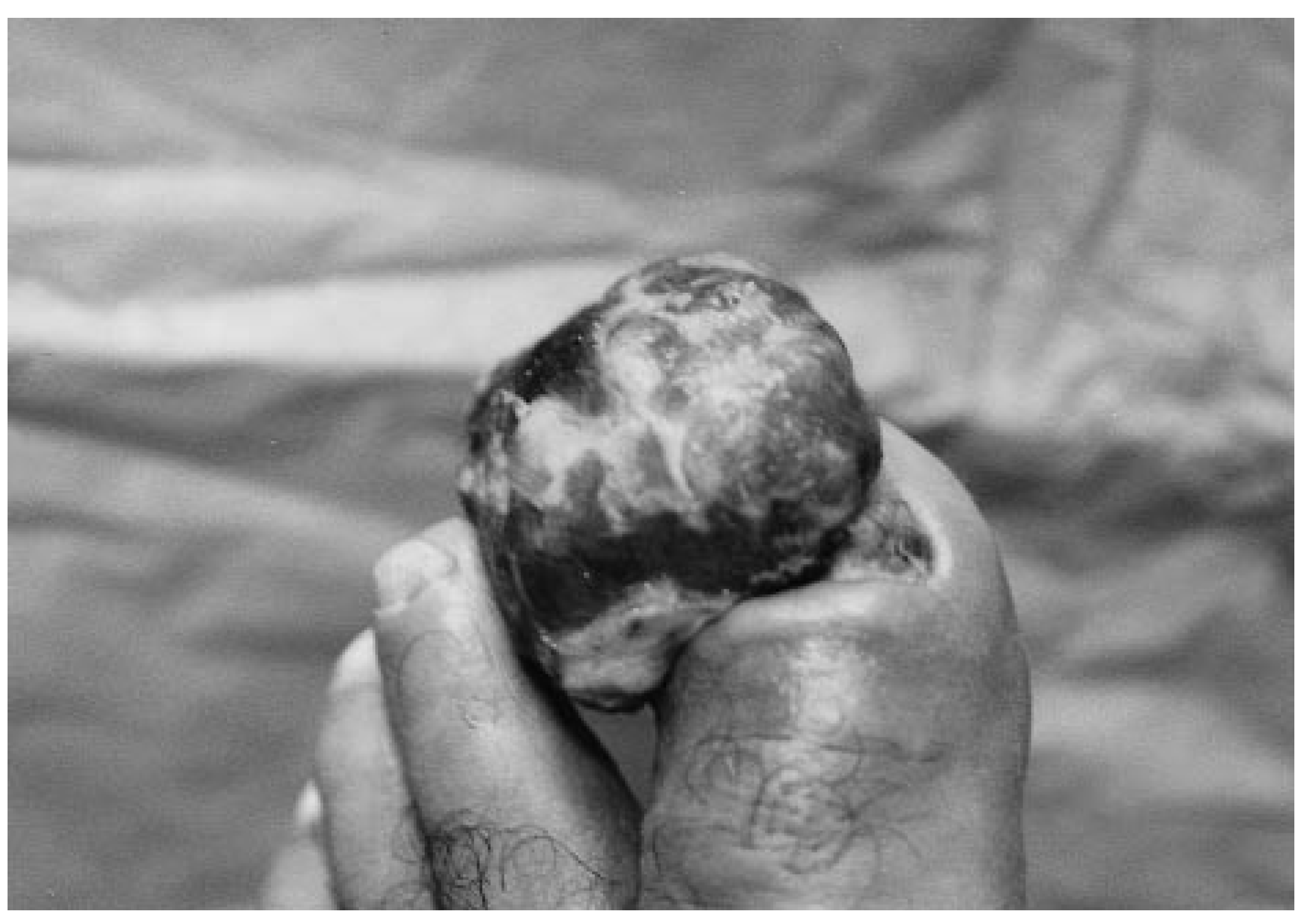
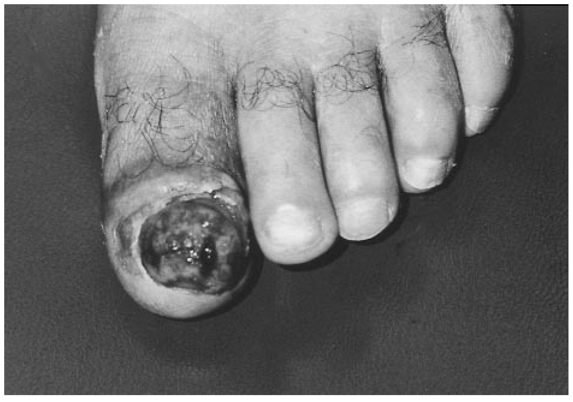
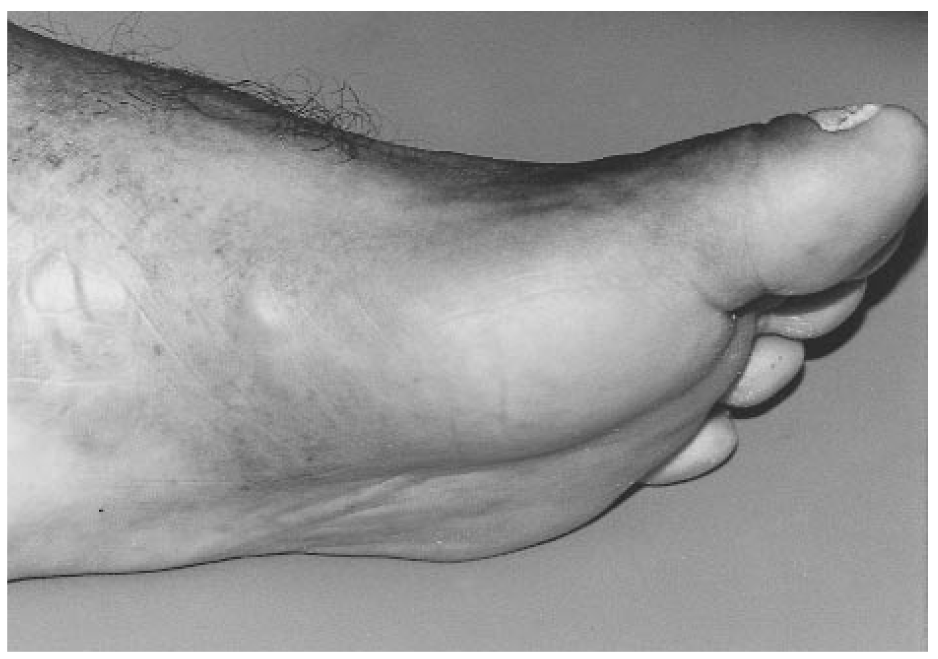
Physical examination revealed an afebrile patient with stable vital signs. The neurovascular status was intact to both feet. The strength of all muscle groups and range of motion to all joints were within normal limits. The left hallux had a red, brown, and yellow tumorous mass protruding dorsolaterally from the hallux nail bed and measuring approximately 2 × 1.5 × 1.5 cm (
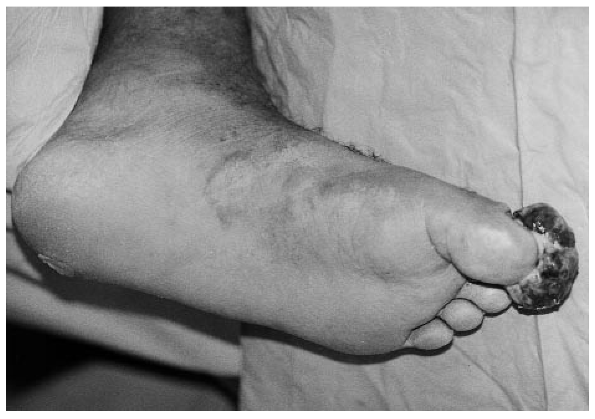
Figure 1). The medial arch of the left foot demonstrated a violaceous patch measuring approximately 4 × 2 × 0.3 cm and there was a similar smaller patch distal to it measuring approximately 1 × 0.5 × 0.1 cm (
Figure 2). The hallux lesion was spongy and produced a malodorous, serosanguineous drainage that appeared to weep from all aspects. The mass was not painful on palpation. Minimal localized erythema and edema surrounded the mass, but did not extend proximal to the interphalangeal joint.
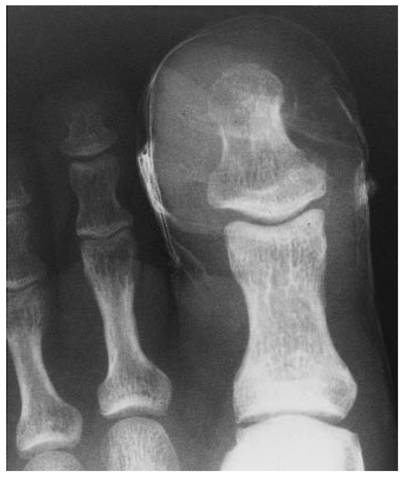
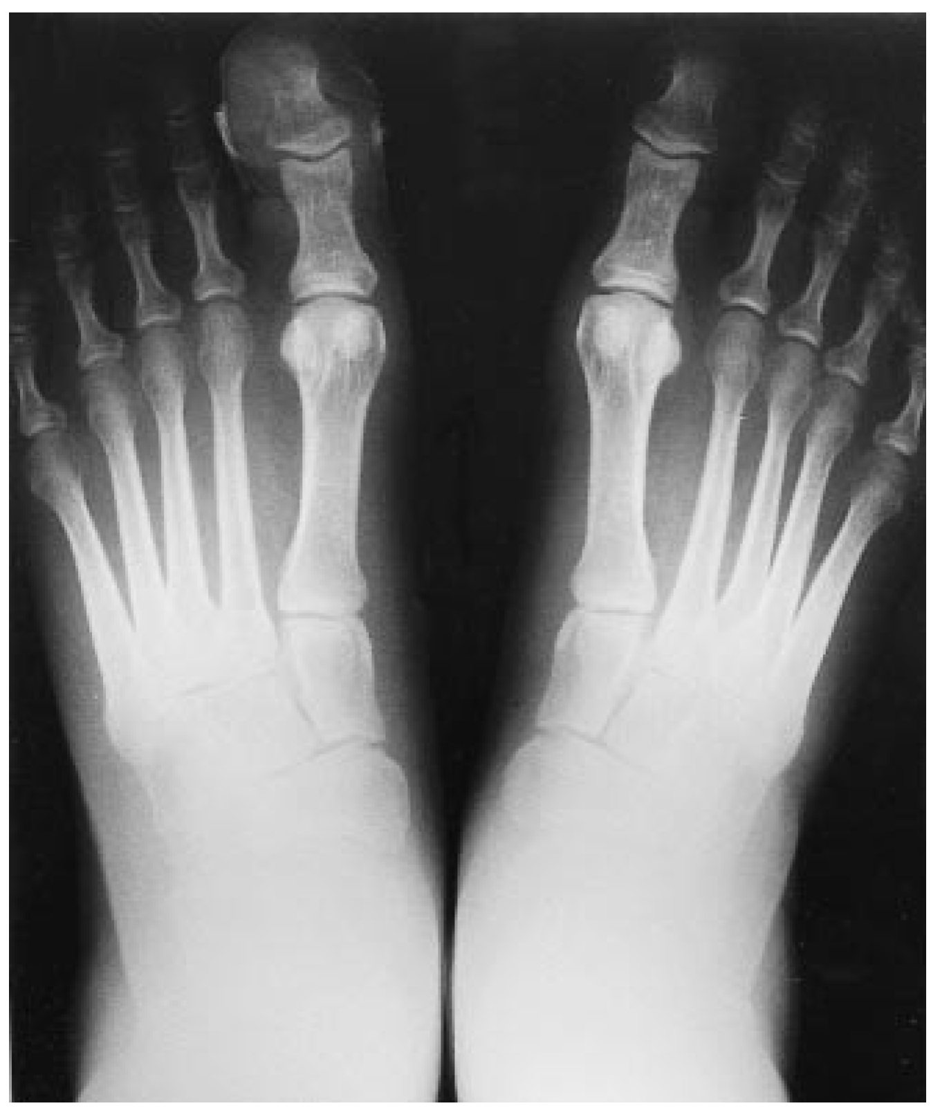
Radiographic examination revealed increased soft-tissue density around the distal phalanx of the left hallux and a small radiolucent region of the distal lateral aspect of the distal phalanx of the hallux. No involvement of the proximal phalanx was noted at this time (
Figure 3 and
Figure 4). Because of the presence of HIV, as well as the clinical appearance of the lesion and the fact that it was not painful, a primary differential diagnosis of AIDS-related Kaposi’s sarcoma was made. Other possible diagnoses included pyogenic granuloma, arteriovenous malformation, angiosarcoma, and hemangioma. The authors decided to order a biopsy of the lesion to make a definitive diagnosis.
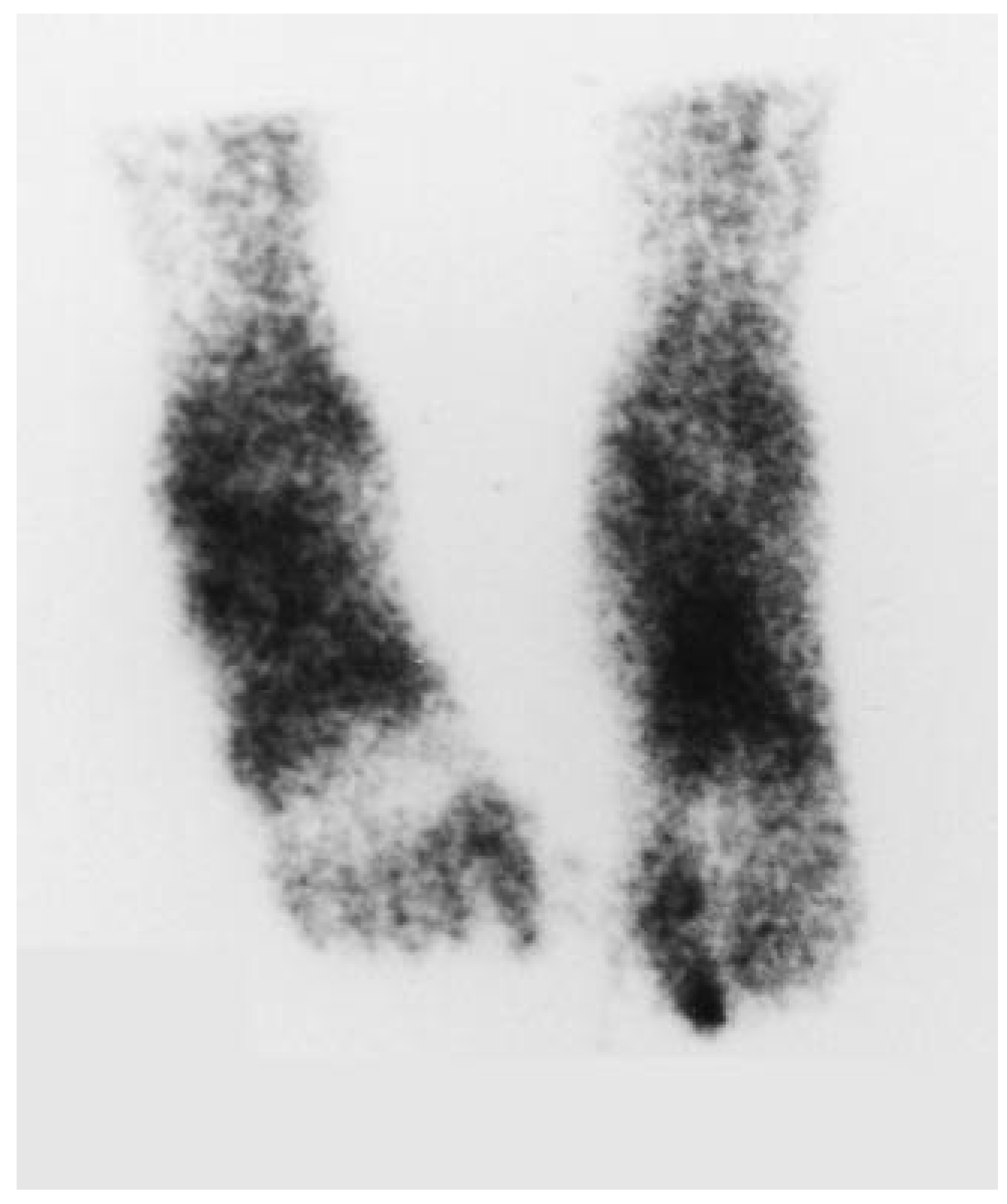
At the initial presentation, a technetium-99 (
99Tc) bone scan was ordered to evaluate the extent of bony involvement of the lesion (
Figure 5). The patient was prescribed a prophylactic regimen of cloxacillin sodium, 500 mg four times a day for 10 days, and instructed to perform daily povidone-iodine wet-todry dressing changes. He was also given a surgical shoe for ambulation. The patient was referred to the oncology department for a second opinion, after which he was requested to return to the podiatric clinic for possible biopsy of the lesion.
The patient returned 2 weeks after initial presentation. The results of the 99Tc bone scan revealed focal increased uptake in the proximal and distal phalanges of the left hallux, most likely representing invasion from the known soft-tissue mass. The oncology department advised an incisional biopsy as the next appropriate step in order to confirm the diagnosis of Kaposi’s sarcoma. The patient was scheduled for evaluation by the immunology department, and was requested to return to the podiatric clinic afterward for biopsy. Unfortunately, as is often the case in clinical settings handling a large indigent population, scheduling problems and patient noncompliance delayed intervention.
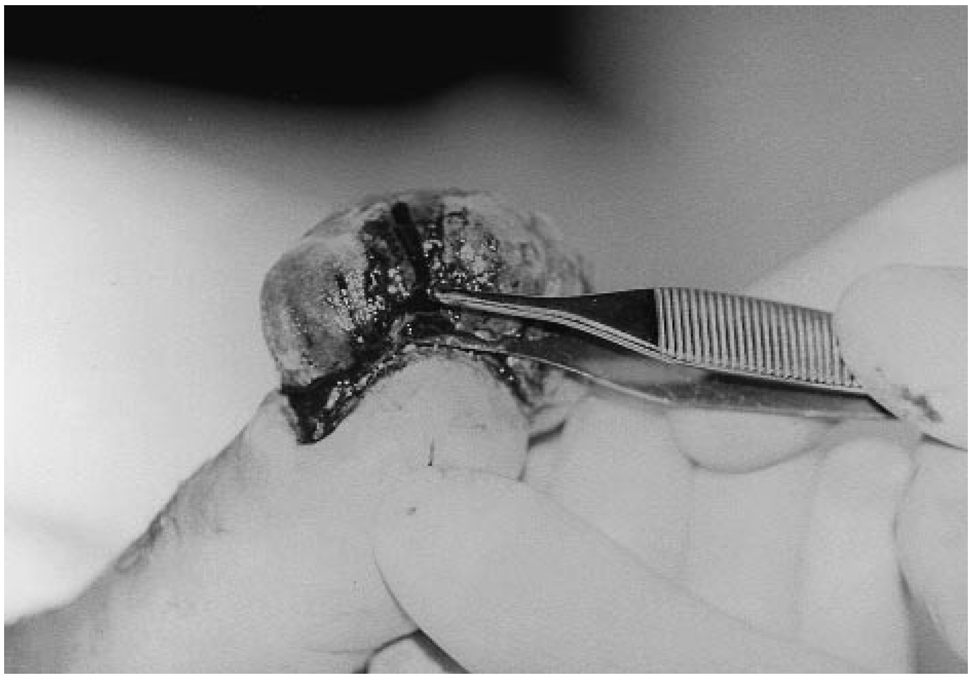
When the patient returned 10 weeks after his initial visit, the mass had more than doubled in size to approximately 4 × 5 × 3 cm, but there was decreased drainage and odor. An incisional biopsy was then performed and the extracted tissue was sent to the pathology department for histopathologic analysis (
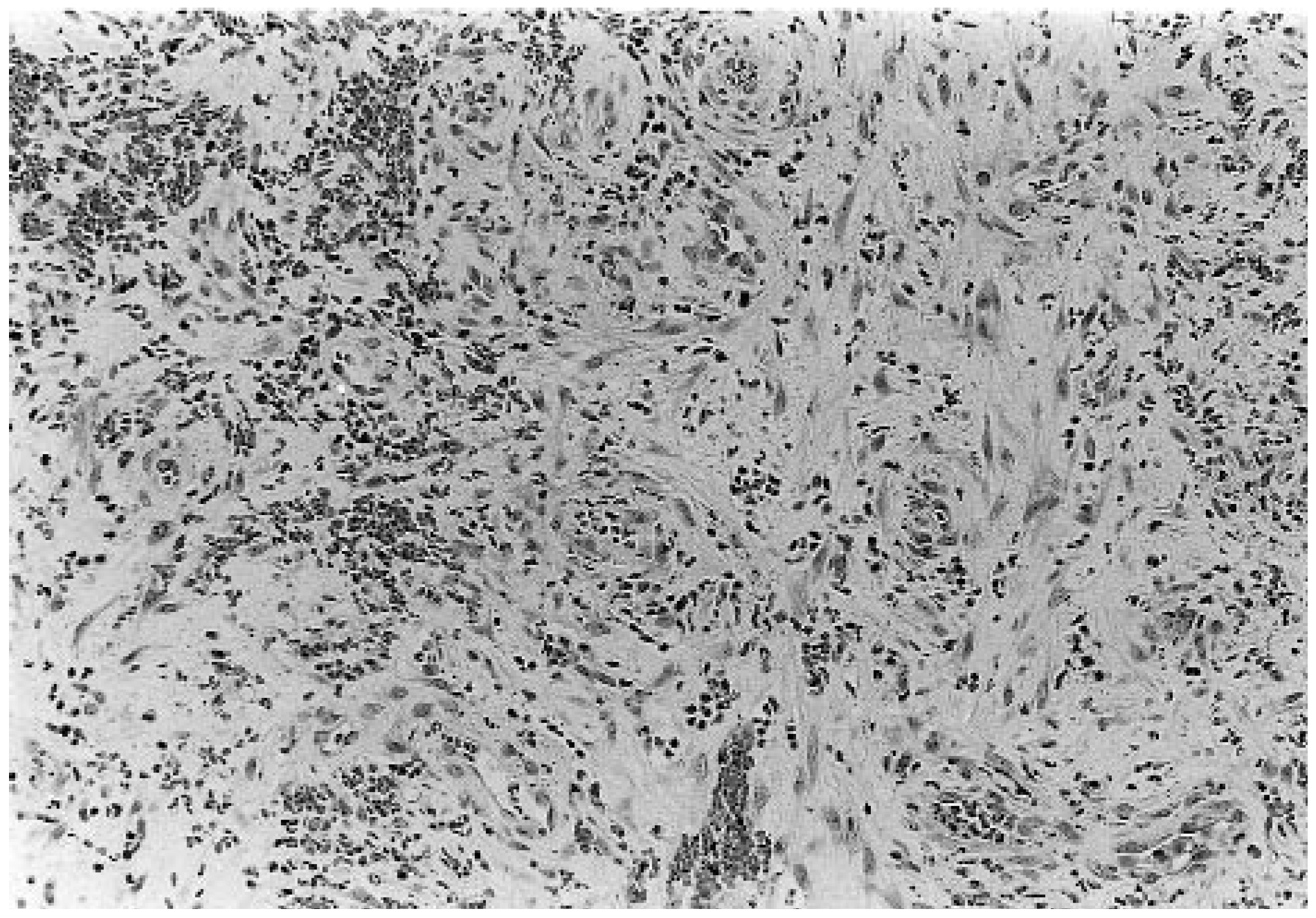
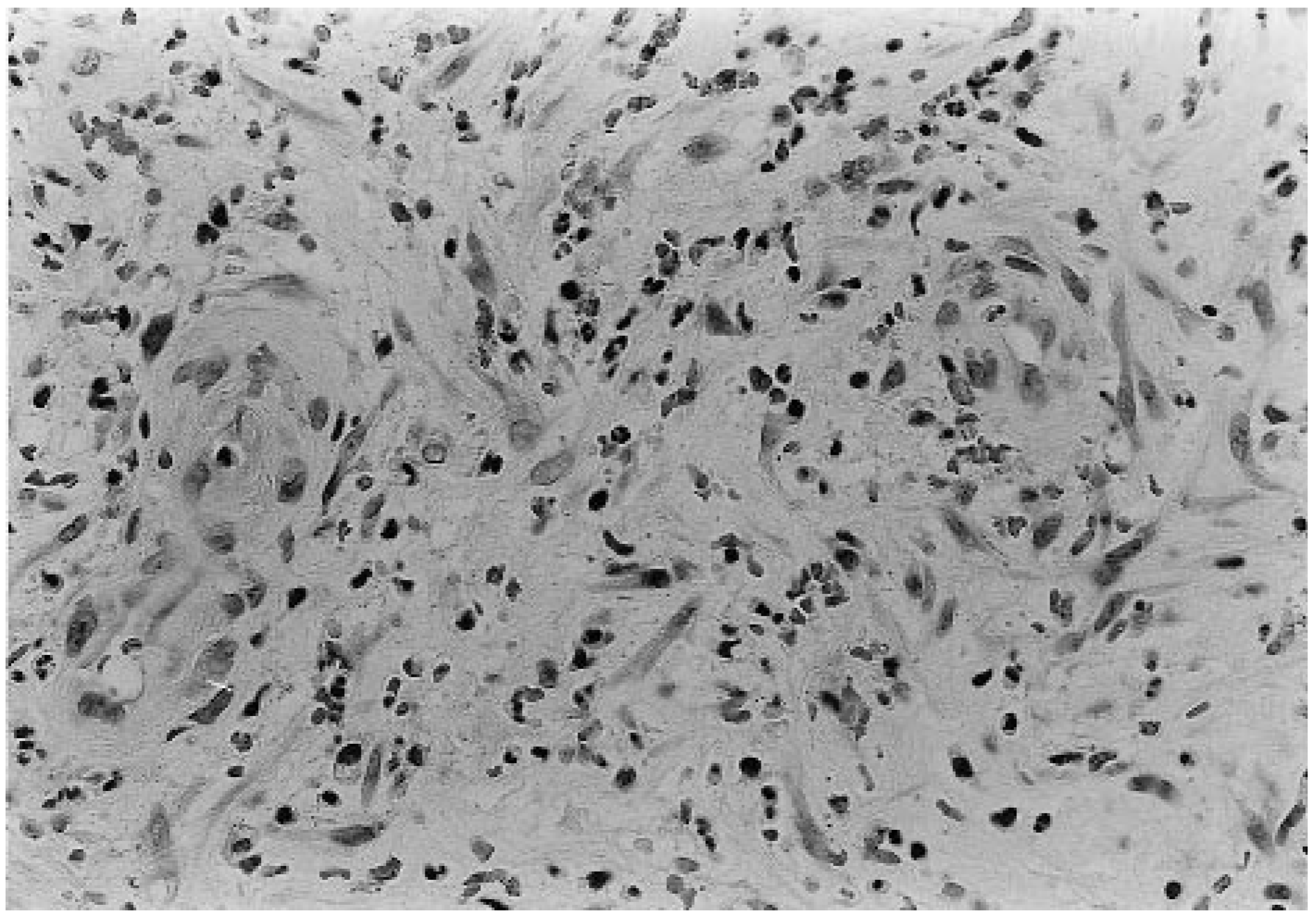
Figure 6). The patient was instructed to proceed with a daily change of dry-gauze dressings. The pathology department confirmed the diagnosis by noting, “soft tissue with vascular proliferation consistent with Kaposi’s sarcoma” (
Figure 7 and
Figure 8). The patient’s immunologist was notified of the findings, and it was the opinion of both the immunologist and the oncology department that radiation therapy was the most appropriate initial intervention. The authors concurred with this decision as being in the best interest of the patient.
The patient underwent ten courses of local radiation therapy over a period of 14 days. The patient’s treatment regimen consisted of 300 rad per visit in a 7 × 12-cm field, totaling 3,000 rad. The patient was examined in the podiatric clinic after eight treatments, and it was noted that the mass had decreased in size to approximately 3 × 4 × 2 cm. A large, black eschar was noted as well, with minimal drainage and odor. Eschar and fibrotic debris were easily removed from the mass by careful debridement. The patient was instructed to follow this daily routine: Soak the foot in warm water with aluminum subacetate solution, remove any debris with gauze, and apply a dry dressing. The lesion on the medial arch was not receiving radiation and had increased in size to approximately 4.5 × 2.5 × 0.5 cm. The more distal arch lesion had increased to 3 × 1 × 0.4 cm. These lesions were left untreated pending further evaluation of the hallux lesion.
The patient returned for follow-up examination 1 month later. The mass on the hallux had decreased to one-third its previous size (
Figure 9). Little drainage was noted, and no odor was detected. Mild, nonpitting edema was present in the entire left foot; the patient reported that it had appeared after radiation treatment began. The medial arch lesions remained unchanged. The patient was instructed to continue the daily soaks and dressing changes. The patient continued to be afebrile, and thus antibiotic therapy, hospital admission, or both were deemed unwarranted.
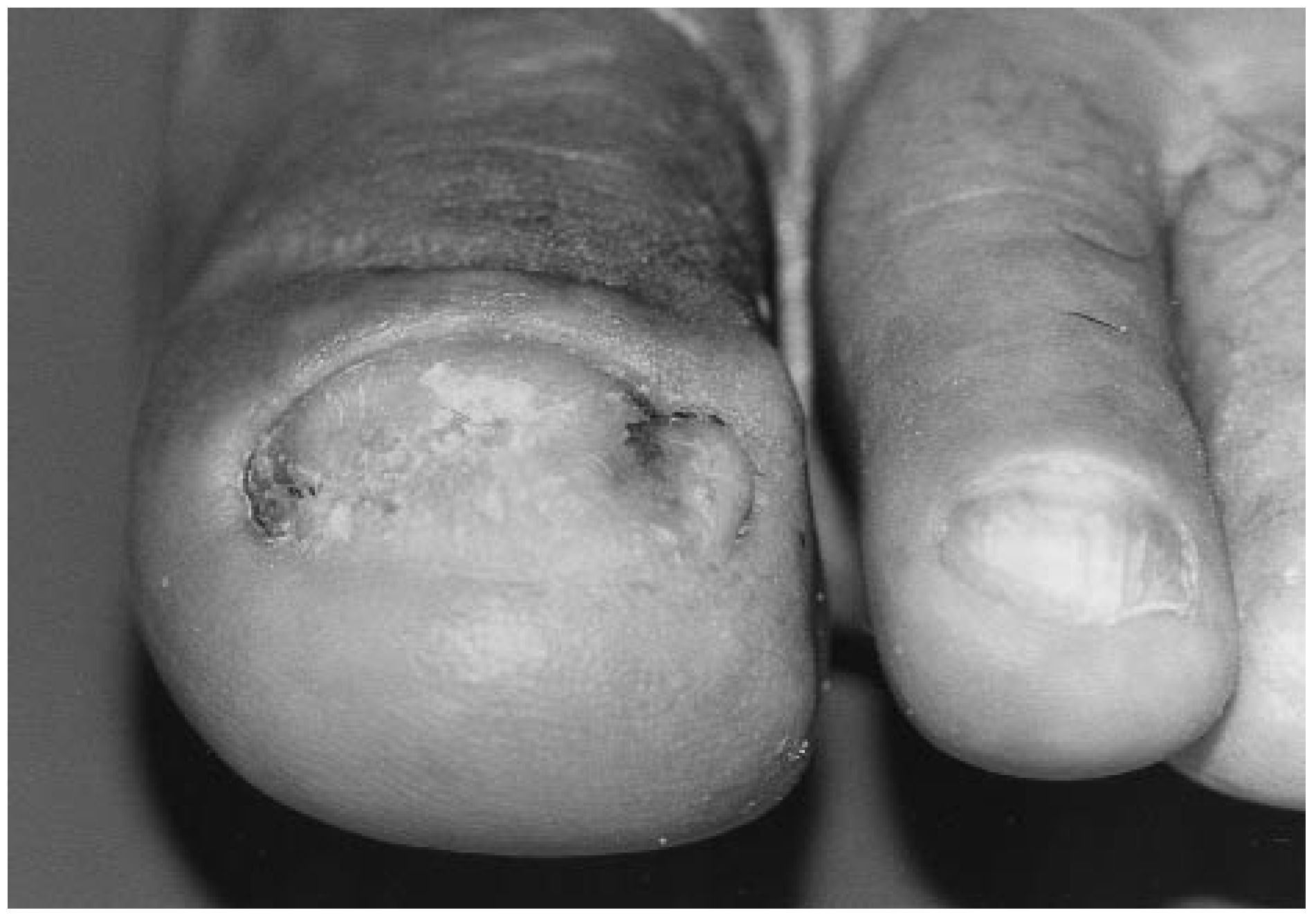
The patient returned 1 month later and there was complete resolution of the Kaposi’s sarcoma (
Figure 10 and
Figure 11). The medial arch lesion remained violaceous and was approximately the same size, with a slight reduction in depth to 0.2 cm. The edema in the foot had completely resolved. The patient was wearing regular footwear for the first time in nearly 6 months. The patient was instructed to continue with follow-up appointments so that the hallux and medial arch lesions could be monitored for any significant changes or recurrence.
Three months later, the patient returned for evaluation and there was no evidence of recurrence of the lesions. A thickened nail plate covering about 80% of the nail bed was present, and the medial arch lesion was reduced to a nonraised patch. At 12 months after initial presentation, there was no recurrence of the lesions and a normal nail plate was evident. The patient was seen at 3, 6, 9, and 12 months for follow-up care. Further visits at 3-month intervals are planned.
Conclusions
The authors have reported an unusual case of trauma-induced Kaposi’s sarcoma of the hallux. Trauma, as observed through Koebner’s phenomenon, should be considered as a mechanism for development of lesions in patients with HIV. Although diagnosis is sometimes difficult, an early diagnosis is critical and can be made through biopsy. Consultations with other specialists such as oncologists and immunologists are recommended. There are many treatment options for cases of Kaposi’s sarcoma, and treatment should be carefully considered based on the overall best interests of the patient. The podiatric physician should be conscious of the potential for development of these lesions following trauma, especially in the at-risk HIV+ population.