Case Report

First Metatarsophalangeal Joint Arthrodesis with Calcaneal Allograft for Failed Joint Arthroplasty

Hunt Regional Medical Center, Greenville, TX 75401, USA
*
Author to whom correspondence should be addressed.
J. Am. Podiatr. Med. Assoc. 2026, 116(5), 61; https://doi.org/10.3390/japma116050061
Submission received: 23 March 2025 / Revised: 27 May 2025 / Accepted: 1 July 2025 / Published: 1 September 2026

Abstract

First metatarsophalangeal joint (MTPJ) arthrodesis is a powerful surgical option for treatment of hallux rigidus, hallux valgus, and revisional first MTPJ surgery. Joint arthroplasty serves as an alternative to arthrodesis for patients who want to preserve joint motion. However, joint arthroplasty has a high rate of failure and can lead to significant bone loss, which makes revisional surgery more challenging. In this study, we present the use of a calcaneal bone block allograft and bone marrow aspirate for fusion of a first MTPJ with a previously failed joint implant. Radiographic fusion was achieved after 6 weeks and ambulation has remained pain-free at 1 year follow-up.

1. Introduction

Hallux rigidus is a degenerative condition leading to stiffness and pain in the first MTPJ, often caused by osteoarthritis [1,2]. It is one of the most common arthritic conditions affecting the foot and can significantly impair gait and daily activities [3]. Conservative treatment options, including orthotics, anti-inflammatory medications, and intra-articular injections, may provide temporary relief, but surgical intervention is frequently required in advanced cases [4,5].
Surgical options for hallux rigidus include joint-preserving procedures, such as cheilectomy or interpositional arthroplasty, and joint-sacrificing procedures, such as arthrodesis or joint replacement [6]. While first MTPJ arthroplasty has been utilized to maintain joint motion, implant survival rates are variable, with some studies reporting high failure rates due to implant loosening, subsidence, and osteolysis [7,8]. When an implant fails, revision surgery is often necessary, and arthrodesis remains the gold standard for restoring function and pain relief [9].
In cases where significant bone loss has occurred due to implant failure, bone block arthrodesis is a valuable technique to restore first ray length and maintain biomechanical integrity [10]. Various grafting options exist, including autografts from the iliac crest or distal tibia, as well as allografts [10]. Calcaneal allografts provide a structural and biologically viable alternative with minimal donor-site morbidity [11]. In this study, we report a case of a patient who had a failed first MTPJ arthroplasty, which was successfully revised using a calcaneal bone allograft and bone marrow aspirate to fuse the first MTPJ.

2. Case Report

A 76 year old female presented to our institution complaining of pain to the right first MTPJ for over a year that had been progressively getting worse. The patient had a past medical history of arthritis, hypertension, and hypothyroidism. The patient had a past surgical history of a right foot first MTPJ implant arthroplasty 20 years prior. The previous implant was a total silastic first MTPJ implant. On physical exam, the patient’s BMI was 32.2, protective sensation was intact, and pedal pulses were palpable bilaterally. On musculoskeletal exam, there was less than 20 degrees of dorsiflexion and 15 degrees of plantarflexion, with pain throughout range of motion. The patient also complained of painful 2nd and 3rd digit hammertoes, which were non-reducible with a Kelekian push-up test on physical exam. Radiographs revealed a first MTPJ implant with component subsidence, decreased joint space, and a shortened first ray (Figure 1). The patient had failed conservative treatment, which included custom orthotics, changes in shoe gear, and physical therapy. As a result, the patient elected to proceed with surgical intervention.

3. Operative Technique and Post-Operative Follow-Up

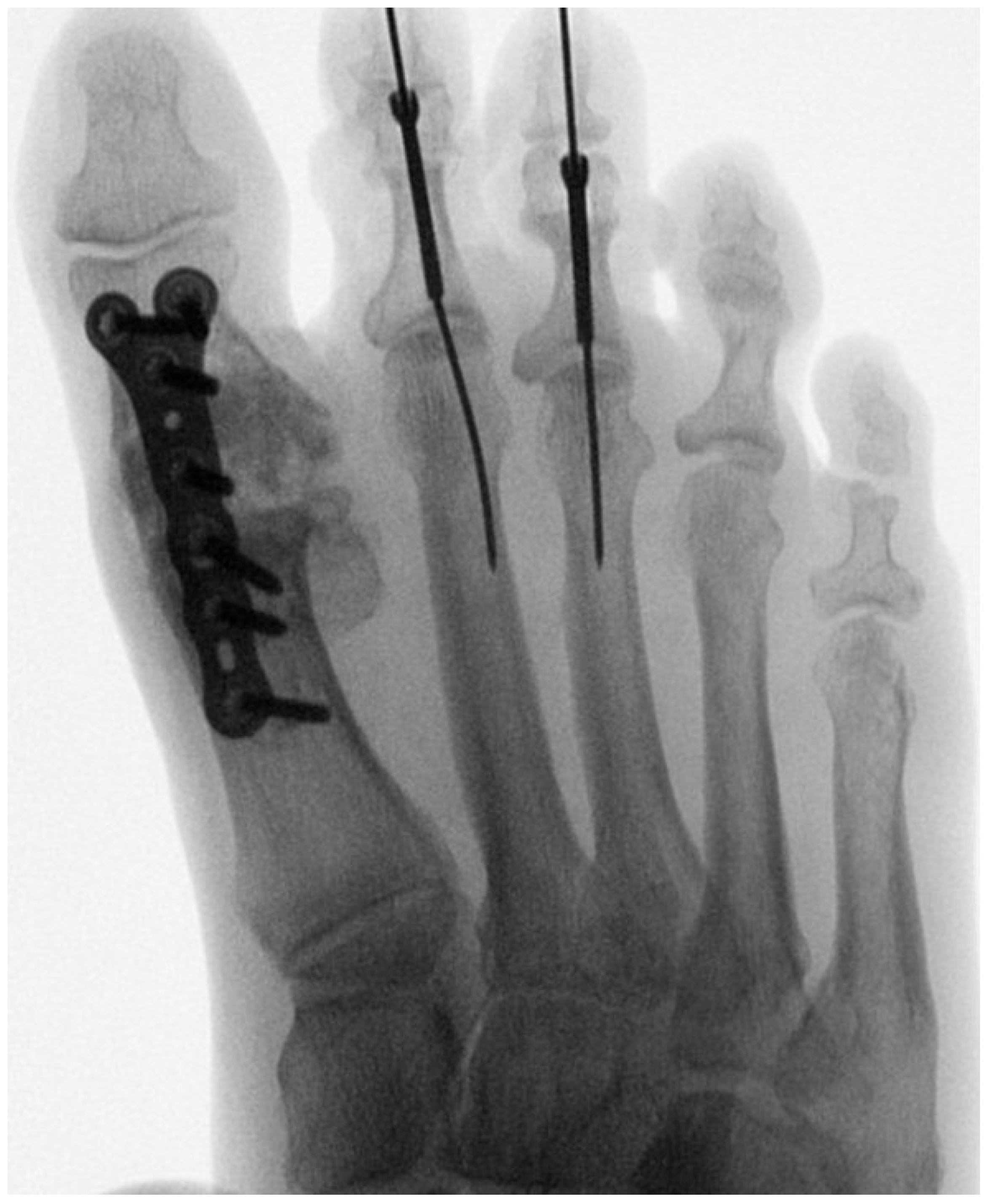
Surgery was performed under intravenous sedation with local block in the supine position. A bone marrow aspirate was acquired from the ipsilateral calcaneal body prior to inflating the thigh tourniquet to 300 mmHg. Next, an 8 cm dorsal linear incision was made medial to the extensor hallucis longus. The incision was deepened to the first MPJ implant, where periprosthetic osteolysis of the first phalanx base and first metatarsal head were noted. The implant was removed in its entirety resulting in a segmental bony deficit. Next, the first MPTJ was prepped for fusion by using a sagittal saw to perform perpendicular osteotomies of the first metatarsal and proximal phalanx to allow complete apposition with the block allograft. Also, the bone was fenestrated with a 2.0 mm drill-bit to increase total surface area of bleeding cancellous bone. A 10.5 mm calcaneal bone block allograft which had been soaked in the acquired bone marrow aspirate was then fitted into the first MTPJ. The first MTPJ was placed in 10 degrees of dorsiflexion, 5 degrees of valgus, and neutral rotation in the frontal plane. Excessive dorsiflexion and valgus was avoided to prevent instability of the allograft while still allowing for adequate toe-off during gait. The allograft was then permanently fixated with a dorsal locking plate (Figure 2). The patient also underwent a standard hammertoe correction of the 2nd and 3rd toes. All alignment and final fixations were confirmed under intra-operative fluoroscopy.
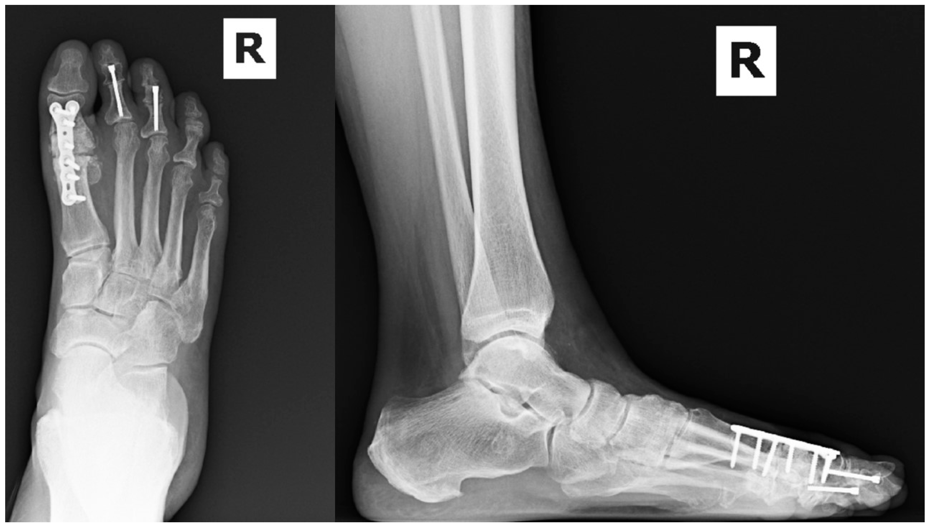
The patient was non-weightbearing for 4 weeks at which time the k-wires from the hammertoes were removed and the patient was transitioned to weightbearing as tolerated (WBAT) in a CAM boot. Post-operative radiographs 6 weeks after surgery revealed adequate callus formation across the first MTPJ fusion site, and the patient was allowed to transition to WBAT in supportive shoe gear (Figure 3). No complications such as infection, wound dehiscence, or non-union were noted during follow-up. Prior to surgery, the patient reported her right foot pain to be a 5 out of 10 on the visual analog scale of pain. At 1 year follow-up, the patient reported 0 out of 10 pain to the right foot. The patient also reported being able to resume her baseline physical activity, which involved walking and strength training.

4. Discussion

Implant arthroplasty, while preserving joint motion, has shown inconsistent long-term outcomes, with some studies reporting implant survival rates as low as 68% at nine years [7]. Brewster et al. performed a systematic review that highlighted that arthrodesis achieves better functional outcomes than total joint replacement [12]. In cases of failed implant arthroplasty, restoration of first ray length is critical for maintaining proper forefoot biomechanics. The use of a structural calcaneal allograft in this case provided a stable scaffold for fusion while minimizing donor-site morbidity associated with iliac crest or calcaneal autografts. In a study comparing mesenchymal stem cell allograft vs. autograft fusion rates, there was no statistically significant difference between union rates, and average radiographic fusion rates were 6.52 weeks for autograft vs. 6.53 weeks for allografts [13].
In cases where the bone defect is larger or there are concerns for infection, the use of the Masquelet technique with an antibiotic spacer has shown high first MTPJ fusion rates [14]. Liu et al. described the use of Masquelet technique for 11 patients with bone defects secondary to gout. Patients had bone defects ranging from 3–6 cm, which were filled with a vancomycin antibiotic spacer. Two months after the initial procedure, the antibiotic spacer was removed while keeping the induced membrane intact. An iliac crest bone block allograft was then implanted into the deficit and fixated with a dorsal locking plate. All 11 patients achieved boney healing at an average time of 3 months [15]. In this case where the defect size was smaller, adequate fusion was achieved without the need for a two-stage procedure. Radiographic union was noted at 6 weeks, and the patient has remained pain-free after one year of follow-up.

5. Conclusions

Calcaneal bone block arthrodesis is an effective salvage procedure for failed first MTPJ implants, offering reliable fusion and improved functional outcomes. The use of structural allografts allows for the restoration of first ray length while avoiding donor-site morbidity associated with autografts. This technique provides a durable and biomechanically sound solution for patients with severe bone loss and deformity following implant failure. Future studies with larger sample sizes and long-term follow-up are needed to further evaluate the efficacy and complication rates associated with this approach.

Author Contributions

Conceptualization, P.T. and P.V.; methodology, P.T. and P.V.; validation, P.T., P.V. and K.H.; investigation, P.T. and P.V.; resources, K.H.; data curation, P.T.; writing—original draft preparation, P.T. and P.V.; writing—review and editing, P.T., P.V. and K.H.; visualization, P.T. and P.V.; supervision, K.H.; project administration, and P.T.; funding acquisition, not applicable. All authors have read and agreed to the published version of the manuscript.

Funding

This research received no external funding.

Institutional Review Board Statement

Ethical review and approval were waived for this study because it is a single-patient case report without prospective intervention, randomization, experimental treatment, or systematic investigation. The manuscript contains no direct patient identifiers or identifiable protected health information.

Informed Consent Statement

Written informed consent has been obtained from the patient to publish this paper.

Data Availability Statement

The original contributions presented in this study are included in the article. Further inquiries can be directed to the corresponding author.

Conflicts of Interest

The authors declare no conflicts of interest.

References

  1. Massimi, S.; Caravelli, S.; Fuiano, M.; Pungetti, C.; Mosca, M.; Zaffagnini, S. Management of high-grade hallux rigidus: A narrative review of the literature. Musculoskelet. Surg. 2020, 104, 237–243. [Google Scholar] [CrossRef] [Scilit] [PubMed]
  2. Cotterill, J.M. The pathology of hallux rigidus. Br. Med. J. 1903, 2, 1400. [Google Scholar] [CrossRef] [Scilit] [PubMed][Green Version]
  3. Bejarano-Pineda, L.; Cody, E.A.; Nunley, J.A., II. Prevalence of hallux rigidus in patients with end-stage ankle arthritis. J. Foot Ankle Surg. 2021, 60, 21–24. [Google Scholar] [CrossRef] [Scilit] [PubMed]
  4. Colò, G.; Fusini, F.; Samaila, E.M.; Rava, A.; Felli, L.; Alessio-Mazzola, M.; Magnan, B. The efficacy of shoe modifications and foot orthoses in treating patients with hallux rigidus: A comprehensive review of literature. Acta. Bio Medica Atenei Parm. 2020, 91, e2020016. [Google Scholar] [CrossRef] [Scilit] [PubMed]
  5. Braun, H.J.; Wilcox-Fogel, N.; Kim, H.J.; Pouliot, M.A.; Harris, A.H.; Dragoo, J.L. The effect of local anesthetic and corticosteroid combinations on chondrocyte viability. Knee Surg. Sports Traumatol. Arthrosc. 2012, 20, 1689–1695. [Google Scholar] [CrossRef] [Scilit] [PubMed]
  6. Galois, L.; Hemmer, J.; Ray, V.; Sirveaux, F. Surgical options for hallux rigidus: State of the art and review of the literature. Eur. J. Orthop. Surg. Traumatol. 2020, 30, 57–65. [Google Scholar] [CrossRef] [Scilit] [PubMed]
  7. Nagy, M.T.; Walker, C.R.; Sirikonda, S.P. Second-generation ceramic first metatarsophalangeal joint replacement for hallux rigidus. Foot Ankle Int. 2014, 35, 690–698. [Google Scholar] [CrossRef] [Scilit] [PubMed]
  8. Jørsboe, P.H.; Pedersen, M.S.; Benyahia, M.; Kallemose, T.; Penny, J.Ø. Mid-term functionality and survival of 116 HemiCAP® implants for hallux rigidus. J. Foot Ankle Surg. 2021, 60, 322–327. [Google Scholar] [CrossRef] [Scilit] [PubMed]
  9. Gross, C.E.; Hsu, A.R.; Lin, J.; Holmes, G.B.; Lee, S. Revision MTP arthrodesis for failed MTP arthroplasty. Foot Ankle Spec. 2013, 6, 471–478. [Google Scholar] [CrossRef] [Scilit] [PubMed]
  10. Usuelli, F.G.; Tamini, J.; Maccario, C.; Grassi, M.; Tan, E.W. Bone-block arthrodesis procedure in failures of first metatarsophalangeal joint replacement. Foot Ankle Surg. 2017, 23, 163–167. [Google Scholar] [CrossRef] [Scilit] [PubMed]
  11. Ahlmann, E.; Patzakis, M.; Roidis, N.; Shepherd, L.; Holtom, P. Comparison of anterior and posterior iliac crest bone grafts in terms of harvest-site morbidity and functional outcomes. J. Bone Jt. Surg. 2002, 84, 716–720. [Google Scholar] [CrossRef] [Scilit] [PubMed]
  12. Brewster, M. Does total joint replacement or arthrodesis of the first metatarsophalangeal joint yield better functional results? A systematic review of the literature. J. Foot Ankle Surg. 2010, 49, 546–552. [Google Scholar] [CrossRef] [Scilit] [PubMed]
  13. Anderson, J.; Jeppesen, N.; Hansen, M.; Brady, C.; Gough, A.; Fowler, Z. First metatarsophalangeal joint arthrodesis: Comparison of mesenchymal stem cell allograft versus autogenous bone graft fusion rates. Surg. Sci. 2013, 4, 263–267. [Google Scholar]
  14. Encinas, R.; Kern, E.; Benjamin Jackson, J., III; Gonzalez, T. The Use of Masquelet Technique and Impaction Bone Grafting for First Metatarsal Phalangeal Joint Fusion With Large Bony Defects: Surgical Technique Tip. Foot Ankle Orthop. 2023, 8, 24730114231169958. [Google Scholar] [CrossRef] [Scilit] [PubMed]
  15. Liu, F.; Huang, R.K.; Xie, M.; Pan, H.; Zhao, J.J.; Lei, B. Use of Masquelet’s technique for treating the first metatarsophalangeal joint in cases of gout combined with a massive bone defect. Foot Ankle Surg. 2018, 24, 159–163. [Google Scholar] [CrossRef] [Scilit] [PubMed]
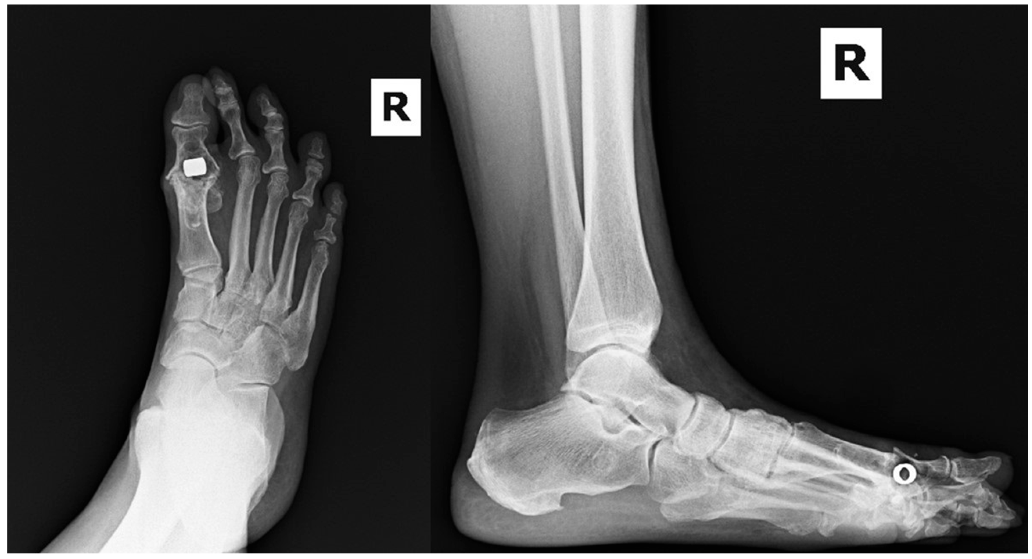
Figure 1. Pre-operative radiographs of painful right foot with 1st MTPJ implant and notable component subsidence.
Japma 116 00061 g001
Figure 2. Intra-operative radiograph of final 1st MTPJ bone block arthrodesis.
Japma 116 00061 g002
Figure 3. Post-operative radiographs 6 weeks after surgery showing union across the 1st MTPJ allograft.
Japma 116 00061 g003
Disclaimer/Publisher’s Note: The statements, opinions and data contained in all publications are solely those of the individual author(s) and contributor(s) and not of MDPI and/or the editor(s). MDPI and/or the editor(s) disclaim responsibility for any injury to people or property resulting from any ideas, methods, instructions or products referred to in the content.