Case Report

An Anomaly of the Pes Anserinus: A Case Study Using Cadaveric Dissection
1
Kent State University College of Podiatric Medicine, Independence, OH 44131, USA
2
Division of Preclinical Sciences, Kent State University College of Podiatric Medicine, Independence, OH 44131, USA
3
Division of Clinical Sciences, Kent State University College of Podiatric Medicine, Independence, OH 44131, USA
*
Authors to whom correspondence should be addressed.
J. Am. Podiatr. Med. Assoc. 2026, 116(5), 60; https://doi.org/10.3390/japma116050060
Submission received: 4 August 2024 / Revised: 18 January 2025 / Accepted: 20 July 2026 / Published: 27 August 2026

Abstract

This case study investigates a pes anserinus anomaly in a male in his seventies who held an occupation in installation, maintenance, or repair. Using whole-body dissection, we found abnormal deposits and synovial tissue in the region of the pes anserinus, possibly suggestive of chronic overuse or injury which would fit with the patient’s occupation. This study could aid healthcare providers in understanding and diagnosing variations and in managing lower limb pathologies, particularly in patients with occupational or repetitive stress-related injuries.

1. Introduction

Some of the first documented cases of pathologies affecting the anserine bursa, reported in 1873, are of syphilitic bursitis [1]. One case was of a law student who after engaging in several hunting parties, developed bilateral “fungous” (Fr. fongueuse) bursitis. The other case was that of a 34-year-old female who developed gummatous bursitis after falling on her knee. These cases highlight how pathologies around the pes anserinus can be linked to trauma or physically demanding occupations.
The anserine bursa (bursa anserina) tends to have an irregularly circular shape, extending to the proximal tibia. The anserine bursa is found deep to the pes anserinus, which is a common tendinous insertion for the sartorius, gracilis, and semitendinosus muscles; “pes anserinus” is Latin for goose’s foot, which it resembles. The anserine bursa separates the tendons of pes anserinus from each other and from the proximal medial tibia. The pes anserinus plays a critical role in lower limb biomechanics. Anomalies in this region can lead to significant alterations in gait and knee mechanics, impacting overall limb function. The muscles surrounding the pes anserine bursa flex and internally rotate the knee. These movements are necessary for normal ambulation and can be compromised by pes anserine bursitis. The insertion site of the pes anserinus tendons varies significantly, which may influence the occurrence of chronic overuse injuries and their clinical presentation [2]. The presence of abnormal anatomical features in the pes anserinus region, such as calcified deposits or abnormal tendon insertions, is rarely documented in the existing literature. Such anomalies are clinically significant as they may contribute to knee dysfunction, altered gait patterns, and discomfort in the affected limb. This condition is linked to knee pathologies such as Osgood-Schlatter syndrome, osteoarthritis, and risk factors like obesity and valgus knee deformity [3]. A multidisciplinary approach in sports medicine is essential, as it emphasizes the importance of thorough physical examination and considering pes anserine bursitis as a differential diagnosis for knee pain [4].
In a case involving a 56-year-old male, a tumor was ultimately diagnosed as secondary to gouty inflammation of the pes anserine bursa, representing a novel manifestation of acute gout [5]. Additionally, a study examining the boundaries of the anserine bursa recommended injection sites for clinical practice and provided critical insights into the anatomical and functional variations in this region. Common pathological conditions affecting the pes anserinus encompass overuse injuries, acute trauma, iatrogenic complications, as well as tumors and tumor-like lesions [6]. Early recognition of anatomical issues in the pes anserinus is crucial for timely intervention, as these conditions may frequently go underdiagnosed. This underscores the need for further research to enhance understanding and improve diagnostic approaches.

2. Methodology

A routine dissection of an embalmed male in his seventies was performed as part of the Lower Extremity Anatomy course at the Kent State University College of Podiatric Medicine (KSUCPM). The donated body was received from the Body Donation Program at Northeast Ohio Medical University (NEOMED) in Rootstown, OH, USA. As far as we know, this body donation program accepts donations from a radius of 75 miles, and potentially farther. During the dissection, an anomaly in the region of the left pes anserinus was identified, prompting further investigation. The body donor held an occupation in installation, maintenance, or repair, and no specific clinical history was available. Upon gross examination, the donated body exhibited a well-developed muscular body habitus with minimal adipose tissue. A scalpel and bone rongeur were used to perform a detailed dissection of the pes anserinus.

3. Results

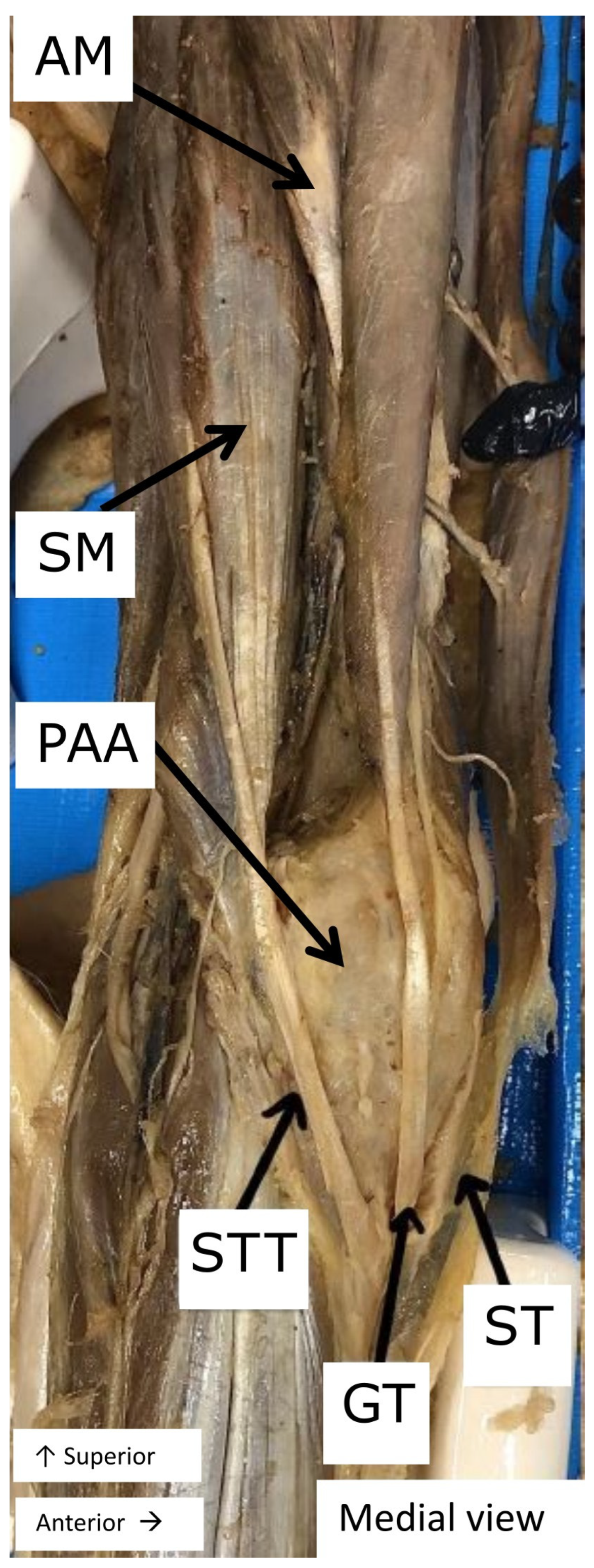
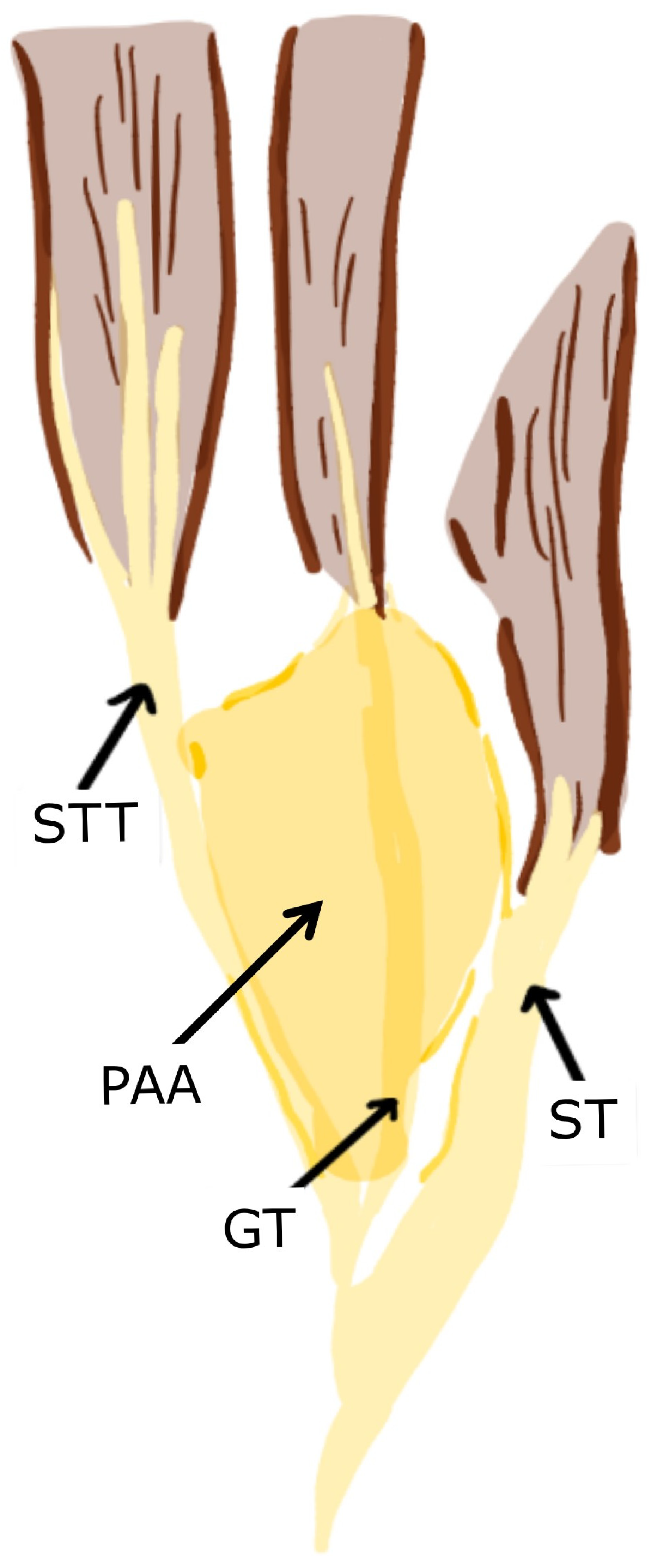
An anomaly of the pes anserinus region of the left lower extremity was identified (Figure 1) and schematically illustrated (Figure 2). Its superior–inferior length was 6 cm, and its anterior–posterior width was 4 cm. It was hard on palpation. The anomaly encompassed a firm grossly observed lesion and synovial tissue. A fluid with a calcification-like consistency exuded from the knee region. The popliteal fossa behind the knee was slightly shifted towards the lateral aspect of the leg. The lesion itself was located deep to the gracilis, semitendinosus, and sartorius muscle tendons. It never extended superiorly to come in contact with the muscle fibers themselves. The right lower extremity showed no evidence of an anserine bursa. It may have been present, but not visible because bursae are difficult to see in dissection.

4. Discussion/Conclusions

A giant soft tissue mass of the pes anserinus region has been described in a case report [7]. Here, we present findings on a firm lesion in the region. Dysfunction in the pes anserinus region, such as pes anserine bursitis, can cause medial knee pain [8]. Overuse, especially in active people, can cause pes anserine bursitis [6]. Additionally, valgus knee deformity is a likely risk factor [9]. Our case presents a condition not routinely focused on in podiatric practice. A firm lesion, like we observed in our dissection, could possibly be of occupational origin and could contribute to medial knee pain, a frequent complaint in the podiatric patient population. Medial knee pain, valgus, and pes planus are often connected and are very relevant to podiatry. In adolescents, reduced physical activity can contribute to both pes planus and genu valgum [10]. In adults, pes planus is associated with frequent knee pain and medial tibiofibular cartilage damage [11].
No living patient was involved in our study; the lesion was discovered during dissection in a Lower Extremity Anatomy course. Therefore, we were limited in the data available about the body donor. Ultimately, there are several conditions that can cause swelling or injury to the anserine bursa [12]. One is patellofemoral syndrome (runner’s knee) which can be caused by overuse and repeated stress from activities such as running or jumping [13]. Septic bursitis is another cause of inflammation in the anserine bursa, and hamstring strain and/or injury to the medial collateral ligament could also cause injury to the anserine bursa. Here, history of present illness, physical examination during life, laboratory studies, and radiographic studies were not available or possible to perform. So, a specific diagnosis would be conjecture at best. From the information we were able to gather, we speculate that this case might involve chronic bursitis that resulted from overuse and may have undergone dystrophic calcification. Further research is encouraged to deepen our understanding of these complex interactions and etiologies in order to optimize patient care strategies.

Author Contributions

Conceptualization, S.M., S.B., I.F. and K.S.; Data curation, S.M.; Formal Analysis, S.M., S.B. and I.F.; Investigation, S.M.; Methodology, S.M., S.B., I.F. and K.S.; Project administration, S.M. and I.F.; Supervision, S.B. and I.F.; Validation, S.M.; Visualization, S.M. and I.F.; Writing—original draft, S.M.; Writing—review and editing, S.M., S.B., I.F. and K.S. All authors have read and agreed to the published version of the manuscript.

Funding

This research received no external funding.

Data Availability Statement

All data relevant to this study can be found in the article.

Acknowledgments

We express our sincere appreciation to the body donor, his family, and loved ones for their generous contribution to medical science. His willingness to donate has greatly contributed to our understanding of pes anserinus anomaly. Thank you to Abe Osbourne and Mary Mooney for their comments and discussion. We gratefully acknowledge the staff at the Edward J. Fiffick, DPM & Family Anatomy Lab at KSUCPM for their essential support and technical assistance during dissection. We thank the reviewers of this manuscript for their helpful comments.

Conflicts of Interest

The authors declare no conflicts of interest.

References

  1. Moreau, L. Des Affections Syphilitiques Tertiaires des Bourses Séreuses: Thèse Pour le Doctorat en Médecine Présentée et Soutenue le 8 Août 1873. 1873. Available online: https://wellcomecollection.org/works/jvg3kh4b (accessed on 10 May 2025).
  2. Lee, J.-H.; Kim, K.-J.; Jeong, Y.-G.; Lee, N.S.; Han, S.Y.; Lee, C.G.; Kim, K.-Y.; Han, S.-H. Pes anserinus and anserine bursa: Anatomical study. Anat. Cell Biol. 2014, 47, 127–131. [Google Scholar] [CrossRef] [Scilit]
  3. Mohseni, M.; Mabrouk, A.; Li, D. Pes Anserine Bursitis. In StatPearls; StatPearls Publishing: Treasure Island, FL, USA, 2025. Available online: https://www.ncbi.nlm.nih.gov/books/NBK532941/ (accessed on 18 January 2025).
  4. Allen, M.F.; Allen, D.E. Pes Anserinus Bursitis: A Case Report. Cureus 2022, 14, e31354. [Google Scholar] [CrossRef] [Scilit] [PubMed]
  5. Grover, R.P.; Rakhra, K.S. Pes anserine bursitis—An extra-articular manifestation of gout. Bull. NYU Hosp. Jt. Dis. 2010, 68, 46–50. [Google Scholar] [PubMed]
  6. Curtis, B.R.; Huang, B.K.; Pathria, M.N.; Resnick, D.L.; Smitaman, E. Pes Anserinus: Anatomy and Pathology of Native and Harvested Tendons. AJR Am. J. Roentgenol. 2019, 213, 1107–1116. [Google Scholar] [CrossRef] [Scilit] [PubMed]
  7. Corominas, H.; Balius, R.; Estrada-Alarcón, P.; Reina, D.; Moya, P.; Videla, M. Giant Pes Anserinus Bursitis: A Rare Soft Tissue Mass of the Medial Knee. Reumatol. Clín. 2020, 17, 420–421. [Google Scholar] [CrossRef] [PubMed]
  8. Rennie, W.J.; Saifuddin, A. Pes anserine bursitis: Incidence in symptomatic knees and clinical presentation. Skelet. Radiol. 2005, 34, 395–398. [Google Scholar] [CrossRef] [Scilit] [PubMed]
  9. Alvarez-Nemegyei, J. Risk Factors for Pes Anserinus Tendinitis/Bursitis Syndrome: A Case Control Study. JCR J. Clin. Rheumatol. 2007, 13, 63–65. [Google Scholar] [CrossRef] [Scilit] [PubMed]
  10. Khalid, Z.; Rai, M.A.; Mobeen, B.; Amjad, I. Pes planus & genu valgum; factors associated. Prof. Med. J. 2015, 22, 1237–1244. [Google Scholar] [CrossRef] [Scilit]
  11. Gross, K.D.; Felson, D.; Niu, J.; Hunter, D.J.; Guermazi, A.; Roemer, F.W.; Dufour, A.; Gensure, R.H.; Hannan, M. Association of flat feet with knee pain and cartilage damage in older adults. Arthritis Care Res. 2011, 63, 937–944. [Google Scholar] [CrossRef] [Scilit] [PubMed]
  12. Glencross, P.M. Pes Anserine Bursitis Differential Diagnoses; Medscape: Newark, NJ, USA, 2023; Available online: https://emedicine.medscape.com/article/308694-differential (accessed on 12 June 2026).
  13. Witvrouw, E.; Callaghan, M.J.; Stefanik, J.J.; Noehren, B.; Bazett-Jones, D.M.; Willson, J.D.; E Earl-Boehm, J.; Davis, I.S.; Powers, C.M.; McConnell, J.; et al. Patellofemoral pain: Consensus statement from the 3rd International Patellofemoral Pain Research Retreat held in Vancouver, 2013. Br. J. Sports Med. 2014, 48, 411–414. [Google Scholar] [CrossRef] [Scilit] [PubMed]
Figure 1. Left lower extremity of a male in his seventies. The pes anserinus, where the semitendinosus (STT), gracilis (GT), and sartorius (ST) tendons insert, had a firm anomaly (pes anserinus anomaly, PAA). The adductor magnus (AM) and semimembranosus muscles (SM) are also visible.
Japma 116 00060 g001
Figure 2. Illustration of Figure 1, depicting the pes anserinus anomaly (PAA) and overlying tendons of the pes anserinus: semitendinosus (STT), gracilis (GT), and sartorius (ST).
Japma 116 00060 g002
Disclaimer/Publisher’s Note: The statements, opinions and data contained in all publications are solely those of the individual author(s) and contributor(s) and not of MDPI and/or the editor(s). MDPI and/or the editor(s) disclaim responsibility for any injury to people or property resulting from any ideas, methods, instructions or products referred to in the content.