Article

Optimizing Ankle Fracture Outcomes with Short-Term Postoperative Immobilization

1
Department of Orthopaedic Trauma, Qinghai University Hospital, Xining 810000, China
2
Department of Sports Medicine, Xining First People’s Hospital, Xining 810000, China
3
Trauma Emergency Center, The Third Hospital of Hebei Medical University, Shijiazhuang 050011, China
*
Author to whom correspondence should be addressed.
J. Am. Podiatr. Med. Assoc. 2026, 116(4), 24128; https://doi.org/10.7547/24-128
Submission received: 25 July 2024 / Revised: 11 October 2024 / Accepted: 16 October 2024 / Published: 13 August 2026

Abstract

Background: This paper aims to investigate the effect of short-term cast immobilization on the prognosis of ankle fractures. Methods: A total of 60 patients who underwent ankle fracture surgery in our hospital from September 2021 to September 2022 were included and divided into a cast immobilization group (n = 30) and a control group (n = 30). Both groups were given open reduction and internal fixation. After the operation, the immobilization group used a plaster cast bandage to fix the ankle joint of the affected side in the functional position for 2 weeks; the control group did not receive cast immobilization and was only bandaged with routine wound dressings. The visual analog scale (VAS) score, ankle joint range of motion (ROM), lower extremity deep venous thrombosis rate, and ankle joint function score were followed up in the two groups. Results: All patients were followed up for 6 months. The VAS scores of the cast immobilization group were lower than the control group at 1, 3 and 7 days post-operation (p < 0.05). At 1, 2 and 3 months post-operation, the dorsiflexion ROM of the ankle joint in the cast immobilization group was larger than control group (p < 0.05). The ankle plantarflexion ROM at postoperative 1 and 2 months were larger than the control group (p < 0.05). The AOFAS of cast immobilization group was larger than the control group at 3 months post-operation (p = 0.002). The postoperative deep venous thrombosis rate was 1/30 (3.3%) in the cast immobilization group and 3/30 (10%) in the control group (p = 0.605). Conclusions: Short-term cast immobilization after ankle fracture surgery can significantly reduce postoperative pain in the early stage, without increasing the recovery and the incidence of deep venous thrombosis.

1. Introduction

Ankle fracture is one of the intra-articular fractures, accounting for about 6.78% of all fractures, with an incidence of about 174/100,000 people [1,2]. Ankle fractures may cause damage to the articular surface and surrounding soft tissues [3]. At present, consensus has been reached on the recovery of joint anatomy in unstable ankle fractures, indicating that fracture sites are treated with incisional reduction and internal fixation to optimize outcome and minimize the risk of post-traumatic arthropathy; therefore, patients can walk again without pain and avoid long-term damage to the ankle joint [4,5,6]. However, even if intraoperative reduction is good, some patients are still at risk of varying degrees of ankle joint dysfunction postoperatively.
To promote Enhanced Recovery After Surgery (ERAS) [7], perioperative patients were intervened with to reduce the difficulty of surgery and reduce complications, thereby promoting the rapid recovery of patients [8,9]. Some scholars [10] pointed out that postoperative immobilization of unstable ankle fractures can reduce the risk of fracture displacement and internal fixation material breakage. In addition, research [11] has shown that reasonable cast support can promote the swelling of the affected limb to subside and reduce pain, and ankle training can be performed after removal of the cast. Therefore, this study aimed to conduct a comparison in the postoperative protocol between cast immobilization and non-immobilization.

2. Methods

2.1. General Information

Patients who underwent ankle fracture surgery in our hospital from September 2021 to September 2022 were retrospectively included and were divided into a cast immobilization group and a control group according to the treatment method.
Inclusion criteria were patients (1) with ankle fractures in line with the diagnostic criteria of the Lauge-Hansen classification [12,13] (2) who were aged 18–65 years (3) with no treatment before admission and 4) who had high compliance with treatment and follow-up.
Exclusion criteria were patients (1) with other combined fractures; (2) with a previous history of ankle fractures; (3) with pathological fractures; (4) associated with abnormal liver, kidney, heart, and lung functions; (5) with immune system diseases; (6) with abnormal coagulation function; and/or (7) with mental illness. The study was approved by the ethics committee of the Third Hospital of Hebei Medical University on May 30, 2016 (K2015-001-12) and was performed according to the Declaration of Helsinki. All patients signed informed consent.

2.2. Treatment

After admission, all patients were treated with detumescence, pain relief, and elevation of the affected limb. Before operation, preoperative X-ray and CT of the ankle joint on the affected side, and color Doppler ultrasound of the deep vein of the lower extremity were performed. These are part of the routine preoperative preparation for ankle fractures and is used to help with surgical anatomy and plate fixation. Surgical treatment was performed after the swelling of the fracture site and the skin.
All operations were performed by the same doctor. According to the AO principles of internal fixation, the fractures were anatomically reduced. For medial malleolus fractures, an arc-shaped incision should be made centered on the tip of the ankle during surgery. The scope of periosteal dissection should be minimized. After the ankle varus was fully exposed, fracture reduction were performed and fixed with 1–2 3.5 mm cannulated lag screws. For patients with comminuted fractures, tension bands and Kirschner wires were also used in the surgical treatment. When dealing with patients with lateral malleolus fractures, 6 to 8-hole lateral plate or screw fixation were selected. For posterior malleolus fractures, T-shaped plate or screw fixation were selected according to the fracture area [14]. Ligament damage during the operation were repaired routinely, and the operation time were recorded.
For the cast immobilization group, the ankle joint on the affected side was fixed with a plaster cast bandage in a functional position immediately after the operation. The fixation methods were as follows. After placing a layer of degreased cotton between the plaster cast bandage and the skin (Figure 1), the cotton and plaster cast bandage were positioned at the bottom of the foot, ensuring that they completely covered the distal toes and the middle and upper one-third of the calf. Finally, the entire assembly was secured with a bandage. The ankle joint was maintained in functional position until plasticity was completed and fixed for 2 weeks. Patients in the control group did not receive cast immobilization after surgery; they were only bandaged with routine wound dressings.

2.3. Postoperative Management

After operation, the affected limbs were routinely raised, and pain relief, swelling reduction, and antithrombotic treatment were given to both groups. In the control group, from the first day after operation, the toes and ankle dorsiflexion, plantar flexion and other activities can be actively moved. The dressing was changed every 2 to 3 days after the operation, and the stitches were removed 2 weeks after the operation. Active dorsiflexion and plantarflexion exercises against the cast were performed in the cast immobilization group on the first day after the operation. After the cast was removed 2 weeks after surgery, active and passive ankle extension and flexion could be performed under non-weight-bearing conditions. Patients in both groups began full weight-bearing and walked without crutches at 10–12 weeks after surgery. Both groups were given ankle pump exercises to prevent lower deep vein thrombosis 300 times a day after surgery with the assistance of a physician.

2.4. Observation Indicators

A Visual Analogue Scale (VAS) was used to evaluate the ankle joint pain on 1 day, 3 days, 7 days, and 14 days postoperatively [15]. The range of motion (ROM) of ankle joint dorsiflexion and plantarflexion were evaluated at postoperative 1, 2, and 3 months [16]. During the measurement, the patient was kept in a sitting position, the knee joint was flexed at 90°, and the ankle joint was kept in the functional position. The goniometer axis was located over the center of the lateral malleolus of the fibula. The fixed goniometer arm was parallel to the longitudinal axis of the fibula and the mobile arm was parallel to the longitudinal axis of the fifth metatarsal. Active dorsiflexion and plantarflexion ROM are measured during maximal ankle dorsiversion followed by active plantarflexion [17].
The patients were followed up at 3 months and 6 months post-operation, and the functional recovery of ankle and foot joints was evaluated by American OrthoPedic Foot and Ankle Society (AOFAS). AOFAS score was considered as excellent (90–100), good (75–89), fair (50–74), and poor (<50) [18]. The incidence of deep venous thrombosis (DVT) of the lower extremities in the two groups was analyzed at 1 month, 2 months, and 3 months post-operation.

2.5. Statistical Analysis

The data was analyzed using SPSS 27.0 statistical software. The numerical data were expressed using mean ± standard deviation (SD), and compared using independent sample t-tests. Classification data were expressed as number and percentage and compared using a χ2 test. p < 0.05 was considered statistically significant.

3. Results

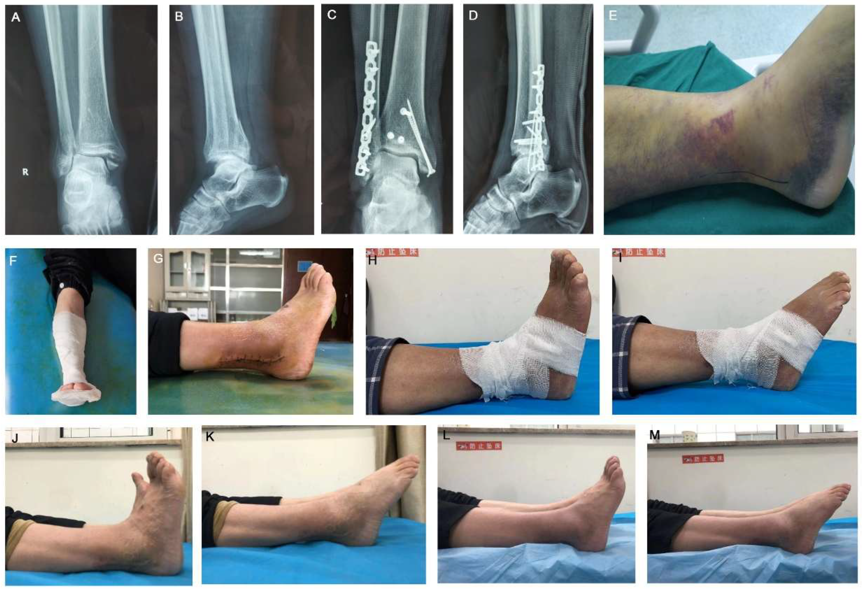
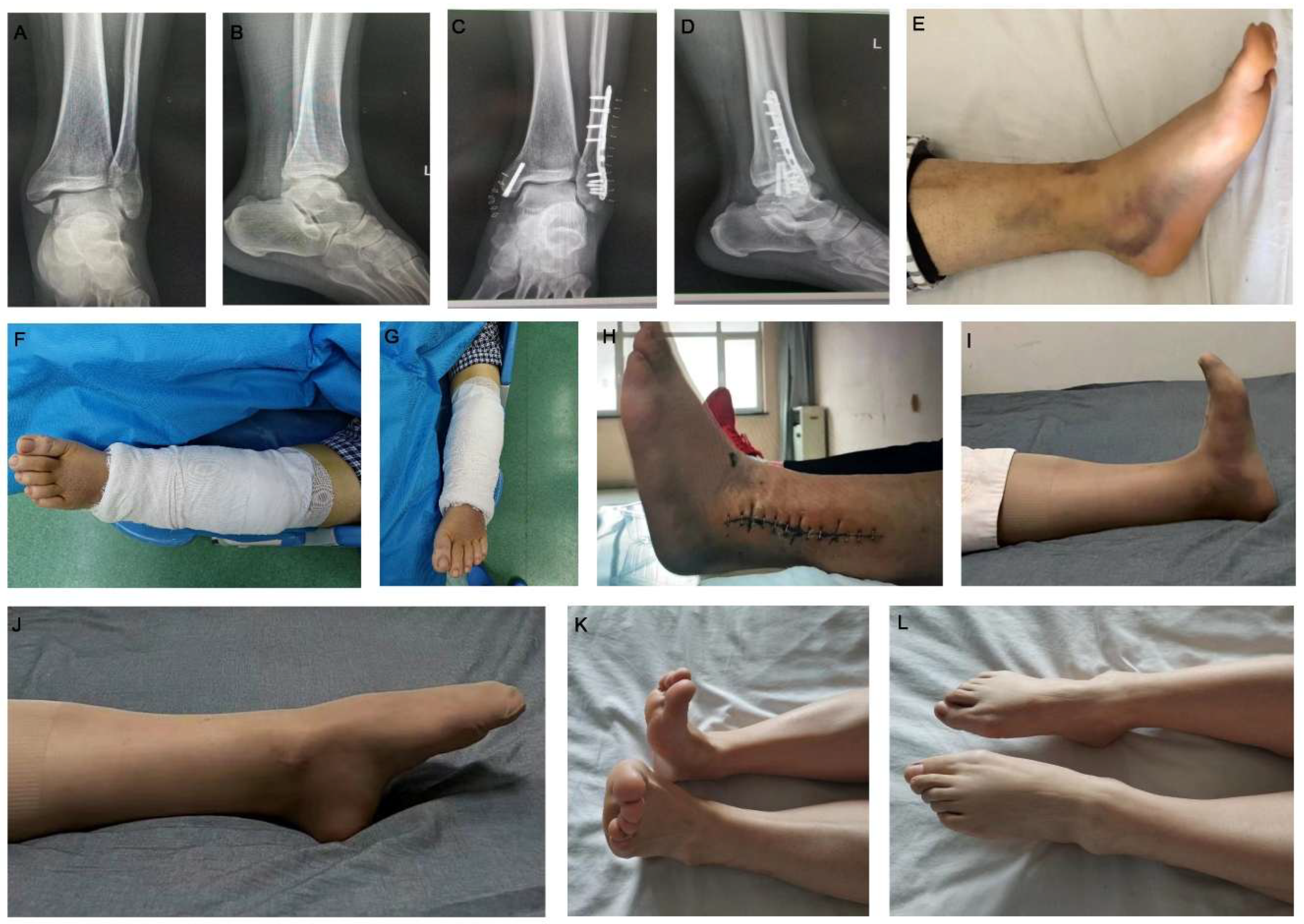
A total of 60 patients were included and divided into cast immobilization group (30 cases) and control group (30 cases). There were no significant differences in the general data including gender, age, height, BMI, facture site, operative time, hospital stay, and Lauge-Hansen type(p > 0.05) (Table 1). In the experimental group, the type of fracture was Lauge-Hansen type III, and in the control group, two fractures were type IV and one was type III. The preoperative and postoperative imaging data and the photos of patient in the experimental group and control group were shown in Figure 2 and Figure 3.

3.1. Comparison of Postoperative Visual Analog Scale (VAS)

The VAS score of cast immobilization group were lower than those of the control group at 1 day (p < 0.001), 3 days (p < 0.001), and 7 days (p = 0.01) post-operation. There were no significant differences in VAS score between the two groups at 14 days post-operation (p = 0.846) (Table 2).

3.2. Comparison of Ankle Joint ROM Between the Two Groups

At 1 month, 2 months, and 3 months post-operation, the dorsiflexion ROM of the ankle joint in cast immobilization group was larger than the control group (p = 0.01, p < 0.001, p < 0.001) (Table 3). The ankle plantarflexion ROM at 1 month and 2 months post-operation were larger than the control group (p < 0.001, p = 0.035). There were no significant differences in the ankle plantarflexion angles between the two groups at 3 months post-operation (p = 0.066) (Table 4).

3.3. AOFAS

The AOFAS of cast immobilization group was larger than the control group at 3 months post-operation (p = 0.002). There were no significant differences in the AOFAS and good to excellent rates between the two groups at postoperative 6 months (p = 0.064) (Table 5).

3.4. Postoperative Deep Venous Thrombosis Rate

The postoperative deep venous thrombosis rate was 1/30 (3.3%) in cast immobilization group and 3/30 (10%) in the control group, and there was no significant difference between the two groups (p = 0.605). The experimental group had intermuscular venous thrombosis of the lower limbs during the follow-up 1 month after the operation, so the vascular surgery consultation was requested. According to the consultation, the patient was given oral rivaroxaban 15 mg/day, and the patient was told to use two crutches to move. After the consultation of vascular surgery, the lower extremity venous thrombosis filter was implanted in the hospital, anticoagulant and thrombolysis were given after operation, and the lower extremity deep vein thrombosis filter was removed after the angiography vessel was unblocked. Three patients in the control group developed deep venous thrombosis of the lower extremity during a 3-month follow-up. Depending on the location of the thrombus, two of the patients had lower limb venous thrombosis filters implanted and were given anticoagulant and thrombolytic therapy. The other patient took rivaroxaban 15 mg/day orally and the thrombus disappeared after 2 months of regular follow-up. At the end of follow-up, thrombosis was controlled in both groups.

4. Discussion

In this study, short-term cast immobilization has a positive effect on reducing pain, eliminating postoperative swelling and restoring ankle dorsiflexion function in the early stage, and does not affect the recovery of postoperative function due to early immobilization. Early functional rehabilitation after ankle fracture surgery is not completely equivalent to early weight-bearing. Instead, early functional rehabilitation training should be guided individually according to the specific conditions of different patients, especially the type of fracture and soft tissue injury.
This study found that there were differences in pain scores between the two groups at 1 d, 3 d, and 7 d postoperatively. A study [19] showed that the skin around the ankle joint was thin, with less blood supply and insufficient soft tissue coverage. Ankle injuries often affect the tendons, nerves, and blood vessels that pass through the ankle joint. During surgical suturing, it is often found that soft tissue swelling is obvious and the tension of the skin edge is high. These factors lead to increased postoperative pain. After the operation, we can increase the comfort of the ankle, reduce the blood flow of the injured part, promote the swelling to subside, and reduce the pain of the affected limb by using cast immobilization.
In addition, there were statistically significant differences in ankle ROM and AOFAS between the two groups within 3 months post-operation. We speculated the main reasons are as follows: (1) Cast immobilization can promote swelling subsidence, avoid soft tissue and joint adhesion, and allow time for soft tissue damage around the fracture to repair, thereby enabling faster recovery. (2) Due to long-term bed rest and non-weight bearing, patients with ankle fractures have their ankles in plantarflexion for a long time, and it is difficult to restore the normal ankle dorsiflexion angle through functional exercises in the later stage. In contrast, patients in the cast immobilization group have a greater advantage in back extension exercises because the ankle joint is fixed in a functional position, which reduces postoperative plantar flexion joint stiffness. (3) Short-term cast immobilization after ankle fracture surgery can effectively avoid pain, inclination and uneven force during early functional exercise, so as to avoid re-injury of the anterior talofibular ligament.
This study also found that there were no significant differences in the postoperative deep venous thrombosis rate in the lower extremities between the two groups, as well as the AOFAS and good to excellent rates at 6 months post-operation. The reported incidence of venous thromboembolism (VTE) in orthopedic foot and ankle surgery is low, ranging from 0.22% to 4% [20,21]. In addition, Huntley et al. [22] confirmed that female sex, increasing age, higher BMI, hospitalization status, and non-elective surgery were associated with an increased risk of VTE after orthopedic foot and ankle surgery.
The meta-analysis by Weisman et al. [23] showed that patients with high risk of VTE should receive prophylaxis. Therefore, patients in the two groups received prophylaxis of thrombosis during treatment. Patients in the cast immobilization group can actively move their toes to promote blood circulation in the lower extremities. Even though the degree of activity is less than that in the control group, cast immobilization does not increase the incidence of deep vein thrombosis in the lower extremities after excluding objective factors. In addition, 2 weeks after the cast was removed, there was no obvious contracture of the Achilles tendon and posterior joint capsule, and the dorsiflexion of the ankle joint recovered quickly. Cast immobilization cannot only achieve early joint function rehabilitation training, but also prevents the possibility of fracture displacement or internal fixation fracture caused by premature weight bearing. After 6 months of functional exercise, there was no significant difference in functional outcomes between the two groups. This is consistent with the report of Godsiff SP et al. [24].
This study has limitations. The sample size is small and there may be selection bias. The follow-up time is short, and further long-term, multi-center, and large-sample-size studies should be conducted in the future. Due to the COVID-19, some postoperative photos were taken by patients at home, so the quality was not good. Additionally, although both groups were all given ankle pump exercises to prevent lower deep vein thrombosis 300 times a day after surgery with the assistance of a physician, there were potential bias regarding the postoperative deep venous thrombosis rate since both groups of patients did not undergo full postoperative weight-bearing. Last, the correlation of Lauge Hansen type and pain, cast or dressing were not explored in the present study, which will be addressed in our future study.

5. Conclusions

Although not statistically significant, short-term cast immobilization after ankle fracture surgery can reduce the occurrence of postoperative deep venous thrombosis rate. It has a positive effect on reducing pain, eliminating postoperative swelling and restoring ankle dorsiflexion function in the early stage, and does not affect the recovery of postoperative function due to early immobilization.

Author Contributions

Conceptualization, Z.D.; methodology, X.Z.; software, W.L.; validation, Z.D., W.L. and X.Z.; formal analysis, Z.D.; investigation, Z.D.; resources, K.L. and X.X.; data curation, B.N.; writing—original draft preparation, Z.D.; writing—review and editing, Z.D.; visualization, W.L.; supervision, X.Z.; project administration, X.Z.; funding acquisition, X.Z. All authors have read and agreed to the published version of the manuscript.

Funding

This study was funded by the 2024 Government-funded Clinical Medicine Talent Training Program of Hebei Province (ZF2024093) and Central Guidance Fund for Local Science and Technology Development of Hebei Province (246Z7758G).

Institutional Review Board Statement

The study was conducted in accordance with the Declaration of Helsinki, and approved by the ethics committee of the Third Hospital of Hebei Medical University on 30 May 2016 (K2015-001-12).

Informed Consent Statement

Informed consent was obtained from all subjects involved in the study.

Data Availability Statement

The data sets used and analyzed in the current study are available from the corresponding author on reasonable request.

Conflicts of Interest

The authors declare no conflicts of interest.

References

  1. Kannus, P.; Palvanen, M.; Niemi, S.; Parkkari, J.; Järvinen, M. Stabilizing incidence of low-trauma ankle fractures in elderly people Finnish statistics in 1970-2006 and prediction for the future. Bone 2008, 43, 340–342. [Google Scholar] [CrossRef] [Scilit] [PubMed]
  2. Schnetzke, M.; El Barbari, J.; Schüler, S.; Swartman, B.; Keil, H.; Vetter, S.; Gruetzner, P.A.; Franke, J. Vascular impulse technology versus elevation for the reduction of swelling of lower extremity joint fractures: Results of a prospective randomized controlled study. Bone Jt. J. 2021, 103-b, 746–754. [Google Scholar] [CrossRef] [Scilit] [PubMed]
  3. Kottmeier, S.A.; Madison, R.D.; Divaris, N. Pilon Fracture: Preventing Complications. J. Am. Acad. Orthop. Surg. 2018, 26, 640–651. [Google Scholar] [CrossRef] [Scilit] [PubMed]
  4. Kemler, E.; van de Port, I.; Schmikli, S.; Huisstede, B.; Hoes, A.; Backx, F. Effects of soft bracing or taping on a lateral ankle sprain: A non-randomised controlled trial evaluating recurrence rates and residual symptoms at one year. J. Foot Ankle Res. 2015, 8, 13. [Google Scholar] [CrossRef] [Scilit] [PubMed]
  5. Goost, H.; Wimmer, M.D.; Barg, A.; Kabir, K.; Valderrabano, V.; Burger, C. Fractures of the ankle joint: Investigation and treatment options. Dtsch. Arztebl. Int. 2014, 111, 377–388. [Google Scholar] [PubMed]
  6. Lübbeke, A.; Salvo, D.; Stern, R.; Hoffmeyer, P.; Holzer, N.; Assal, M. Risk factors for post-traumatic osteoarthritis of the ankle: An eighteen year follow-up study. Int. Orthop. 2012, 36, 1403–1410. [Google Scholar] [CrossRef] [Scilit] [PubMed]
  7. Kehlet, H. Multimodal approach to control postoperative pathophysiology and rehabilitation. Br. J. Anaesth. 1997, 78, 606–617. [Google Scholar] [CrossRef] [Scilit] [PubMed]
  8. Smith, T.W., Jr.; Wang, X.; Singer, M.A.; Godellas, C.V.; Vaince, F.T. Enhanced recovery after surgery: A clinical review of implementation across multiple surgical subspecialties. Am. J. Surg. 2020, 219, 530–534. [Google Scholar] [CrossRef] [Scilit] [PubMed]
  9. Debono, B.; Sabatier, P.; Boniface, G.; Bousquet, P.; Lescure, J.P.; Garnaud, V.; Hamel, O.; Lonjon, G. Implementation of enhanced recovery after surgery (ERAS) protocol for anterior cervical discectomy and fusion: A propensity score-matched analysis. Eur. Spine J. Off. Publ. Eur. Spine Soc. Eur. Spinal Deform. Soc. Eur. Sect. Cerv. Spine Res. Soc. 2021, 30, 560–567. [Google Scholar] [CrossRef] [Scilit] [PubMed]
  10. Thomas, G.; Whalley, H.; Modi, C. Early mobilization of operatively fixed ankle fractures: A systematic review. Foot Ankle Int. 2009, 30, 666–674. [Google Scholar] [CrossRef] [Scilit] [PubMed]
  11. Lin, C.W.; Donkers, N.A.; Refshauge, K.M.; Beckenkamp, P.R.; Khera, K.; Moseley, A.M. Rehabilitation for ankle fractures in adults. Cochrane Database Syst. Rev. 2012, 11, Cd005595. [Google Scholar] [PubMed]
  12. Tartaglione, J.P.; Rosenbaum, A.J.; Abousayed, M.; DiPreta, J.A. Classifications in Brief: Lauge-Hansen Classification of Ankle Fractures. Clin. Orthop. Relat. Res. 2015, 473, 3323–3328. [Google Scholar] [CrossRef] [Scilit] [PubMed]
  13. Hermans, J.J.; Wentink, N.; Beumer, A.; Hop, W.C.; Heijboer, M.P.; Moonen, A.F.; Ginai, A.Z. Correlation between radiological assessment of acute ankle fractures and syndesmotic injury on MRI. Skelet. Radiol. 2012, 41, 787–801. [Google Scholar] [CrossRef] [Scilit] [PubMed]
  14. Persigant, M.; Colin, F.; Noailles, T.; Pietu, G.; Gouin, F. Functional assessment of transplantar nailing for ankle fracture in the elderly: 48 weeks’ prospective follow-up of 14 patients. Orthop. Traumatol. Surg. Res. OTSR 2018, 104, 507–510. [Google Scholar] [CrossRef] [Scilit] [PubMed]
  15. Hawker, G.A.; Mian, S.; Kendzerska, T.; French, M. Measures of adult pain: Visual Analog Scale for Pain (VAS Pain), Numeric Rating Scale for Pain (NRS Pain), McGill Pain Questionnaire (MPQ), Short-Form McGill Pain Questionnaire (SF-MPQ), Chronic Pain Grade Scale (CPGS), Short Form-36 Bodily Pain Scale (SF-36 BPS), and Measure of Intermittent and Constant Osteoarthritis Pain (ICOAP). Arthritis Care Res. 2011, 63, S240–S252. [Google Scholar] [CrossRef] [Scilit] [PubMed]
  16. Fong, C.M.; Blackburn, J.T.; Norcross, M.F.; McGrath, M.; Padua, D.A. Ankle-dorsiflexion range of motion and landing biomechanics. J. Athl. Train. 2011, 46, 5–10. [Google Scholar] [CrossRef] [Scilit] [PubMed]
  17. Eliasziw, M.; Young, S.L.; Woodbury, M.G.; Fryday-Field, K. Statistical methodology for the concurrent assessment of interrater and intrarater reliability: Using goniometric measurements as an example. Phys. Ther. 1994, 74, 777–788. [Google Scholar] [CrossRef] [Scilit] [PubMed]
  18. Sieradzki, J.P.; Larsen, N.; Wong, I.; Ferkel, R.D. Symptom and Disability Measurement by Common Foot and Ankle-Specific Outcome Rating Scales. Foot Ankle Int. 2020, 41, 849–858. [Google Scholar] [CrossRef] [Scilit] [PubMed]
  19. Poon, H.; Le Cocq, H.; Mountain, A.J.; Sargeant, I.D. Dermal Fenestration With Negative Pressure Wound Therapy: A Technique for Managing Soft Tissue Injuries Associated With High-Energy Complex Foot Fractures. J. Foot Ankle Surg. Off. Publ. Am. Coll. Foot Ankle Surg. 2016, 55, 161–165. [Google Scholar] [CrossRef] [Scilit] [PubMed]
  20. Hanslow, S.S.; Grujic, L.; Slater, H.K.; Chen, D. Thromboembolic disease after foot and ankle surgery. Foot Ankle Int. 2006, 27, 693–695. [Google Scholar] [CrossRef] [Scilit] [PubMed]
  21. Solis, G.; Saxby, T. Incidence of DVT following surgery of the foot and ankle. Foot Ankle Int. 2002, 23, 411–414. [Google Scholar] [CrossRef] [Scilit] [PubMed]
  22. Huntley, S.R.; Abyar, E.; Lehtonen, E.J.; Patel, H.A.; Naranje, S.; Shah, A. Incidence of and Risk Factors for Venous Thromboembolism After Foot and Ankle Surgery. Foot Ankle Spec. 2019, 12, 218–227. [Google Scholar] [CrossRef] [Scilit] [PubMed]
  23. Weisman, M.H.S.; Holmes, J.R.; Irwin, T.A.; Talusan, P.G. Venous Thromboembolic Prophylaxis in Foot and Ankle Surgery: A Review of Current Literature and Practice. Foot Ankle Spec. 2017, 10, 343–351. [Google Scholar] [CrossRef] [Scilit] [PubMed]
  24. Godsiff, S.P.; Trakru, S.; Kefer, G.; Maniar, R.N.; Flanagan, J.P.; Tuite, J.D. A comparative study of early motion and immediate plaster splintage after internal fixation of unstable fractures of the ankle. Injury 1993, 24, 529–530. [Google Scholar] [CrossRef] [Scilit] [PubMed]
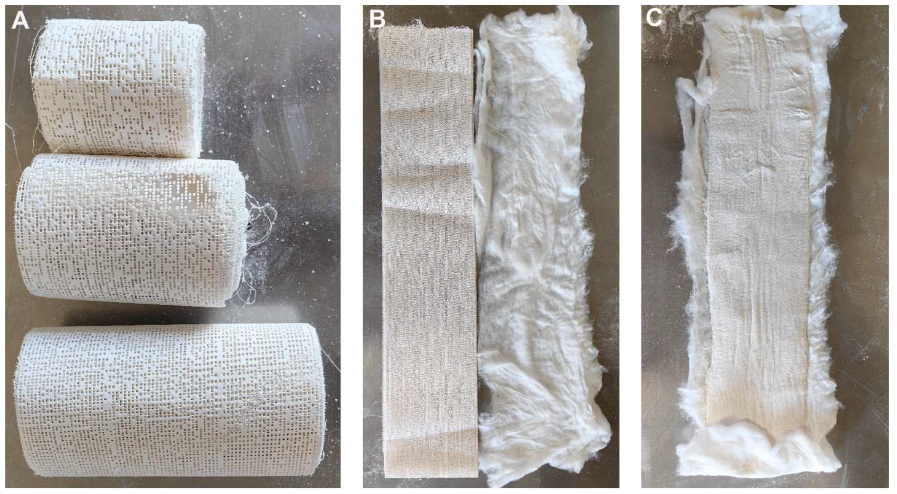
Figure 1. (A). The picture of plaster cast bandage. (B). The picture of absorbent cotton and plaster cast bandage. (C). A layer of degreased cotton was placed between the plaster cast bandage and the skin to prevent skin damage from the cast.
Japma 116 24128 g001
Figure 2. (A,B). Preoperative X-ray anteroposterior and lateral films after injury showed right ankle fracture (supination and external rotation type); (C,D). X-ray anteroposterior and lateral films on the day after operation showed good fracture reduction and satisfactory screw placement; (E,F). Photos of the ankle before and after the operation; (G). Photos of the cast removed at 14 days after the operation; (H,I). Photos of the dorsiflexion and plantar flexion of the ankle joint at 1 month post-operation; (J,K). Photos of ankle dorsiflexion and plantarflexion at 3 months post-operation; (L,M). Photos of ankle dorsiflexion and plantarflexion at 6 months post-operation.
Japma 116 24128 g002
Figure 3. (A,B). Preoperative X-ray anteroposterior and lateral films after injury showed left ankle fracture (supination and external rotation type); (C,D). X-ray anteroposterior and lateral films on the day after operation showed good fracture reduction and satisfactory screw placement; (E). Photos of the ankle before and after the operation; (F,G). Photos of the ankle after the operation; (H). Photos of the bandaging removed at 14 days after the operation; (I,J). Photos of the dorsiflexion and plantar flexion of the ankle joint at 3 months post-operation; (K,L). Photos of ankle dorsiflexion and plantarflexion at 3 months post-operation.
Japma 116 24128 g003
Table 1. General data.
IndexCast Immobilization Group
(n = 30)
Control Group
(n = 30)
p Value
Male, n (%)17(56.67)15(50)0.612
Age39.53 ± 15.0536.83 ± 13.140.462
Height168.83 ± 9.92168.20 ± 8.030.787
BMI23.54 ± 3.4122.31 ± 2.690.128
Fracture site, n (%) 0.610
Left18(60)16(53.33)
Right12(40)14(46.67)
Operative time (min)70.30 ± 10.8774.10 ± 16.250.291
Hospital stay (d)11.16 ± 2.2111.10 ± 2.630.916
Lauge-Hansen type, n (%) 0.489
Supination adduction6(20)7(23.33)
Supination external rotation13(43.33)15(50)
Pronation abduction7(23.33)5(16.67)
Pronation external rotation4(13.34)3(10)
Table 2. Comparison of postoperative visual analog scale (VAS).
IndexCast Immobilization Group
(n = 30)
Control Group
(n = 30)
p Value
1 day post-operation5.10 ± 1.156.63 ± 1.15<0.001
3 days post-operation4.33 ± 1.155.50 ± 1.10<0.001
7 days post-operation2.73 ± 1.203.70 ± 0.910.01
14 days post-operation1.23 ± 0.721.26 ± 0.580.846
Table 3. Comparison of postoperative dorsiflexion range of motion (ROM) between the two groups.
IndexCast Immobilization Group
(n = 30)
Control Group
(n = 30)
p Value
1 month post-operation7.66 ± 1.516.16 ± 1.890.01
2 months post-operation12.00 ± 1.5910.06 ± 1.16<0.001
3 months post-operation15.70 ± 1.2114.56 ± 1.19<0.001
Table 4. Comparison of postoperative plantarflexion range of motion (ROM) between the two groups.
IndexCast Immobilization Group
(n = 30)
Control Group
(n = 30)
p Value
1 month post-operation15.72 ± 1.4812.42 ± 1.68<0.001
2 months post-operation25.23 ± 3.0023.66 ± 2.590.035
3 months post-operation31.30 ± 1.8930.43 ± 1.670.066
Table 5. Comparison of ankle joint function scores (AOFAS) and the good to excellent rate at last follow-up between the two groups.
IndexCast Immobilization Group
(n = 30)
Control Group
(n = 30)
p Value
3 months post-operation71.76 ± 6.1066.60 ± 6.310.002
6 months post-operation86.00 ± 8.2982.03 ± 7.960.064
Good to excellent rate83.3%24(80%)0.739
Disclaimer/Publisher’s Note: The statements, opinions and data contained in all publications are solely those of the individual author(s) and contributor(s) and not of MDPI and/or the editor(s). MDPI and/or the editor(s) disclaim responsibility for any injury to people or property resulting from any ideas, methods, instructions or products referred to in the content.