Article

Balance, Gait and Foot Pressure Distribution in Neuropathic Pain Associated with Lumbar Disc Degeneration

by
Suleyman Korkusuz
1,
Büşra Seckinogullari Korkusuz
2,*,
Zeliha Ozlem Yuruk
3,
Sibel Kibar
4 and
Ferdi Yavuz
5
1
Department of Physiotherapy and Rehabilitation, Faculty of Health Sciences, Atılım University, 06830 Ankara, Turkey
2
Department of Therapy and Rehabilitation, Kızılcahamam Vocational School of Health Services, Ankara University, 06890 Ankara, Turkey
3
Department of Physiotherapy and Rehabilitation, Faculty of Health Sciences, Baskent University, 06790 Ankara, Turkey
4
Department of Therapy and Rehabilitation, Vocational School of Health Services, Atılım University, 06830 Ankara, Turkey
5
Department of Physiotherapy and Rehabilitation, Faculty of Health Sciences, European University of Lefke, 99728 Lefke, Cyprus
*
Author to whom correspondence should be addressed.
J. Am. Podiatr. Med. Assoc. 2026, 116(2), 24138; https://doi.org/10.7547/24-138
Submission received: 11 August 2024 / Revised: 23 January 2025 / Accepted: 28 January 2025 / Published: 21 April 2026

Abstract

Background: This study aimed to evaluate the effects of NP associated with LDD on balance, gait and foot pressure distribution. Methods: This prospective controlled study was conducted on 42 individuals aged between 40 and 70 years. There were 3 groups in the study: individuals diagnosed with NP associated with LDD (n = 14), individuals with LDD without NP (n = 14), and the control group (n = 14). The Force Plate system and Core Balance System measured static and dynamic postural balance and stability limits. Gait and dynamic plantar pressure distribution analyses were performed with a computerized gait evaluation system. Results: The Leeds Assessment of Neuropathic Signs and Symptoms (LANSS), VAS during gait, and Oswestry Disability Index (ODI) scores were higher in the LDD with NP group than in the LDD without NP group (p < 0.05). It was found that LDD with NP group had backward dynamic balance control (p < 0.05). There was no significant difference in balance control, dynamic plantar pressure distribution, and spatiotemporal gait parameters between the groups (p > 0.05). Conclusions: Although participants with NP had higher levels of pain severity in gait and disability, there was no difference in postural balance, dynamic plantar pressure distribution, and spatiotemporal gait parameters compared to participants with LDD without NP and healthy individuals. All participants with LDD were unilaterally affected. Therefore, postural balance and gait tasks would be able to compensate for the unaffected limb.

1. Introduction

Neuropathic pain (NP) is pain caused by damage or disease affecting the somatosensory system [1]. The NP may be associated with abnormal sensations called dysesthesia or pain from normally non-painful stimuli (allodynia). It may have continuous and/or episodic components. Common qualities include burning or coldness, “pins and needles” sensations, numbness, and itching [2].
The NP may result from disorders of the peripheral nervous system or the central nervous system [3]. Any condition that damages or impairs the function of nerve tissue can cause NP [4]. Low back pain and neck pain constitutes approximately 2/3 of NP. The neural structures surrounding the disc may be compressed or mechanically irritated and causing chemical inflammation in lumbar disc degeneration (LDD) [5]. The conduction block in the axons or roots of the spinal nerves causes loss of sensory and motor function and radicular pain.
Chronic LDD characterized by pain, paresthesia, and muscle weakness in the lower extremities, is one of the most common NP syndromes [6,7]. The NP impairs the patients’ quality of life, and the treatment is complicated. Gait and postural balance are both critical for independence in activities of daily living. Impaired proprioception due to NP, an asymmetrical load of the lower extremities, and the timing of dysfunctional muscle activation, sequencing, and asymmetry in plantar pressure may result in alterations in gait and balance control, with an increased risk of falls [8,9,10]. The most common method of measuring the degree of symmetry between the extremities is to measure foot pressure distribution using a force or pressure platform mounted on a specially constructed treadmill [11].
In the literature, most of the studies were conducted on diabetic NP. Studies on diabetic patients showed that NP impairs postural balance and gait. However, there is limited study on the effects of balance control and gait of LDD. Also, the impact of NP associated with LDD on balance control and gait is not clear [12,13,14,15].
This study aimed to evaluate the effects of NP associated with LDD on postural balance, gait and foot pressure distribution. The hypotheses of the study are as follows:
H1: 
Neuropathic pain has an effect on static balance in patients with lumbar disc degeneration.
H2: 
Neuropathic pain has an effect on dynamic balance in patients with lumbar disc degeneration.
H3: 
Neuropathic pain has an effect on spatiotemporal gait parameters in patients with lumbar disc degeneration.

2. Materials and Methods

2.1. Study Design

The study was a prospective controlled study. The study was carried out at the Private Physical Therapy and Rehabilitation Center Gait Analysis Laboratory Unit in Ankara. This study was approved by the Baskent University Medical and Health Sciences Research Board and Non-Interventional Clinical Research Ethics Committee. All participants provided formal written informed consent. The clinical trial registration identifier is NCT05223439. Informed consent was obtained from the participants.

2.2. Participants

Patients with LDD who were diagnosed with Magnetic Resonance Imaging at least 3 months ago and were routinely followed up in the clinic were included in the study. LDD patients included in the study were divided into two groups, LDD with NP and LDD without NP, based on clinical examination performed by the physician. At the same time, healthy individuals were participating as the control group. The study groups are described below:
Group 1: Individuals diagnosed with NP associated with LDD (n = 14).
Group 2: Individuals with LDD without NP (n = 14).
Group 3: Healthy control group (n = 14).
Inclusion criteria for individuals with NP associated with LDD [16]: Individuals were previously diagnosed with NP and scored above the threshold on the LANSS scale.
  • Individuals between the ages of 40 and 70 years old.
  • Individuals had LDD symptoms for at least 6 months.
  • Individuals had NP at least 3 months.
  • Individuals had 12 points or more from the Leeds Assessment of Neuropathic Signs and Symptoms (LANSS) scale.
Inclusion criteria for individuals with LDD without NP [17]:
  • Individuals between the ages of 40 and 70 years old.
  • Individuals had LDD symptoms for at least 6 months.
  • Individuals had below 12 points from the Leeds Assessment of Neuropathic Signs and Symptoms (LANSS) scale.
Inclusion criteria of healthy individuals in the study:
  • Individuals between the ages of 40–70 years old.
  • Individuals had no pain (Taking 1 point or less according to the Visual Pain Scale).
Exclusion criteria [10]:
  • Individuals had pain symptoms from different etiology.
  • Individuals had diabetes mellitus.
  • Presence of neurological disease that may cause central NP such as stroke, traumatic brain injury, multiple sclerosis.
  • Individuals underwent orthopedic surgery in the last 6 months.
  • Individuals had severe vision problems.
  • Individuals had a diagnosis of vestibular disorders (Bening Paroxysmal Positional Vertigo, Meniere’s Disease, etc.).

2.3. Outcome Measures

The assessments were performed barefoot. The devices were explained to the patient and the patients were allowed to try them on before the assessments. After each assessment, patients were allowed to rest for 5–10 min according to their needs. All assessments lasted a maximum of 60 min.

2.3.1. Sociodemographic Form

The participants fulfilled a sociodemographic form. The form included individual and clinical characteristics of individuals such as age, gender, height, body weight, marital status, education level, duration of symptoms, and dominant side.

2.3.2. Leeds Assessment of Neuropathic Symptoms and Signs (LANSS) Pain Questionnaire

The LANSS is a validated questionnaire that consists of 7 items, can distinguish NP from nociceptive pain, and is easy to score in clinical settings. The questionnaire is scored between 0 and 24 points. Twelve points and above indicate NP. In the study, the Turkish version of the LANNS questionnaire was administered [18,19].

2.3.3. Visual Analog Scale (VAS)

The VAS is a scale that evaluates pain on a horizontal 100 mm line. On the line, the left side indicates ‘no pain’ and the right side indicates ‘the most severe pain imaginable’. Pain level was measured in centimeters starting from the left to the marked point [4].

2.3.4. Oswestry Disability Index (ODI)

The ODI is a self-administered questionnaire that gives a subjective percentage score of level of function (disability) in activities of daily living in those rehabilitating from low back pain. It consists of 10 sections. Each section is scored on a 0–5 scale, with 5 representing the highest disability. The index is calculated by dividing the summed score by the total possible score, multiplied by 100, and expressed as a percentage. Higher scores indicate lower functionality [20,21].

2.3.5. Static and Dynamic Postural Balance

Computerized balance assessment tests are a valid and reliable method for the evaluation of patients with LDD [22,23].
Stability limits were assessed with the E-LINK FP3 Force Plate (Biometrics Ltd., UK) system, and static and dynamic balance control was assessed with the KoreBalance System.
E-LINK FP3 Force Plate System
We measured the weight distribution in the front, back, left and right directions and the patient’s stability limits. The patient’s average stability limit (%) was calculated [24].
KoreBalance System
The system, consisting of a computer and a platform where participants can place their feet, assesses static and dynamic balance control [23].
In the static balance measurement, participants placed their feet on the marked places on the platform and tried to keep the object on the computer screen at a fixed point for 30 s without support from their hands. As a result of the test, static total score and static balance distribution (front–back) were obtained. An increase in the static total score indicates an increase in the risk of falling, and static balance distribution indicates that the individual’s balance distribution in the static stance is in the front or back direction.
For the dynamic balance test, participants transferred their body weights and followed a moving object on the computer screen for 30 s. As a result of the test, the dynamic total score and dynamic balance distribution (front–back) were obtained from the device, which indicate the individual’s risk of falling. An increase in dynamic total scores means that the risk of falling during the activity increases. Dynamic balance distribution shows whether the individuals’ balance is in the front or back direction during the activity [23].

2.3.6. Gait Analysis

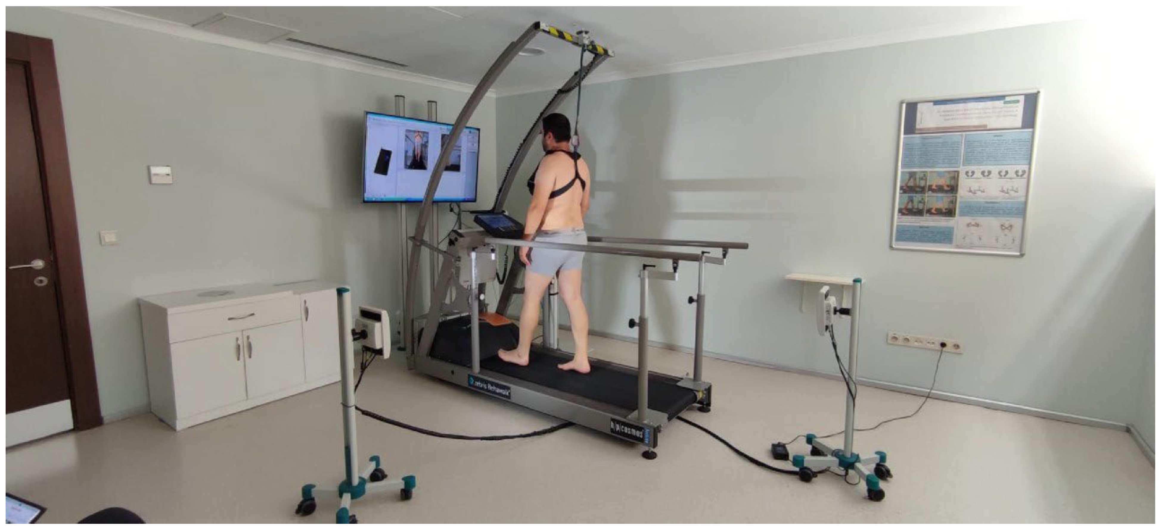
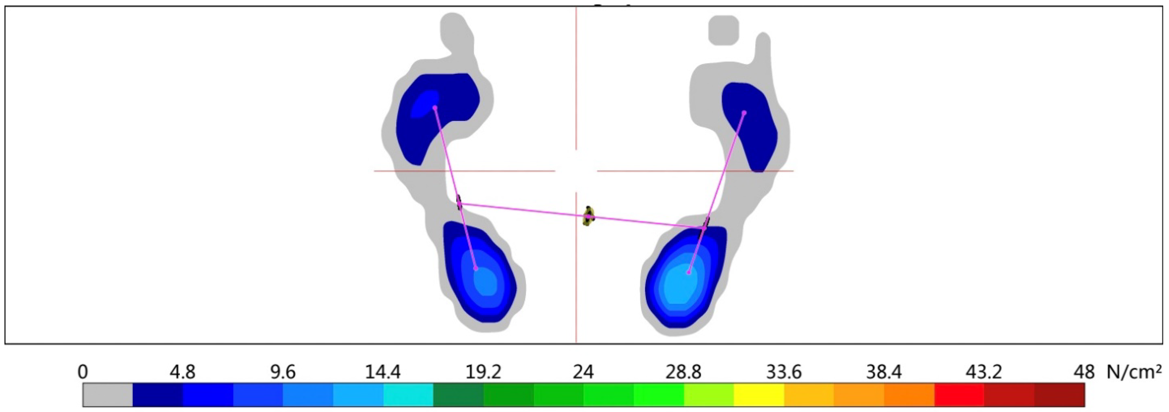
The Zebris Rehawalk (Zebris Medical GmbH, Germany) [25] computerized gait evaluation system was used to assess the spatiotemporal characteristics of the gait as well as the dynamic plantar pressure distribution during gait. The system consists of a treadmill and a computer. Dynamic plantar pressure distribution was analyzed by pedobarography integrated into this system. The computer automatically detects the patient’s body structures and measures an individual’s plantar pressure on the ground. For gait assessment, participants walked barefoot at their preferred comfortable gait speed (Figure 1) [26,27]. After two trials, their third gait was recorded. The system provided gait speed, stride length, step length, cadence, stance, and swing phase outputs, as well as forefoot and backfoot dynamic plantar pressure distribution. The foot pressure distribution obtained from the system is given in Figure 2.

2.4. Statistical Analysis

Sample size calculation (G*Power Ver. 3.0.10, Franz Faul, Universität Kiel, Germany) was performed based on the “step length” parameter according to Awotidebe et al. [17]. The analysis was carried out with an effective width of f = 0.59, a power of 90%, and a margin of error of 0.05. It was found that 14 individuals in each of the three groups (a total of 42 individuals) should be included in the study.
The study data were analyzed by using the Statistical Program for Social Sciences (SPSS) version 23.0 (IBM SPSS Statistics for Windows, Armonk, NY, USA: IBM Corp.). The Levene test evaluated the homogeneity of the groups. The descriptive data were shown as mean and standard deviation, and minimum–maximum values were given. The qualitative variables were given with frequency and percentage values. The Kruskal–Wallis Test was used for the comparison of independent samples. To compare the clinical characteristics of the LDD with NP and LDD without NP groups the Mann–Whitney U test was used. The Chi-square test was used to determine whether there is a statistically significant difference between the frequencies. All the statistical analyses were set a priori at an alpha level of p < 0.05.

3. Results

The mean age, gender, body weight, height, and body mass index of the participants were similar across the groups (p > 0.05) (Table 1). The clinical characteristics of the LDD patients are shown in Table 2. The symptom duration, affected extremity, and VAS at rest score were similar between the groups (p > 0.05). The LANSS, VAS during gait, and ODI scores were higher in the LDD with NP group than in the LDD without NP group (p < 0.05).
Table 3 presents the comparison of static and dynamic balance values between the groups. It was found that LDD with NP group had balance towards the back (p < 0.05). On the other hand, there was no significant difference in balance scores between the groups (p > 0.05). The dynamic plantar pressure distribution was similar across the groups (p > 0.05) (Table 4). The gait analysis scores are shown in Table 5. There was no significant difference in spatiotemporal gait scores between the groups (p > 0.05).

4. Discussion

The study examined the effects of NP due to LDD on postural balance and gait. Individuals with NP, without NP, and healthy control group participated in the study. As a result, there was no difference in balance control and spatiotemporal gait parameters between individuals with and without NP and healthy individuals.
The groups were homogeneous in terms of age, gender, and physical characteristics. Participants had chronic lumbar disc degeneration and radiculopathy. The NP was diagnosed by a physician and participants had NP symptoms for more than 3 months. Deane et al. [28] included participants with LDD for at least 3 months. We included participants who had LDD for more than 6 months. We thought that a longer symptom duration could expose the alteration of balance and gait.
There are many scales for assessing NP [4]. The LANNS is a valid and reliable scale for NP. The LANSS was used to evaluate NP in the study [29,30]. Pain at rest and during gait was evaluated with VAS. Participants with NP had higher pain intensity during gait. Pain intensity at rest was similar in participants with and without NP. In the study, participants with NP were also more negatively affected functionally.
Visual, vestibular, and somatosensory systems transmit their inputs to the central nervous system, revealing optimal muscle strength and body reactions to keep the center of gravity within the base of support, thus providing adequate postural control [31]. Numerous factors, such as aging and neurological or musculoskeletal disorders, and decreased sensory–motor system performance impair postural balance. Feedback defects of proprioceptors in the lumbar spine, trunk, or lower extremities can affect postural control. The LDD and NP could impair somatosensorial inputs and muscle strength. These changes might cause an asymmetrical load of the lower extremities, dysfunctional muscle activation, and asymmetric dynamic plantar pressure in balance and gait [13,32].
When static balance distributions were examined in the study, no difference was observed between the groups. However, when dynamic balance distributions were examined, it was seen that the participants with NP had a more backward tendency. This might be related to the impairment in the forefoot sensation of participants with NP and participants might have difficulty in forefoot loading. In the study pinprick and touch sensation were evaluated within LANSS assessment. However, the threshold values were not recorded. Frost et al. [33] investigated the relationship between foot skin sensitivity and standing balance control in chronic low back pain (LBP) with radiculopathy. The researchers assessed foot skin sensitivity with a vibratory threshold test and monofilaments. Static and dynamic balance was tested on a force plate. They found that skin sensitivity of the foot correlated with static balance. The authors concluded that reduced foot skin sensitivity might be associated with poorer balance control in LBP patients. Frost et al. tested static balance with eyes closed. We evaluated static balance with eyes open. In Brumagne et al. [34], visual information contributed to postural balance in LBP. Therefore, we might not have found a difference in static balance distribution.
Participants with NP and LDD without NP did not demonstrate balance impairments, as compared to the healthy control group. Previous research has shown that LBP participants, with eyes closed in static standing, had balance impairments [35,36]; however, LBP patients have also demonstrated no difference in balance, as compared to controls [37]. A recent review of the LBP balance literature concluded that there were no systemic differences between control and LBP participants in static balance [38]. Frost et al. [33] could not find a difference in static and dynamic balance scores between LBP and healthy control groups. Frost et al. [33] did not evaluate NP. However, the results of the study support our results. Thus, it may not be surprising that we did not find statistically significant differences. Additionally, in the current study all LDD patients were unilaterally affected; therefore, bilateral stance would be able to compensate for the unaffected limb.
Deane et al. [28] examined differences in anticipatory and compensatory postural adjustments, between symptomatic LDD patients and asymptomatic controls (no pain) during postural perturbation. The authors found that the symptomatic group exhibited higher compensatory lumbar displacement than the asymptomatic group in the predicted condition. However, there was no difference observed in the unpredicted condition. The static and dynamic balance assessment protocol was explained to the participants before the test in our study. The test protocol did not involve reactive balance tasks. So, the participants performed the test under predicted conditions. We did not find a difference in the static or dynamic balance between the symptomatic and control groups. The results of our study were similar to those of Deane et al. [28].
In our study, plantar foot pressure was not different in participants with LDD and healthy controls. This is an expected result parallel to the balance and gait results of the participants.
Anukoolkarn et al. [39] examined the characteristics of the plantar pressure distribution patterns during the mid-stance phase of the gait cycle in 17 chronic LBP and 17 asymptomatic participants. They found that the plantar pressure was unequally distributed in the LBP group at the mid-stance phase of gait. The authors explain that altered foot contact in the participants with LBP might be used to avoid pain or to compensate for limited mobility of the lower limbs in the pre-swing phase. Although the VAS score of the participants was similar to our study, our results were different from Anukoolkarn et al. [39].
Previous studies suggest LDD has unique patterns of gait deterioration [15,40]. Spatiotemporal gait deterioration in gait velocity, cadence with increased double-support duration, and gait variability are distinguishing features in LDD. In a study conducted by Bonab et al. [41] including 59 individuals with LDD, it was reported that the spatiotemporal gait parameters of individuals with LDD were different from healthy controls. In another study conducted on 70 participants with chronic LBP, the spatiotemporal gait parameters were more significantly decreased in LBP than in the healthy control group [15]. The LBP group had significantly longer step duration, gait cycle duration, double stop duration, swing duration, shorter step length, gait cycle length, slower walking speed (velocity), and less cadence than the healthy control group. This result was associated with pain intensity in the study. On the other hand, Gombatto et al. [42] suggested that there was no statistically significant difference in bilateral gait cycle duration, bilateral step length, bilateral double step length, and velocity, and these gait parameters were similar between patients with LBP and healthy control group. In the literature, most of the studies were conducted on LBP. Spatiotemporal gait parameters were only investigated in diabetic NP. In our study, cadence and swing phase were less than LDD without NP and control group in NP group. However, there was no significant difference between the groups. We also did not find any spatiotemporal gait alteration in LDD groups. In our study pain intensity during gait was higher in the NP group. However higher pain scores did not affect gait. In the gait, analysis participants walked at their preferred comfortable walking speed. Comfortable walking speed may have minimized observed effects, and there was limited power to detect movement-based subgroup differences.
In the literature, studies on diabetic NP are more common. However, no study on NP due to LDD has been found. There were 3 groups in the study and postural balance and gait were evaluated with objective methods. These are the strengths of the study. Therefore, the study may contribute to the literature in this respect. The study also had some limitations. Muscle strength is a major determinant of balance and gait, which are the main functions of the lower extremity. Muscle strength was not evaluated in the study. Also, foot deformities are related to balance and gait alterations. Foot deformities could have been evaluated in the study. Pinprick and touch sensation were evaluated within the LANSS scale. However, the threshold values were not recorded. Monofilaments and vibratory testing could be used.

5. Conclusions

Although participants with NP had higher levels of pain severity in gait and disability, there was no difference in postural balance, dynamic plantar pressure distribution, and spatiotemporal gait parameters compared to participants with LDD without NP and healthy individuals. However, it was observed that participants with NP had a backward tendency in their dynamic balance distributions. Considering this information, it is thought that it may be beneficial for clinicians to include exercises to regulate balance distribution and improve balance in the rehabilitation programs of individuals with NP associated with LDD. All participants with LDD were unilaterally affected. Therefore, postural balance and gait tasks would be able to compensate for the unaffected limb. The authors suggest that future studies can evaluate muscle strength, detailed sensory examination, and foot deformities beyond postural balance and gait.

Author Contributions

Conceptualization, S.K. (Suleyman Korkusuz) and Z.O.Y.; methodology, S.K. (Suleyman Korkusuz) and B.S.K.; formal analysis, S.K. (Suleyman Korkusuz), B.S.K. and F.Y.; investigation, S.K. (Suleyman Korkusuz), B.S.K. and S.K. (Sibel Kibar); data curation, S.K. (Suleyman Korkusuz) and B.S.K.; writing—original draft preparation, S.K. (Suleyman Korkusuz) and B.S.K.; writing—review and editing, Z.O.Y. and F.Y.; supervision, Z.O.Y., S.K. (Suleyman Korkusuz) and F.Y. All authors have read and agreed to the published version of the manuscript.

Funding

This research received no external funding.

Institutional Review Board Statement

The study was conducted in accordance with the Declaration of Helsinki and approved by the Baskent University Medical and Health Sciences Research Board and Non-Interventional Clinical Research Ethics Committee (protocol code KA21/489 and date of approval 29 December 2021).

Informed Consent Statement

Informed consent was obtained from all subjects involved in the study. Written informed consent has been obtained from the patient to publish this paper.

Data Availability Statement

The data presented in this study are available on request from the corresponding author due to privacy and ethical restrictions.

Conflicts of Interest

The authors declare no conflicts of interest.

References

  1. Haanpää, M.L.; Backonja, M.-M.; Bennett, M.I.; Bouhassira, D.; Cruccu, G.; Hansson, P.T.; Jensen, T.S.; Kauppila, T.; Rice, A.S.; Smith, B.H.; et al. Assessment of neuropathic pain in primary care. Am. J. Med. 2009, 122, S13–S21. [Google Scholar] [CrossRef] [Scilit] [PubMed]
  2. Bouhassira, D.; Lantéri-Minet, M.; Attal, N.; Laurent, B.; Touboul, C. Prevalence of chronic pain with neuropathic characteristics in the general population. Pain 2008, 136, 380–387. [Google Scholar] [CrossRef] [Scilit]
  3. Davis, M.P. What is new in neuropathic pain? Support Care Cancer 2007, 15, 363–372. [Google Scholar] [CrossRef] [Scilit]
  4. Harden, R.N. Chronic neuropathic pain. Mechanisms, diagnosis, and treatment. Neurologist 2005, 11, 111–122. [Google Scholar] [CrossRef] [Scilit]
  5. Al Nezari, N.H.; Schneiders, A.G.; Hendrick, P.A. Neurological examination of the peripheral nervous system to diagnose lumbar spinal disc herniation with suspected radiculopathy: A systematic review and meta-analysis. Spine J. 2013, 13, 657–674. [Google Scholar] [CrossRef] [Scilit]
  6. Khoromi, S.; Patsalides, A.; Parada, S.; Salehi, V.; Meegan, J.M.; Max, M.B. Topiramate in chronic lumbar radicular pain. J. Pain 2005, 6, 829–836. [Google Scholar] [CrossRef] [Scilit]
  7. Keynan, O.; Mirovsky, Y.; Dekel, S.; Gilad, V.H.; Gilad, G.M. Safety and Efficacy of Dietary Agmatine Sulfate in Lumbar Disc-associated Radiculopathy. An Open-label, Dose-escalating Study Followed by a Randomized, Double-blind, Placebo-controlled Trial. Pain Med. 2010, 11, 356–368. [Google Scholar] [CrossRef] [Scilit]
  8. Allet, L.; Armand, S.; De Bie, R.A.; Golay, A.; Pataky, Z.; Aminian, K.; De Bruin, E.D. Clinical factors associated with gait alterations in diabetic patients. Diabet. Med. 2009, 26, 1003–1009. [Google Scholar] [CrossRef] [Scilit] [PubMed]
  9. Morrison, S.; Colberg, S.R.; Parson, H.K.; Vinik, A.I. Exercise improves gait, reaction time and postural stability in older adults with type 2 diabetes and neuropathy. J. Diabetes Complicat. 2014, 28, 715–722. [Google Scholar] [CrossRef] [Scilit]
  10. Allet, L.; Armand, S.; de Bie, R.A.; Pataky, Z.; Aminian, K.; Herrmann, F.R.; de Bruin, E.D. Gait alterations of diabetic patients while walking on different surfaces. Gait Posture 2009, 29, 488–493. [Google Scholar] [CrossRef] [Scilit] [PubMed]
  11. VanZant, R.S.; McPoil, T.G.; Cornwall, M.W. Symmetry of plantar pressures and vertical forces in healthy subjects during walking. J. Am. Podiatr. Med. Assoc. 2001, 91, 337–342. [Google Scholar] [CrossRef] [Scilit]
  12. Sipko, T.; Kuczyński, M. The effect of chronic pain intensity on the stability limits in patients with low back pain. J. Manip. Physiol. Ther. 2013, 36, 612–618. [Google Scholar] [CrossRef] [Scilit]
  13. Takla, M. Alterations of static and dynamic balance in patients with lumbar radiculopathy. Bull. Fac. Phys. Ther. 2019, 24, 49. [Google Scholar] [CrossRef] [Scilit]
  14. Morag, E.; Hurwitz, D.E.; Andriacchi, T.P.; Hickey, M.; Andersson, G.B. Abnormalities in muscle function during gait in relation to the level of lumbar disc herniation. Spine 2000, 25, 829–833. [Google Scholar] [CrossRef] [Scilit]
  15. Bonab, M.; Colak, T.K.; Toktas, Z.O.; Konya, D. Assessment of Spatiotemporal Gait Parameters in Patients with Lumbar Disc Herniation and Patients with Chronic Mechanical Low Back Pain. Turk. Neurosurg. 2020, 30, 277–284. [Google Scholar] [CrossRef] [Scilit]
  16. Zivi, I.; Maffia, S.; Ferrari, V.; Zarucchi, A.; Molatore, K.; Maestri, R.; Frazzitta, G. Effectiveness of aquatic versus land physiotherapy in the treatment of peripheral neuropathies: A randomized controlled trial. Clin. Rehabil. 2018, 32, 663–670. [Google Scholar] [CrossRef] [Scilit]
  17. Awotidebe, T.O.; Ativie, R.N.; Oke, K.I.; Akindele, M.O.; Adedoyin, R.A.; Olaogun, M.O.; Olubayode, T.E.; Kolawole, B.A. Relationships among exercise capacity, dynamic balance and gait characteristics of Nigerian patients with type-2 diabetes: An indication for fall prevention. J. Exerc. Rehabil. 2016, 12, 581–588. [Google Scholar] [CrossRef] [Scilit]
  18. Bennett, M. The LANSS Pain Scale: The Leeds assessment of neuropathic symptoms and signs. Pain 2001, 92, 147–157. [Google Scholar] [CrossRef] [Scilit]
  19. Yucel, A.; Senocak, M.; Kocasoy Orhan, E.; Cimen, A.; Ertas, M. Results of the Leeds assessment of neuropathic symptoms and signs pain scale in Turkey: A validation study. J. Pain 2004, 5, 427–432. [Google Scholar] [CrossRef] [Scilit]
  20. Fairbank, J.C.; Pynsent, P.B. The Oswestry Disability Index. Spine 2000, 25, 2940–2952; discussion 2952. [Google Scholar] [CrossRef] [Scilit]
  21. Yakut, E.; Düger, T.; Öksüz, Ç.; Yörükan, S.; Üreten, K.; Turan, D.; Frat, T.; Kiraz, S.; Krd, N.; Kayhan, H.; et al. Validation of the Turkish version of the Oswestry Disability Index for patients with low back pain. Spine 2004, 29, 581–585; discussion 585. [Google Scholar] [CrossRef] [Scilit]
  22. Stienen, M.N.; Ho, A.L.; Staartjes, V.E.; Maldaner, N.; Veeravagu, A.; Desai, A.; Gautschi, O.P.; Bellut, D.; Regli, L.; Ratliff, J.K.; et al. Objective measures of functional impairment for degenerative diseases of the lumbar spine: A systematic review of the literature. Spine J. 2019, 19, 1276–1293. [Google Scholar] [CrossRef] [Scilit]
  23. Dogruoz Karatekin, B.; Yasin, S.; Yumusakhuylu, Y.; Bayram, F.; Icagasioglu, A. Validity of the Korebalance(®) Balance System in Patients with Postmenopausal Osteoporosis. Medeni. Med. J. 2020, 35, 79–84. [Google Scholar] [CrossRef] [Scilit]
  24. Samira, B.; Afsoon Hassani, M.; Mahdi, D.; Parvin, R. Virtual Reality Practice, Computer Games, and Improvement of Cerebral Palsy Balance: A Single Subject Study. J. Mod. Rehabil. 2017, 11, 23–30. [Google Scholar] [CrossRef] [Scilit]
  25. Van Alsenoy, K.; Thomson, A.; Burnett, A. Reliability and validity of the Zebris FDM-THQ instrumented treadmill during running trials. Sports Biomech. 2019, 18, 501–514. [Google Scholar] [CrossRef] [Scilit]
  26. Available online: https://www.zebris.de/medizin/rehawalkr-ganganalyse-und-gangtraining-in-der-rehabilitation (accessed on 20 March 2023).
  27. Kibar, S.; Yavuz, F.; Balaban, B. An Accelerated Multi-Modality Rehabilitation Protocol Combined with Botulinum Toxin—A Injection in Adult Idiopathic Toe Walking: Case Report. J. Clin. Diagn. Res. 2016, 10, YD01–YD03. [Google Scholar] [CrossRef] [Scilit]
  28. Deane, J.A.; Lim, A.K.P.; Phillips, A.T.M.; McGregor, A.H. Symptomatic individuals with Lumbar Disc Degeneration use different anticipatory and compensatory kinematic strategies to asymptomatic controls in response to postural perturbation. Gait Posture 2022, 94, 222–229. [Google Scholar] [CrossRef] [Scilit]
  29. Dolgun, H.; Turkoglu, E.; Kertmen, H.; Gurer, B.; Yilmaz, E.R.; Comoglu, S.S.; Sekerci, Z. Gabapentin versus pregabalin in relieving early post-surgical neuropathic pain in patients after lumbar disc herniation surgery: A prospective clinical trial. Neurol. Res. 2014, 36, 1080–1085. [Google Scholar] [CrossRef] [Scilit]
  30. Vagaska, E.; Litavcova, A.; Srotova, I.; Vlckova, E.; Kerkovsky, M.; Jarkovsky, J.; Bednarik, J.; Adamova, B. Do lumbar magnetic resonance imaging changes predict neuropathic pain in patients with chronic non-specific low back pain? Medicine 2019, 98, e15377. [Google Scholar] [CrossRef] [Scilit]
  31. Carver, S.; Kiemel, T.; Jeka, J.J. Modeling the Dynamics of Sensory Reweighting. Biol. Cybern. 2006, 95, 123–134. [Google Scholar] [CrossRef] [Scilit]
  32. Mientjes, M.I.; Frank, J.S. Balance in chronic low back pain patients compared to healthy people under various conditions in upright standing. Clin. Biomech. 1999, 14, 710–716. [Google Scholar] [CrossRef] [Scilit]
  33. Frost, L.R.; Bijman, M.; Strzalkowski, N.D.; Bent, L.R.; Brown, S.H. Deficits in foot skin sensation are related to alterations in balance control in chronic low back patients experiencing clinical signs of lumbar nerve root impingement. Gait Posture 2015, 41, 923–928. [Google Scholar] [CrossRef] [Scilit] [PubMed]
  34. Brumagne, S.; Janssens, L.; Janssens, E.; Goddyn, L. Altered postural control in anticipation of postural instability in persons with recurrent low back pain. Gait Posture 2008, 28, 657–662. [Google Scholar] [CrossRef] [Scilit]
  35. Mann, L.; Kleinpaul, J.F.; Pereira Moro, A.R.; Mota, C.B.; Carpes, F.P. Effect of low back pain on postural stability in younger women: Influence of visual deprivation. J. Bodyw. Mov. Ther. 2010, 14, 361–366. [Google Scholar] [CrossRef] [Scilit]
  36. Hamaoui, A.; Do, M.C.; Bouisset, S. Postural sway increase in low back pain subjects is not related to reduced spine range of motion. Neurosci. Lett. 2004, 357, 135–138. [Google Scholar] [CrossRef] [Scilit]
  37. Brumagne, S.; Janssens, L.; Knapen, S.; Claeys, K.; Suuden-Johanson, E. Persons with recurrent low back pain exhibit a rigid postural control strategy. Eur. Spine J. 2008, 17, 1177–1184. [Google Scholar] [CrossRef] [Scilit]
  38. Mazaheri, M.; Coenen, P.; Parnianpour, M.; Kiers, H.; van Dieën, J.H. Low back pain and postural sway during quiet standing with and without sensory manipulation: A systematic review. Gait Posture 2013, 37, 12–22. [Google Scholar] [CrossRef] [Scilit]
  39. Anukoolkarn, K.; Vongsirinavarat, M.; Bovonsunthonchai, S.; Vachalathiti, R. Plantar Pressure Distribution Pattern during Mid-Stance Phase of the Gait in Patients with Chronic Non-Specific Low Back Pain. J. Med. Assoc. Thai. 2015, 98, 896–901. [Google Scholar]
  40. Natarajan, P.; Fonseka, R.D.; Kim, S.; Betteridge, C.; Maharaj, M.; Mobbs, R.J. Analysing gait patterns in degenerative lumbar spine diseases: A literature review. J. Spine Surg. 2022, 8, 139–148. [Google Scholar] [CrossRef] [Scilit] [PubMed]
  41. Bonab, M.A.R.; Sener, S.; Colak, T.K.; Amirrashedi, M.; Yeldan, I.; Konya, D.; Toktas, Z.O. Spatiotemporal Gait Parameters and Gait Asymmetry in Patients with Lumbar Disc Herniation, Treated with Microdiscectomy: A Prospective, Observational Study. Neurospine 2023, 20, 947–958. [Google Scholar] [CrossRef] [Scilit] [PubMed]
  42. Gombatto, S.P.; Brock, T.; DeLork, A.; Jones, G.; Madden, E.; Rinere, C. Lumbar spine kinematics during walking in people with and people without low back pain. Gait Posture 2015, 42, 539–544. [Google Scholar] [CrossRef] [Scilit] [PubMed]
Figure 1. Zebris Rehawalk computerized gait evaluation system.
Japma 116 24138 g001
Figure 2. Foot pressure distribution image as a result of analysis.
Japma 116 24138 g002
Table 1. The descriptive characteristics of the participants.
Descriptive CharacteristicsLDD with NP Group
(n = 14)
LDD Without NP Group
(n = 14)
Control Group
(n = 14)
p
Age, (years, X ± SD)56.64 ± 9.754.79 ± 9.2961.93 ± 6.760.134 ɭ
(Min.–max.)(40–70)(40–70)(50–70)
Gender, (n%) 0.493 ¥
  Female7 (50)4 (28.6)5 (35.7)
  Male7 (50)10 (71.4)9 (64.3)
Body Weight, (kg, X ± SD)83.10 ± 4.7481.62 ± 8.6178.0 ± 8.770.458 ɭ
(Min.–max.)(77–92)(60–94)(65–95)
Height, (cm, X ± SD)167.14 ± 9.0173.64 ± 8.62167.28 ± 9.260.072 ɭ
(Min.–max.)(153–178)(155–185)(147–183)
Body Mass Index, (kg/m2 X ± SD)29.03 ± 3.8927.20 ± 2.9328.19 ± 5.410.267 ɭ
(Min.–max.)(21.46–38.20)(23.44–31.63)(23.59–39.07)
ɭ Kruskal–Wallis Test; ¥ Chi-square test; p < 0.05; n: number; %: percentage; min: minimum; max: maximum; x: mean; SD: standard deviation; kg: kilogram; cm: centimeter. LDD: lumbar disc degeneration; NP: neuropathic pain.
Table 2. Clinical characteristics of the participants.
Clinical CharacteristicsLDD with NP Group
(n = 14)
LDD Without NP Group
(n = 14)
p
LDD symptom duration, (month, X ± SD)80.8 ± 10.575.6 ± 8.40.207 β
(Min.–max.)(60–96)(48–96)
NP symptom duration, (month, X ± SD)8.12 ± 6.29--
(Min.–max.)(3–30)
Affected Extremity, (n%) 0.287 ¥
  Right6 (42.9)10 (71.4)
  Left8 (57.1)4 (28.6)
LANNS, (X ± SD)16.36 ± 2.843.86 ± 3.710.000 *β
(Min.–max.)(12–21)(0–10)
VAS at rest, (X ± SD)3.65 ± 2.022.17 ± 1.830.059 β
(Min.–max.)(1–6)(1–6.6)
VAS during gait, (X ± SD)5.36 ± 2.703.21 ± 2.140.034 *β
(Min.–max.)(1.87–10)(1.1–7.2)
ODI, (X ± SD)52.57 ± 11.5931.28 ± 6.400.000 *β
(Min.–max.)(26–76)(20–38)
* p < 0.05; β Mann–Whitney U; ¥ Chi-square test; n: number; %: percentage; min: minimum; max: maximum; x: mean; SD: standard deviation; LDD: lumbar disc degeneration; NP: neuropathic pain; LANNS: Leeds Assessment of Neuropathic Symptoms and Signs pain questionnaire; VAS: Visual Analog Scale; ODI: Oswestry Disability Index.
Table 3. Static and dynamic balance of the participants.
Balance ParametersLDD with NP Group
(n = 14)
LDD Without NP Group
(n = 14)
Control Group
(n = 14)
p
Static balance distribution (n%)
  Front6 (42.9)10 (71.4)9 (64.3)
  Back8 (57.1)4 (28.6)5 (35.7)0.277¥
Dynamic balance distribution (n%)
  Front4 (28.6)8 (57.1)11 (78.6)
  Back10 (71.4)6 (42.9)3(21.4)0.029 ¥
Static total score, (X ± SD)742.6 ± 273.6727.0 ± 331.8669.0 ± 323.90.661 ɭ
(Min.–max.)(326–1167)(239–1309)(315–1349)
Dynamic total score, (X ± SD)1591.9 ± 417.01509.2 ± 553.61507.6 ± 457.90.628 ɭ
(Min.–max.)(921–2303)(839–2668)(1012–2231)
Stability Limits, (X ± SD)81.43 ± 8.1183.29 ± 8.5787.50 ± 7.300.130 ɭ
(Min.–max.)(69–97)(65–97)(72–98)
ɭ Kruskal–Wallis Test; ¥ Chi-square test; p < 0.05; n: number; %: percentage; min: minimum; max: maximum; x: mean; SD: standard deviation; LDD: lumbar disc degeneration; NP: neuropathic pain.
Table 4. Dynamic plantar pressure distribution scores of the participants.
Pressure Distribution ParametersLDD with NP Group
(n = 14)
LDD Without NP Group
(n = 14)
Control Group
(n = 14)
p
Forefoot score, (X ± SD)588.5 ± 156.1616.1 ± 106.5653.0 ± 106.00.528 ɭ
(Min.–max.)(315–795)(378–752)(455–792)
Backfoot score, (X ± SD)1591.9 ± 417.01509.2 ± 553.61507.6 ± 457.90.067 ɭ
(Min.–max.)(921–2303)(839–2668)(1012–2231)
ɭ Kruskal–Wallis Test; min: minimum; max: maximum; x: mean; SD: standard deviation; kg: kilogram; cm: centimeter. LDD: lumbar disc degeneration; NP: neuropathic pain.
Table 5. Spatiotemporal gait characteristics of the participants.
Gait CharacteristicsLDD with NP Group
(n = 14)
LDD Without NP Group
(n = 14)
Control Group
(n = 14)
p ɭ
Step Length, (cm, X ± SD)31.0 ± 12.533.5 ± 11.233.9 ± 12.20.819
(Min.–max.)(−3.0–49)(12–51)(16–52)
Stride Length, (cm, X ± SD)68.0 ± 21.969.1 ± 23.971.2 ± 22.80.936
(Min.–max.)(27–106)(30–104)(34–130)
Step Width, (cm, X ± SD)14.5 ± 3.713.5 ± 2.512.4 ± 3.20.276
(Min.–max.)(6–19)(8–18)(7–17)
Step Time, (sec, X ± SD)0.5 ± 0.060.5 ± 0.10.6 ± 0.090.113
(Min.–max.)(0.40–0.69)(0.41–0.83)(0.48–0.79)
Cadance, (step/min, X ± SD)81.43 ± 8.1183.29 ± 8.5787.50 ± 7.300.137
(Min.–max.)(76–119)(73–151)(83–148)
Velocity, (km/h, X ± SD)2.0 ± 0.72.1 ± 0.62.1 ± 0.40.918
(Min.–max.)(0.7–3.1)(0.7–3.1)(1.5–3.1)
Foot Rotation, (degree, X ± SD)11.9 ± 5.712.3–5.214.1 ± 4.00.538
(Min.–max.)(−4.1–18.5)(3–19)(5.4–20.3)
Stance Phase, (%, X ± SD)67.5 ± 4.068.0 ± 4.665.7 ± 2.80.640
(Min.–max.)(62.8–78.0)(63.7–81.0)(57.9–70.8)
Load Response, (%, X ± SD)17.9 ± 4.817.9 ± 4.016.5 ± 1.40.795
(Min.–max.)(13–31.5)(13.6–28)(13.9–19)
Mid Stance, (%, X ± SD)31.9 ± 4.831.85 ± 3.5733.62 ± 1.340.504
(Min.–max.)(18.5–36.9)(23.3–36.8)(31.4–35.5)
Pre-Swing, (%, X ± SD)18.2 ± 4.017.6 ± 4.116.2 ± 1.70.422
(Min.–max.)(13.3–29.3)(12.9–28.1)(14.0–20.4)
Swing Phase, (%, X ± SD)31.9 ± 4.632.3 ± 4.034.2 ± 2.80.618
(Min.–max.)(19–36.3)(22–37.2)(29.2–42.1)
Double Stance, (%, X ± SD)36.2 ± 8.135.6 ± 8.932.7 ± 2.90.684
(Min.–max.)(26.9–57.5)(25.9–59.6)(28.7–39.5)
Length of Gait Line (Butterfly parameters), (mm, X ± SD)174.6 ± 20.6171.9 ± 31.3165.6 ± 33.20.910
(Min.–max.)(145.4–216.4)(109–211.9)(105.7–204.9)
Single Support Line (Butterfly parameters), (mm, X ± SD)74.9 ± 33.675.3 ± 33.478.0 ± 23.20.996
(Min.–max.)(26–115.2)(11.9–132.3)(47.7–136.4)
Ant/Post position (Butterfly parameters), (mm, X ± SD)164.7 ± 18.1169.5 ± 21.8173.5 ± 29.40.800
(Min.–max.)(138.6–216.8)(143.2–231.0)(136.7–244.5)
Lateral Symmetry, (Butterfly parameters), (mm, X ± SD)−4.1 ± 11.3−1.95 ± 6.90.9 ± 10.40.722
(Min.–max.)(−28.7–11.5)(−13.7–10.0)(−13.6–23.5)
Max Gait Line Velocity, (Butterfly parameters), (cm/sec, X ± SD)183.8 ± 88.3217.3 ± 89.2234.7 ± 133.90.400
(Min.–max.)(110.3–467.6)(76.6–423.2)(96.6–458.2)
ɭ Kruskal–Wallis Test; n: number; %: percentage; min: minimum; max: maximum; x: mean; SD: standard deviation; LDD: lumbar disc degeneration; NP: neuropathic pain.
Disclaimer/Publisher’s Note: The statements, opinions and data contained in all publications are solely those of the individual author(s) and contributor(s) and not of MDPI and/or the editor(s). MDPI and/or the editor(s) disclaim responsibility for any injury to people or property resulting from any ideas, methods, instructions or products referred to in the content.