Different morphologic and functional factors can alter the biomechanics of the first metatarsophalangeal joint and reduce the range of hallux dorsiflexion. Among such factors are tightness of the soft tissues plantar to the first metatarsophalangeal joint,[
1] inability of the peroneus longus to stabilize the first ray on the ground during the push-off phase of gait,[
2] hypermobility of the first ray,[
3,
4] alterations in the length of the first metatarsal and hallux,[
5] glenosesamoid alterations either by hypertrophy of the sesamoids or by a localization that is too proximal from the joint,[
6] tightness of the posterior muscles of the lower extremity (hamstrings and gastrocnemius),[
7] and the squared form of the first metatarsal head.[
8–
10]
During our clinical activity over the years, we have observed that patients with limited hallux dorsiflexion also show a lesser capacity of internal rotation of the lower limb than do patients with a normal hallux dorsiflexion. These observations led us to postulate that the reduction in the capacity of internal rotation of the lower extremity might be related to hallux limitus. In addition, an increase in the foot progression angle has traditionally been associated with the deformity of hallux rigidus, as a mechanism of compensation to prevent pain in the first metatarsophalangeal joint during the stance phase of gait.[
11–
14] It is known that alterations in the normal values of rotation of the hip may affect the foot progression angle[
15,
16] such that a limitation of the internal rotation of the hip or an increase in the external rotation could result in an increased foot progression angle. Also, these alterations in the reference range of internal and external rotation of the lower limb have been established as risk factors for several pathologic abnormalities and orthopedic problems of the lower extremity.[
17–
20] However, we have not found in the literature references on the existence of a relationship between limited hallux dorsiflexion and alterations in the normal values of rotation of the lower limb.
The fact that many patients who show limitation of the internal rotational pattern of the lower limb present mild and symptom-free hallux limitus led us to theorize whether such limitation of internal rotation of the lower extremity could be related to the etiology of hallux limitus and so constitute a cause and not just a consequence of the hallux rigidus deformity. We are unaware of the existence of studies that have examined whether the increased foot progression angle and the limitation in the capacity of the internal rotational pattern of the lower extremity are present in the incipient phase of hallux limitus, thereby suggesting that these factors could be related to the etiology of hallux limitus besides being a consequence of this deformity. Thus, we performed this work with the following aims: 1) to determine whether individuals with mild hallux limitus demonstrate a decrease in the internal rotational pattern of the lower limb compared with those who do not have hallux limitus and 2) to evaluate whether individuals with mild hallux limitus demonstrate an increase in the foot progression angle. The null hypotheses of this study are as follows: 1) the internal rotational pattern of the lower limb is similar in patients with mild hallux limitus and in patients with normal feet and 2) in patients with mild hallux limitus, there is an increase in the foot progression angle.
Materials and Methods
The design of this work corresponds to a transverse correlational study. The study was performed in the Podiatric Clinical Area of the University of Seville (Seville, Spain) during 2006 and 2007. The sample size was calculated with the software G*Power version 3.0.10 (Franz Faul, Universität Kiel, Kiel, Germany). Using this software, it was calculated a priori that for a two-tailed contrast we would need a minimum of 62 subjects or cases in each group, 124 total, to be able to detect a significant difference in means of 0.6 (effect size: large), with a type I error rate of 5% (α = 0.05) and a power of 90% (1 – b = 0.90). The sample comprised 80 volunteers (35 people in a control group and 45 patients with slight hallux limitus). Fifteen of these volunteers were podiatric medical students at the University of Seville, ten were podiatric medical lecturers at the University of Seville, and 55 were patients attending the Podiatric Clinical Area of the University of Seville. Note that references are always to lower limbs, rather than to persons, because the clinical manifestations of the two metatarsophalangeal joints (right and left) and the two hips could be different in the same patient, and in clinical practice it is often necessary to evaluate each extremity separately. As Menz and Munteanu[
21] explain, the main conceptual and statistical problems caused by this type of design occur when the inferences deriving from such studies are made with respect to persons, with feet or extremities being used as the unit of analysis. Because the aim of this study was to analyze and relate the morphologic characteristics of the extremity and not of the person, we used lower limbs as units of the sample, and not the individual study participants; thus, we considered the sample to comprise finally 160 lower extremities: 70 in the control group and 90 in the hallux limitus group. Because the examination maneuvers to which the subjects of the sample were subjected entailed no health risk, evaluation of this study by an ethics research committee was not required. Nevertheless, the work was approved by the head of the Podiatric Clinical Area of the University of Seville, and all of the patients were informed of the nature of the study and gave their written consent to take part in it.
The inclusion criteria for the control group were a minimum age of 20 years (so that the growth physes were closed) and a maximum age of 45 years (because it is known that the range of movement of the first metatarsophalangeal joint decreases with age)[
22] and presenting more than 65° of hallux dorsiflexion; the inclusion criteria for patients with mild hallux limitus were age 20 to 45 years with a bilateral hallux dorsiflexion of 35° to 55° (this being the range of mobility that Dananberg et al[
23] consider slight hallux limitus). The exclusion criteria were osteoarticular surgery of the lower extremity, serious trauma to the lower extremity in the past 12 months, serious alterations or deformities in the foot, any abnormality that might limit the mobility of the hip, and neuromuscular imbalance.
The main variables recorded were the hip joint external rotational pattern, the hip joint internal rotational pattern, hallux dorsiflexion, and the foot progression angle; all of these are continuous quantitative variables measured in degrees. Other variables recorded were sex and age. All of the quantitative variables were measured three times, and the resulting mean was used in the statistical analysis.
To establish a reliable simple measurement to determine the capacity of the lower extremity in pointing the foot inward or outward, we proposed the concept of the rotational pattern of the lower limb, ie, the capacity of the rotation movement of the lower extremity, taking as the neutral position and reference point the second ray in the vertical position and the knee and ankle in the neutral position.[
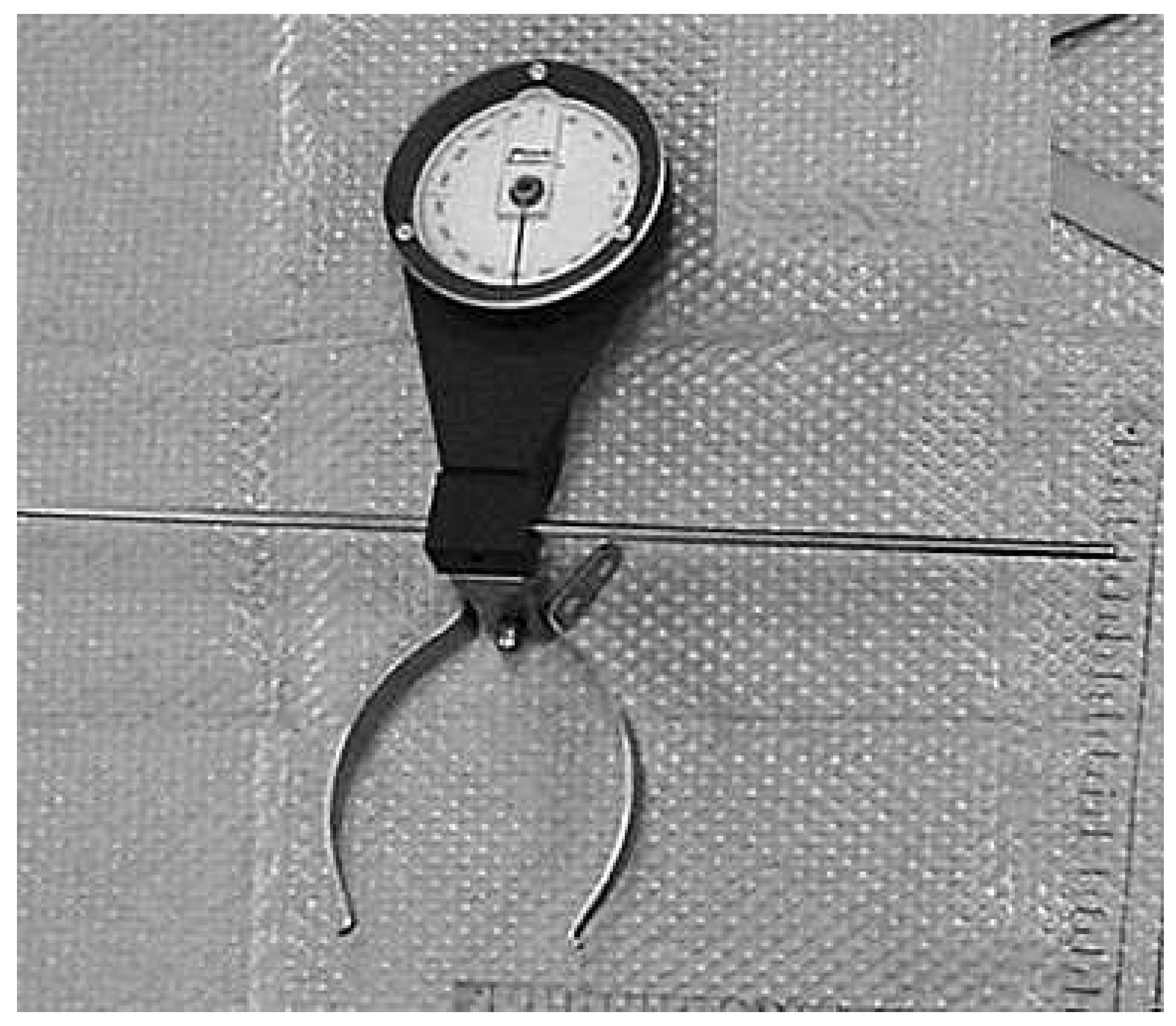
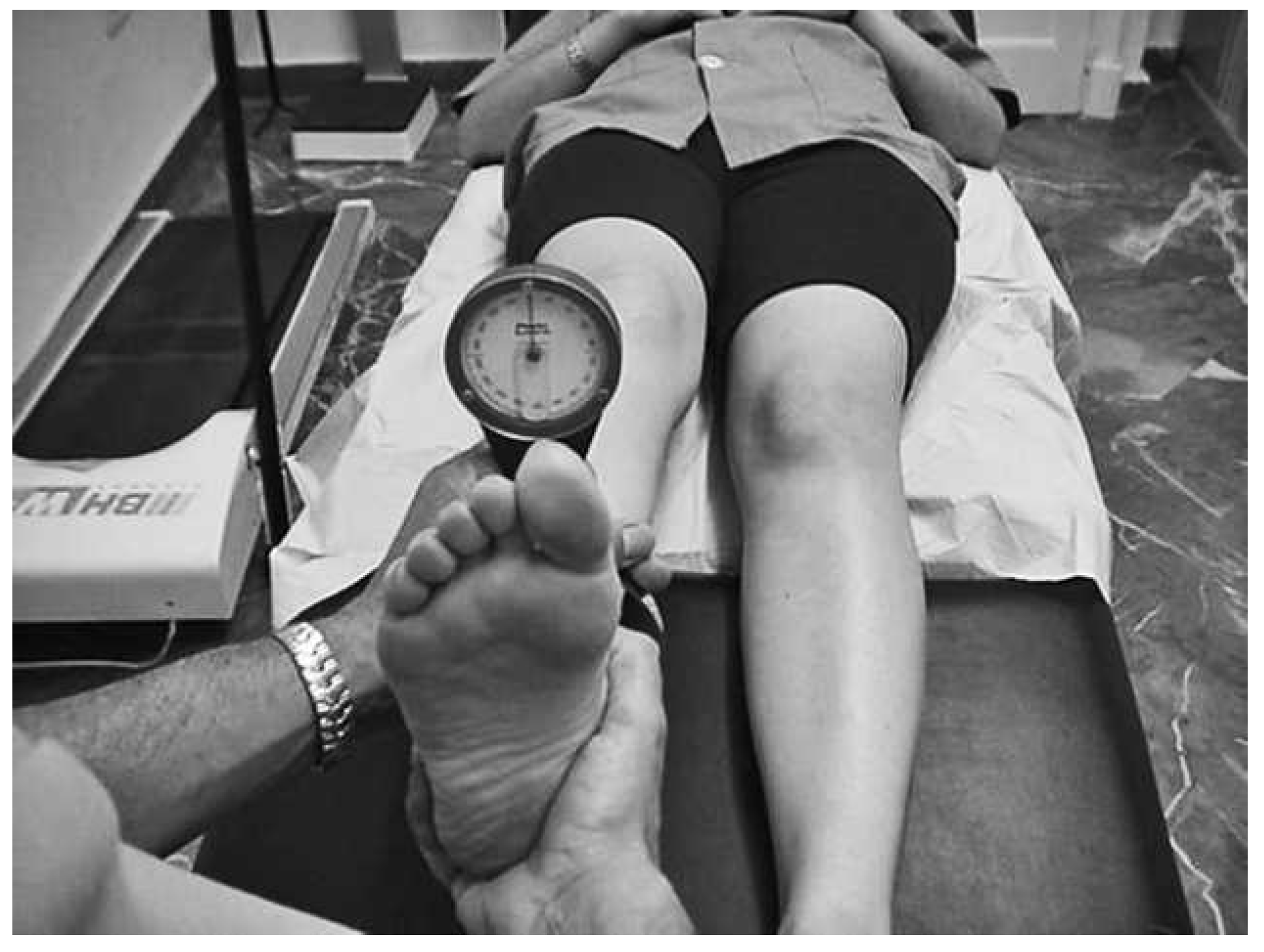
24] This method of measuring the rotational ability of the lower extremity considers all of the parameters that contribute to the capacity of the lower extremity to point the foot inward (adduction) and outward (abduction). For the measurement, the study participant was placed on the examination table in the supine position with the knee extended, the ankle in the neutral position, and the second ray coinciding with the vertical. An inclinometer, coupled to a holding system, was placed in the supramaleolar region to quantify the movement (
Figs. 1 and
2). The specific maneuver consisted of turning the foot inward (in adduction, toward the midaxis of the body) and outward (in abduction, away from the midaxis of the body) up to the limit of mobility to obtain the degrees of internal and external rotational pattern, respectively. The rotation of the pelvis was controlled in the measurement to prevent patients from increasing their measured internal rotation by lifting their pelvis off the table.
Hallux dorsiflexion was measured following the method proposed by Buell et al[
22] but taking as the neutral position the relaxed position of the hallux with respect to the first metatarsal. This method has been used previously in other studies.[
5]
Figure 1.
Inclinometer coupled to a system of holding pins.
Figure 1.
Inclinometer coupled to a system of holding pins.
Figure 2.
Measurement of the rotational pattern.
Figure 2.
Measurement of the rotational pattern.
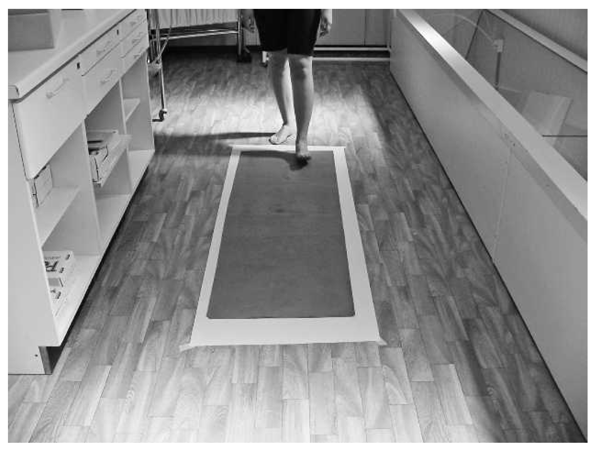
The foot progression angle was measured with the participant unshod. Each participant was asked to walk over a 2-mm-thick, 120-cm-long, and 50-cm-wide latex sheet. This latex sheet was inked on its underside and placed on a porous paper of greater length and 60 cm wide, positioned parallel to the line of progression of the individual so that when the participant walked on the latex sheet, a plantar impression was left on the paper, and the sides of the paper coincided with the line of gait (
Fig. 3). This line of gait and the longitudinal axis of the foot obtained from the plantar impression were used to determine the foot progression angle. This procedure for measuring the foot progression angle has been used previously in other studies.[
24,
25]
Figure 3.
Method of obtaining the foot progression angle.
Figure 3.
Method of obtaining the foot progression angle.
The data were analyzed with a commercially available computer program (SPSS 15.0 for Windows; SPSS Inc, Chicago, Illinois). On checking whether the data followed a gaussian distribution, using the Kolmogorov-Smirnov test, we found that the only quantitative variable that was distributed normally in the two groups was the foot progression angle. Thus, besides the means, standard deviations, and 95% confidence intervals for the main quantitative variables, the average ranges of the main quantitative variables not distributed normally were obtained. The values for the foot progression angle were compared between the two study groups with the Student
t test for independent samples (with the Levene test to check the equality of variance), and the values for the remaining quantitative variables were compared between the two groups with the nonparametric Mann-Whitney
U test. Comparisons were considered significant when the value of
P was less than 0.05. The correlation between the quantitative variables was also studied using the Spearman correlation coefficient Rho (ρ) in a bilateral significance test. These tests also gave values for the effect size for the correlations and for the comparisons, considering that for the correlations it is small when ρ = 0.10, medium when ρ = 0.30, and large when ρ ≥ 0.50 and for the comparisons it is small when
R2 = 0.01, medium when
R2 = 0.06, and large when
R2 ≥ 0.14.[
26] For the calculation of
R2, the values of the internal rotational pattern were transformed to ranges, and
R2 was obtained using a one-way analysis of variance between the ranges for the normal cases and the hallux limitus cases. To check whether the two groups were different regarding age and sex, a Student
t test for independent samples and a
χ2 test, respectively, were performed. On finding that there were significant differences between the groups with respect to sex, the same comparisons described previously herein (after finding that the only variable that followed a gaussian distribution in the two groups was the foot progression angle for men and women) were made but for men and women independently. Thus, it was evaluated whether the results obtained for the overall sample were similar when men and women were analyzed separately.
Results
The control group comprised 35 individuals (11 men and 24 women; mean ± SD age, 27.03 ± 7.86 years) for a total of 70 lower extremities. The group of patients with slight hallux limitus comprised 45 people (27 men and 18 women; mean ± SD age, 29.29 ± 6.60 years) for a total of 90 lower extremities. The mean ± SD values of the four main quantitative variables and the average ranges of the three main quantitative variables that did not follow a normal distribution are given in
Table 1 for the control group and patients with mild hallux limitus.
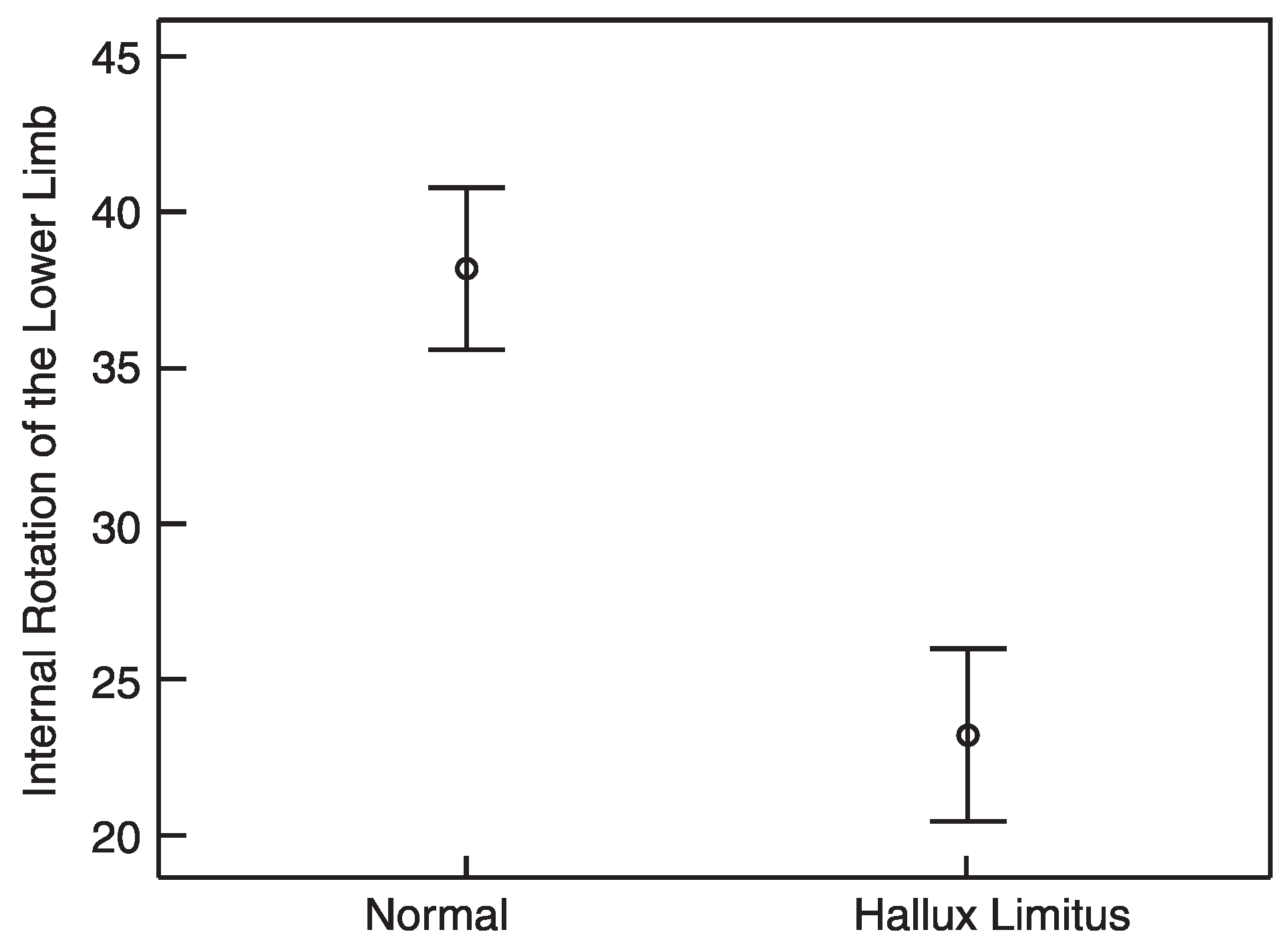
Figure 4.
Bar graph of error comparing the mean and the 95% confidence interval between the control group and the hallux limitus group. Units of measure of the vertical axis are degrees.
Figure 4.
Bar graph of error comparing the mean and the 95% confidence interval between the control group and the hallux limitus group. Units of measure of the vertical axis are degrees.
Table 1.
Mean ± SD Values and 95% CIs for the Main Quantitative Variables and Mean Ranges of Those Not Distributed Normally.
Table 1.
Mean ± SD Values and 95% CIs for the Main Quantitative Variables and Mean Ranges of Those Not Distributed Normally.
The difference in the foot progression angle between the two groups was nonsignificant (
P = .115). However, the differences in hallux dorsiflexion (
P < .0001), the internal rotational pattern (
P < .0001), and the external rotational pattern (
P =.027) between the two groups were significant. Patients with mild hallux limitus had lesser capacity to internally rotate the lower limb than did individuals in the control group (
Fig. 4). Given that the comparison between the internal rotational pattern of the lower extremity of individuals with and without hallux limitus was one of the aims of this study, the effect size for this comparison was calculated, and it was large (
R2 corrected = 0.266).[
26] This finding, together with the statistical significance obtained, could be interpreted as showing a very high probability that the effect exists in the population.
The study of the correlations showed a strong significant positive correlation between hallux dorsiflexion and the internal rotational pattern; ie, the greater the capacity of internal rotation of the lower limb, the greater the hallux dorsiflexion, and vice versa, the lesser the capacity of internal rotation of the lower limb, the lesser the hallux dorsiflexion. The value of ρ and the significance are given in
Table 2. The effect for the correlation between hallux dorsiflexion and the internal rotational pattern is large (ρ > 0.5),[
26] which, together with the statistical significance obtained, could also be interpreted as showing a high probability that the effect is present in the population.
Table 2.
The Spearman Rho (ρ) and Significance Values for the Correlations Between the Main Quantitative Variables.
Table 2.
The Spearman Rho (ρ) and Significance Values for the Correlations Between the Main Quantitative Variables.
The groups did not show significant differences regarding age (
P = .05). However, they showed a significant difference regarding the distribution by sex (
P < .0001) (
Table 3). The mean ± SD values and the
P values for the comparisons of the main variables performed separately in men and women are displayed in
Table 4. The behaviors of hallux dorsiflexion, the internal rotational pattern, and the foot progression angle are similar when the two groups are compared overall and when the men of the control group are compared separately with the men of the hallux limitus group and the women of the control group with the women of the hallux limitus group. The external rotational pattern is the only variable that shows a different behavior, being significantly lesser in women with hallux limitus than in women in the control group.
Table 3.
Distribution by Sex in the Study Groups.
Table 3.
Distribution by Sex in the Study Groups.
Table 4.
Mean ± SD and P Values for the Comparisons of the Main Variables Performed Separately in Men and Women.
Table 4.
Mean ± SD and P Values for the Comparisons of the Main Variables Performed Separately in Men and Women.
Discussion
In our clinical experience, many patients observed with mild hallux limitus further presented with limited internal rotation of the lower limb, leading us to consider the possibility that these two limitations might be related. From the results obtained, we rejected the first null hypothesis and accepted the second; ie, we can state that in the study sample, the capacity of internal rotation of the lower extremity was lesser in the study participants who had mild hallux limitus than in those without this abnormality and that this limitation did not affect the foot progression angle because this variable was similar in the two groups of study participants.
This work has some limitations. One of these is not having studied patients with more advanced hallux limitus, or even hallux rigidus, to check whether the increased foot progression angle claimed on various occasions by different authors is found in the most serious phases of the deformity, which would suggest that this is a consequence of hallux limitus progression; this limitation could be overcome by including a third study group of patients with hallux rigidus and evaluating the capacity of internal rotation of the lower limb and the foot progression angle. Another limitation is that the study groups presented a different distribution regarding the sex of the study participants, which could suggest that the results obtained are attributable to differences related to sexual dysmorphisms. In an attempt to diminish this slant, the men and women of the two groups were analyzed separately, with the comparisons of hallux dorsiflexion, internal rotational pattern, external rotational pattern, and foot progression angle being made separately between the men of the two groups and between the women of the two groups. As has been seen, the limitation of the internal rotational pattern was significantly different in men and women, and the foot progression angle was similar in men and women. This means that despite the χ2 test having shown significant differences in the distribution by sex for the two groups, this difference did not affect the results obtained for the overall sample.
Over the years, Dananberg[
27] studied and described the compensations that develop in the foot and the locomotor apparatus as a consequence of hallux limitus. Some of these alterations have their origin in a blocking of the movement of progression in the sagittal plane that enables the body to move forward, as in, for example, the collapse of the mediotarsal joint, the deficient extension of the hip during the propulsion phase of gait, or the reduction of lumbar lordosis.[
28] However, to our knowledge, the relationship between hallux limitus and other bodily alterations in the opposite direction has not been studied; ie, we do not know of any descriptions of alterations at the level of the hip or the low back that originate a limitation of hallux dorsiflexion. An exception could be one study in which a moderate direct correlation was found between hallux dorsiflexion and the internal rotational pattern (
r = 0.506;
P < .01); ie, the lesser the capacity of internal rotation of the lower extremity, the lesser the hallux dorsiflexion.[
25]
Alterations in the reference range of internal and external rotation of the lower limb, above all of the hip joint, have been related to various orthopedic problems of the lower extremity.[
17] Gelberman et al[
18] demonstrated that a limitation of the internal rotation of the hip associated with a reduced femoral anteversion angle was a risk factor for slipped capital femoral epiphysis of the hip. Tonnis and Heinecke[
20] state that there is a relationship between the limitation of internal rotation of the hip and degenerative disease of the hip. Ito et al[
19] demonstrated that patients with signs of a labral tear of the hip had a decreased femoral anteversion angle associated with a reduction in the internal rotation of the lower limb. Other studies have demonstrated the association between an increase or decrease in the femoral anteversion angle (which, in turn, affects the range of rotational movement of the lower limb) and an in-toeing or out-toeing gait.[
15,
29–
31] Although there have been many studies relating variations in the range of internal or external rotation of the lower extremity to various orthopedic problems, we have not found any study associating a rotational problem of the lower extremity, such as a limitation of the internal rotational pattern, with the pathologic findings of hallux limitus.
The fact that individuals with incipient hallux limitus have less movement of internal rotation of the lower limb than do patients without this abnormality leads us to postulate that the lesser capacity to point the foot toward the midaxis of the body might be related to the etiology of hallux limitus because this decrease in rotation has been observed in mild cases of hallux limitus. It is known that in the first half of the stance phase of gait the lower limb rotates internally, helping the whole sole of the foot, including the plantar face of the head of the first metatarsal, make contact with the ground and adapt to variations in it. A reduction in the movement of internal rotation of the lower limb might result in the head of the first metatarsal not being able to bear the amount of load that it would under normal conditions, making the first ray behave as a dorsiflexed first ray, and making the proximal phalange of the hallux become plantar-flexed to substitute the distal point of support of the medial column of the foot. This could generate a retrograde force that secondarily increases the elevation of the first ray. Remember that a dorsiflexed first ray and a plantarflexed proximal phalange of the hallux, together with a hyperextended distal phalange, is the structural scheme that is characteristic of the hallux limitus deformity.[
32–
34] Taking this hypothesis into account, addition of elements that limit an existent limitation of internal motion further could be contraindicated in orthotic management of hallux limitus. This could be a research objective because it would further differentiate orthosis prescription writing based on the presence of hallux limitus.
A relationship between hallux limitus or rigidus and an increase in the foot progression angle has also been reported elsewhere.[
12,
13,
35] To our knowledge, such a relationship between the foot progression angle and the limitation of hallux dorsiflexion always refers to this increase in the foot progression angle being produced as a consequence of the limitation of hallux dorsiflexion, sometimes as a mechanism to prevent pain in the first metatarsophalangeal joint during the stance phase of gait. The impossibility of adequately dorsiflexing the first metatarsophalangeal joint gives rise to compensations such as supination of the forefoot, collapse of the longitudinal arch, compensatory pronation during push-off, and absence of propulsion of the foot because there is a failure to raise the heel during push-off,[
28] which could result in the establishment of a pattern of gait with the feet turned outward. Our initial idea that an increase in the foot progression angle might favor the development of hallux limitus, constituting its cause and not its consequence, is not sustained by the results of this work.