Next Article in Journal
Hallux Limitus and Its Relationship with the Internal Rotational Pattern of the Lower Limb
Previous Article in Journal
Lengthening of the Midfoot in a Case of Lower-extremity Hemimelia
 
 
Journal of the American Podiatric Medical Association is published by MDPI from Volume 116 Issue 1 (2026). Previous articles were published by another publisher in Open Access under a CC-BY (or CC-BY-NC-ND) licence, and they are hosted by MDPI on mdpi.com as a courtesy and upon agreement with American Podiatric Medical Association.
Article

Mimicry in Older Patients: Tophaceous Pseudogout as a Tumorlike Lesion. A Case Report

by
Matthias A. Erhardt
,
Sven Y. Vetter
,
Arnold J. Suda
*,
Andreas Wentzensen
and
Christian B. Frank
Department of Trauma and Orthopaedic Surgery, BG Trauma Center Ludwigshafen, Ludwig-Guttmann-Strasse 13, 67071 Ludwigshafen, Germany
*
Author to whom correspondence should be addressed.
J. Am. Podiatr. Med. Assoc. 2011, 101(5), 462-465; https://doi.org/10.7547/1010462
Published: 1 September 2011

Abstract

Pseudogout is frequently seen in painful arthritis of larger joints, especially the knees and shoulders. This case report describes a rare clinical entity of soft-tissue calcium pyrophosphate dihydrate crystal deposition disease surrounding the second metatarsal bone of an 81-year-old man with moderate painful swelling of the right forefoot for 2 years. Diagnostic imaging demonstrated a calcified soft-tissue tumor adjacent to the second metatarsal bone by considering a neoplastic process in the differential diagnosis. Excisional biopsy revealed a chalklike material on the calcified soft tissue that was pathognomonic for chondrocalcinosis or pseudogout. The excisional biopsy findings led to stagnation of the clinical symptoms, and no recurrence of the initial tumor was seen during 12-month follow-up. In the literature, only 28 cases of tophaceous pseudogout tumors in the extremities have been described. In these cases, the lesions were suspected of being chondrosarcomas. For this reason, a correct diagnosis has to be pursued by performing a biopsy to treat the disease correctly.

Tumorlike calcium pyrophosphate dihydrate crystal deposition associated with pseudogout is seldom encountered. Mainly, these tumors are localized in the temporomandibular joint, the cervical spine, and the joints of the hand.[1,2] In diagnostic imaging and during clinical investigation, they can be misinterpreted as malignant tumors, especially chondrosarcomas.[3,4]
Twenty-eight cases of tophaceous pseudogout tumors in and near the extremities have been reported since 1983.[414] In those 28 cases, the tumorlike lesions were suspected of being chondrosarcomas because of the clinical and radiologic appearance and the patients’ history of painless tumoral growth over the years. In these patients, the need for surgical therapy seems obvious. Without clear pathohistologic findings, wrong decisions could be made concerning the operation and adjuvant therapies. In the worst case, this may lead to mutilation or needless resection.

Case Report

An 81-year-old man (height, 164 cm; weight, 78 kg) presented with moderate concerns about the right metatarsus without a recent trauma. The patient’s history revealed intermittent pain and chronic swelling of the right forefoot for the past 2 years, especially after strenuous exercise. He was limited in his activities of daily living because of discomfort after physical strain. The examination showed a man in regular condition with a slightly remarkable gait pattern. There was diffuse soft-tissue swelling in the region of the right metatarsus. The surrounding skin was mildly discolored but was not warm, thickened, or indurated. The range of motion of the toes and the upper and lower ankle joints was not affected and was within the reference range.
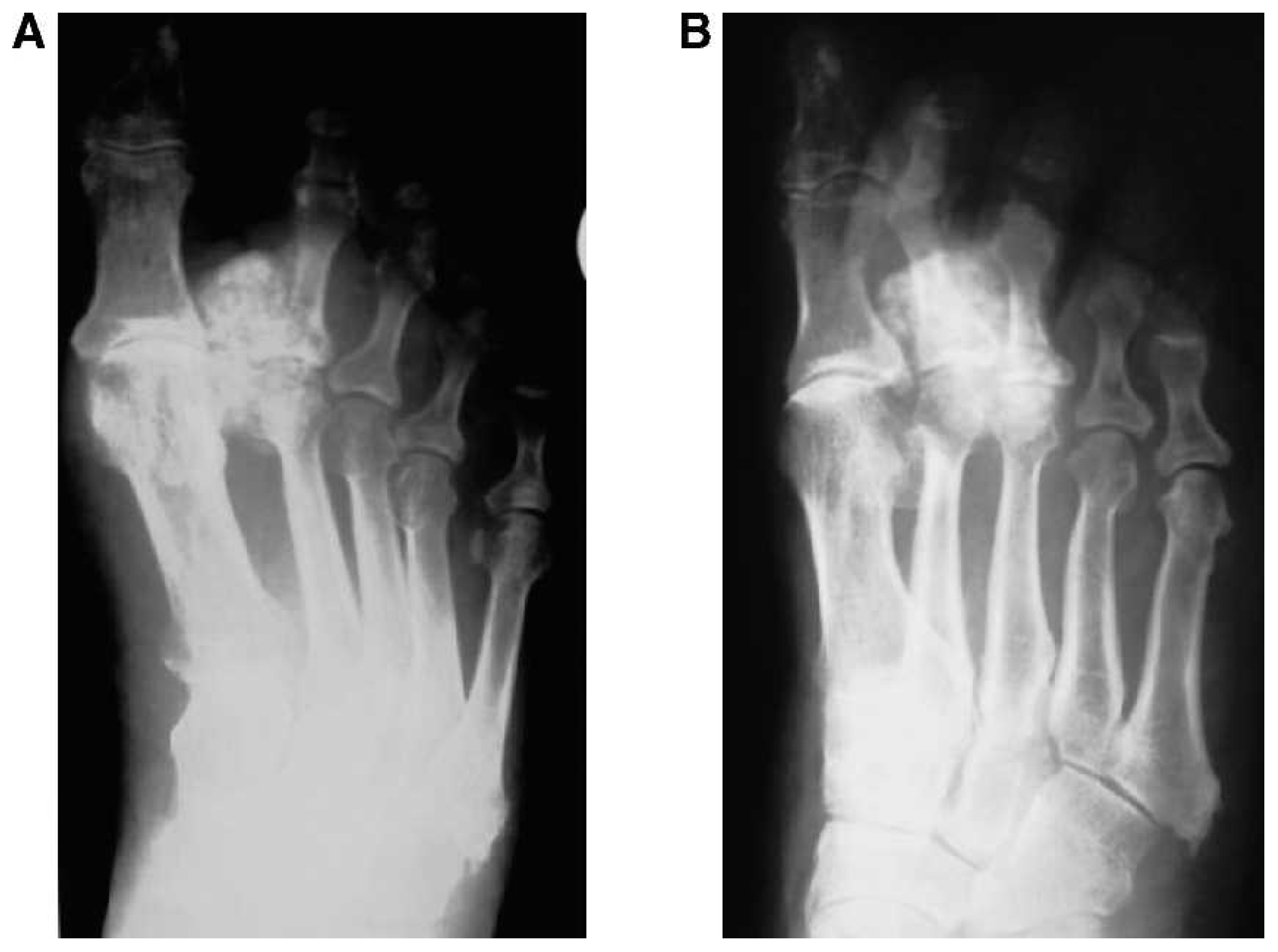
Radiographs revealed a radiopaque shadow of the soft tissue between the first and second metatarsal bones (Fig. 1). This finding was confirmed with magnetic resonance imaging (MRI), which demonstrated an oval hypointense mass adjacent to the second metatarsal bone and high signal intensity of the lobulated soft-tissue lesion (Fig. 2). These MRI patterns were similar to those of cartilaginous lesions, and synovial chondromatosis, tumoral calcinosis, chondroma, and chondrosarcoma could not be excluded as the tumor entity preoperatively.
Figure 1. Anteroposterior (A) and oblique (B) radiographs of the right forefoot demonstrate the soft-tissue lesion adjacent to the second metatarsal bone.
Japma 101 00462 g001
Because the lesions were highly suspected of being malignant, the regular staging examinations were performed. Scintigraphy showed increased metabolism in all three phases in the suspected area corresponding to a mesenchymal tumor and in the second lumbar vertebral body. The findings in the lumbar spine were suspected of being a metastasis. Other unspecific increased metabolism could be detected in both acromioclavicular joints, related to degenerative changes.
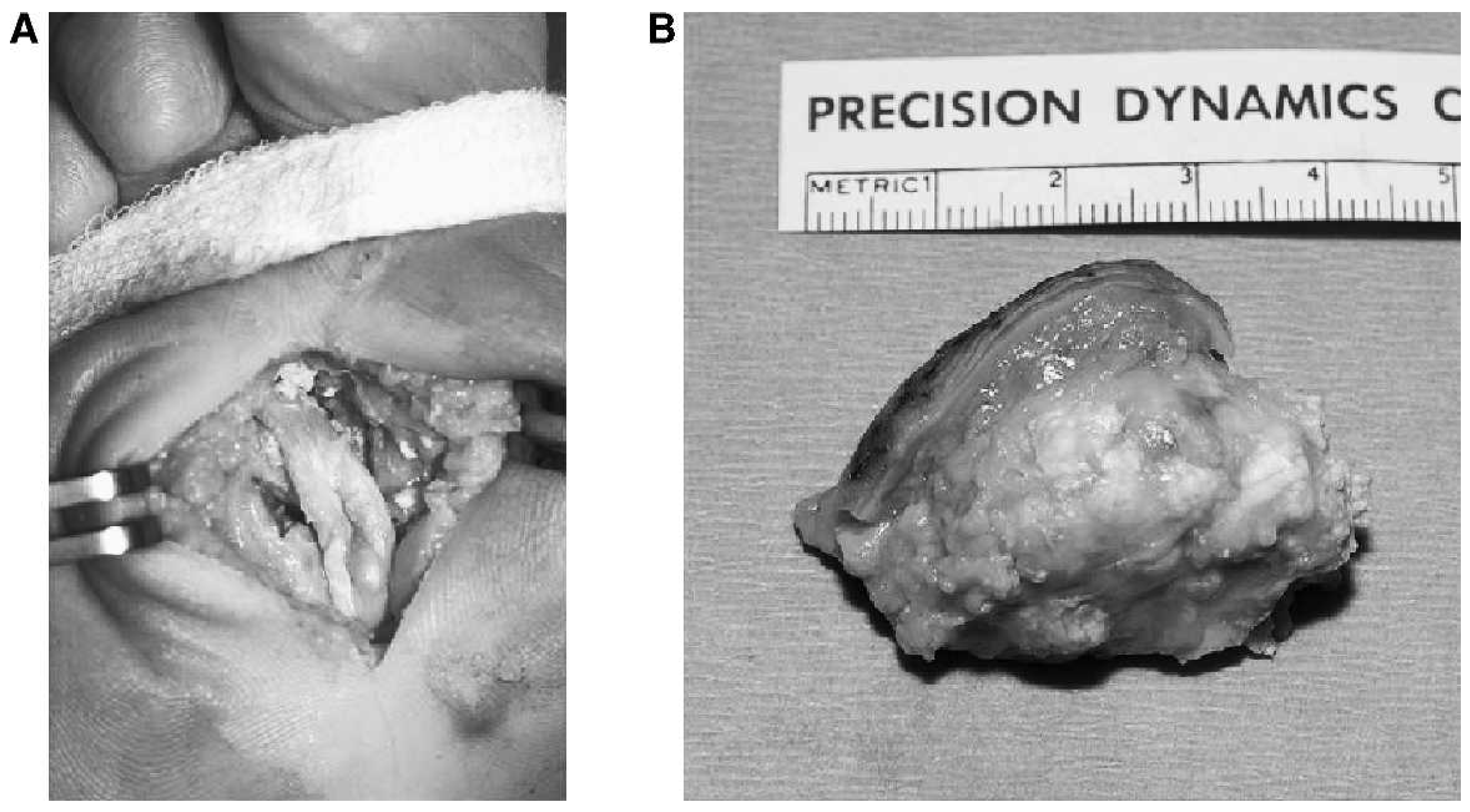
An incisional biopsy was performed to confirm the etiology and pathogenesis of the calcified soft-tissue lesion of the right metatarsus. Histologic examination of the biopsy material then demonstrated extracellular crystals and abundant amorphous acellular material surrounded by chronic inflammatory cells, histiocytes, and foreign-body giant cells. The crystals were birefringent when examined in polarized light. The finding was considered to be typical for tophaceous pseudogout because examination of the material under polarized light showed a crystalline pattern. In a second operation, an excisional biopsy was performed (Fig. 3).
Follow-up care for 12 months postoperatively included anti-inflammatory medication, physical examination, and radiographic control. Physical examination showed an absence of clinical inflammatory signs of the metatarsus and of tumor recurrence. Radiographs after 12 months remained unchanged since the operative procedure, without signs of tumor recurrence.
Figure 2. (A) T1-weighted, and (B) T2-weighted magnetic resonance images show a heterogenous mass in the inferior portion of the metatarsus between the first and second metatarsal bones.
Japma 101 00462 g002
Figure 3. (A) in vivo site and (B) excisional biopsy showing a chalklike, pasty, caseous mass with brown stains.
Japma 101 00462 g003

Discussion

Tophaceous pseudogout, or calcium pyrophosphate dihydrate deposition disease, has lately been designated to the same pathologic findings encompassing tumorous formation and microscopic shape. Calcium pyrophosphate dihydrate deposition disease is a rare, benign crystalline lesion of unknown cause and is commonly observed in elderly patients.[15] The deposition of calcium pyrophosphate crystals in the soft tissue is a rare presentation.[5,10,1618]
A common and a tumoral formation of calcium pyrophosphate crystal deposition disease has been described. Whereas the common type often affects larger joints subsequent to trauma, surgery, or ischemic heart disease, the tumoral type is mainly localized in the temporomandibular joint, the cervical spine, and the joints of the hand.[10,16]
The main differential diagnosis of calcium pyrophosphate dihydrate deposition disease includes tumoral calcinosis, synovial chondromatosis, benign and malignant tumors, and tophaceous gout. Commonly, untreated hyperuricemia leads to intra-and extra-articular deposition of urate crystals, causing inflammatory processes with histologically detectable macrophages and foreign-body giant cells, causing pain and limitation of range of motion of the affected joint.[1921] The uric acid crystals observed in gout are needle shaped and are rarely birefringed in polarized light, in contrast to the calcium crystals of calcium pyrophosphate dihydrate deposition disease, which have blunt ends and regularly show weak birefringence under polarized light.
Common radiologic characteristics of calcium pyrophosphate dihydrate deposition disease include joint space narrowing, articular surface sclerosis, subchondral cyst formations, and, in some cases, soft-tissue calcification. Soft-tissue tumoral calcium pyrophosphate dihydrate deposition disease has rarely been described in the literature. In all cases, exclusion of a malignant neoplasm (ie, chondrosarcoma) had to be accomplished.[2,5,8]
A previous case report[22] described cystic pseudotumors in both upper ankle joints in a patient with gouty arthritis. As well as other entities, this lesion can be misinterpreted in considering a neoplastic process. To our knowledge, pseudogout tumors mimicking chondrosarcomas of the foot were found in only six cases. Including the present case, a total of seven cases in 29 soft-tissue tumor calcium pyrophosphate dihydrate deposition disease of the extremities are reported; 25% of the soft-tissue calcium pyrophosphate dihydrate deposition disease would appear in the lower extremity, mainly in the metatarsus and toes. It is unclear whether these tumors appear more often nowadays because of demographic changes in industrialized countries and increasing metabolic disorders, such as gout and pseudogout. It is known that such metabolic diseases are more frequent in the older population.[23] Chondrosarcomas are also seen in the older population, with a peak after the fourth to seventh decades. Therefore, especially in the older population, this kind of benign soft-tissue tumor has to be excluded before definite surgery.
In the present case, an 81-year-old white man with moderate swelling and pain in the forefoot was diagnosed as having a remarkable calcified soft-tissue lesion adjacent to the second metatarsus. Radiologic findings suggested a malignant neoplasm. Biopsy revealed a chalky white material, and a diagnosis of goutlike tumor was offered. The histopathologic findings confirmed the diagnosis and were pathognomonic for tumoral calcium pyrophosphate dihydrate deposition disease.
However, in this rare case with a remarkable soft-tissue lesion of the forefoot, the therapeutic possibilities in addition to excisional biopsy are limited. In the present case, during 12-month follow-up, the patient’s initial concerns no longer existed.
Before marginal tumor excision, incisional biopsy should reveal the definite histopathologic diagnosis. If in doubt, the normal staging examinations should be performed, and if malignant metaplasia is suspected, the biopsy should be sent to a histopathologic reference center for investigation.
Financial Disclosure: None reported.
Conflict of Interest: None reported.

References

  1. Naqvi, AH, JLAbraham, RMKellman, et al. :Calcium pyrophosphate dihydrate deposition disease (CPPD)/ pseudogout of the temporomandibular joint: FNA findings and microanalysis. .Cytojournal5::8. ,2008.
  2. Yu, SL, RZLi, and ZHBian. :Tumoral calcium pyrophosphate dihydrate deposition disease. .Chin Med J (Engl)105::780. ,1992.
  3. Doherty, M, PDieppe, and IWatt. :Pyrophosphate arthropathy: a prospective study. .Br J Rheumatol32::189. ,1993.
  4. Lambrecht, N, SDNelson, LSeeger, et al. :Tophaceous pseudogout: a pitfall in the diagnosis of chondrosarcoma. .Diagn Cytopathol25::258. ,2001.
  5. Ishida, T, HDDorfman, and PGBullough. :Tophaceous pseudogout (tumoral calcium pyrophosphate dihydrate crystal deposition disease). .Hum Pathol26::587. ,1995.
  6. Havitçioğlu, H, HTatari, OBaran, et al. :Calcium pyrophosphate dihydrate crystal deposition disease mimicking malignant soft tissue tumor. .Knee Surg Sports Traumatol Arthrosc11::263. ,2003.
  7. Ammar, A, KBen Romdhane, RKhattech, et al. :Tumoral calcinosis: a clinical and pathological study of 8 cases reported in Tunisia. .Rev Chir Orthop Reparatrice Appar Mot80::261. ,1994.
  8. Sissons, HA, GCSteiner, FBonar, et al. :Tumoral calcium pyrophosphate deposition disease. .Skeletal Radiol18::79. ,1989.
  9. Staub-Zaehner, T, DGarzoni, CFretz, et al. :Pseudotumor of gout in the patella of a kidney transplant recipient. .Nat Clin Pract Nephrol3::345. ,2007.
  10. Yamakawa, K, HIwasaki, YOhjimi, et al. :Tumoral calcium pyrophosphate dihydrate crystal deposition disease: a clinicopathologic analysis of five cases. .Pathol Res Pract197::499. ,2001.
  11. Ohshio, I, TOgino, SSatoh, et al. :Tumourous deposition of calcium pyrophosphate dihydrate crystals in the wrist. .J Hand Surg Br16::219. ,1991.
  12. Luisiri, P, JBlair, and MHEllman. :Calcium pyrophosphate dihydrate deposition disease presenting as tumoral calcinosis (periarticular pseudogout). .J Rheumatol23::1647. ,1996.
  13. Martens, HA, HRLebbink, and MDPosthumus. :Clinical images: a patient with a tumor of the foot. .Arthritis Rheum58::1085. ,2008.
  14. Dagregorio, G and YSaint-Cast. :Tumoral calcium pyrophosphate dihydrate crystal deposition in the fifth metacarpophalangeal region. .Orthopedics29::161. ,2006.
  15. Terkeltaub, R. :“Diseases Associated with Articular Deposition of Calcium Pyrophosphate Dehydrate,”. inKelley’s Textbook of Rheumatology, ,7th Ed. , ed byHarrisJr, ED, RCBudd, GSFirestein, et al. ,Elsevier. ,Philadelphia. ,2005.
  16. Ling, D, WAMurphy, and MKyriakos. :Tophaceous pseudogout. .AJR Am J Roentgenol138::162. ,1982.
  17. Richman, KM, RDBoutin, LMVaughan, et al. :Tophaceous pseudogout of the sternoclavicular joint. .AJR Am J Roentgenol172::1587. ,1999.
  18. Kinoshita, TM, SMaruoka, TYamazaki, et al. :Tophaceous pseudogout of the spine: MR image and bone scintigraphy findings. .Eur J Radiol27::271. ,1998.
  19. Vukmir, R, LWeiss, and VPVerdile. :Polyarticular symmetric tophaceous joint inflammation as the initial presentation of gout. .Am J Emerg Med8::43. ,1990.
  20. Palmer, D, JHighton, and PAHessian. :Development of the gout tophus. .Am J Clin Pathol91::190. ,1989.
  21. Giannestras, N. :Food Disorders: Medical and Surgical Management, ,2nd Ed. ,Lea & Febinger. ,Philadelphia. ,1973.
  22. Vetter, SY, RSimon, Jvon Recum, et al. :Cystic pseudotumours in both upper ankle joints in gouty arthritis. .Foot Ankle Surg14::229. ,2008.
  23. Richette, P, TBardin, and MDoherty. :An update on the epidemiology of calcium pyrophosphate dihydrate crystal deposition disease. .Rheumatology (Oxford)48::711. ,2009.