Next Article in Journal
Mimicry in Older Patients: Tophaceous Pseudogout as a Tumorlike Lesion. A Case Report
Previous Article in Journal
Bilateral Tibial Hallux Sesamoid Agenesis and Fibular Hallux Sesamoid Hypoplasia in a Patient with Bilateral Hallux Valgus
 
 
Journal of the American Podiatric Medical Association is published by MDPI from Volume 116 Issue 1 (2026). Previous articles were published by another publisher in Open Access under a CC-BY (or CC-BY-NC-ND) licence, and they are hosted by MDPI on mdpi.com as a courtesy and upon agreement with American Podiatric Medical Association.
Article

Lengthening of the Midfoot in a Case of Lower-extremity Hemimelia

by
Tanja Kraus
*,
Ernst B. Zwick
,
Martin Svehlik
and
Wolfgang E. Linhart
Medical University Graz, Pediatric Orthopedics, Auenbruggerpaltz 34, Graz, 8036, Austria
*
Author to whom correspondence should be addressed.
J. Am. Podiatr. Med. Assoc. 2011, 101(5), 456-461; https://doi.org/10.7547/1010456
Published: 1 September 2011

Abstract

Hemimelia of the lower limb belongs to the group of congenital deficiency disorders. The clinical spectrum ranges from minimal shortening of the long bones to severe deficiencies of the extremities. Several etiologies, such as X-rays or drugs, have been implicated to be responsible for hemimelia. In the present report the clinical course and the long-term follow-up of a patient with transverse terminal hemimelia of the left foot at the level of the basis of the metatarsals is described. Due to frequent episodes of pain, development of pressure sores, and an increasing psychological burden, operative intervention consisting of a lengthening procedure using an Ilizarov fixator was indicated. Long-term outcome was good; the patient is now able to painlessly wear conventional shoes and displays a normal gait pattern.

Hemimelia of the lower limb belongs to the group of congenital deficiency disorders. Although there are a few limb abnormalities with genetic bases, most limb deformities develop sporadically with no identifiable environmental factors, trauma, or familial incidence. Nevertheless, several etiologies, such as X-rays or drugs, have been implicated to be responsible for hemimelia.
At birth, some form of limb deficiency is seen in 23% of boys and 18% of girls.[1] The majority of these deficiencies, however, do not cause functional or cosmetic problems and there is no need for treatment. Severe deficiencies are seen in 1 of 200 newborns, resulting in an overall incidence of 0.5%.[1] Thalidomide and several other teratogenic products are well-known contributors to the development of growth disturbances in the growing limb buds.[2,3]
In the present case report, we describe the clinical course, surgical treatment, and outcome of a forefoot lengthening procedure using callotaxis with an Ilizarov-fixator in a case of transverse terminal hemimelia of the foot.

Case Report

Clinical History

The male patient was born in 1987 with a congenital defect of the left forefoot at the level of the basis of the metatarsals. Family history revealed that one uncle and one aunt had congenital defects of the hand and the foot, respectively. The patient first presented as an infant from the birth clinic. He did not display any additional disorders. The other ipsilateral proximal long bones and joints showed normal development at birth. We have followed him from infancy as an outpatient in our department at the Medical University Graz, Graz, Austria and subsequent follow-ups in our outpatient clinic showed normal development and normal milestones. At the time the patient developed walking ability, the first shoe modifications were done.
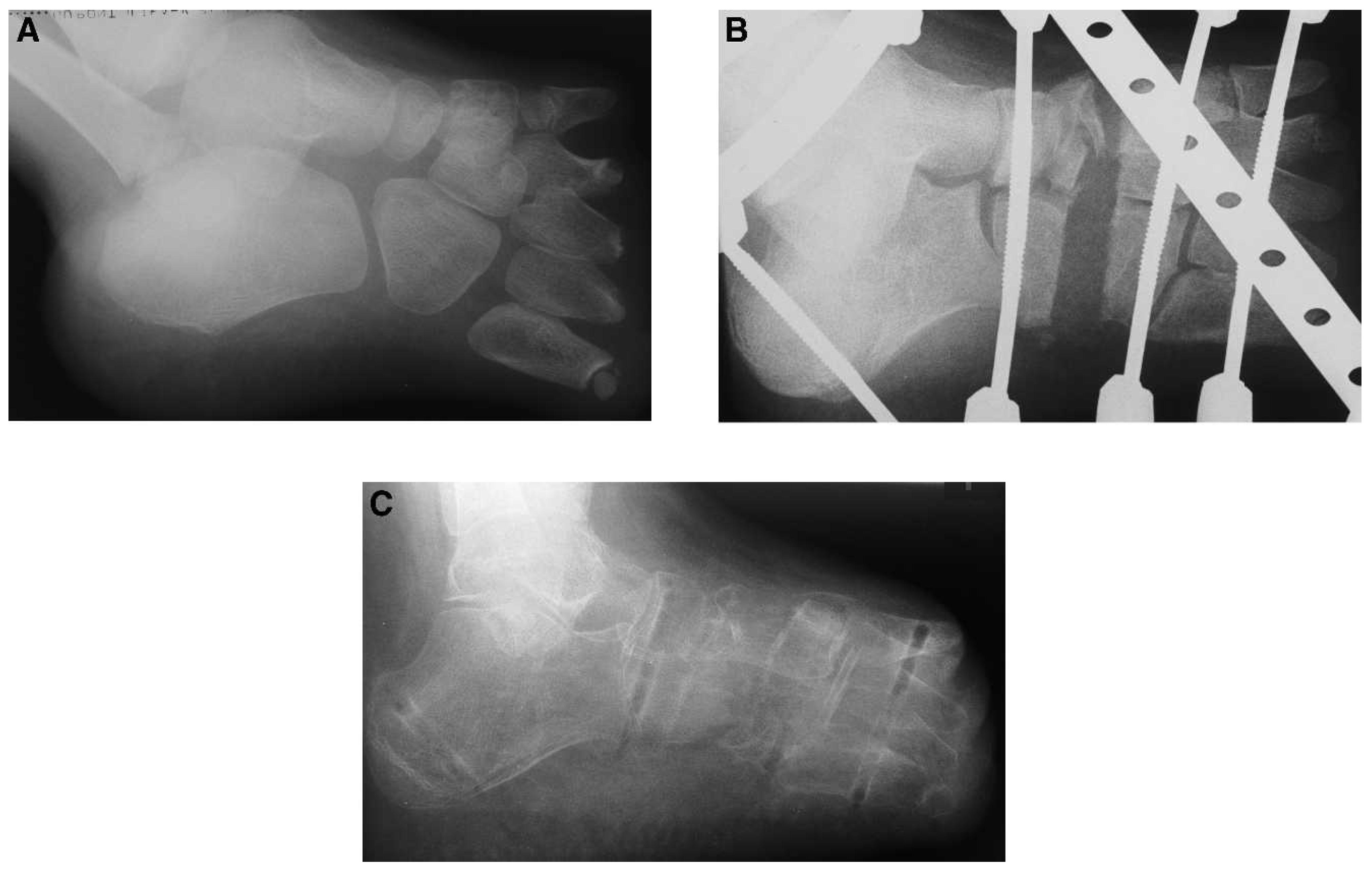
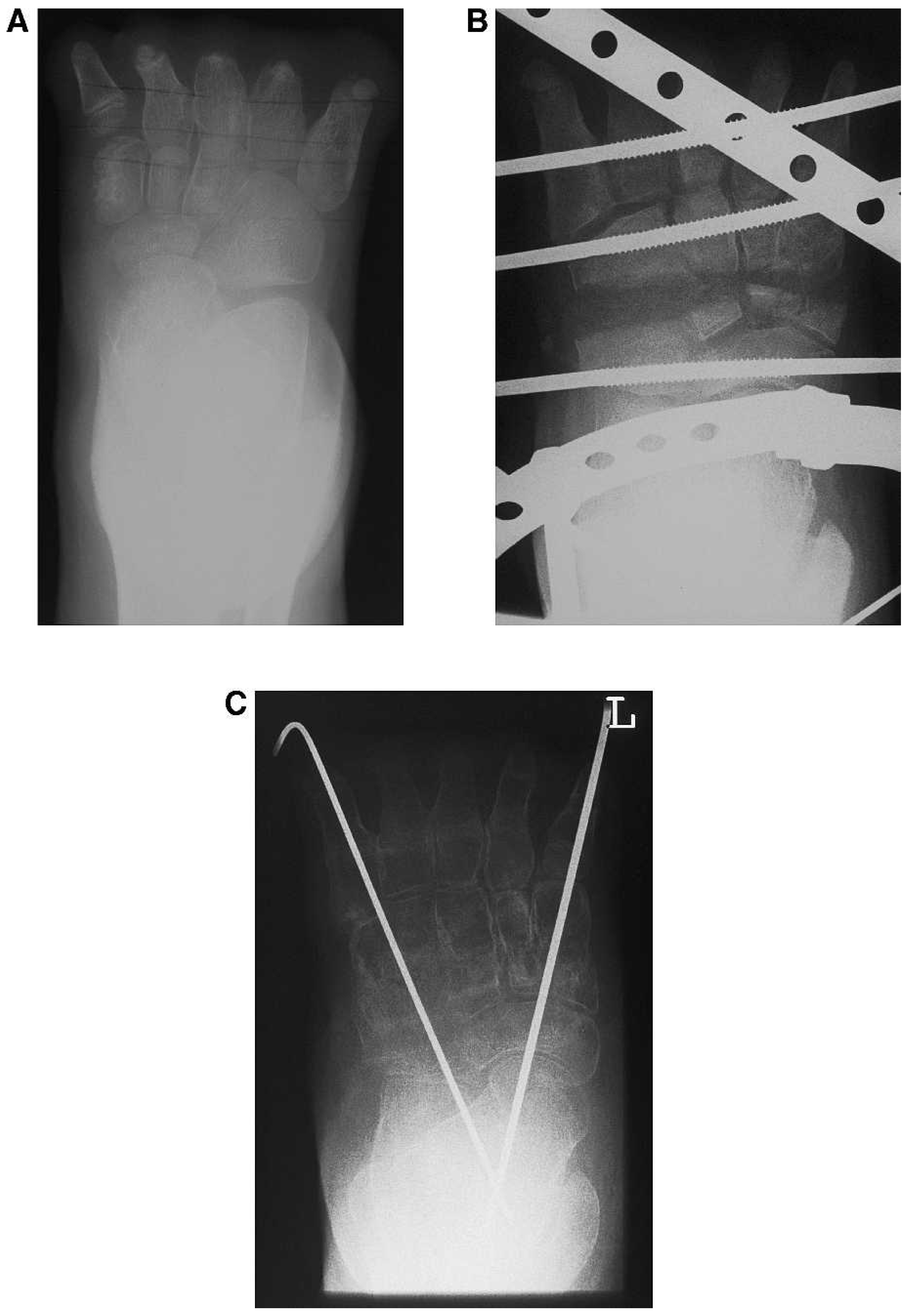
The patient displayed a terminal transverse hemimelia of the foot at the level of the forefoot according to the classification of Frantz and O’Rahilly[4] (Figs. 1A and 2A). For this deficiency we did not expect the need for surgical intervention because most of these patients are well managed with orthopedic shoes or shoe adaptations.[5] Follow-ups demonstrated a proportional growth of the rest of the foot and the lower limb.
Figure 1. Lateral radiograph showing A, hemimelic foot before treatment; B, during treatment with Ilizarov device; and C, after removal of Ilizarov after 15 weeks.
Japma 101 00456 g001
The patient was well managed with orthopedic shoes until the age of ten when he started to complain about sweating and constantly expressed the need for new adapted shoes. Simultaneously, he developed painful pressure sores on and a feeling of weakness in the affected foot. Inflammation often followed pressure sores and was repeatedly treated using local antibiotics and new shoe adaption.
Additionally, the foot deformity itself and frequent hospital visits caused an increasing psychological burden for the patient. At the request of the patient and his parents, we reevaluated the situation in 1999. At this time the patient was 12 years old and did not want to continue wearing orthopedic shoes. Owing to the foot deformity, he had problems wearing conventional shoes.
Following reevaluation, we decided to perform a midfoot lengthening with the goal to improve stability of the foot by lengthening the forefoot lever arm to eliminate the need for orthopedic shoes and to prevent soft-tissue breakdown.

Operative Treatment and Postoperative Evaluation

Surgery was performed when the patient was 13 years old. The osteotomy level was chosen to transverse the cuneiforms and the cuboid. Lengthening was performed using an Ilizarov fixator at a rate of 0.5 mm per day (Figs. 1B and 2B). A total lengthening of 3 cm was achieved. During the postoperative management, several soft-tissue infections were encountered. All of them were successfully treated with local antibiotics. Three weeks postoperatively, a superficial skin necrosis of approximately 2 × 2 cm developed, necessitating debridement and a full-thickness skin graft from the groin, performed under general anesthesia. Two weeks later, the patient left the hospital and was treated on an outpatient basis. The bone regenerate had good quality, and the patient walked with full weightbearing during the rest of the lengthening procedure.
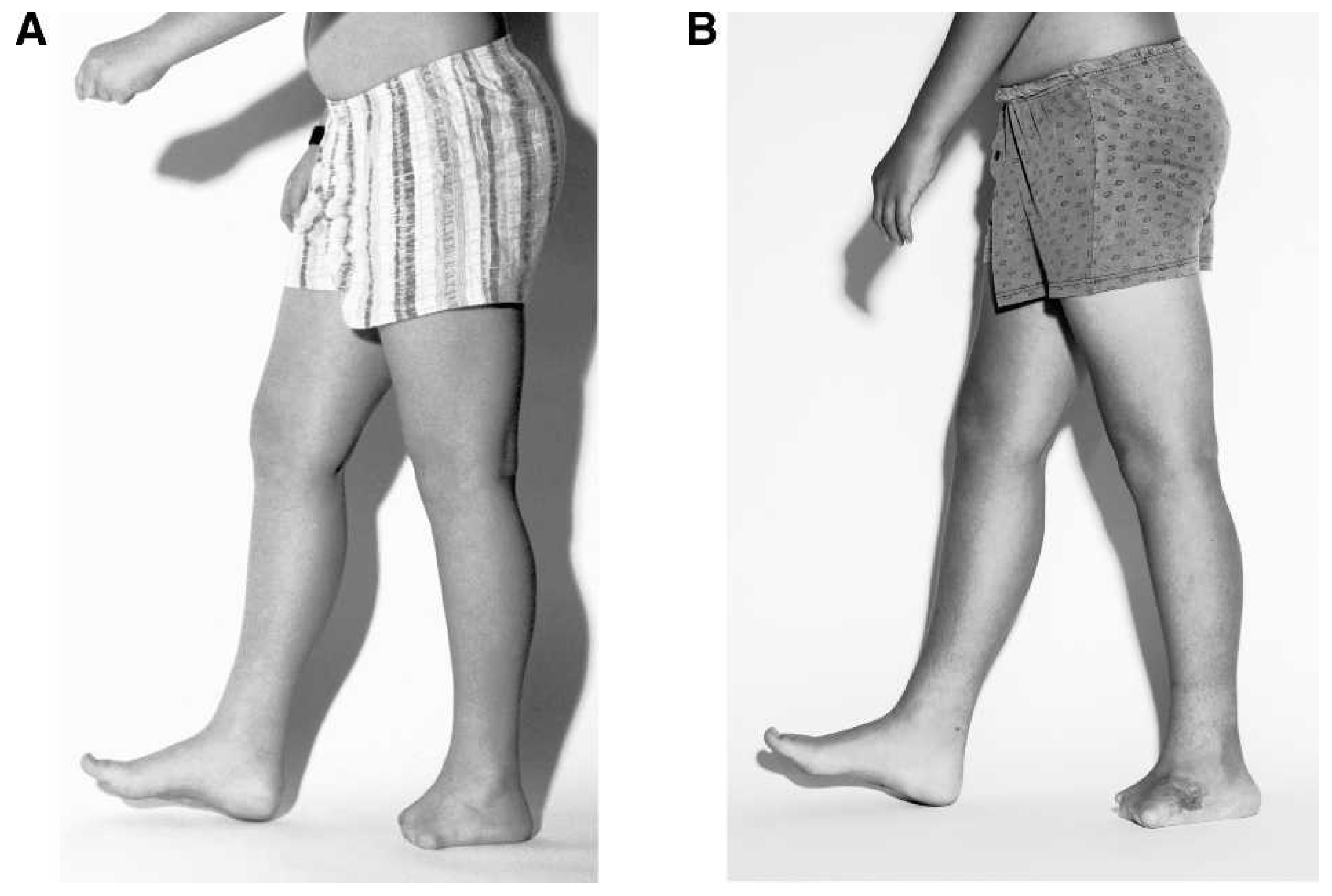
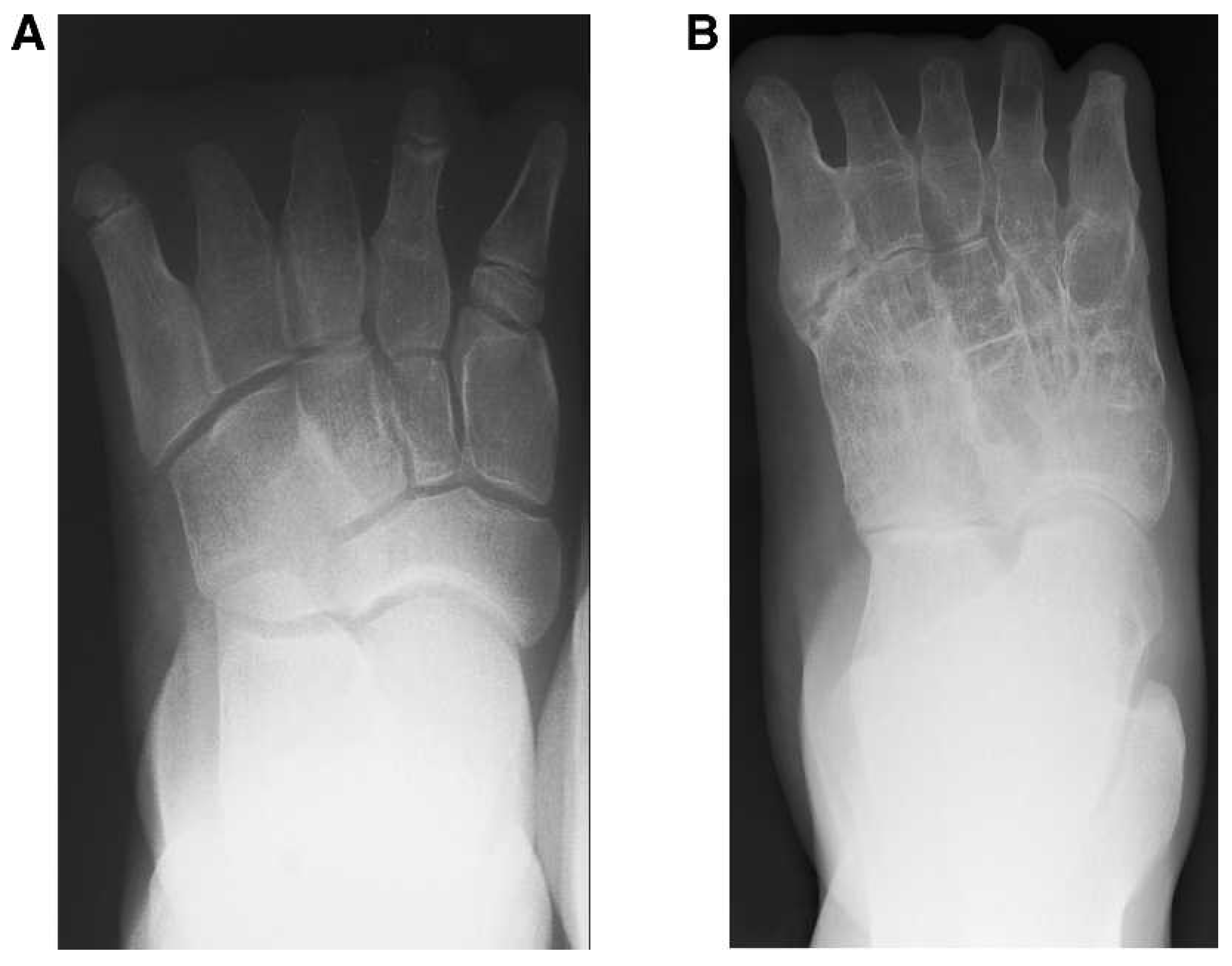
The external fixator could be removed after 15 weeks (Fig. 1C) and the midfoot was temporarily stabilized with Kirschner wires (Fig. 2C). The patient was treated with a below-knee walking plaster cast for 7 weeks until the Kirschner wires were removed. After removal of the Kirschner wires and a total treatment time of 5.5 months, the primary goal was achieved and the patient was able to wear conventional shoes without shoe modifications (Figs. 3 and 4). Moreover, the patient did not have pain during weightbearing.
Figure 2. Anteroposterior radiograph showing A, hemimelic foot before treatment; B, during treatment with Ilizarov device; and C, after removal of Ilizarov after 15 weeks and with temporary fixation with Kirschner wires.
Japma 101 00456 g002

Postoperative Follow-up

The patient was called in for a long-term follow-up 8 years postoperatively in May 2007. He was 20 years old and worked as a medical attendant. He was able to wear normal footwear and did not complain about shoe-related problems. He occasionally described discomfort in the midfoot, especially after extensive activity and sports, but the pain resolved with rest and no analgesic medication was needed.
Figure 3. Photograph showing (A) the patient before surgery and (B) after surgery.
Japma 101 00456 g003
Clinical examination revealed a normal gait pattern with symmetric step lengths. Loading of both lower extremities was symmetrical, and the development of the musculature of both lower limbs was comparable.
We applied standardized evaluation protocols to objectively evaluate the patient’s treatment outcome. Application of the Short Musculoskeletal Function Assessment Questionnarie in German (SMFA-D)[6,7] resulted in full score points. However, the American Orthopaedic Foot and Ankle Society score[8,9] showed 90 points (some midfoot malalignment limited the full score) for the ankle-hindfoot-scale and 83 points for the midfoot scale (occasional pain and some degree of midfoot malalignment limited the full score).
Figure 4. Radiograph showing A, the foot before treatment and B, after treatment.
Japma 101 00456 g004

Discussion

Some form of limb deficiency is seen in 23% of boys and 18% of girls at birth.[1] The majority of these deficiencies, however, do not cause functional or cosmetic problems and often do not require treatment. Severe forms of limb deficiencies are rare. For more severe lower-limb deficiencies, complex treatment is necessary and surgical interventions should be planned early.[5,10] In a case of a terminal hemimelia, conservative treatment with shoe adaption usually is sufficient. After initial treatment with shoe adaption, our patient developed painful pressure sores and an increased need for adapted shoes. Frequent hospital visits caused a severe psychological burden. Because of these problems, and at the request of both the patient and his parents, we decided to perform a midfoot lengthening procedure when the patient was 12 years old.
Various methods for short bone lengthening have been described. The most frequently used techniques are one-stage lengthening with an intercalary bonegraft[11,12] and gradual lengthening by callotasis.[12] llizarov described his method of bone lengthening after corticotomy by callus distraction using a ring-fixator.[13] Matev was the first to report on the lengthening of short bones.[14,15] Lengthening of metatarsals and metacarpals was reported using the same principles with a mini-fixator.[16,17] The application of the Ilizarov method can and has been applied for lengthening of long bones. The main advantages of using an Ilizarov external fixator are the ability to gradually correct deformity in the postoperative period and maintain this stable correction while weightbearing.[18] Disadvantages are that duration of this mode of treatment is long, with many fixator-related problems, of which pin-tract infection is the most common.[1923] Pin-tract infection frequently occurs when fixators are mounted on feet and weightbearing is allowed. In the initial stages of treatment with a fixator, soft-tissue breakdown attributable to vascular compromise is a well-known complication. The main complication in our patient was pin-tract infection associated with skin necrosis.
Nevertheless, the goal of treatment for our patient was reached: shoe adaption is no longer needed, the patient has a plantigrade foot, and he is pain free. This type of treatment requires good patient compliance and cooperation for the management of inevitable pin-tract infections and soft-tissue problems.

Conclusion

Foot lengthening after transverse osteotomy of the midfoot is difficult and unpredictable. Surgery indication is controversial. Despite several soft-tissue problems during the lengthening of the foot in our case, the primary goal of treatment was achieved. The patient is now able to painlessly wear normal shoes. He can pursue the profession of his choice and is active in light-to-moderate sports activities.
Financial Disclosure: None reported.
Conflict of Interest: None reported.

References

  1. Rogala, EJ, RWynne-Davies, ALittlejohn, et al. :Congenital limb anomalies: frequency and aetiological factors. Data from the Edinburgh Register of the Newborn (1964–68). .J Med Genet11::221. ,1974.
  2. Frantz, C. :The upsurge in phocomelic congenital anomalies .Available at: http://www.acpoc.org/library/1962_10_001.asp. .Accessed October 16, 2009.
  3. Frantz, C. :The increase in the incidence of malformed babies in the German federal Republic during the years 1959–1962 .Available at: http://www.edric.info/fileadmin/downloads/archive/holistic_case_management/PD0190.pdf. .Accessed October 16, 2009.
  4. Frantz, HC and RO’Rahilly. :Congenital skeletal limb deficiencies. .J Bone Joint Surg Am43::1202. ,1961.
  5. Herring, JA. :“Transverse Deficiencies,”. inTachdjian’s Pediatric Orthopedics, ,3rd Ed. , Vol.3:, ed byHerring, JA. p1782. ,WB Saunders. ,Philadelphia. ,2002.
  6. Konig, A, SKirschner, MWalther, et al. :Cultural adaptation, practicability and reliability evaluation of the Musculoskeletal Functional Assessment Questionnaire. .Z Orthop Ihre Grenzgeb138::295. ,2000.
  7. Konig, A, MWalther, MMatzer, et al. :Validity and sensitivity to change of the Musculoskeletal Functional Assessment Questionnaire in primary gonarthrosis and total endoprosthetic joint replacement. .Z Orthop Ihre Grenzgeb138::302. ,2000.
  8. Kitaoka, HB, IJAlexander, RSAdelaar, et al. :Clinical rating systems for the ankle-hindfoot, midfoot, hallux and lesser toes. .Foot Ankle Int15::349. ,1994.
  9. Ibrahim, T, ABeiri, MAzzabi, et al. :Reliability and validity of the subjective component of the American Orthopaedic Foot and Ankle Society clinical rating scales. .J Foot Ankle Surg46::65. ,2007.
  10. Hefti, F. :Malformations of the lower extremities. .Orthopade37::381. ,2008.
  11. Baek, GH and MSChung. :The treatment of congenital brachymetatarsia by one-stage lengthening. .J Bone Joint Surg Br80::1040. ,1998.
  12. Choi, IH, MSChung, GHBaek, et al. :Metatarsal lengthening in congenital brachymetatarsia: one-stage lengthening versus lengthening by callotasis. .J Pediatr Orthop19::660. ,1999.
  13. Ilizarov, GA, AADeviatov, and VGTrokhova. :Surgical lengthening of the shortened lower extremities. .Vestn Khir Im I I Grek108::100. ,1972.
  14. Matev, IB. :Thumb reconstruction after amputation at the interphalangeal joint by gradual lengthening of the proximal phalanx. A case report. .Hand11::302. ,1979.
  15. Matev, IB. :Thumb reconstruction through metacarpal bone lengthening. .J Hand Surg Am5::482. ,1980.
  16. Magnan, B, ABragantini, DRegis, et al. :Metatarsal lengthening by callotasis during the growth phase. .J Bone Joint Surg Br77::602. ,1995.
  17. Levine, SE, RSDavidson, JPDormans, et al. :Distraction osteogenesis for congenitally short lesser metatarsals. .Foot Ankle Int16::196. ,1995.
  18. Aaron, AD and REEilert. :Results of the Wagner and Ilizarov methods of limb-lengthening. .J Bone Joint Surg Am78::20. ,1996.
  19. Aquerreta, JD, FForriol, and JCanadell. :Complications of bone lengthening. .Int Orthop :18::299. ,1994.
  20. Glorion, C, JCPouliquen, and JLanglais. :Femoral lengthening using the callotasis method: study of the complications in a series of 70 cases in children and adolescents. .J Pediatr Orthop16::161. ,1996.
  21. Paley, D. :Problems, obstacles, and complications of limb lengthening by the Ilizarov technique. .Clin Orthop Relat Res250::81. ,1990.
  22. Garcia-Cimbrelo, E, BOlsen, MRuiz-Yague, et al. :Ilizarov technique. Results and difficulties. .Clin Orthop Relat Res283::116. ,1992.
  23. Hrutkay, JM and REEilert. :Operative lengthening of the lower extremity and associated psychological aspects: the Children’s Hospital experience. .J Pediatr Orthop10::373. ,1990.