Article

Antiproliferative Effect of Clitoria ternatea Ethanolic Extract against Colorectal, Breast, and Medullary Thyroid Cancer Cell Lines

1
Department of Biology, College of Science, University of Jeddah, Jeddah 21959, Saudi Arabia
2
Department of Biology, College of Science, Princess Nourah bint Abdulrahman University, Riyadh 11671, Saudi Arabia
3
Biochemistry Division, Department of Pathology, College of Medicine, Jouf University, Sakaka 72388, Saudi Arabia
4
Department of Clinical Pathology, El Ahrar Educational Hospital, Ministry of Health, Zagazig 44511, Egypt
5
Department of Laboratory Medicine, Faculty of Applied Medical Sciences, Albaha University, Albaha 65431, Saudi Arabia
6
Department of Clinical Laboratory Sciences, College of Applied Medical Sciences, Taif University, Taif 21944, Saudi Arabia
7
Department of Genetics, Faculty of Agriculture and Natural Resources, Aswan University, Aswan 81528, Egypt
8
College of Biotechnology, Misr University for Science and Technology, Giza 12563, Egypt
*
Author to whom correspondence should be addressed.
Separations 2022, 9(11), 331; https://doi.org/10.3390/separations9110331
Submission received: 21 September 2022 / Revised: 6 October 2022 / Accepted: 7 October 2022 / Published: 31 October 2022
(This article belongs to the Special Issue Extraction and Analysis of Compounds in Food Samples)

Abstract

:
Clitoria ternatea is a native plant with medicinal and nutritive significance in Asia. The goal of this work was to examine the antiproliferative role of Clitoria ternatea against colorectal (HCT116), breast (MCF-7), and thyroid (TT) cancer cell lines at cellular and molecular levels. A phytochemical analysis, the cytotoxic effect, an apoptotic induction cell cycle analysis, and the expression level of GAX, DIABLO, and NAIP1 genes were assessed. The plant extract exhibited a clear cytotoxic action against the utilized cancer cell lines via a low IC50, foremost by means of cell cycle arrest at the pre-G0, G1, and S phases associated with an apoptotic induction. An apparent raise in the mRNA levels of GAX and DIABLO and a concomitant decrease in the NAIP1 mRNA level were observed in the used cancer cells treated with the IC50 of the plant extract. This study concluded that an ethanolic extract of Clitoria ternatea induced apoptotic cell death, suggesting that it could possibly be utilized as a new source of an apoptosis-inducing anticancer agent for colon, breast, and medullary thyroid cancer cell line treatments with further detailed studies.

1. Introduction

Recently, cancer has become a major health problem, with a progressive social and financial burden on healthcare systems worldwide [1]. Globally, cancer is the leading cause of death—ranked first or second in many countries—for populations less than 70 years of age, whilst the mortality rate of coronary heart disease and strokes are declining [2,3,4,5]. In 2020, new cancer cases were estimated at 19.3 million, with 10.0 million estimated deaths. Moreover, global cancer cases are projected to be 28.4 million cases in 2040, a 37% increment from the 2020 estimate [4]. Breast cancer is currently the most common primary cancer and the second most common cause of death from cancer, with a 90% 5-year survival rate in developed countries [6]. Breast cancer is projected to be the most common cancer in 2040, with an optimistic decline in breast cancer-related deaths [7].
In the past decades, tremendous progress has been demonstrated in cancer research in the development of new imaging techniques, the sophisticated characterization of cancer cells, and several novel therapeutic approaches [8]. However, the cutting edge of cancer detection, diagnosis, and management has failed short of impacting several types of cancer such as intrinsic pontine gliomas (diffuse midline gliomas) [9], subsets of melanomas [10], and pancreatic [11] and lung cancers [12]. Furthermore, quality of life is severely impaired despite new chemotherapeutic medications due to non-cancer cell toxicity [13,14,15]. Moreover, the troublesome “late effect” of cancer survivors adds a greater burden to healthcare systems [16,17]. In addition, patients with cancer living in areas with poor or limited resources suffer from low rates of survival [18,19]. Therefore, there is an unmet need for extensive research into more targeted and less toxic anticancer agents.
Plants have been considered to be a unique approach to extracting anticancer therapies that are more targeted toward cancer cells than normal cells, thus reducing medication toxicity [20]. Flavonoids have been recognized as the most promising plant-extracted metabolites for cancer treatments. The preparation of plant extracts is relatively simple and inexpensive [21]. In addition, the administration of crude extracts is oral; therefore, no invasive procedures (e.g., a needle injection) are required. However, safety and efficacy investigations are mandatory to detect undesirable or harmful compounds within the crude plant extract [22]. Several chemical plant-extracted compounds have been identified with anticancer properties, including, but not limited to, genistein, lycopene, resveratrol (investigated for breast cancer), and pregnane glycosides as well as alkaloids [23,24,25].
Clitoria ternatea (butterfly pea) is a perennial leguminous herbaceous plant belonging to the Fabaceae family. The genus Clitoria is widely distributed in tropical and subtropical environments worldwide. C. ternatea-derived anthocyanins (ternatins) give the flowers their characteristic vivid blue color. Moreover, the distinctive anthocyanins in C. ternatea, along with other metabolites, have potential benefits in agriculture and medicine. C. ternatea is used as a forage and fodder crop, a natural food colorant, and a nutritive-enriching food additive [26]. In addition, C. ternatea extract contains cyclotide molecules (ultra-stable cyclic plant defense peptides) that are responsible for eco-friendly pesticide properties [27]. A large body of literature has described the pharmacological activities of C. ternatea that pertain to the anti-inflammatory, antibacterial, antioxidant, antipyretic, analgesic, diuretic, and antidiabetic properties of the herb [28,29,30]. Several compounds have been claimed to be responsible for the pharmacological activities of C. ternatea other than anthocyanins, including flavonols [26], cyclotides [31], and delphinidin [32].
In the field of cancer therapy, earlier studies showed that petroleum ether and ethanolic flower extracts of Clitoria ternatea Linn exhibited an in vitro cytotoxicity on Dalton’s lymphoma ascites cells. Several phytochemical compounds have been found in petroleum ether extract, including steroids, triterpenoids, tannins, and saponins. Flavonoids were abundant in the ethanol extract [33]. Moreover, water extracts of C. ternatea showed an antiproliferative activity against the hormone-dependent breast cancer cell line, MCF-7. Mome inositol and pentanal were the most abundant compounds in a water extraction [34]. Furthermore, recent studies showed that C. ternatea extracts contain an abundance of polyphenolic compounds—including anthocyanins, which have an antioxidant activity—with implications in cancer therapy [35,36,37]. Taraxerol, a naturally occurring compound in several plants, including C. ternatea, was found to have a cytotoxic activity against a squamous carcinoma cell line (A431) [38] and a human gastric epithelial cell line [39], with a comparable activity to cisplatin [40]. Genetically transformed root cultures were reported to produce a pentacyclic triterpenoid taraxerol, a cancer therapeutic phytochemical compound, as an alternative to the naturally obtained root extract of C. ternatea [41].
Given the existing literature, extracts of C. ternatea are a focus of concern as a potential source of anticancer therapies with a high safety profile and efficacy. Therefore, the current study was conducted, aiming to assess the antiproliferative capacity of an ethanolic extract of C. ternatea leaves against the cell lines of colon, breast, and thyroid cancer in Saudi Arabia. The results of the study may pave the way for more extensive preclinical and clinical research to produce a cutting-edge anticancer agent.

2. Results

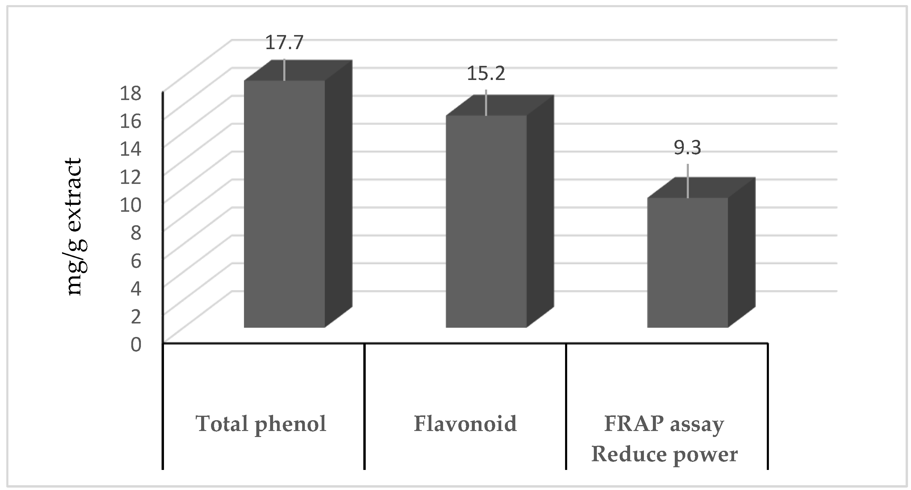
2.1. Antioxidants, Flavonoids, and Phenolics

The chemical composition of the Clitoria ternatea samples, ascertained using an HPLC chromatograph and a GC-TSQ mass spectrometer, is stated in Supplementary Figure S1. An analysis of the antioxidants (ferric-reducing activity), total flavonoid content (TFC), and total phenolic content (TPC) of the Clitoria ternatea extract is demonstrated in Figure 1. In the used ethanolic extract, the TPC was 17.7 mg/g and the TFC was 15.5 mg/g, accompanied by antioxidants of 9.3 mg/kg.

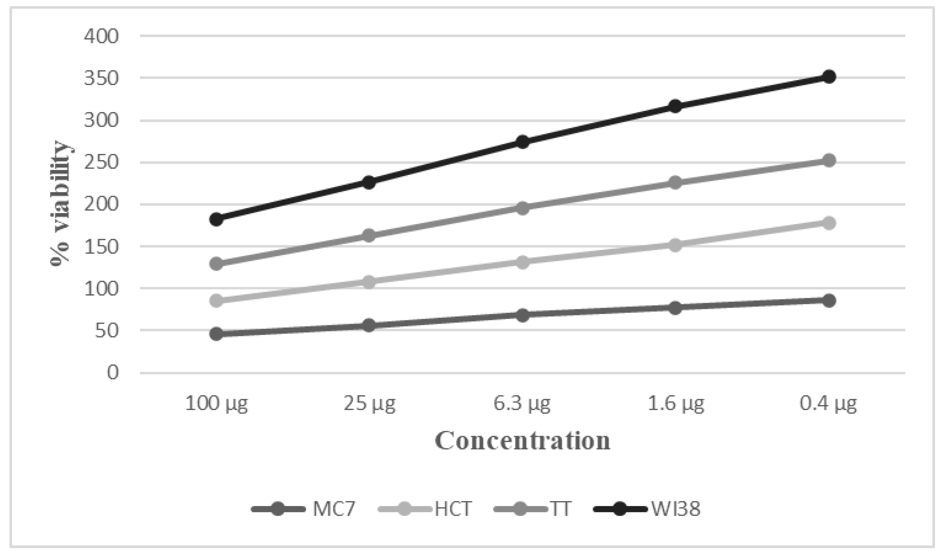
2.2. Cytotoxic Effects

Using an MTT assay, the cytotoxic effect of the Clitoria ternatea extract on the development of the utilized cell lines was assessed. A dose-reliant response was observed for the plant extract and staurosporine between 0.4 and 100 µg/mL, respectively (the standard control); the count of the viable cells declined with raising concentrations of both substances (Figure 2). An estimation of the IC50 value was conducted (Table 1).
IC50 values were applied to characterize the cell susceptibility to the Clitoria ternatea extract (Table 1 and Figure 2). HCT116 had the lowest IC50 value for Clitoria ternatea (29.2 µg/mL) compared with the MCF-7 and TT cell lines (55.7 µg/mL and 61.6 µg/mL, respectively).

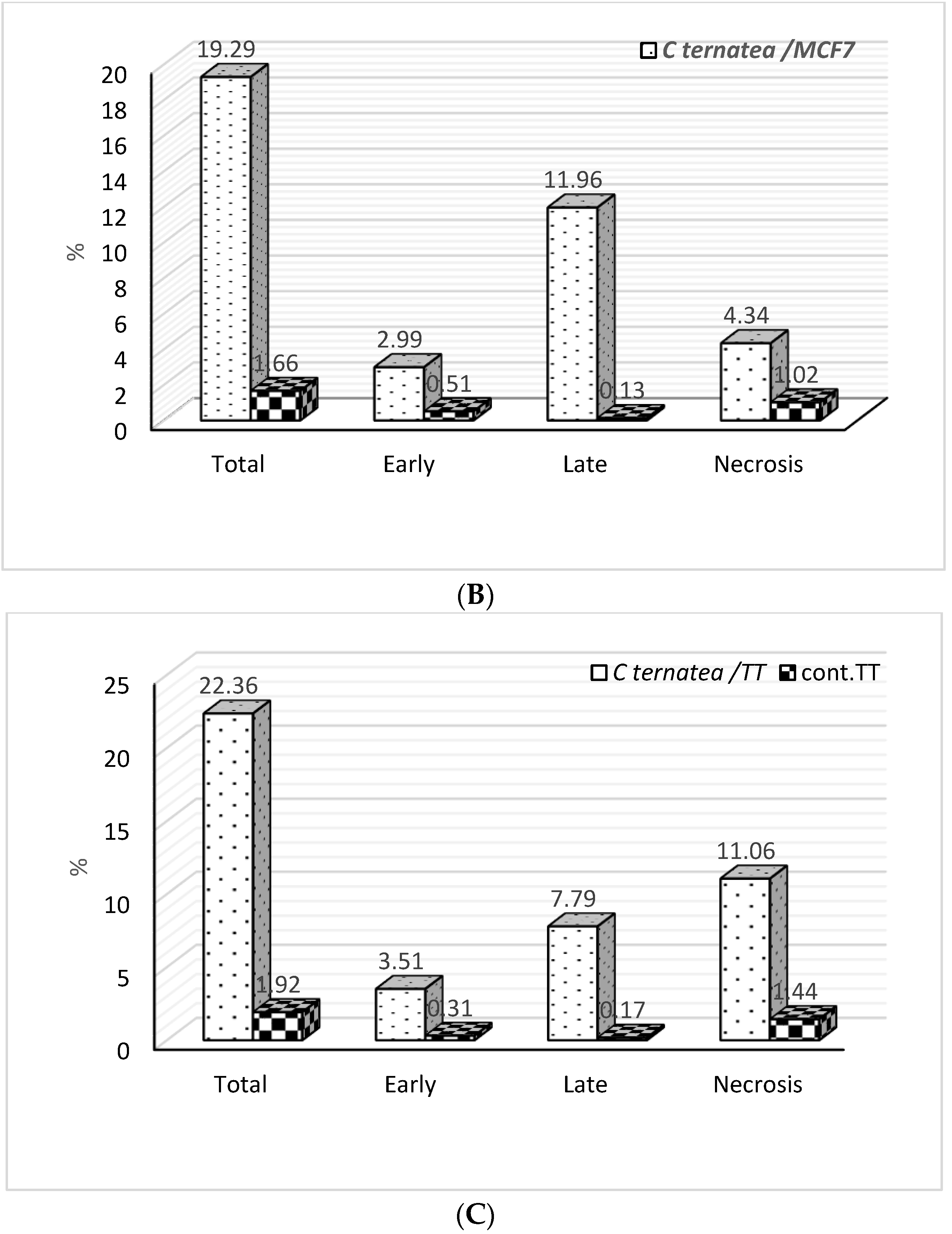
2.3. Apoptosis Detection

The potential of the extract to stimulate apoptosis in cancer cells was evaluated using the IC50 concentration. The cells were preserved for 48 hours with the IC50 (Figure 3 and Figure 4). The MCF-7, HCT116, and TT cell lines undertook a late apoptosis and mild necrosis at IC50 concentrations. The highest percentage of induced apoptosis due to a Clitoria ternatea treatment was detected with the HCT116 cells (24.51%); this was followed by 22.36% with the TT cells and finally 19.29% with the MCF-7 cells. Moreover, late apoptosis events were more increased than early apoptotic events with all cancer cell lines. The apoptotic and necrotic cell populations significantly increased in the HCT116, TT, and MCF-7 cells due to the treatment with the ethanol extracts of Clitoria ternatea when compared with the control. However, the apoptosis percent was significantly increased compared with necrosis in all treatments of the utilized cancer cells.

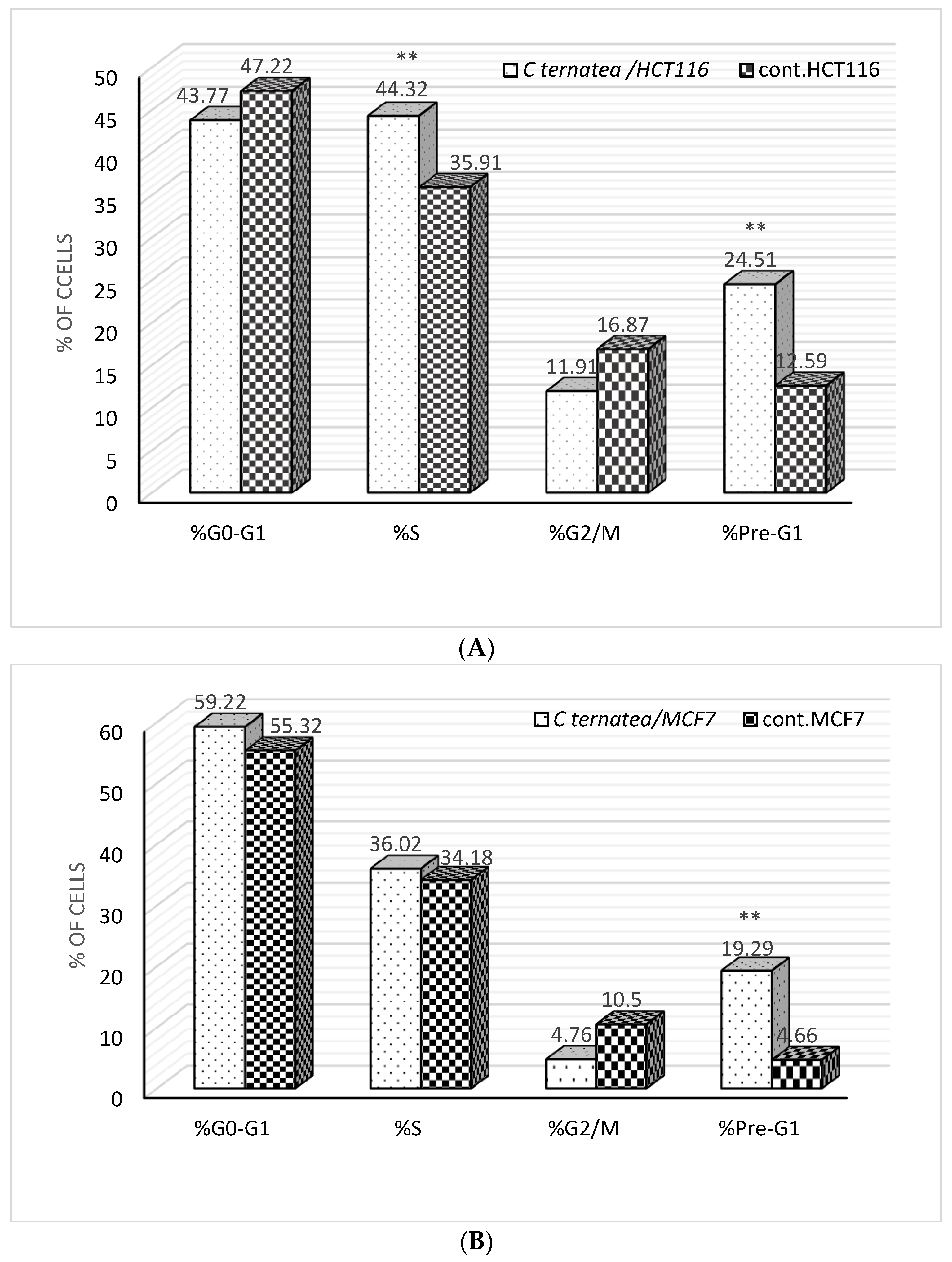
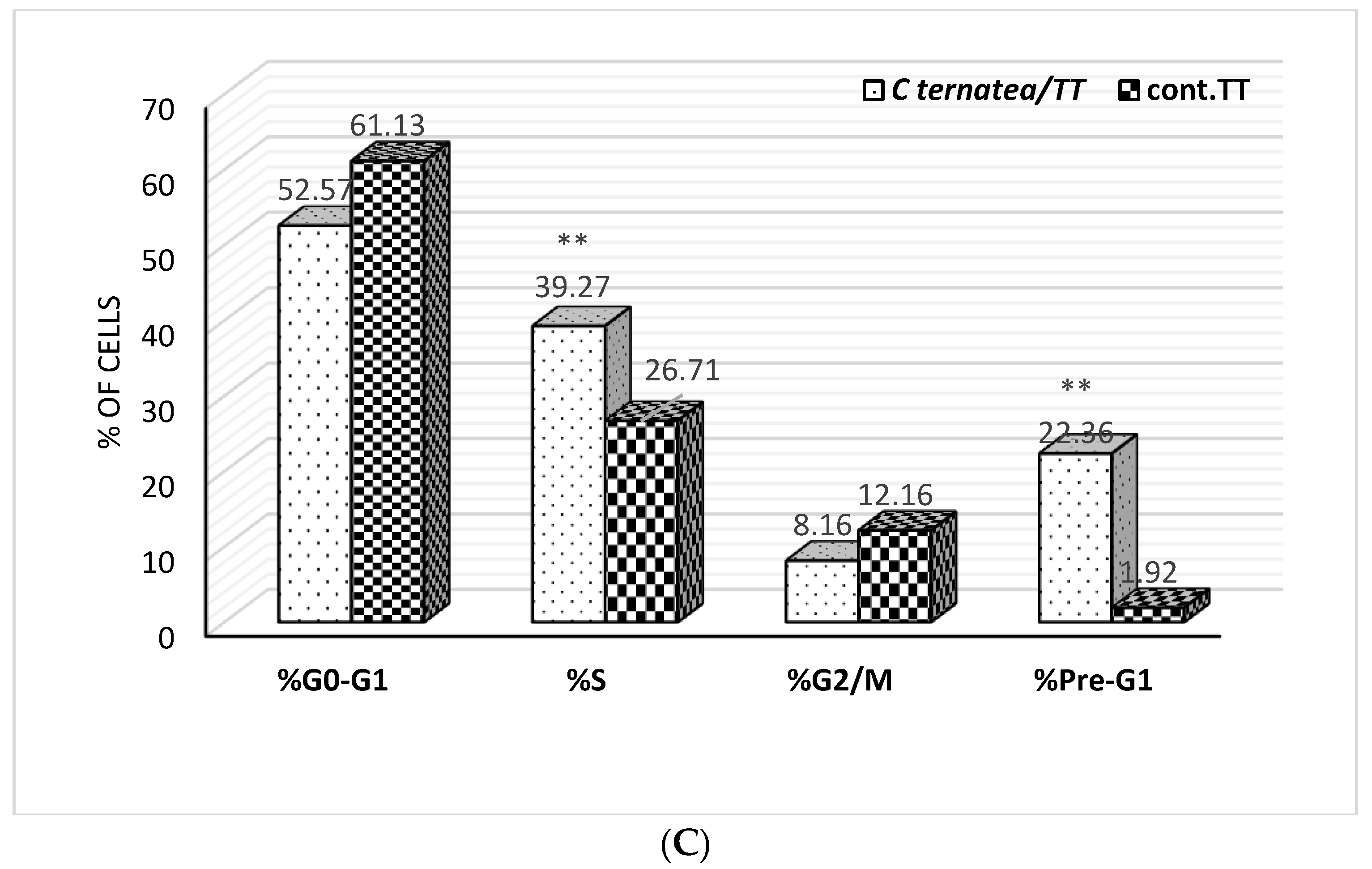
2.4. Cell Cycle Analysis

The influence of the C. ternatea extract on the cell cycle progression of the HCT116, MCF-7, and TT cell lines was determined. The findings indicated that the treatments induced cell cycle detention in the pre-G1 and S stages for the HCT116 and TT cell lines, and in the pre-G1 stage for the MCF-7 cell line (Figure 5). The treatment of the HCT116 and TT cells with the C. ternatea extract induced a statistically significant greater percentage of cells in the S and pre-G1 cell cycle phases and only in the pre-G1 phase with the MCF-7 cells in comparison with the control group.

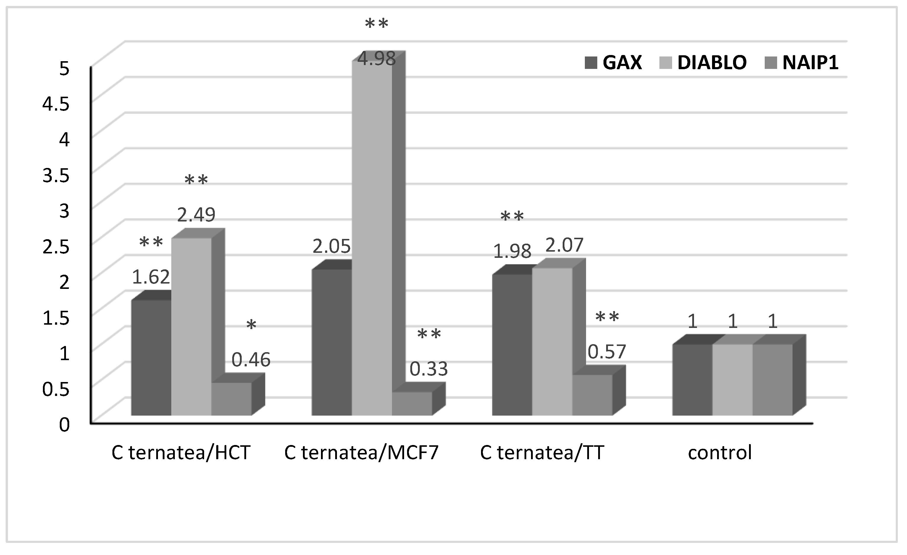
2.5. Gene Expression Analysis

Among the HCT116, MCF-7, and TT cells, the expression level of GAX, DIABLO, and NAIP1 genes was studied in response to the treatment with the plant extract and compared with the untreated cells (control). The results exhibited that, due to the treatment, both the GAX and DIABLO genes were upregulated whilst the NAIP1 gene was significantly downregulated (Figure 6).
The GAX mRNA level was highly significantly increased (p-value ≤ 0.01) due to the treatment with the extract among the HCT116 and TT cells; however, no significant increment was observed with MCF-7. The DIALBO gene expression level was highly significantly upregulated with the HCT116 and MCF-7 cells, with no significance in the case of the TT cells. In contrast, the NAIP1 gene expression results exhibited a highly significant downregulation with the HCT116 and MCF-7 cells as well as a significant (p-value ≤ 0.05) downregulation with the TT cells.

3. Discussion

Clitoria ternatea is a perennial leguminous Ayurvedic medicinal plant that has been used as a traditional medicine in China. Extracts of C. ternatea roots, seeds, leaves, and flowers have been used for diverse indications, including memory-enhancing, anxiolytic, nootropic, antistress, antidepressant, and anticonvulsant therapies to name a few. Recently, C. ternatea was documented to have antioxidant, apoptotic, and antiproliferative activities, which render the plant a promising anticancer therapy.
The antioxidant activity of C. ternatea was determined by the presence of an abundance of polyphenolic and flavonoid contents. Phenolics are the largest group of phytochemicals that account for most of the antioxidant activity in plants and their extracts. Flavonoids are the largest group of naturally occurring phenolic compounds, which are present in a free state and as glycosides. Several studies have documented the abundance of flavonoids and phenolics with a significant antioxidant activity and high IC50 values [42]. When an optimum extraction condition of a C. ternatea flower based on response surface methodology was applied to determine the total phenolic and flavonoid contents, it was concluded that the extractable phenolic and flavonoid compounds implied that C. ternatea was an excellent natural antioxidant source [43]. In accordance with the literature, the current study detected the phenolic and flavonoid compounds with a proven antioxidant activity.
In addition, C. ternatea (flower and leaf) was found to have a cytotoxic activity with a specific predilection toward breast cancer cell line MCF-7 and MDA-MB-231 with IC50 values of 110 and 490 mg/mL, respectively [44]. The potential cytotoxic capacity demonstrated by C. ternatea extracts has encouraged suggestions to incorporate C. ternatea as an anticancer drug [45,46]. In the current study, the IC50 values detected for the HCT116, MCF-7, and TT cell lines represented a moderate cytotoxicity according to the National Cancer Institute (NCI), the standard of cytotoxicity in the USA [47]. However, the IC50 values were much higher than those for the normal cell line WI38. Therefore, the cytotoxic and anticancer properties of C. ternatea showed a considerable predilection toward cancer cells rather than normal cells.
Apoptosis has been investigated in several studies using extracts from different parts of the C. ternatea plant. A recent study investigating an ethyl acetate fraction of the C. ternatea flower failed to detect apoptosis in zebrafish models [48]. However, an investigation into the hydrophilic extract of C. ternatea seeds and petals showed a reduced laryngeal cancer cell (Hep-2) viability, down to 7.2% and 17.2%, respectively. Phenolic, flavonoids and ternatin compounds were detected in the seeds and petal extracts. Both α- and γ-tocopherols were found in the seeds and petals; the level of γ-tocopherol was abundant in the seeds rather than in the petals [49]. A recent study conducted on an MCF-7 HER2-positive breast cancer cell line found that using a C. ternatea crude flower extract below the IC50 (detected to be 862 ug/mL) showed a 50% reduction in the migration activity of the MCF-7 HER2-positive breast cancer cell line. Therefore, it was concluded that C. ternatea (Butterfly pea) was effective in holding the metastatic activity of the breast cancer cell line in vitro [50]. In line with Asysyifa et al. [50], our study showed that a late apoptosis and mild necrosis occurred in the MCF-7, HVT116, and TT cell lines at IC50 concentrations. Therefore, the anticancer activity of C. ternatea enhanced apoptosis.
To recognize the mechanisms of the documented cytotoxicity of the C. ternatea plant, in-depth studies were needed at molecular and cellular levels. The molecular level revealed the antioxidant and apoptotic effects of the C. ternatea extract. At the cellular level, the C. ternatea extract exhibited great potential in arresting the cell cycle at different phases of cell division, including the pre-G1 and S phases. The phytochemical compounds in the ethanolic extract of the plant parts were documented to have an antiproliferative activity and a signal transduction modulation where the flavonoids could arrest cell growth in the G1 phase in human gastric carcinoma cells [51]. These phytochemical compounds, the phenolics, and flavonoids are considered to be apoptosis-modulating agents that stimulate the release of apoptosis modifiers into the cytosol, impacting the mitochondrial functionality and enhancing the translocation of apoptosis-modulating compounds into the nucleus to act on the DNA [52,53]. Therefore, the mechanism of action of flavonoids and phenols can be explained by the apoptosis and induction of cell cycle arrest at different phases, including the S/G2, G1, and S phases [54]. In line with the literature, C. ternatea petal flower extract was reported to enhance cell cycle arrest by an inversion of the cycle from the G2/M to the G0/G1 phases through impacting the mRNA gene expression [55]. In accordance with the literature, the results of the current study showed that C. ternatea extract was able to induce cell cycle arrest in the pre-G1 and S stages of the HCT116 and TT cell lines and in the pre-G1 phase for the MCF-7 cell line. These results support the anticancer activity of the C. ternatea extract.
The growth arrest-specific homeobox (GAX) is a nuclear transcription inhibitor gene found in abundant levels in quiescent cells. The GAX gene function is the inhibition of the proliferation of vascular endothelial cells and smooth muscle cells. Recently, the GAX gene showed the ability to disrupt the signaling pathway controlling adventitial fibroblast activities, thus blocking angiogenesis [56]. A recent study concluded that GAS plays a role in lung cancer angiogenesis and, hence, cancer cell progression [57]. Therefore, GAX is considered to be a promising target for lung cancer treatments. Moreover, the DIABLO gene encodes the Smac/DIABLO proapoptotic protein, which is released from the mitochondria and inactivates the inhibitor of apoptosis proteins (IPAs) [58,59]. An overexpression of the DIABLO gene was found to inhibit prostate cancer (PC3) cell lines [60]. An earlier study showed that DIABLO inhibits clonogenic cancer colon growth by suppressing migration and proliferation and augmenting apoptosis. DIABLO was proposed to enhance cytochrome c-dependent caspase activity by releasing IAP inhibition [58]. A low level of DIABLO expression was observed in bladder, renal, and estrogen-negative lung cancers, which inferred a poor prognosis [61].
On the other hand, NLR family apoptosis inhibitory protein (NAIP) is an apoptosis inhibitor that promotes cancer cell growth. NAIP is targeted by microRNAs (miRs), including miR-1 and miR-145. Therefore, the oncogenic activity of miRs in colon cancer can be attributed to the suppression of NAIP [62]. NAIP has been reported to be implicated in other types of cancer, including breast cancer, melanomas, gliomas, and colorectal cancer [63].
Concurrent with the literature, this study found that GAX (an inhibitor of the proliferation of the fibroblast) and DIABLO (an IPA inhibitor) were upregulated and NAIP1 (an apoptosis inhibitor) was downregulated. Consequently, the C. ternatea extract has the potential to be an anticancer agent by inhibiting cell proliferation and promoting the apoptosis of cancer cells.

4. Materials and Methods

4.1. Plant Materials

The plant samples were gathered and dried in the Aljouf region of Saudi Arabia. The dried plant samples were pulverized into a powder. The institutional botanist (Ahmed Elbanhawy) identified the plants and confirmed them to be Clitoria ternatea.

4.2. Research Design and Statistical Analysis

The cancer cells were cured with various levels of Clitoria ternatea extract to determine the IC50 compared with untreated cells to investigate the antiproliferative activity of the plant extract. The IC50 method was applied to examine the ability of the plant extract to stimulate cell cycle arrest, apoptosis, and gene expression alterations in the untreated and treated cells. The significance of variations between the untreated and treated cells was determined using a Student’s t-test. SPSS version 22.0 was utilized to perform the statistical analysis (IBM, Chicago, IL, USA). Statistically, p-values of ≤0.05 and ≤0.01 were considered to be significant and highly significant, respectively.

4.3. Ethanolic Extraction

The Soxhlet method for extraction was applied to formulate the ethanolic plant extract from the dry plant material. In a rotary evaporator, the evaporation to dryness and the concentration of the ethanolic extracts were obtained at 40 to 50 °C under pressure. The extracts were gathered and kept in airtight dark containers until applied [64]. All subsequent examinations were conducted in triplicate.

4.4. Antioxidant, Phenolic, and Flavonoid Influences of the Plant Extract

A gas chromatography–mass spectrometry (GC-MS) analysis, the total flavonoid content (TFC), and the total phenolic content (TPC) were determined in the ethanolic extracts by employing a colorimetric check, based on the methods defined by Singleton et al. [65] and Chang et al. [66]), respectively. The gallic acid equivalent mg/g (GAE) represented the total content of the phenolic. The total flavonoid content (mg/g) was identified by means of a quercetin calibration curve and presented as mg equivalents of quercetin. The antioxidant activity of the collected extracts was quantified using the approach described by Oyaizu et al. [67] with minor modifications, as implemented by Puranik et al. [68]. Briefly, various concentrations (100–500 L) of methanolic extracts were combined with 0.2 mM PBS (2.5 mL, pH 7.4) and 1% potassium ferricyanide (2.5 mL). This mixture was then kept for 20 minutes at 500 °C. After adding 2.5 mL (w/v) of 10% trichloroacetic acid, 2.5 mL of distilled water, and 0.5 mL of 0.1% ferrous chloride (w/v), the mixture was centrifugated at 3500 rpm for 8 minutes. The absorbance at 700 nm was determined and ascorbic acid was employed as a reference standard.

4.5. Cell Cultures and Lines

Colon (HCT116), breast (MCF-7), and TT (medullary thyroid) cell lines of cancer, in addition to the normal cell line WI38, were utilized. Egypt’s VACSERA was the source of the utilized cell lines. The cells were cultured in a Gibco RPMI BRL 1640 medium (Thermo Fisher Scientific, Carlsbad, CA, USA). The medium was augmented with 1% penicillin–streptomycin (Invitrogen, Grand Island, NY, USA), and FBS (Sijixin Inc., Dalian, China) and kept at 37 °C in a CO2 incubator comprising 5% CO2.

4.6. In Vitro Cytotoxicity Examination

The cytotoxicity was examined by seeding 1 × 10⁵ cells mL−1 (100 µg/well) into 96-well tissue culture plates; these were incubated for 24 h at 37 °C to generate a full monolayer. Each cell line was incubated at 37 °C with and without ethanol extracts at various concentrations ranging from 0.4 to 100 µg/mL. After an incubation of 72 h, the cytotoxicity was evaluated employing the MTT test, as defined in [69].

4.7. Apoptosis Detection

A staining of Annexin V-fluorescein isothiocyanate (FITC) and propidium-iodide (PI) was applied to distinguish the apoptotic cells following the medication of the cancer cells with the C. ternatea extract IC50 level compared with the untreated cells.

4.8. Cell Cycle Analysis

Flow cytometry with a BD FACS Array Bioanalyzer was used to analyze the plant extract-treated and untreated cells for the phase of the arrested cell cycle.

4.9. Expression Level Detection of GAX, DIABLO, and NAIP1 Genes

Comparing the untreated and IC50-treated HCT116, MCF-7, and TT cells, variations in the mRNA of the apoptosis-associated genes GAX, DIABLO, and NAIP1 were explored using a quantitative polymerase chain reaction (qPCR). For 48 h, the cancer cells were cured with IC50 applications. The total RNA was extracted following the instructions from the triazole reagent manufacturer.

4.10. Real-Time Quantitative PCR (qPCR)

A total of 1 µg of RNA was reverse transcripted to the cDNA first strand. As previously reported [70], the achieved cDNA was expanded in order to assess the expressions of the GAX, DIABLO, and NAIP1 genes. The DIABLO gene was examined using the DIABLO Human qPCR Primer Pair (NM 019887) of OriGene Technologies. β-actin was used as a benchmark for the RT-PCR reaction as an internal control. R 5′-ACAGGATGTCAAAACTGCCC-3′ and F 5′-GTGACATCCACACCCAGAGG-3′ were the primers used to amplify β-actin. A quantitative real-time PCR (qRT-PCR) was employed to measure the mRNA levels of GAX, DIABLO, NAIP1, and β-actin. To specify the amplification, the PCR products for the individual primer pairs were obtained using a melting curve analysis. The obtained data were evaluated using the2(-Delta Delta C(T)) (2CT) method. The findings were explained as a fold change (RFC) in comparison with the negative control.

5. Conclusions

In conclusion, C. ternatea root extract is a promising anticancer agent with antioxidant, apoptotic, and cell cycle arrest activities. The abundance of flavonoids and phenolics can be attributed to the anticancer properties of C. ternatea. Moreover, C. ternatea extract acts at the gene level by upregulating the GAX and DIABLO genes, thus suppressing fibroblast angiogenesis and enhancing cancer cell apoptosis, respectively. On the other hand, C. ternatea extracts downregulate NAIP1, unleashing apoptosis in cancer cell lines. The phytochemical compounds yielded by the extract have been documented to be implicated in the anticancer property of C. ternatea. Future research may be directed to optimize the conditions of C. ternatea extracts for use as an anticancer agent.

Supplementary Materials

The following supporting information can be downloaded at: https://www.mdpi.com/article/10.3390/separations9110331/s1, Figure S1: The chemical composition of Clitoria ternatea. (A) HPLC Chromatograph Report. (B) GC-TSQ mass spectrometer.

Author Contributions

Conceptualization, S.M.A., F.A.S., M.A.M., R.M.S. and N.S.A.; methodology, S.M.A., F.A.S., M.A.M., R.M.S. and N.S.A.; software, M.A.M., R.M.S., A.A. and D.S.A.; validation, S.M.A., F.A.S., M.A.M., R.M.S., A.A., D.S.A. and N.S.A.; formal analysis, S.M.A., F.A.S., M.A.M. and N.S.A.; investigation, R.M.S., A.A., D.S.A. and N.S.A.; resources, S.M.A., F.A.S., R.M.S., A.A., D.S.A. and N.S.A.; data curation, S.M.A., F.A.S., M.A.M., R.M.S., A.A., D.S.A. and N.S.A.; writing—original draft preparation, M.A.M., R.M.S., A.A. and N.S.A.; writing—review and editing, S.M.A., F.A.S., M.A.M., R.M.S., A.A., D.S.A. and N.S.A.; visualization, R.M.S., A.A. and D.S.A.; supervision, R.M.S., A.A., D.S.A. and N.S.A.; project administration, S.M.A., F.A.S., M.A.M. and N.S.A.; funding acquisition, S.M.A. and F.A.S. All authors have read and agreed to the published version of the manuscript.

Funding

This research was funded by Princess Nourah bint Abdulrahman University Researchers Supporting Project, number PNURSP2022R318, Princess Nourah bint Abdulrahman University, Riyadh, Saudi Arabia.

Informed Consent Statement

Not applicable.

Data Availability Statement

Not applicable.

Acknowledgments

We want to thank to Princess Nourah bint Abdulrahman University Researchers Supporting Project, number PNURSP2022R318, Princess Nourah bint Abdulrahman University, Riyadh, Saudi Arabia. Also we want to extend out thanks to Jouf University, Saudi Arabia and Aswan University, Egypt for their support during the practical work.

Conflicts of Interest

The authors declare no conflict of interest.

References

  1. Bray, F.; Laversanne, M.; Weiderpass, E.; Soerjomataram, I. The Ever-Increasing Importance of Cancer as a Leading Cause of Premature Death Worldwide. Cancer 2021, 127, 3029–3030. [Google Scholar] [CrossRef] [PubMed]
  2. GBD 2019 Adolescent Young Adult Cancer Collaborators. The Global Burden of Adolescent and Young Adult Cancer in 2019: A Systematic Analysis for the Global Burden of Disease Study 2019. Lancet Oncol. 2022, 23, 27–52. [Google Scholar] [CrossRef]
  3. GBD 2019 Stroke Collaborators. Global, Regional, and National Burden of Stroke and Its Risk Factors, 1990-2019: A Systematic Analysis for the Global Burden of Disease Study 2019. Lancet Neurol. 2021, 20, 795–820. [Google Scholar] [CrossRef]
  4. Sung, H.; Ferlay, J.; Siegel, R.L.; Laversanne, M.; Soerjomataram, I.; Jemal, A.; Bray, F. Global Cancer Statistics 2020: GLOBOCAN Estimates of Incidence and Mortality Worldwide for 36 Cancers in 185 Countries. CA A Cancer J. Clin. 2021, 71, 209–249. [Google Scholar] [CrossRef]
  5. World Health Organization (WHO). Global Health Estimates: Leading Causes of Death. Available online: https://www.who.int/data/gho/data/themes/mortality-and-global-health-estimates/ghe-leading-causes-of-death (accessed on 22 June 2022).
  6. Afifi, A.M.; Saad, A.M.; Al-Husseini, M.J.; Elmehrath, A.O.; Northfelt, D.W.; Sonbol, M.B. Causes of Death after Breast Cancer Diagnosis: A US Population-Based Analysis. Cancer 2020, 126, 1559–1567. [Google Scholar] [CrossRef]
  7. Rahib, L.; Wehner, M.R.; Matrisian, L.M.; Nead, K.T. Estimated Projection of US Cancer Incidence and Death to 2040. JAMA Netw. Open 2021, 4, e214708. [Google Scholar] [CrossRef]
  8. Kaspers, G.J.L. 20 Years of Expert Review of Anticancer Therapy. Expert Rev. Anticancer Ther. 2022, 22, 1–2. [Google Scholar] [CrossRef]
  9. Renzi, S.; Michaeli, O.; Ramaswamy, V.; Huang, A.; Stephens, D.; Maguire, B.; Tabori, U.; Bouffet, E.; Bartels, U. Causes of Death in Pediatric Neuro-Oncology: The Sickkids Experience from 2000 to 2017. J. Neurooncol. 2020, 149, 181–189. [Google Scholar] [CrossRef]
  10. van Niekerk, C.C.; Groenewoud, H.M.M.; Verbeek, A.L.M. Trends and Projections in Cutaneous Melanoma Death in the Netherlands from 1950 to 2045. Medicine 2021, 100, e27784. [Google Scholar] [CrossRef]
  11. Chen, X.; Zeh, H.J.; Kang, R.; Kroemer, G.; Tang, D. Cell Death in Pancreatic Cancer: From Pathogenesis to Therapy. Nat. Rev. Gastroenterol. Hepatol. 2021, 18, 804–823. [Google Scholar] [CrossRef]
  12. Wu, G.X.; Ituarte, P.H.G.; Ferrell, B.; Sun, V.; Erhunmwunsee, L.; Raz, D.J.; Kim, J.Y. Causes of Death and Hospitalization in Long-Term Lung Cancer Survivors: A Population-Based Appraisal. Clin. Lung Cancer 2020, 21, 204–213. [Google Scholar] [CrossRef]
  13. Neibart, S.S.; Manne, S.L.; Jabbour, S.K. Quality of Life After Radiotherapy for Rectal and Anal Cancer. Curr. Color. Cancer Rep. 2020, 16, 1–10. [Google Scholar] [CrossRef] [PubMed]
  14. Polanski, J.; Jankowska-Polanska, B.; Rosinczuk, J.; Chabowski, M.; Szymanska-Chabowska, A. Quality of Life of Patients with Lung Cancer. OncoTargets Ther. 2016, 9, 1023–1028. [Google Scholar] [CrossRef] [Green Version]
  15. Suarez-Almazor, M.; Pinnix, C.; Bhoo-Pathy, N.; Lu, Q.; Sedhom, R.; Parikh, R.B. Quality of Life in Cancer Care. Med 2021, 2, 885–888. [Google Scholar] [CrossRef] [PubMed]
  16. Cox, A.; Faithfull, S. “They’re Survivors Physically but We Want Them to Survive Mentally as Well”: Health Care Professionals’ Views on Providing Potential Late Effect Information. Support. Care Cancer 2013, 21, 2491–2497. [Google Scholar] [CrossRef] [Green Version]
  17. McKenna, N.P.; Bews, K.A.; Yost, K.J.; Cima, R.R.; Habermann, E.B. Bowel Dysfunction after Low Anterior Resection for Colorectal Cancer: A Frequent Late Effect of Surgery Infrequently Treated. J. Am. Coll. Surg. 2022, 234, 529–537. [Google Scholar] [CrossRef]
  18. GBD 2013 Mortality and Causes of Death Collaborators. Global, Regional, and National Age-Sex Specific All-Cause and Cause-Specific Mortality for 240 Causes of Death, 1990-2013: A Systematic Analysis for the Global Burden of Disease Study 2013. Lancet 2015, 385, 117–171. [Google Scholar] [CrossRef] [Green Version]
  19. Walli-Attaei, M.; Joseph, P.; Rosengren, A.; Chow, C.K.; Rangarajan, S.; Lear, S.A.; AlHabib, K.F.; Davletov, K.; Dans, A.; Lanas, F.; et al. Variations between Women and Men in Risk Factors, Treatments, Cardiovascular Disease Incidence, and Death in 27 High-Income, Middle-Income, and Low-Income Countries (PURE): A Prospective Cohort Study. Lancet 2020, 396, 97–109. [Google Scholar] [CrossRef]
  20. Mandlik Ingawale, D.S.; Namdeo, A.G. Pharmacological Evaluation of Ashwagandha Highlighting Its Healthcare Claims, Safety, and Toxicity Aspects. J. Diet. Suppl. 2021, 18, 183–226. [Google Scholar] [CrossRef]
  21. Wang, K.; Chen, Q.; Shao, Y.; Yin, S.; Liu, C.; Liu, Y.; Wang, R.; Wang, T.; Qiu, Y.; Yu, H. Anticancer Activities of TCM and Their Active Components against Tumor Metastasis. Biomed. Pharm. 2021, 133, 111044. [Google Scholar] [CrossRef]
  22. Buyel, J.F. Plants as Sources of Natural and Recombinant Anti-Cancer Agents. Biotechnol. Adv. 2018, 36, 506–520. [Google Scholar] [CrossRef] [PubMed]
  23. Illam, S.P.; Kandiyil, S.P.; Raghavamenon, A.C. Targeting Histone Onco- Modifications Using Plant-Derived Products. Curr. Drug Targets 2021, 22, 1317–1331. [Google Scholar] [CrossRef] [PubMed]
  24. Raees, M.A.; Hussain, H.; Al-Rawahi, A.; Csuk, R.; Muhammad, S.A.; Khan, H.Y.; Rehman, N.U.; Abbas, G.; Al-Broumi, M.A.; Green, I.R.; et al. Anti-Proliferative and Computational Studies of Two New Pregnane Glycosides from Desmidorchis Flava. Bioorg. Chem. 2016, 67, 95–104. [Google Scholar] [CrossRef] [PubMed]
  25. Schnekenburger, M.; Dicato, M.; Diederich, M. Plant-Derived Epigenetic Modulators for Cancer Treatment and Prevention. Biotechnol. Adv. 2014, 32, 1123–1132. [Google Scholar] [CrossRef]
  26. Oguis, G.K.; Gilding, E.K.; Jackson, M.A.; Craik, D.J. Butterfly Pea (Clitoria ternatea), a Cyclotide-Bearing Plant With Applications in Agriculture and Medicine. Front. Plant Sci. 2019, 10, 645. [Google Scholar] [CrossRef] [Green Version]
  27. Oguis, G.K.; Gilding, E.K.; Huang, Y.-H.; Poth, A.G.; Jackson, M.A.; Craik, D.J. Insecticidal Diversity of Butterfly Pea (Clitoria ternatea) Accessions. Ind. Crop. Prod. 2020, 147, 112214. [Google Scholar] [CrossRef]
  28. Devi, B.P.; Boominathan, R.; Mandal, S.C. Anti-Inflammatory, Analgesic and Antipyretic Properties of Clitoria ternatea Root. Fitoterapia 2003, 74, 345–349. [Google Scholar] [CrossRef]
  29. Singh, N.K.; Garabadu, D.; Sharma, P.; Shrivastava, S.K.; Mishra, P. Anti-Allergy and Anti-Tussive Activity of Clitoria ternatea L. in Experimental Animals. J. Ethnopharmacol. 2018, 224, 15–26. [Google Scholar] [CrossRef]
  30. Sreekala, S.; Muraleedharan, U.D. Cationic Clitoria ternatea Seed Peptide as a Potential Novel Bioactive Molecule. Protein Pept. Lett. 2021, 28, 1259–1271. [Google Scholar] [CrossRef]
  31. Thell, K.; Hellinger, R.; Sahin, E.; Michenthaler, P.; Gold-Binder, M.; Haider, T.; Kuttke, M.; Liutkevičiūtė, Z.; Göransson, U.; Gründemann, C.; et al. Oral Activity of a Nature-Derived Cyclic Peptide for the Treatment of Multiple Sclerosis. Proc. Natl. Acad. Sci. USA 2016, 113, 3960–3965. [Google Scholar] [CrossRef]
  32. Harada, G.; Onoue, S.; Inoue, C.; Hanada, S.; Katakura, Y. Delphinidin-3-Glucoside Suppresses Lipid Accumulation in HepG2 Cells. Cytotechnology 2018, 70, 1707–1712. [Google Scholar] [CrossRef] [PubMed]
  33. Jacob, L.; Latha, M. Anticancer Activity of Clitoria ternatea Linn. against Dalton’s Lymphoma. Int. J. Pharmacogn. Phytochem. Res. 2012, 4, 207–212. [Google Scholar]
  34. Neda, G.D.; Mohd Salleh, R.; Ong, M.T. Chemical Composition and Anti-Proliferative Properties of Flowers of Clitoria ternatea. Int. Food Res. J. 2013, 20, 1229–1234. [Google Scholar]
  35. Escher, G.B.; Marques, M.B.; do Carmo, M.A.V.; Azevedo, L.; Furtado, M.M.; Sant’Ana, A.S.; da Silva, M.C.; Genovese, M.I.; Wen, M.; Zhang, L.; et al. Clitoria ternatea L. Petal Bioactive Compounds Display Antioxidant, Antihemolytic and Antihypertensive Effects, Inhibit α-Amylase and α-Glucosidase Activities and Reduce Human LDL Cholesterol and DNA Induced Oxidation. Food Res. Int. 2020, 128, 108763. [Google Scholar] [CrossRef]
  36. Fu, X.; Wu, Q.; Wang, J.; Chen, Y.; Zhu, G.; Zhu, Z. Spectral Characteristic, Storage Stability and Antioxidant Properties of Anthocyanin Extracts from Flowers of Butterfly Pea (Clitoria ternatea L.). Molecules 2021, 26, 7000. [Google Scholar] [CrossRef]
  37. López Prado, A.S.; Shen, Y.; Ardoin, R.; Osorio, L.F.; Cardona, J.; Xu, Z.; Prinyawiwatkul, W. Effects of Different Solvents on Total Phenolic and Total Anthocyanin Contents of Clitoria ternatea L. Petal and Their Anti-Cholesterol Oxidation Capabilities. Int. J. Food Sci. Technol. 2019, 54, 424–431. [Google Scholar] [CrossRef]
  38. Mus, A.A.; Goh, L.P.W.; Marbawi, H.; Gansau, J.A. The Biosynthesis and Medicinal Properties of Taraxerol. Biomedicines 2022, 10, 807. [Google Scholar] [CrossRef] [PubMed]
  39. Tan, B.; Shi, H.; Ji, G.; Xie, J. Effects of taraxerol and taraxerol acetate on cell cycle and apoptosis of human gastric epithelial cell line AGS. Zhong Xi Yi Jie He Xue Bao 2011, 9, 638–642. [Google Scholar] [CrossRef] [PubMed]
  40. Csupor-Löffler, B.; Hajdú, Z.; Zupkó, I.; Molnár, J.; Forgo, P.; Vasas, A.; Kele, Z.; Hohmann, J. Antiproliferative Constituents of the Roots of Conyza Canadensis. Planta Med. 2011, 77, 1183–1188. [Google Scholar] [CrossRef] [Green Version]
  41. Swain, S.S.; Rout, K.K.; Chand, P.K. Production of Triterpenoid Anti-Cancer Compound Taraxerol in Agrobacterium-Transformed Root Cultures of Butterfly Pea (Clitoria ternatea L.). Appl. Biochem. Biotechnol. 2012, 168, 487–503. [Google Scholar] [CrossRef]
  42. Lakshan, S.A.T.; Jayanath, N.Y.; Abeysekera, W.P.K.M.; Abeysekera, W.K.S.M. A Commercial Potential Blue Pea (Clitoria ternatea L.) Flower Extract Incorporated Beverage Having Functional Properties. Evid. Based Complement. Altern. Med. 2019, 2019, 2916914. [Google Scholar] [CrossRef] [PubMed] [Green Version]
  43. Jaafar, N.F.; Ramli, M.E.; Mohd Salleh, R. Optimum Extraction Condition of Clitorea ternatea Flower on Antioxidant Activities, Total Phenolic, Total Flavonoid and Total Anthocyanin Contents. Trop Life Sci. Res. 2020, 31, 1–17. [Google Scholar] [CrossRef] [PubMed]
  44. Akter, R.; Uddin, S.J.; Grice, I.D.; Tiralongo, E. Cytotoxic Activity Screening of Bangladeshi Medicinal Plant Extracts. J. Nat. Med. 2014, 68, 246–252. [Google Scholar] [CrossRef]
  45. Deepika, S.; Selvaraj, C.I.; Anbalagan, M. Phytochemical Characterization and Cancer Cell Line Cytotoxicity of Clitoria ternatea. Bangladesh J. Pharmacol. 2018, 13, 349–352. [Google Scholar] [CrossRef]
  46. Zakaria, N.N.A.; Okello, E.J.; Howes, M.-J.; Birch-Machin, M.A.; Bowman, A. In Vitro Protective Effects of an Aqueous Extract of Clitoria ternatea L. Flower against Hydrogen Peroxide-Induced Cytotoxicity and UV-Induced MtDNA Damage in Human Keratinocytes. Phytother. Res. 2018, 32, 1064–1072. [Google Scholar] [CrossRef] [PubMed] [Green Version]
  47. Ramos-Silva, A.; Tavares-Carreón, F.; Figueroa, M.; De la Torre-Zavala, S.; Gastelum-Arellanez, A.; Rodríguez-García, A.; Galán-Wong, L.J.; Avilés-Arnaut, H. Anticancer Potential of Thevetia Peruviana Fruit Methanolic Extract. BMC Complement. Altern. Med. 2017, 17, 241. [Google Scholar] [CrossRef] [Green Version]
  48. Mat Zian, N.F.A.; Swain, P.; Mohd Faudzi, S.M.; Zakaria, N.; Wan Ibrahim, W.N.; Abu Bakar, N.; Shaari, K.; Stanslas, J.; Choi, T.-I.; Kim, C.-H. Mapping Molecular Networks within Clitoria ternatea Linn. against LPS-Induced Neuroinflammation in Microglial Cells, with Molecular Docking and In Vivo Toxicity Assessment in Zebrafish. Pharmaceuticals 2022, 15, 467. [Google Scholar] [CrossRef]
  49. Shen, Y.; Du, L.; Zeng, H.; Zhang, X.; Prinyawiwatkul, W.; Alonso-Marenco, J.; Xu, Z. Butterfly Pea (Clitoria ternatea) Seed and Petal Extracts Decreased HEp-2 Carcinoma Cell Viability. Int. J. Food Sci. Technol. 2016, 51, 1860–1868. [Google Scholar] [CrossRef]
  50. Asysyifa, A.; Agustiningtyas, A.; Nurgina, A.I. Butterfly Pea (Clitoria ternatea Linn.) Flower Extract Prevents MCF-7 HER2-Positive Breast Cancer Cell Metastasis in-Vitro. Ann. Oncol. 2020, 31, S1266. [Google Scholar] [CrossRef]
  51. Weber, G. Down-Regulation of Increased Signal Transduction Capacity in Human Cancer Cells. Adv. Enzym. Regul. 2005, 45, 37–51. [Google Scholar] [CrossRef]
  52. Lee, J.-H.; Jeong, Y.-J.; Lee, S.-W.; Kim, D.; Oh, S.-J.; Lim, H.-S.; Oh, H.-K.; Kim, S.-H.; Kim, W.-J.; Jung, J.-Y. EGCG Induces Apoptosis in Human Laryngeal Epidermoid Carcinoma Hep2 Cells via Mitochondria with the Release of Apoptosis-Inducing Factor and Endonuclease G. Cancer Lett. 2010, 290, 68–75. [Google Scholar] [CrossRef] [PubMed]
  53. Zhao, J.; Blayney, A.; Liu, X.; Gandy, L.; Jin, W.; Yan, L.; Ha, J.-H.; Canning, A.J.; Connelly, M.; Yang, C.; et al. EGCG Binds Intrinsically Disordered N-Terminal Domain of P53 and Disrupts P53-MDM2 Interaction. Nat. Commun. 2021, 12, 986. [Google Scholar] [CrossRef] [PubMed]
  54. Hazafa, A.; Rehman, K.-U.-; Jahan, N.; Jabeen, Z. The Role of Polyphenol (Flavonoids) Compounds in the Treatment of Cancer Cells. Nutr. Cancer 2020, 72, 386–397. [Google Scholar] [CrossRef] [PubMed]
  55. Chayaratanasin, P.; Caobi, A.; Suparpprom, C.; Saenset, S.; Pasukamonset, P.; Suanpairintr, N.; Barbieri, M.A.; Adisakwattana, S. Clitoria ternatea Flower Petal Extract Inhibits Adipogenesis and Lipid Accumulation in 3T3-L1 Preadipocytes by Downregulating Adipogenic Gene Expression. Molecules 2019, 24, 1894. [Google Scholar] [CrossRef] [Green Version]
  56. Liu, P.; Zhang, C.; Feng, J.B.; Zhao, Y.X.; Wang, X.P.; Yang, J.M.; Zhang, M.X.; Wang, X.L.; Zhang, Y. Cross Talk among Smad, MAPK, and Integrin Signaling Pathways Enhances Adventitial Fibroblast Functions Activated by Transforming Growth Factor-Beta1 and Inhibited by Gax. Arter. Thromb. Vasc. Biol. 2008, 28, 725–731. [Google Scholar] [CrossRef] [Green Version]
  57. Wang, W.; Hong, G.; Wang, S.; Gao, W.; Wang, P. Tumor-Derived Exosomal MiRNA-141 Promote Angiogenesis and Malignant Progression of Lung Cancer by Targeting Growth Arrest-Specific Homeobox Gene (GAX). Bioengineered 2021, 12, 821–831. [Google Scholar] [CrossRef]
  58. Anguiano-Hernandez, Y.M.; Chartier, A.; Huerta, S. Smac/DIABLO and Colon Cancer. Anticancer Agents Med. Chem. 2007, 7, 467–473. [Google Scholar] [CrossRef] [PubMed]
  59. Qiu, W.; Zhou, J.; Zhu, G.; Zhao, D.; He, F.; Zhang, J.; Lu, Y.; Yu, T.; Liu, L.; Wang, Y. Sublytic C5b-9 Triggers Glomerular Mesangial Cell Apoptosis via XAF1 Gene Activation Mediated by P300-Dependent IRF-1 Acetylation. Cell Death Dis. 2014, 5, e1176. [Google Scholar] [CrossRef] [Green Version]
  60. Üyetürk, U.; Ocak, Z.; Acar, M.; Özbey, I.; Tekelioğlu, V. The Effects of Salidrosid on DIABLO and XAF1 Gene Expression in PC3 Prostat Cancer Cells. Acta Med. Anatolia 2014, 2, 133. [Google Scholar] [CrossRef]
  61. Espinosa, M.; Lizárraga, F.; Vázquez-Santillán, K.; Hidalgo-Miranda, A.; Piña-Sánchez, P.; Torres, J.; García-Ramírez, R.A.; Maldonado, V.; Melendez-Zajgla, J.; Ceballos-Cancino, G. Coexpression of Smac/DIABLO and Estrogen Receptor in Breast Cancer. Cancer Biomark. 2021, 30, 429–446. [Google Scholar] [CrossRef]
  62. Xu, X.; Wu, X.; Jiang, Q.; Sun, Y.; Liu, H.; Chen, R.; Wu, S. Downregulation of MicroRNA-1 and MicroRNA-145 Contributes Synergistically to the Development of Colon Cancer. Int. J. Mol. Med. 2015, 36, 1630–1638. [Google Scholar] [CrossRef] [PubMed] [Green Version]
  63. Kay, C.; Wang, R.; Kirkby, M.; Man, S.M. Molecular Mechanisms Activating the NAIP-NLRC4 Inflammasome: Implications in Infectious Disease, Autoinflammation, and Cancer. Immunol. Rev. 2020, 297, 67–82. [Google Scholar] [CrossRef] [PubMed]
  64. Mandal, S.C.; Nayak, A.K.; Dhara, A.K. Herbal Biomolecules in Healthcare Applications; Academic Press: London, UK, 2022; ISBN 978-0-323-90080-5. [Google Scholar]
  65. Singleton, V.L.; Rossi, J.A. Colorimetry of Total Phenolics with Phosphomolybdic-Phosphotungstic Acid Reagents. Am. J. Enol. Vitic. 1965, 16, 144–158. [Google Scholar]
  66. Chang, C.-C.; Yang, M.-H.; Wen, H.-M.; Chern, J.-C. Estimation of Total Flavonoid Content in Propolis by Two Complementary Colometric Methods. J. Food Drug Anal. 2020, 10, 3. [Google Scholar] [CrossRef]
  67. Oyaizu, M. Studies on Products of Browning Reaction Prepared from Glucose Amine Products Derived from Bees. J. Pharm. Biomed. Anal. 1986, 41, 1220–1234. [Google Scholar]
  68. Puranik, S.I.; Hiremath, M.; Nerli, R.B.; Ghagane, S.C. Evaluation of in Vitro Antioxidant and Anticancer Activity of Tabernaemontana divaricata Leaf Extracts Against T-24 Human Bladder Cancer Cell Lines. Int. J. Cancer Res. 2018, 14, 100–108. [Google Scholar] [CrossRef] [Green Version]
  69. Khan, S.; Ansari, A.A.; Khan, A.A.; Abdulla, M.; Al-Obaid, O.; Ahmad, R. In Vitro Evaluation of Cytotoxicity, Possible Alteration of Apoptotic Regulatory Proteins, and Antibacterial Activity of Synthesized Copper Oxide Nanoparticles. Colloids Surf. B Biointerfaces 2017, 153, 320–326. [Google Scholar] [CrossRef]
  70. Morón-Calvente, V.; Romero-Pinedo, S.; Toribio-Castelló, S.; Plaza-Díaz, J.; Abadía-Molina, A.C.; Rojas-Barros, D.I.; Beug, S.T.; LaCasse, E.C.; MacKenzie, A.; Korneluk, R.; et al. Inhibitor of Apoptosis Proteins, NAIP, CIAP1 and CIAP2 Expression during Macrophage Differentiation and M1/M2 Polarization. PLoS ONE 2018, 13, e0193643. [Google Scholar] [CrossRef]
Figure 1. Analysis of total phenolic content, total flavonoid content, and their antioxidant capacity of the C. ternatea extract.
Separations 09 00331 g001
Figure 2. IC50 values of Clitoria ternatea ethanolic extract against HCT116, MCF-7, and TT cell lines and staurosporine.
Separations 09 00331 g002
Figure 3. Impacts of IC50 concentrations of C. ternatea on the stimulation of apoptosis along with (A) HCT116, (B) MCF-7, and (C) TT cell lines.
Separations 09 00331 g003aSeparations 09 00331 g003b
Figure 4. IC50 in (A) untreated (1) HCT116, (2) MCF-7, and (3) TT cell lines compared to (B) IC50 of C. ternatea extract induces apoptosis in (1) HCT116, (2) MCF-7, and (3).
Separations 09 00331 g004
Figure 5. Cell cycle analysis of HCT116, MCF-7, and TT cells after treatment by IC50 of C. ternatea extract relative to the untreated control (C). (A) HCT116, (B) MCF-7, and (C) TT cell lines. ** p-value < 0.01 represent highly significant statistical differences.
Separations 09 00331 g005aSeparations 09 00331 g005b
Figure 6. Alterations in the mRNA levels of the GAX, DIABLO, and NAIP1 genes in HCT116, MCF-7, and TT cell lines as a result of medication with the IC50 of C. ternatea extract compared with the control group. The statistically significant variations are indicated by * (p-value ≤ 0.05) and highly significant variations are implied by ** (p-value ≤ 0.01).
Separations 09 00331 g006
Table 1. Inhibition concentration (IC50) of Clitoria ternatea versus HCT116, MCF-7, and TT cell lines.
SerCytotoxicity IC50 µg/mL
CodeMCF-7HCT116TTWI38
1BTL61.6 ± 3.3229.2 ± 1.5755.7 ± 3124 ± 6.67
2Staurosporine12.7 ± 0.686.99 ± 0.387.39 ± 0.430.4 ± 1.64
Publisher’s Note: MDPI stays neutral with regard to jurisdictional claims in published maps and institutional affiliations.

