Article

Two Eco-Friendly Chromatographic Methods Evaluated by GAPI for Simultaneous Determination of the Fluoroquinolones Moxifloxacin, Levofloxacin, and Gemifloxacin in Their Pharmaceutical Products

by
Eman A. Abdel Hameed
1,*,
Zaitona A. Abd El-Naby
2,
Alaa El Gindy
2,
Roshdy E. Saraya
1,
Aisha Nawaf Al balawi
3,
Sawsan A. Zaitone
4,5 and
Gasser M. Khairy
6
1
Department of Pharmaceutical Analytical Chemistry, Faculty of Pharmacy, Port Said University, Port Fouad 42526, Egypt
2
Department of Pharmaceutical Analytical Chemistry, Faculty of Pharmacy, Suez Canal University, Ismailia 41522, Egypt
3
Biology Department, University College of Haqel, University of Tabuk, Tabuk 71491, Saudi Arabia
4
Department of Pharmacology and toxicology, Faculty of Pharmacy, University of Tabuk, Tabuk 71491, Saudi Arabia
5
Department of Pharmacology and toxicology, Faculty of Pharmacy, Suez Canal University, Ismailia 41522, Egypt
6
Department of Chemistry, Faculty of Science, Suez Canal University, Ismailia 41522, Egypt
*
Author to whom correspondence should be addressed.
Separations 2022, 9(11), 330; https://doi.org/10.3390/separations9110330
Submission received: 26 September 2022 / Revised: 13 October 2022 / Accepted: 21 October 2022 / Published: 28 October 2022
(This article belongs to the Special Issue Application of Chromatography in Analytical Chemistry)

Abstract

:
In this paper, novel green HPLC and HPTLC chromatographic methods were developed for the concurrent determination of moxifloxacin, levofloxacin, and gemifloxacin in bulk and pharmaceutical products. The green HPLC method was used on Thermo C18 (4.6 × 250 mm, 5 µm). By mixing ethanol and 20 mM sodium dihydrogen phosphate dihydrate (pH 5) in a ratio of 25:75, v/v, the mobile phase was created using isocratic elution. The flow rate was 1 mLmin−1. The studied antibiotics were separated well within 9.5 min. The green HPTLC method was used on coated HPTLC aluminum sheets with Silica gel 60 F254 using a mobile phase mixture of water: acetone: ammonia (8:1:1, v/v/v). Compact and well-resolved peaks were obtained under chamber-saturation circumstances for the standard fluoroquinolone antibiotics. Both methods were optimized individually, validated by ICH, and assessed using the Green analytical procedure index (GAPI). The methods were applied to pharmaceutical products and compared with the published methods for the determination of each of these antibiotics individually, using Student’s t-test. They can be used by quality-control laboratories in pharmaceutical factories as sensitive eco-friendly methods for the analysis of these drugs and for the detection of cross-contamination during manufacturing processes.

Graphical Abstract

1. Introduction

Quinolones are antibiotics that are widely utilized in both human and veterinary medicine. These medications hinder essential bacterial enzymes (DNA gyrase and topoisomerase IV) that unwind the DNA helix to replicate and transcribe [1]. They have been used as antibiotics for more than five decades owing to their strong potency, excellent absorption, broad spectrum of activity, convenient preparations, high serum concentrations, and low frequency of side effects [1]. First-generation medications, such as nalidixic acid, have low serum concentrations. Second-generation quinolones, such as ciprofloxacin, have greater Gram-negative and systemic activity. Third-generation medications, such as levofloxacin and moxifloxacin, and fourth-generation medications, such as gemifloxacin, have increased activity against Gram-positive bacteria and atypical pathogens. These novel fluoroquinolones have broad-spectrum bactericidal action, good tissue penetration, and favorable safety and tolerability profiles [2]. Levofloxacin hemihydrate, (LV), is (-)-(S)-9-fluoro-2,3-dihydro-3-methyl-10-(4-methyl -1-piperazinyl]-7-oxo-7H-Pyridol [1,2,3-di]-1,4-benzoxazine-6-carboxylic acid hemihydrate [3]. The optical S-(-) isomer of racemic ofloxacin is LV. LV has prolonged activity against Gram-positive bacteria and atypical pathogens [2]. It is used to treat bacterial infections in adults, including those that affect the skin, urinary system, prostate, and upper and lower respiratory tracts. Moxifloxacin hydrochloride (MX) is {1-cyclopropyl-7-[2,8-diazobicyclo (4.3.0) nonane]-6-fluoro- 8-methoxy-1,4 dihydro-4-oxo-3-quinoline carboxylic acid hydrochloride. It is considered a broad-spectrum antibiotic that inhibits cell reproduction by hindering DNA gyrase [3]. It is used to treat lung and sinus infections such as pneumonia, sinusitis, bacterial conjunctivitis, and subsequent infections in chronic bronchitis [3]. Gemifloxacin mesylate (GM) is (R,S)-7-[(4Z)-3- (aminomethyl)-4- (methoxyimino) -1-pyrrolidinyl]-1-cyclopropyl-6-fluoro-1,4-dihydro-4-oxo-1,8-naphthyridine-3- carboxylic acid methane sulfonate [3], which has favorable safety and tolerability characteristics, significant tissue penetration, and broad-spectrum antibacterial action.
It is used to treat mild to moderate pneumonia as well as acute bacterial exacerbations of chronic bronchitis [3]. Common adverse effects of fluoroquinolones include tendon injury and risk of myocardial arrhythmias [4].
Different analytical approaches have been proposed for determining these fluoroquinolones in pure dosage forms and biological fluids. Spectrophotometric methods included determining MX [5,6], MX and GM [7], LV [8], GM with ambroxol or linezolid [9,10], LV with the fluorescence method [11], and MX with LV [12]. Electrochemical methods were also reported for the determination of quinolones [13,14]. The reported HPTLC methods included the determination of MX in tablets [15], MX with other fluoroquinolone antibiotics in industrial wastewater [16], LV with ornidazole or cefpodoxime [17,18] and GM [19,20]; the reported HPLC methods involved the determination of MX [21,22,23], LV [24,25], MX by stability studies [26], LV in urine [27], and GM in pharmaceutical and biological matrices [28,29,30,31,32,33].
These fluoroquinolone antibiotics have relatively similar chemical structures, and distinguishing between them is considered a challenge. Many pharmaceutical factories produce these antibiotics; therefore, there is a real need for rapid, sensitive, and eco-friendly analytical methods for their simultaneous analysis to prevent cross-contamination during manufacturing processes and also for cleaning-validation processes. According to a literature survey, no report was found for the simultaneous determination of these antibiotics in tablet dosage forms by means of HPLC and HPTLC methods.
Reversed-phase HPLC is the analytical method that is most frequently used in the research and manufacturing of pharmaceutical drugs, including the quality control of bulk medications and pharmaceutical formulations. Organic solvents, including acetonitrile, hydrocarbons, and dichloromethane, are combustible, poisonous, and costly to buy and dispose of. In our investigation, ethanol was employed, which is environmentally benign, because greening analytical methodologies have attracted significant concern in the field of pharmaceutical analysis. These methodologies help to decrease negative environmental effects, avoid the use or production of hazardous materials, and improve analyst health safety [34].
In order to improve the resolution of the compounds to be separated and to enable quantitative analysis of the compounds, the analytical technique HPTLC, based on TLC, has been improved. Some of the improvements include the use of higher-quality TLC plates in the stationary phase with finer particle sizes, which allows for better resolution. The separation can be enhanced further by developing the plate many times with multiple development tools. Consequently, HPTLC provides higher resolution and a lower detection limit (LOD) [35].
Therefore, the aim of this study was to create two new methods, HPLC and HPTLC, for the concurrent identification and quantification of these fluoroquinolone antibiotics to assist pharmaceutical factories in detecting and preventing cross-contamination and to have the opportunity to choose any of these two eco-friendly methods. Both methods were optimized, validated, and applied to pharmaceutical products.

2. Experimental

2.1. Instrumentation

A Camag HPTLC system (Muttenz, Switzerland) was employed. It included Linomat 5 sample applicator, HPTLC plate heater III, TLC scanner 3, UV cabinet, 100 μL Hamilton Syringe (Bonaduz, Schweiz), twin trough development chambers (for 10 cm × 10 cm sheets), and winCATS 1.3.4 software, used for analytical purposes. Coated HPTLC aluminum sheets with Silica gel 60 F254 (Darmstadt, Germany) were used as the stationary phase. Mettler Toledo balance (OH, USA) model XP 205 was used for weighing the chemicals and reagents. An Agilent 1220 Infinity LC system (G4294B configuration; Agilent Technologies, Santa Clara, CA, USA) was used, which consisted of a dual-solvent delivery system, an autosampler, and a diode array detector (DAD).

2.2. Chemicals and Reagents

E.G.P.I. (Egyptian group for pharmaceutical industries) company, Egypt, supplied pharmaceutical standards of LV and MX; they were certified to contain 99.9% and 99.8%, respectively. GM was provided by Utopia pharmaceutical company, Egypt. Ethanol was HPLC-grade from Chem-Lab grade; methanol was HPLC-grade (CHROMASOLV®, Seelze, Germany). Acetic acid, ethyl acetate, acetone, ammonia, acetonitrile, tetrahydrofuran (THF), toluene, and hexane were analytical grade. De-ionized water was made using the Millipore water purification system. Sodium dihydrogen orthophosphate dihydrate, phosphoric acid, sodium lauryl sulfate, sodium hydroxide, triethanolamine (TEA), and triethyl amine were analytical grade.

2.3. Pharmaceutical Samples

Advancrib® tablets (batch No. 042) (E.G.P.I, Egypt), 400 mg MX, Monosho® tablets (batch no. 042) (E.G.P.I, Egypt), 400 mg LV, and Quinabiotic® tablets (batch no. 042) (Utopia Pharmaceuticals, Egypt), 320 mg GM, were the tablets used to prepare samples.

2.4. Chromatographic Conditions

2.4.1. For HPLC Method

Thermo C18 column (4.6 × 250 mm, 5 µm) was used for the green HPLC method of analysis. By mixing ethanol and 20 mM sodium dihydrogen phosphate dihydrate (pH was adjusted to 5 using NaOH) in a ratio of 25:75, v/v, the mobile phase was made using isocratic elution. The injection volume was 20 µL, and the flow rate was 1 mL/ min. The detector was set to wavelength at 350 nm. All measurements were made at a temperature of 25 °C.

2.4.2. For HTPLC Method

As bands, sample and standard solutions were put on HPTLC plates. The mobile phase was a tertiary combination of water, acetone, and ammonia (8:1:1 v/v/v). The development chamber was saturated with the mobile phase for 20 min. Prior to sample application, all HPTLC plates were activated for 10 min at 60 °C. Bands of 3 mm length, 8 mm spacing, and 10 mm from the plate’s bottom were used. The HPTLC plate was air-dried and scanned at 260 nm after developing over an 8 cm distance. The scan length and width were adjusted to cover the entire band.

2.5. Standard Solutions and Calibration

2.5.1. For HPLC Method

MX, LV, and GM were each dissolved in ethanol to a concentration of 0.2 mg mL−1 before being stored at 4 °C to create separate stock solutions of the analytes for use in the subsequent creation of the working solutions.
The standard stock solutions were further diluted with ethanol to produce working solutions, which had a concentration range of 0.1–25 µg mL−1 for each drug separately.
The injected volume of 20 µL was performed three times for each concentration and analyzed under the previously reported chromatographic conditions. The values of peak area were plotted versus the concentrations. Linear correlations were discovered.

2.5.2. For HPTLC Method

Stock standard solutions for each drug, 100 μg mL−1, were prepared. In order to achieve final concentration ranges of 0.1 to 3 µg/band for GM and 0.05 to 2 µg/band for LV and MX, various volumes of stock standard solutions ranging from 1 to 30 μL for GM and from 0.5 to 20 μL for LV and MX were spotted on the TLC plates. The plates were created using the mobile phase that was previously described. By relating the integrated area under the peak to the relevant concentrations as µg/band, calibration curves were constructed.

2.6. Analysis of the Studied Antibiotics in Their Dosage Forms by Proposed Methods

Each commercial product’s twenty tablets were weighed and ground separately to create the samples. For each drug, an amount of powder equal to 20 mg was carefully weighed, deposited in a 100 mL volumetric flask, and dissolved in ethanol by means of the ultrasonic bath for 20 min and then cooled to room temperature. With the same solvent, the solution was then diluted to volume before being filtered through 0.45 µm membrane filters. The initial portion of the filtrate was discarded, and the remaining filtrate was utilized to create a stock sample solution. Using ethanol, more dilution was created to achieve the concentration range of 0.1–25.0 µg mL−1 for each drug separately for the HPLC method and concentration range of 0.1–3.0 µg/band for GM, and 0.05–2.00 µg/band for LV and MX, for the HPTLC method. The general procedures for both methods previously investigated have been made and the concentrations of the antibiotics found were calculated.

3. Results and Discussion

3.1. Method Development and Optimization

The goal of this approach was to develop accurate, sensitive, and time-saving LC methods for the simultaneous quantification of these antibiotics in raw materials and tablets. Therefore, the DAD responses of these drugs were studied. The suitable wavelength for the HPLC method that enables effective drug separation with sufficient sensitivity was determined to be 350 nm, and 260 nm was chosen for the HPTLC method for its good separation of these antibiotics with reasonable sensitivity.
Several changes in the mobile phase composition were studied to increase the performance of the chromatographic method. Different aqueous phases were tested for the HPLC method with ethanol and sodium lauryl sulfate, in which LV and GM showed forked peaks, and poor separation was achieved; also, water containing 0.05% triethanolamine was examined: the three antibiotics had broad peaks and poor signal. The most appropriate buffer was sodium dihydrogen phosphate, dehydrated at 20 mM concentration. The pH of the buffer was also studied, and three pH values were examined. The pH 3.0 resulted in peak broadening of the three antibiotics, while pH 7.0 resulted in high retardation to the appearance of the peaks, and pH 5.0 was found to be the most appropriate pH. The effect of ethanol concentration was also changed for examination. It was discovered that decreasing the acetonitrile concentration to less than 15% caused an increase in the retention time of the antibiotic peaks, resulting in peak broadening and excessive tailing, whereas increasing the acetonitrile concentration to more than 35% caused a high overlap between MX and LV. The best separation was obtained by using 25% ethanol. Using isocratic elution, acceptable results were obtained with a mobile phase comprised of ethanol: 20 mM sodium dihydrogen phosphate dihydrate, pH 5 (25:75, v/v). The flow rate was 1 mLmin−1, and the maximum HPLC response was obtained at the wavelength of 350 nm. The studied antibiotics were well separated within 9.5 min.
For the HPTLC method, several mobile phases were tried, including water: ethyl acetate: ammonia (8:1:1, v/v/v), in which LV and GM moved to the same distance away from the baseline; acetone: ammonia (7:3, v/v), ethanol: water: acetone (6:1:1, v/v/v); ethanol: ammonia: acetone (9:1:1, v/v/v); ethanol: water: ammonia: acetone (8:1:1:1, v/v/v/v); methanol: dichloromethane: ethyl acetate (6:3:1, v/v/v); acetone: ammonia: THF: water (6:2:1:1, v/v/v/v); and water: ammonia (9:1, v/v), in which all analytes remained at the baseline. These mobile phases have not achieved separation for the selected drugs. The most appropriate mobile phase was water: acetone: ammonia (8:1:1, v/v/v). The chromatogram was obtained under chamber saturation circumstances for the analysis of standard fluoroquinolone antibiotics and showed symmetrical, compact, and well-resolved peaks with Rf values of 0.55 ± 0.02, 0.75 ± 0.02, 0.83 ± 0.02 for MX, LV, and GM, respectively. The densitometric estimation of antibiotics was performed at 260 nm.
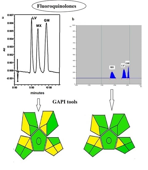
The chromatograms for both methods are shown in Figure 1, where there was a total separation of the investigated fluoroquinolones. Sharp peaks with distinct baseline separation were present. Table 1 demonstrates the system suitability results of both methods.

3.2. The Validation of the Methods

3.2.1. Linearity

In order to evaluate the linearity of the HPLC and HPTLC procedures for determining MX, LV, and GM, a series of various drug concentrations were examined. Seven concentrations were chosen, ranging between 0.1–25.0 µg mL−1 for the HPLC method and 0.1 to 3.0 µg/band for GM and 0.05–2.00 µg/band for LV and MX for the HPTLC method. To provide information about variation in peak area values between samples of the same concentration, each concentration was repeated three times. The linearity of the calibration graphs of the drugs was 0.1–25.0 µg mL−1 for the HPLC method and 0.1–3.0 µg/band for GM and 0.05–2.00 µg/band for LV and MX for the HPTLC method. The high value of the correlation coefficient confirmed validation (Table 2). The lack-of-fit statistical test was employed to check linearities and evaluate the variance of the residual values (Table S1) [36].

3.2.2. Detection and Quantitation limits

The International Conference on Harmonization (ICH) recommendations was followed [37], and the detection and quantitation limits were calculated using the standard deviation (S.D.) of the response and the slope. The theoretical values were evaluated practically and given in Table 2.

3.2.3. Selectivity

By creating eight laboratory-prepared mixes of the studied chemicals at various concentrations within the linearity range, the selectivity of the methods was assessed. Using the previously stated approaches, the laboratory-prepared mixtures were analyzed. The outcomes were satisfactory. Acceptable drug recoveries were from 98.85% to 99.41% for the HPLC approach and from 99.03% to 100.29% for the HPTLC method, suggesting the good selectivity of the developed methods for concurrent determination of MX, LV, and GM (Table S2).

3.2.4. Accuracy

Three marketed pharmaceutical formulations comprising these drugs were exposed to the standard addition procedure. The mean percentage recoveries and standard deviations for the proposed procedures were computed for six replicates. The antibiotic percent recoveries ranged from 99.29% to 99.70% for the HPLC method and from 99.01% to 101.00% for the HPTLC method, indicating good accuracy for these methods (Table S3).

3.2.5. Precision

For each compound, repeatability and intermediate precision were tested at three concentration levels. Through one-way ANOVA, the data were analyzed for each concentration level. An 8-day × 2-replicate design was used (Table S4). The p-value of the F-test was used to compare the results statistically. For each concentration level, three univariate analyses of variance were performed. Because the p-value of the F-test is consistently more than 0.05, no statistically significant difference in mean findings found from one level of day to another was found at the 95% confidence level.

3.3. Analysis of Pharmaceutical Products

The two developed methods were applied to determine LV, MX, and GM in their pharmaceutical products, Monosho®®, Advancrib®®, and Quinabiotic®®, respectively. Seven determinations were carried out, and the results achieved were acceptable for each antibiotic with regard to the labeled amount (Table 3). The chromatograms of the studied fluoroquinolones in pharmaceutical products are shown in Figures S1 and S2. The proposed methods were compared to previously published methods for the determination of each of these fluoroquinolone antibiotics by HPLC and HPTLC using Student’s t-test and F-ratio at a 95% confidence level, and no significant variation in results was found (Table 3). Furthermore, in Table 4, a comparison of figures of merit between the two developed methods and previously published methods is constructed, revealing that both developed methods have nearly the lowest limits of detection for the determination of these fluoroquinolones, in addition to their ability to quantify them simultaneously.

3.4. Evaluation of the Greenness of the Proposed Analytical Methods

There are already several methods available to examine and compare different analytical procedures in terms of their greenness. In comparison to the previous analytical eco-scale, the new Green Analytical procedure index (GAPI) [38,39] has the advantage of spanning the entire analytical technique [40,41]. It is made up of five pentagrams, each representing a step in the analytical procedure, such as sample collection, sample preparation, chemicals and solvents utilized, apparatus, and the aim of the analytical method. GAPI has three color codes, with red representing a high risk to the environment and yellow and green representing lower risk and better greenness.
Figure 2 shows the GAPI index evaluating the proposed methods for the determination of the studied antibiotics. As seen in (Figure 2a), the proposed green HPTLC method has 11 green and 4 yellow pentagrams, and no red color pentagrams appear in its GAPI index; the proposed HPLC method has 7 green and 8 yellow pentagrams, and no red color pentagrams appear in its GAPI index (Figure 2b). The GAPI pentagrams indicate that the suggested methods were green and eco-friendly due to the lack of an extraction stage and the usage of a greener solvent. The HPTLC method was logically greener than the HPLC method due to the use of instruments of lower energy and low solvent consumption and generation.

4. Conclusions

Our work offers two new HPLC and HPTLC methods for the separation and concurrent determination of the fluoroquinolone antibiotics (moxifloxacin HCl, levofloxacin hemihydrate, and gemifloxacin mesylate) in their bulk and pharmaceutical products. The conditions of both techniques, as well as the mobile phase solvents, offered a good resolution for these antibiotics. Both methods were eco-friendly according to GAPI. Furthermore, the developed HPLC method had a low separation time. ICH criteria were used to validate the methods. The methods were accurate, precise, specific, and sensitive, with low limits of detection. The methods were applied to pharmaceutical formulations. They can be safely and efficiently used by quality-control laboratories in pharmaceutical factories as fast, sensitive methods for the analysis of these drugs and for the detection of cross-contamination during manufacturing processes.

Supplementary Materials

The following supporting information can be downloaded at: https://www.mdpi.com/article/10.3390/separations9110330/s1, Table S1: ANOVA (showing lack-of-fit calculation) for the selected antibiotics using the proposed HPLC and HPTLC methods; Table S2: Determination of LV, MX, and GM (mixture) in laboratory-prepared mixtures using the proposed A) HPLC and B) HPTLC methods; Table S3: Application of standard addition technique to the analysis of the studied antibiotics using the proposed HPLC and HPTLC methods; Table S4: Analysis of variance for repeatability and intermediate precision for antibiotic mixture using the proposed HPLC and HPTLC methods; Figure S1: HPLC chromatograms of: a) Monosho ® tablets sample containing 2.5 µg mL−1 of LV, b) Advancrib ® tablets sample containing 2.5 µg mL−1 of MX, and c) Quinabiotic® tablets sample containing 2.5 µg mL−1 of GM using the proposed HPLC method; Figure S2: HPTLC densitogram of: a) Advancrib ® tablets sample containing 0.5 µg/band of MX, b) Monosho® tablets sample containing 0.5 µg/band of LV, and c) Quinabiotic® tablets sample containing 0.6 µg/band of GM using the proposed HPTLC method.

Author Contributions

Conceptualization, A.E.G. and E.A.A.H.; methodology, A.E.G., E.A.A.H. and G.M.K.; software, R.E.S. and Z.A.A.E.-N.; validation, E.A.A.H., A.E.G. and R.E.S.; formal analysis, Z.A.A.E.-N., G.M.K. and E.A.A.H.; investigation, G.M.K., Z.A.A.E.-N. and R.E.S.; resources, Z.A.A.E.-N., A.N.A.b. and S.A.Z.; data curation, R.E.S., S.A.Z. and A.N.A.b.; writing—original draft preparation, E.A.A.H., Z.A.A.E.-N. and G.M.K.; writing—review and editing, S.A.Z. and A.N.A.b.; visualization, A.N.A.b. and S.A.Z.; supervision, A.E.G., E.A.A.H. and G.M.K. All authors have read and agreed to the published version of the manuscript.

Funding

This research was funded by Suez Canal University, grant number 1.

Data Availability Statement

All data are available from the corresponding author upon request.

Acknowledgments

This work was supported by the Graduate Studies Sector at Suez Canal University for the research groups: “Chemo- and bio-sensors Development for Environmental Management Research Group”.

Conflicts of Interest

The authors declare no conflict of interest.

References

  1. Pharm, T.D.M.; Ziora, Z.M.; Blaskovich, M.A.T. Quinolone antibiotics. Med. Chemcomm. 2019, 10, 1719–1739. [Google Scholar]
  2. King, D.; Malone, R.; Lilly, S. New Classification and Update on the Quinolone Antibiotics. Am. Fam. Physician 2000, 61, 2741–2748. [Google Scholar] [PubMed]
  3. Wishart, D.S.; Feunang, Y.D.; Guo, A.C.; Lo, E.J.; Marcu, A.; Grant, J.R.; Sajed, T.; Johnson, D.; Li, C.; Sayeeda, Z.; et al. Drug Bank 5.0: A major update to the Drug Bank database for 2018. Nucleic Acids Res. 2018, 46, 1074–1082. [Google Scholar] [CrossRef] [PubMed]
  4. Abdelrady, A.M.; Zaitone, S.A.; Farag, N.E. Cardiotoxic effect of levofloxacin and ciprofloxacin in rats with/without acute myocardial infarction: Impact on cardiac rhythm and cardiac expression of Kv4.3, Kv1.2 and Nav1.5 channels. Biomed. Pharmacother. 2017, 92, 196–206. [Google Scholar] [CrossRef] [PubMed]
  5. Elbashir, A.; Ebraheem, S.; Elwagee, A.; Aboul-Enein, H. New spectrophotometric methods for the determination of moxifloxacin in pharmaceutical formulations. Acta Chim. Slov. 2013, 60, 159–165. [Google Scholar] [PubMed]
  6. Kamruzzaman, M.; Alam, A.; Lee, S.; Ragupathy, D.; Kim, Y. Spectrofluorimetric study of the interaction between europium (III) and moxifloxacin in micellar solution and its analytical application. Spectrochim. Acta A Mol. Biomol. Spectrosc. 2012, 86, 375–380. [Google Scholar] [CrossRef] [PubMed]
  7. Gouda, A.A.; Amin, A.S.; El-Sheikh, R.; Yousef, A.G. Spectrophotometric Determination of Gemifloxacin Mesylate, Moxifloxacin Hydrochloride and Enrofloxacin in Pharmaceutical Formulations Using Acid Dyes. J. Anal. Methods Chem. 2014, 2014, 286379. [Google Scholar] [CrossRef] [PubMed]
  8. El-Hamshary, M.S.; Fouad, M.A.; Hanafi, R.S.; Al-Easa, H.S.; El-Moghazy, S.M. Screening and optimization of samarium-assisted complexation for the determination of norfloxacin, levofloxacin and lomefloxacin in their corresponding dosage forms employing spectrofluorimetry. Spectrochim. Acta A Mol. Biomol. Spectrosc. 2019, 206, 578–587. [Google Scholar] [CrossRef] [PubMed]
  9. Wankhede, S.B.; Mahajan, A.M.; Chitlange, S.S. Simultaneous spectrophotometric estimation of Gemifloxacin mesylate and Ambroxol hydrochloride in tablets. Pharma Chem. 2011, 3, 269–273. [Google Scholar]
  10. Moussa, B.A.; Mahrouse, M.A.; Hassan, M.A. Spectrofluorimetric determination of gemifloxacin mesylate and linezolid in pharmaceutical formulations: Application of quinone-based fluorophores and enhanced native fluorescence. Acta Pharm. 2014, 64, 15–28. [Google Scholar] [CrossRef] [Green Version]
  11. Ren, Q.; Zhu, X. Methyl-β-Cyclodextrin /Cetyltrimethyl Ammonium Bromide Synergistic Sensitized Fluorescence Method for the Determination of Levofloxacin. J. Fluoresc. 2016, 26, 671–677. [Google Scholar] [CrossRef] [PubMed]
  12. Ocaña, J.A.; Barragá, F.J.; Callejo, M.; De la Rosa, F. Application of Lanthanide-Sensitised Chemiluminescence to the Determination of Levofloxacin, Moxifloxacin and Trovafloxacin in Tablets. Microchim. Acta 2004, 144, 207–213. [Google Scholar] [CrossRef]
  13. Kergaravat, S.V.; Gagneten, A.M.; Hernandez, S.R. Development of an electrochemical method for the detection of quinolones: Application to cladoceran ecotoxicity studies. Microchem. J. 2018, 141, 279–286. [Google Scholar] [CrossRef]
  14. Radi, A.; Wahdan, T.; Anwar, Z.; Mostafa, H. Electrochemical determination of gatifloxacin, moxifloxacin and sparfloxacin fluoroquinolonic antibiotics on glassy carbon electrode in pharmaceutical formulations. Drug Test Anal. 2010, 2, 397–400. [Google Scholar] [CrossRef]
  15. Vandana, D.; Chaudhary, A.K. A Validated HPTLC Method for Estimation of Moxifloxacin Hydrochloride in Tablets. Pharm. Methods 2010, 1, 54–56. [Google Scholar]
  16. Khattab, F.I.; Salem, H.; Riad, S.M. Determination of Fluoroquinolone Antibiotics in Industrial Wastewater by High-Pressure Liquid Chromatography and Thin-Layer Chromatography–Densitometric Methods. J. Planar Chromatogr. 2014, 27, 287–293. [Google Scholar] [CrossRef]
  17. Chepurwar, S.B.; Shirkhedkar, A.A.; Bari, S.B. Validated HPTLC Method for Simultaneous Estimation of Levofloxacin Hemihydrate and Ornidazole in Pharmaceutical Dosage Form. J. Chromatogr. Sci. 2007, 45, 531–536. [Google Scholar] [CrossRef] [Green Version]
  18. Vaidya, H.; Chorawala, H.; Dedania, Z. Development of Validated HPTLC method for simultaneous determination of levofloxacin hemihydrate and cefpodoxime proxetil in synthetic mixture and tablet dosage form. Pharma Sci. Monit. 2014, 5, 29–34. [Google Scholar]
  19. Mahmoud, A.M.; Atia, N.N.; El-Shabouri, S.R. Development and Validation of Stability Indicating HPTLC Assay for Determination of Gemifloxacin Mesylate in Dosage Forms. Am. J. Anal. Chem. 2015, 6, 85–97. [Google Scholar] [CrossRef] [Green Version]
  20. Narayan, U.L.; Garnaik, B.; Patro, S.K. HPTLC Methods for Determination of Gemifloxacin Mesylate in Rabbit Plasma. Br. J. Pharm. Res. 2014, 4, 1707–1714. [Google Scholar] [CrossRef]
  21. Razzaq, S.N.; Ashfaq, M.; Khan, I.U. Simultaneous determination of dexamethasone and moxifloxacin in pharmaceutical formulations using stability indicating HPLC method. Arab. J. Chem. 2017, 10, 321–328. [Google Scholar] [CrossRef]
  22. Maddala, V.L.; Ray, P.C.; Bulusu, L.S. Development and Validation of a RP-HPLC method for fast, sensitive and simultaneous determination of few (eight) active pharmaceutical ingredient residues on stainless steel surface in manufactureing plants. Rasayan J. Chem. 2015, 8, 316–320. [Google Scholar]
  23. Mangukiya, R.P.; Patela, R.A.; Shah, P.A. Chromatographic Methods for Simultaneous Determination of Moxifloxacin Hydrochloride and Difluprednate in Ophthalmic Dosage Form. Acta Chromatogr. 2015, 27, 495–508. [Google Scholar] [CrossRef] [Green Version]
  24. Belal, F.F.; El-Din, M.K.S.; Saad, S. Micellar liquid chromatographic method for the simultaneous determination of Levofloxacin and Ambroxol in combined tablets: Application to biological fluids. Chem. Cent. J. 2013, 7, 162. [Google Scholar] [CrossRef] [PubMed] [Green Version]
  25. Bivha, D.; Sujata, M.; Prabhakar, T. Formulation and analysis of levofloxacin hemihydrate by RP-HPLC in bulk and tablet dosage form. Chemistry 2014, 3, 97594519. [Google Scholar]
  26. Priya, M.V.; Madhavan, P.; Kumar, P. Avalidated RP- HPLC method for the analysis of Moxifloxacin Hydrochloride in pharmaceutical dosage forms and stability studies. J. Chem. Pharm. 2015, 7, 836–841. [Google Scholar]
  27. EL-Gindy, A.; Emara, S.; Mostafa, A. UV Partial Least-Squares Calibration and Liquid Chromatographic Methods for Direct Quantitation of Levofloxacin in Urine. J. AOAC Int. 2007, 90, 1258–1265. [Google Scholar] [PubMed]
  28. Durmusa, Z.; Tekkeib, S.E.K.; Kizitasb, M.V. Magnetic Solid Phase ExtractIon for A New HPLC Method for the DeterminatIon of GemifloxacIn in Human Plasma and Breast Milk. J. Chil. Chem. Soc. 2017, 62, 3483–3489. [Google Scholar] [CrossRef] [Green Version]
  29. Roy, B.; Das, A.; Bhaumik, U.; Sarkar, A.K.; Bose, A.; Mukharjee, J. Determinationof gemifloxacin in different tissues of rat after oral dosing of gemifloxacin mesylate by LC-MS/MS and its application in drug tissue distribution study. J Pharm. Biomed. Anal. 2010, 52, 216–226. [Google Scholar] [CrossRef]
  30. Ranjane, P.N.; Gandhi, S.V.; Kadukar, S.S. Stability Indicating RP-LC Method for the Determination of Gemifloxacin Mesylate. Chromatographia 2010, 71, 1113–1117. [Google Scholar] [CrossRef]
  31. Sagirli, O.; Demirci, S.; Önal, A. A very simple high-performance liquid chromatographic method with fluorescence detection for the determination of gemifloxacin in human breast milk. Luminescence 2015, 30, 1326–1329. [Google Scholar] [CrossRef] [PubMed]
  32. Grunspan, L.D.; Kaiser, M.; Hurtado, F.K.; Costa, T.D.; Tasso, L. HPLC Determination of Gemifloxacin in Different Tissues of Rats Under Normobaric and Hyperbaric Exposure. Chromatographia 2012, 75, 253–262. [Google Scholar] [CrossRef]
  33. Ganapathy, S.; Raju, G.V.H.; Sankar, D.G. LC Determination of Gemifloxacin in Bulk and Pharmaceutical Formulation. Asian J. Chem. 2009, 21, 6121–6129. [Google Scholar]
  34. Yabré, M.; Ferey, L.; Gaudin, K. Greening Reversed-Phase Liquid Chromatography Methods Using Alternative Solvents for Pharmaceutical Analysis. Molecules 2018, 23, 1065. [Google Scholar] [CrossRef] [Green Version]
  35. Attimarad, M.; Ahmed, K.K.M.; Aldhubaib, B.E. High-performance thin layer chromatography: A powerful analytical technique in pharmaceutical drug discovery. Pharm. Methods 2011, 2, 71–75. [Google Scholar] [CrossRef] [Green Version]
  36. Sprent, P.; Draper, N.R.; Smith, H. Applied Regression Analysis. Biometrics 1981, 37, 863. [Google Scholar] [CrossRef]
  37. ICH, Q8(R2): Pharmaceutical development. In Proceedings of the International Conference on Harmonization, August 2009.
  38. Płotka-Wasylka, J. A new tool for the evaluation of the analytical procedure: Green Analytical Procedure. Index. Talanta 2018, 181, 204–209. [Google Scholar] [CrossRef]
  39. Susdorf, K.; Bevanda, A.M.; Talić, S. Green analytical chemistry: Social dimension and teaching. TrAC Trends Anal. Chem. 2019, 111, 185–196. [Google Scholar] [CrossRef]
  40. Gałuszka, A.; Migaszewski, Z.; Namieśnik, J. The 12 principles of green analytical chemistry and the Significance mnemonic of green analytical practices. TrAC Trends Anal. Chem. 2013, 50, 78–84. [Google Scholar] [CrossRef]
  41. Gałuszka, A.; Migaszewski, Z.M.; Konieczka, P. Analytical Eco-Scale for assessing the greenness of analytical procedures. TrAC Trends Anal. Chem. 2012, 37, 61–72. [Google Scholar] [CrossRef]
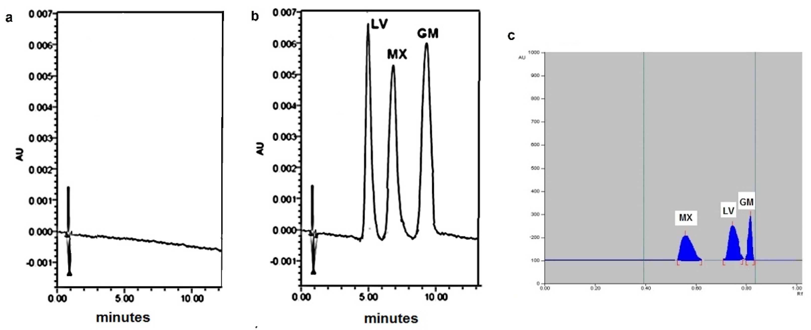
Figure 1. (a) Typical HPLC chromatogram of diluent, (b) Typical HPLC chromatogram of 20 μL injection of prepared mixture containing 5 µg mL−1 of each of LV, MX, and GM using the proposed HPLC method; (c) HPTLC densitogram of prepared mixture containing 1 µg /band of MX, 1 µg/band of LV, and 0.7 µg/band of GM using the proposed HPTLC method.
Separations 09 00330 g001
Figure 2. GAPI pictograms for (a) the proposed green HPTLC method and (b) the proposed green HPLC method.
Separations 09 00330 g002
Table 1. The system suitability test results of the developed (A) HPLC method and (B) HPTLC for determination of the selected antibiotics.
(A) HPLC
CompoundRetention Time
(min) a
%RSD b of Retention TimeCapacity Factor
(K′)
Selectivity
(α) c
Resolution
(Rs) c
Tailing FactorPlate Count
LV4.981.204.471.55 (a1)2.10 (b1)1.101841.02
MX6.861.406.541.48 (a2)2.28 (b2)0.992820.15
GM9.340.449.26 1.204118.54
(B) HPTLC
CompoundRetardation Factor (Rf)Capacity Factor
(K′)
Selectivity
(α) c
Resolution
(Rs) c
Tailing Factor
MX0.550.820.41 (a3)1.60 (b3)1.3
LV0.750.330.61 (a4)1.00 (b4)1.2
GM0.830.21 0.9
a: The retention time of unretained peak is 0.91 min. b: Relative standard deviation. c: a1, b1 are α and Rs calculated for MX-LV for HPLC method; a2, b2 are α and Rs calculated for GM-MX for HPLC method; a3, b3 are α and Rs calculated for LV-MX for HPTLC method; a4, b4 are α and Rs calculated for GM-LV for HPTLC method.
Table 2. Characteristic parameters of the calibration equations for the developed HPLC and HPTLC methods for determination of the selected antibiotics.
HPLC
ParametersLVMXGM
Calibration range (µg mL−1)0.1–25.00.1–25.00.1–25.0
Detection limit (µg mL−1)0.030.040.02
Quantitation limit (µg mL−1)0.090.140.08
Regression equation (Y) a:
Slope (b)
17313.917405.312060.3
Standard deviation of the slope (Sb)210.9305.09890.17
Relative standard deviation slope (%)1.221.751.44
Intercept (a)1069.8471.59290.6
Correlation coefficient (r)0.99990.99980.9999
HPTLC
ParametersLVMXGM
Calibration range (µg /band)0.05–2.000.05–2.000.1–3.0
Detection limit (ng /band)15.0014.4043.92
Quantitation limit (ng /band)50.0148.00146.40
Regression equation (Y) a:
Slope (b)
20.4831.2022.54
Standard deviation of the slope (Sb)0.200.230.30
Intercept (a)−0.936.233.47
Correlation coefficient (r)0.99970.99980.9996
a Y = a + bC, where C is the concentration in μg mL−1 for HPLC method or µg/band for HPTLC method, and Y is the peak area.
Table 3. Determination of GM, LV, and MX in tablet dosage forms using (a) the proposed HPTLC in comparison with reported HPTLC methods and (b) the proposed HPLC in comparison with reported HPLC methods.
(a) HPTLC Method%Recovery a ± SDt-Value bF-Value b
ProposedReported
Quinabiotic ®® tablets (GM)100.88 ± 1.85101.88 ± 0.85 c [19]1.300.98
Monosho ®® tablets(LV)98.60 ± 2.1099.79 ± 0.22 c [17]1.490.98
Advancrib ®® tablets (MX)101.74 ± 0.85101.2 ± 1.66 c [15]0.770.99
(b) HPLC Method%Recovery a ± SD
ProposedReported
Monosho ®® tablets(LV)97.3 ± 1.1098.16 ± 0.46 c [25]1.910.98
Advancrib ®® tablets(MX)100.96 ± 1.90100.65 ± 1.3 c [26]0.360.99
Quinabiotic ®® tablets(GM)98.99 ± 1.0099.20 ± 0.84 c [30]0.431.00
a The values are mean of seven determinations. b The tabulated t- and F-values at 95% confidence limit are 2.18 and 4.28, respectively. c Reported HPTLC and HPLC methods.
Table 4. A comparison of figures of merit between (a) the proposed green HPLC and the previously reported HPLC methods and (b) the proposed green HPTLC and the previously reported HPTLC methods.
(a) HPLC
CompoundMethodLinearity Range
(µg mL−1)
LOD
(µg mL−1)
LOQ
(µg mL−1)
MatrixRef.
LVProposed0.1–25.00.030.09Dosage form
Reported 50.0–150.00.030.09Tablet dosage form[25]
Reported1.0–44.00.260.8Dosage forms and plasma[24]
Reported0.5–16.50.030.09 Urine[27]
MXProposed0.1–25.00.040.14Dosage form
Reported20.0–60.01.85.6Dosage form[26]
Reported50.0–350.00.321.01Dosage form[21]
Reported5.0–15.00.72.1Cleaning validation in factories [22]
Reported1.0–50.00.842.56Industrial wastewater[16]
GMProposed0.1–25.00.020.08Dosage form
Reported5.0–25.00.1710.518Dosage form [30]
Reported0.1–2.50.632.1Human breast milk[31]
Reported0.2–30.00.060.2Rat tissues—lung, liver, and kidney[32]
Reported10.34–82.720.040.13Bulk and pharmaceutical formulation[33]
(b) HPTLC
CompoundMethodLinearity Range
(µg/Band)
LOD
(ng/Band)
LOQ
(ng/Band)
MatrixRef.
GMProposed0.1–3.043.92146.40Dosage form
Reported0.1–0.7 45.00150.00Rabbit plasma[20]
Reported0.002–0.1800.28 0.86Dosage form[19]
LVProposed0.05–2.0014.4048.00Dosage form
Reported0.05–0.250.110.36Dosage forms[17]
Reported0.13–0.75--Tablet dosage form[18]
MXProposed0.05–2.0015.0050.01Dosage form
Reported 9.0–54.0--Dosage form[15]
Reported1.2–2.033.31100.95Ophthalmic Dosage form[23]
Publisher’s Note: MDPI stays neutral with regard to jurisdictional claims in published maps and institutional affiliations.