Article

Effect of 8 Weeks Aerobic Training and Saffron Supplementation on Inflammation and Metabolism in Middle-Aged Obese Women with Type 2 Diabetes Mellitus

1
Department of Exercise Physiology, Faculty of Educational Sciences and Psychology, University of Mohaghegh Ardabili, Ardabil 5619913131, Iran
2
Department of Exercise Physiology, Faculty of Physical Education and Sport Science, University of Tehran, Tehran 141556619, Iran
3
Neuroscience Research Center, Institute of Neuropharmacology, Department of Physiology and Pharmacology, Afzalipour School of Medicine, Kerman University of Medical Sciences, Kerman 7616914115, Iran
4
Student Research Committee, Kerman University of Medical Sciences, Kerman 7619813159, Iran
5
Faculty of Sport Sciences, Waseda University, Tokorozawa 359-1192, Japan
*
Authors to whom correspondence should be addressed.
Sports 2022, 10(11), 167; https://doi.org/10.3390/sports10110167
Submission received: 3 August 2022 / Revised: 4 October 2022 / Accepted: 27 October 2022 / Published: 30 October 2022

Abstract

:
Background: This study aimed to investigate the effects of 8-week aerobic training (AT) and saffron supplementation on inflammation and metabolism in middle-aged obese women with type 2 diabetes mellitus (T2DM). Methods: Thirty-two obese women with T2DM were randomly divided into four groups (n = 8 in all groups): saffron + training (ST), placebo + training (PT), saffron supplementation (SS), and placebo (P). The ST and PT groups performed eight weeks of aerobic training (AT) (three sessions/week at 60–75% HRmax). A daily dose of 400 mg saffron powder was consumed by the ST and SS groups for 8 weeks. Blood samples were taken after 12 h of fasting, 48 h before the first AT session, 48 h and two weeks after the last AT session. Results: AT, saffron supplementation, and their combination affected body mass index (BMI), homeostatic model assessment for insulin resistance (HOMA-IR), and serum levels of insulin, adiponectin, interleukin-6 (IL-6), high-density lipoprotein cholesterol (HDL-C), cholesterol, and triglyceride (TG) (p < 0.05). However, body weight, body fat percentage, and serum levels of glucose, resistin, tumor necrosis factor-alpha (TNF-α), irisin, and low-density lipoprotein cholesterol (LDL-C) showed significant changes in the ST group only (p < 0.05). In addition, a significant difference was seen between all factors in post-training and follow-up in the ST group (p < 0.05). Conclusions: Saffron supplementation at a dose of 400 mg/day, when combined with AT, could improve inflammation, metabolism, glycemic status, and lipid profile in T2DM patients, and these changes are sustainable at up to 2 weeks of detraining.

1. Introduction

Diabetes mellitus (DM) is a global health problem mainly caused by a sedentary lifestyle, poor dietary habits, and obesity [1,2,3]. Obesity can change adipokine secretion and lead to insulin resistance; a close connection has been reported between obesity and type 2 diabetes mellitus (T2DM) [4]. Adiponectin, the most crucial insulin-sensitizing, anti-inflammatory adipokine, and resistin, a newly discovered adipokine directly related to insulin resistance, are the main adipokines influenced by obesity [5,6]. Adiponectin reduces glucose output from the liver by increasing liver sensitivity to insulin [4,6] and increases glucose uptake in the muscles, thus reducing blood glucose [7]. Similar to adiponectin, resistin regulates glucose homeostasis and acts as a physiological antagonist to insulin action in hepatocytes [5].
Another aspect of obesity and T2DM is the increase in the serum levels of inflammatory cytokines such as interleukin-6 (IL-6) and tumor necrosis factor-alpha (TNF-α) [8]. It has been shown that serum levels of IL-6 and TNF-α are correlated with body mass index (BMI) and obesity [4,9]. IL-6 and TNF-α have an inhibitory effect on glucose transporter type-4 (GLUT-4) expression, and their increase leads to insulin resistance [10,11,12]. Furthermore, IL-6 and TNF-α enhance the survival of inflammatory factors such as resistin, reinforcing insulin resistance [11]. Thus, increased resistin and decreased adiponectin secretion, along with increased pro-inflammatory cytokines, such as IL-6 and TNF- α, are the preliminary properties of T2DM [4,11].
Much like adipose tissue, muscles can secrete metabolically active proteins called myokines. Irisin is a myokine produced by the fibronectin type III domain-containing protein-5 (FNDC5) gene, and its levels decrease in T2DM. Irisin stimulates uncoupling protein-1 (UCP1) production, helping white-to-brown adipose tissue conversion. Considering UCP1′s role in controlling blood glucose, insulin sensitivity, mitochondrial density, and fat metabolism [13], brown adipose tissue is thought to prevent obesity and diabetes [14].
Sedentary behavior and physical inactivity are the main risk factors for T2DM. Thus, aerobic training (AT), the most popular training method in the world [15], could be an essential strategy to counter T2DM [16]. Aerobic training reduces the risk of many conditions. These conditions include obesity, heart disease, high blood pressure, T2DM, metabolic syndrome, stroke, and certain types of cancer [16]. It has been reported that AT could increase the number of GLUT-4 [1]. Thus, it can reduce fasting blood glucose and insulin resistance and reduce inflammation by decreasing serum levels of IL-6, TNF-α, and resistin and increasing serum levels of adiponectin and irisin, which ultimately improve insulin sensitivity [1]. However, some studies have reported no change or increase in the serum levels of inflammatory factors after AT [17].
Supplementation is another strategy to fight T2DM [18]. However, studies have reported that taking chemical supplements may lead to unfavorable side effects, and this has shifted researchers’ attention towards using organic supplements with no or fewer side effects [19]. One of the most popular organic supplements with various medical applications is saffron. The scientific name of saffron is Crocus sativus L. from the Iridaceae family. The most important compounds in saffron are aldehydes (picrocrocin and safranal) and flavonoids (crocetin and crocin) [20]. Research has shown that saffron supplementation improves diabetes by activating the AMP-activated protein kinase (AMPK)/GLUT-4 pathway [21]. Furthermore, it modifies inflammation and oxidative stress [22]. However, studies showed that the interaction of saffron supplementation and combined training has more efficient effects on anti-inflammatory status [23] and pulmonary volume and capacities [24] compared to saffron supplementation or training alone.
Based on the promising effects of both AT and saffron supplementation in improving diabetes-induced inflammation and metabolic disorders, we hypothesized that participating in AT along with saffron supplementation could be even more effective. Therefore, this study aimed to investigate the effect of 8-week AT and saffron supplementation on inflammation and metabolism in middle-aged obese women with T2DM.

2. Materials and Methods

This was a quasi-experimental study with repeated measures design: 1: pre-test (48 h before the first AT session), 2: post-test (48 h after the last AT session), and 3: follow-up (2 weeks after the last AT session). Participants consisted of 32 middle-aged obese women with T2DM who lived in Kermanshah Province, Iran. They were selected by convenience sampling method according to the inclusion criteria. The inclusion criteria included no cardiovascular and musculoskeletal disorders, glycosylated hemoglobin lower than 9.9%, no diabetic complications (neuropathy, nephropathy, or retinopathy), no regular AT, no smoking, diabetes history less than 5 years, and a maximum of one type of oral anti-diabetic tablet a day. Participants with blood glucose levels above 120 mg/dL were considered DM. Participants were divided into four groups: saffron + training (ST) (n = 8), placebo + training (PT) (n = 8), saffron supplementation (SS) (n = 8), and placebo (P) (n = 8). The goals and procedure of the study were explained, and written consent was obtained from the participants. During this study, there were no significant changes in the prescription of drugs to control blood glucose or lipids, as reported by the participants’ physicians. The Ethics Committee approved the current study of Mohaghegh Ardabili University (Ethics No: 1605/A. D; date 6 November 2017).

2.1. Preparation and Administration of Saffron and Placebo Capsules

Powdered saffron (400 mg) [16] (Food and Drug Organization of Iran Ministry of Health ID: 1021/50 and 111191/50) was placed in capsules and used for two months by the ST and SS groups (once a day at 6 pm). Similar placebo capsules containing 400 mg of wheat flour were prepared for the P and PT groups with the same color and shape as the saffron capsule. All participants were asked to use no drug, except metformin, during the study if possible to control confounding and interfering factors. Major metabolites of saffron were determined using high-performance liquid chromatography.

2.2. Controlling Participant Diet

Nutritional data were collected using the 24 h recall diet (two non-holiday days a week and one day at the weekend to determine the mean of nutrient intake). All participants were asked to list all foods and beverages consumed within the last 24 h. All participants completed the questionnaire 30 non-consecutive times (three times a week during the study).

2.3. Training Program

An 8 week AT program was performed by the ST and PT groups. Each training session consisted of 10 min warm-up at 55% of target heart rate (THR), 10 min of combined hand-leg movements at 55–60% THR, 30 min of running at 60–75% of THR, and 5 min cool down at 50% THR. Target heart rate was calculated using Karvonen’s formula.
THR = [(MHR − RHR) × %Intensity] + RHR
MHR: heart rate maximum, RHR: resting heart rate.
Participant heart rate was monitored using a polar watch during AT sessions (Polar Electro Oy Professoriate 5, Polar, US6584344, FI-90440 Kempele, Finland). Due to the lack of regular AT and the low physical fitness of participants, the AT program consisted of fast walking in the first two weeks (intensity of 60% THR, duration of 15 to 20 min). The intensity and duration gradually increased each week for the remaining 6 weeks (Table 1). All participants were encouraged to walk and combine hand-leg movements until the end of the session. The P and SS groups did no AT during this period.

2.4. Measurement of Variables

A body composition analyzer (In Body version 3.0, made in Seoul, South Korea) was used to measure body weight, Body mass index (BMI), and body fat percentage. Each participant stood on the device without any metal objects and with minimum clothing holding the device electrodes. Measurement was performed at least 3 h after breakfast. Blood sampling was conducted after 10 h of fasting (10 cc venous blood sample from the left radial artery) in 3 stages: pre-test (48 h before the first AT session), post-test (48 h after the last AT session), and follow-up (2 weeks after the last AT session). Serum levels of adiponectin, resistin, IL-6, TNF-α, and irisin were calculated using their optical density at 450 nm by an ELISA reader and using standard curves and applying dilution markers. The ELISA kits were used as follows: adiponectin (Mercodia, Stockholm, Sweden), TNF-α (Orgenium, Helsinki, Finland), IL-6 (Boster, Pleasanton, CA, USA), resistin and irisin (Cusabio, Beijing, China), insulin (Diaplus, North York, ON, Canada), and glucose (Pars Azmun, Tehran, Iran). Serum levels of lipid profile–cholesterol, triglyceride (TG), low-density lipoprotein-cholesterol (LDL-C), and high-density lipoprotein-cholesterol (HDL-C) were also measured enzymatically (Pars Azmun, Tehran, Iran). The insulin resistance index was calculated using homeostatic model assessment for insulin resistance (HOMA-IR) with the following formula [25]:
HOMA-IR = fasting blood glucose (mmol/L) × fasting insulin (μu/mL)/22.5

2.5. Statistical Analysis
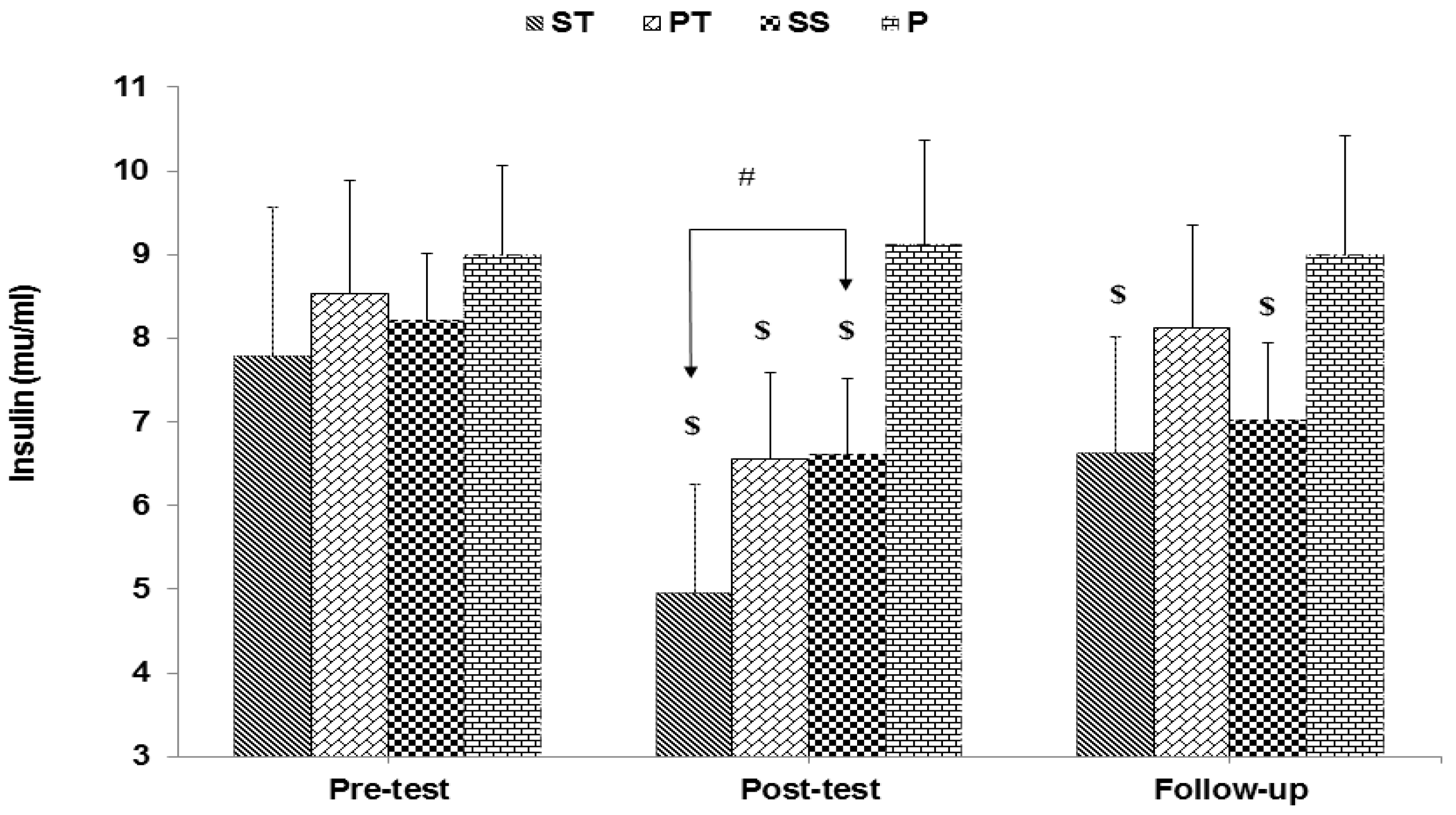
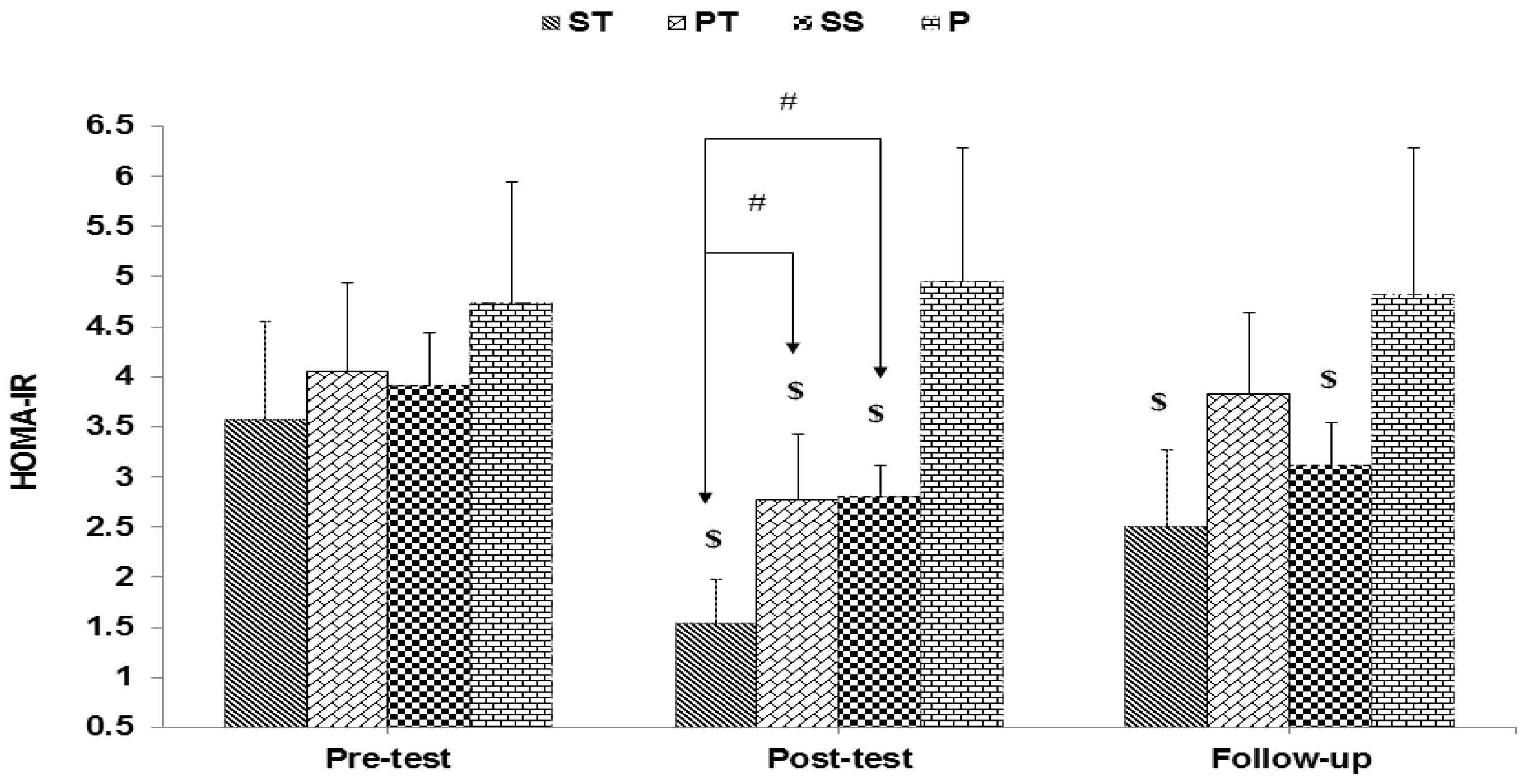
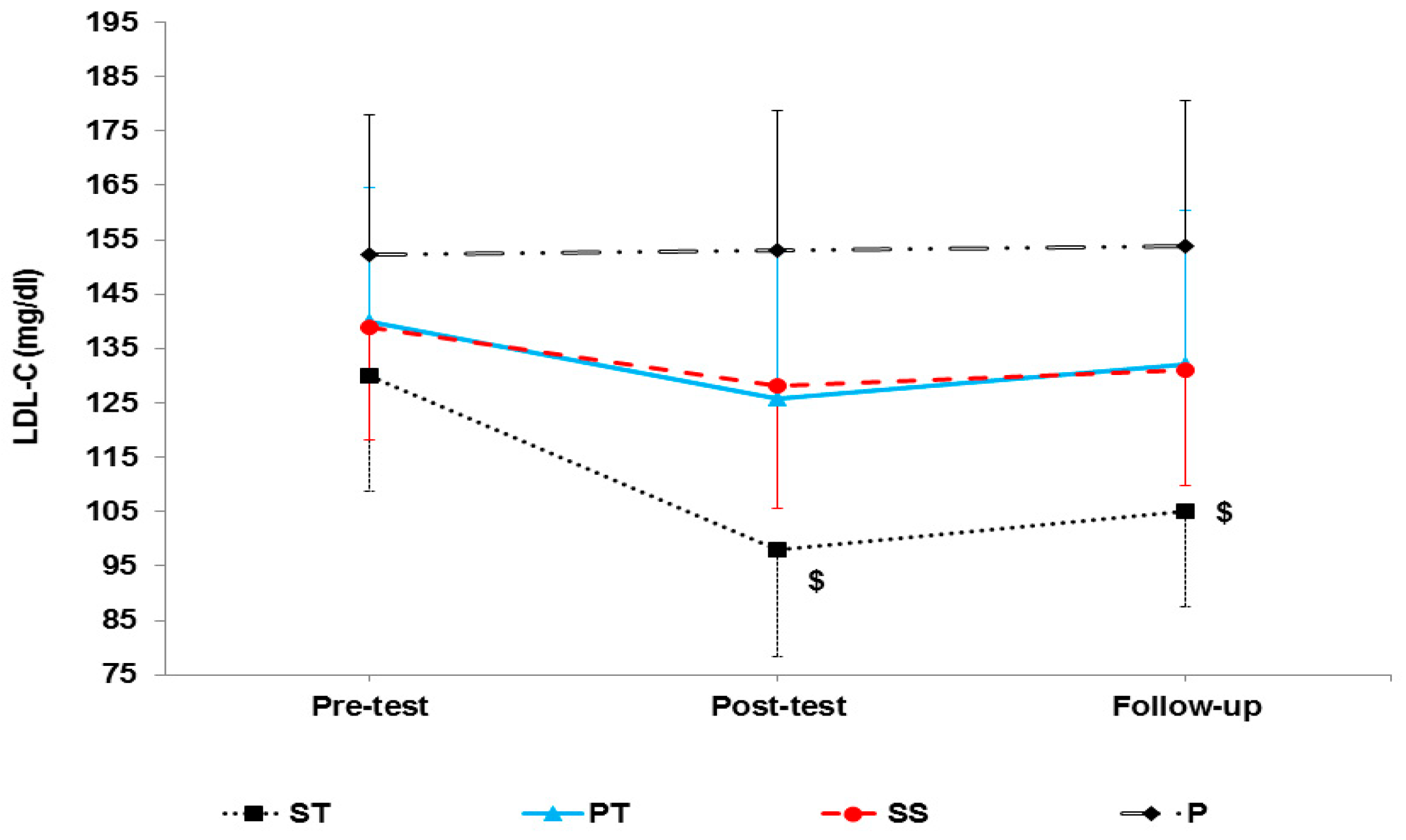
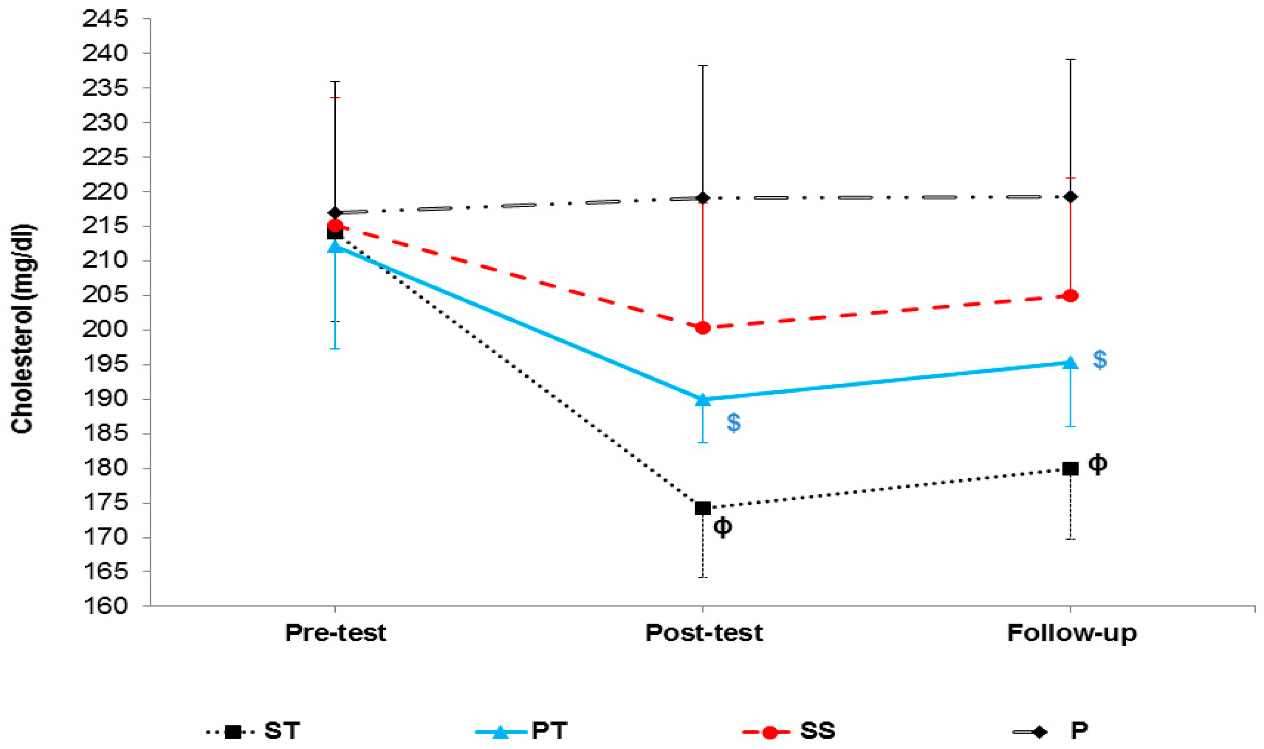
Data were expressed as mean ± standard deviation (SD). Normal data distribution and homogeneity of variances were assessed using the Shapiro–Wilk and Levene tests, respectively. A mixed-model ANOVA with repeated measures across time and group was used to test the main effects and interactions. The Bonferroni post-hoc test was used for checking significant differences among the main effects of each dependent variable. Cohen’s d test was used to estimate the effect size. An effect size of less than 0.2 was considered negligible, between 0.2 and 0.5 small, between 0.5 and 0.8 moderate, and more than 0.8 high. Statistical analysis was performed using SPSS version 22 at a significance level of 5%.

3. Results

As shown in Table 2, there was no significant difference between the groups regarding age, energy, and nutrient intake.
The result of the HPLC test is shown in Table 3, in which saffron composition is shown.
Mixed ANOVA showed a significant interaction between groups and time for body weight (F 6, 56 = 11.101, p < 0.001), BMI (F 6, 56 = 10.18, p < 0.001), and fat percentage (F 6, 56 = 8.98, p < 0.001) (Table 4).
The results of mixed ANOVA showed a significant interaction between groups and time for glucose (F 6, 56 = 22.88, p < 0.001) (Figure 1), insulin (F 6, 56 = 11.52, p < 0.001) (Figure 2), and HOMA-IR (F 6, 56 = 19.34, p < 0.001) (Figure 3).
The results of mixed ANOVA showed a significant interaction between groups and time for adiponectin (F 6, 56 = 11.39, p < 0.001), resistin (F 6, 56 = 7.55, p < 0.001), TNF-α levels (F 6, 56 = 5.87, p < 0.001), IL-6 levels (F 6, 56 = 13.86, p < 0.001), and irisin (F6, 56 = 9.73, p < 0.001) (Table 5).
The results of mixed ANOVA showed that there was a significant interaction between groups and time in LDL (F 6, 56 = 15.42, p < 0.001) (Figure 4), HDL-C (F 6, 56 = 24.91, p < 0.001) (Figure 5), cholesterol (F 6, 56 = 5.72, p < 0.001) (Figure 6), and TG (F 6, 56 = 13.65, p < 0.001) (Figure 7).

4. Discussion

The present study aimed to investigate the effects of 8-week AT and saffron supplementation on inflammation and metabolism in middle-aged obese women with T2DM. A growing body of evidence has revealed that saffron supplementation could be considered an essential nutraceutical for T2DM treatment due to its hypoglycemic and anti-inflammatory properties [21,22]. Inhibition of oxidative stress, amelioration of ß–cell function, repression of inflammatory pathways, improvement in insulin sensitivity, and stimulation of GLUT-4 expression could be affected by saffron supplementation [21,22,26]. Our results showed that when AT was combined with saffron supplementation, adiponectin, irisin, and HDL-C increased, but resistin, TNF-α, IL-6, body weight, BMI, body fat percentage, LDL-C, cholesterol, blood glucose, insulin, and HOMA-IR decreased. This means that combining AT with saffron supplementation could reduce inflammation, improve metabolism, and ameliorate insulin resistance and body composition.
The present study results indicated that insulin levels and resistance decreased after eight weeks of AT and saffron supplementation, but their combination led to better results. These results are consistent with several studies [15,27] but inconsistent with some others [28,29]. For instance, in a study by Choi et al. [29], where the participants were obese women who performed 45 min/session, 300 kcal/session AT, and 20 min/session, 100 kcal/day strength training, five times a week for three months, a decrease in insulin levels and insulin resistance was not seen. Different diets, the age and sex of participants, and the type of AT protocol may explain this inconsistency [30].
Blood glucose levels only decreased in the ST group. This means that neither AT nor saffron supplementation could affect blood glucose alone. In line with this result, Cauza et al. [31] and Bello et al. [32] did not report significant changes in blood glucose after 16 and 8 weeks of AT, respectively. It has been suggested that AT cannot significantly affect blood glucose in the middle-aged and the elderly [33]. In addition, our participants had a long history of diabetes and a high level of insulin resistance so they may have needed a longer AT period. These two reasons may explain why we could not see significant changes in the PT or SS groups. However, combining AT and saffron supplementation provided sufficient stimulation for upregulating AMPK signaling, which resulted in increasing GLUT-4 translocation into the cell membrane and therefore increased glucose uptake and insulin sensitivity [34].
In line with other studies [35,36], the present study showed that while eight weeks of AT and saffron supplementation increased serum adiponectin levels, combining these interventions led to better results. A study by Rezaeeshirazi [36] showed that AT is more effective in ameliorating insulin resistance in T2D. Yokoyama et al. [37] reported that improvement in insulin sensitivity induced by diet combined with AT (40 min/session, five sessions/week cycling, and 10,000 steps/session walking) for three weeks could not alter adiponectin levels in T2DM patients, which is inconsistent with our results. The reasons for the inconsistency may be the different types of AT and participants’ sex [30] (i.e., female in our study versus both male and female in Yokoyama et al.); high levels of testosterone in men compared to women could inhibit production and secretion of adiponectin [38]. The increase in adiponectin levels in the ST group could be explained by the association of adiponectin with lipid profile (LDL-C, HDL-C, cholesterol, and TG). Adiponectin concentration is directly related to HDL-C levels and inversely related to LDL-C and TG levels [39,40]. This study showed that LDL-C, cholesterol, and TG decreased, and HDL-C increased after combining eight weeks of AT with saffron supplementation.
Furthermore, increased plasma-free fatty acids can stimulate adiponectin secretion. Aerobic training, by increasing lipolysis [39,40] and crocin (one of saffron’s components) by inhibiting pancreatic lipase, which indirectly leads to malabsorption of fat, could lead to an increase in plasma-free fatty acids and stimulate adiponectin secretion [41]. In addition, decreasing insulin resistance might be another reason for increasing adiponectin. Based on this result, we concluded that obesity and T2DM are associated with a decrease in adiponectin, which reduces insulin sensitivity. Conversely, the enhancement of serum levels of adiponectin resulting from AT and saffron supplementation could increase insulin sensitivity through insulin-mimicking, insulin-sensitizing, and anti-inflammatory properties [42,43].
Resistin is another crucial link between insulin resistance and obesity. Our results showed no significant decrease in serum resistin levels after eight weeks of AT and saffron supplementation alone but did show a decrease when they were used in combination. Kadoglou et al. [44] reported that after 16 weeks of AT (i.e., 45–60 min running with 50–85% of vo2max, four sessions/week), serum levels of resistin decreased in T2DM patients. However, no significant changes were reported in other studies after AT [45,46]. Different AT types, durations, and intensities might explain this inconsistency [30]. Many studies have shown that resistin is directly related to changes in body fat, body weight, BMI, HOMA-IR, and TG [47], which is in line with our results since all the above factors showed significant changes in the ST group. Another important factor is diet, as it is the primary regulator of resistin. This is why we supervised the participants’ diets, where we observed no significant change in nutrient uptake.
Another insulin resistance influencer is an adipo-myokine, irisin, which has a vital role in energy homeostasis and metabolism [48]. While eight weeks of AT and saffron supplementation could not affect irisin serum levels alone, its levels showed a significant increase in the ST group. This result showed that the isolated effect of AT or saffron supplementation is not enough to increase irisin levels, but the combined effects are needed. However, some studies showed that AT could increase irisin serum levels in diabetic patients [49,50]. Although some studies have suggested a positive correlation between plasma irisin and lactate levels [51], our AT protocol did not have enough load to induce changes in irisin levels.
Our results also showed that while both AT and saffron supplementation could reduce serum IL-6 levels, combining these interventions led to better results. The antioxidant properties of saffron [22] and AT [52] together can reduce reactive oxygen species levels, which play an essential role in the production of pro-inflammatory cytokines [53]. In fact, crocin improves the expression of phosphorylated Akt (p-Akt) and phosphatidylinositol 3-kinase (PI3K). PI3K/Akt signaling plays a significant role in blocking oxidative stress and suppressing the pro-inflammatory response to oxidative stress [54]. The effectiveness of 8 weeks of AT on IL-6 serum levels can also be due to body weight loss, mainly due to the reduced adipose tissue content [55]. IL-6 is secreted by adipose tissue as adipokines, and a decrease in fat mass is associated with a decrease in IL-6 levels [55]. Some studies have reported no significant changes in IL-6 levels after AT [56,57], but others have shown decreased inflammatory and increased anti-inflammatory cytokines in T2DM patients [58].
Another pro-inflammatory cytokine, TNF-α, showed a significant reduction in the ST group only, indicating that it may need a higher amount of AT or saffron to induce isolated changes. In other words, IL-6 seems to be more sensitive to our interventions than TNF-α. The lipid profile, including LDL-C, cholesterol, and TG serum levels, showed a significant reduction in the ST group, and HDL-C showed a significant increase in the ST, SS, and PT groups with no significant difference between them. However, an additive effect was seen for cholesterol and TG highlighting their sensitiveness to AT and saffron. In line with this study, other studies have shown that both saffron and AT [59,60] could improve the lipid profile. Based on previous evidence, saffron supplementation causes an increase in the activity of lecithin-cholesterol acyltransferase (LCAT) [61], which is involved in the regulation of blood lipid metabolism [62]. In addition, regular AT leads to increased daily energy output and fat oxidation and reduces fat mass [63]. As our results show, body weight, BMI, and body fat percentage significantly decreased after eight weeks of AT with saffron supplementation.
The follow-up results (after two weeks) showed that the adaptations disappeared in the PT, SS, and P groups but not in the ST group. It seems that the adaptations in the ST group were high enough to still be detectable two weeks after detraining.

5. Conclusions

Saffron supplementation at a dose of 400 mg/day, when combined with AT, could improve inflammation, metabolism, glycemic status, and lipid profile in T2DM patients, and these changes are sustainable at up to 2 weeks of detraining.

6. Limitations and Strength

We gave the saffron/placebo capsules to participants and asked them to take them according to the study plan and followed up with them through phone call. This could be a potential limitation in our study, and a future study should ask the participants to take the supplement in front of the researcher. In addition, we collected participants’ nutritional data three days per week, which might have led to a misinterpretation of the participants’ diets. What we consider the strength in our study is using a free-style rather than traditional training method. Furthermore, measuring various factors that affect metabolism and inflammation helped us to better interpret our data, which could be another strength of this study.

Author Contributions

Conceptualization, A.R., M.S., M.K. and A.A. writing—original draft preparation, K.K.; writing—review and editing, K.K. and K.S. All authors have read and agreed to the published version of the manuscript.

Funding

This research received no external funding.

Institutional Review Board Statement

The animal study protocol was approved by the Institutional Review Board of Mohaghegh Ardabili university (Ethics No: 1605/A. D; date 6 November 2017).

Informed Consent Statement

Informed consent was obtained from all subjects involved in the study.

Data Availability Statement

Conflicts of Interest

The authors declare no conflict of interest.

References

  1. Messina, G.; Palmieri, F.; Monda, V.; Messina, A.; Dalia, C.; De Luca, V. Exercise causes muscle GLUT4 translocation in an insulin. Biol. Med. 2015, 1, 1–4. [Google Scholar]
  2. Khoramipour, K.; Sandbakk, Ø.; Keshteli, A.H.; Gaeini, A.A.; Wishart, D.S.; Chamari, K. Metabolomics in exercise and sports: A systematic review. Sports Med. 2021, 52, 547–583. [Google Scholar] [CrossRef] [PubMed]
  3. Bragazzi, N.L.; Khoramipour, K.; Chaouachi, A.; Chamari, K. Toward sportomics: Shifting from sport genomics to sport postgenomics and metabolomics specialties. Promises, challenges, and future perspectives. Int. J. Sports Physiol. Perform. 2020, 15, 1201–1202. [Google Scholar] [CrossRef]
  4. Vasseur, F.; Helbecque, N.; Dina, C.; Lobbens, S.; Delannoy, V.; Gaget, S.; Boutin, P.; Vaxillaire, M.; Leprêtre, F.; Dupont, S.; et al. Single-nucleotide polymorphism haplotypes in the both proximal promoter and exon 3 of the APM1 gene modulate adipocyte-secreted adiponectin hormone levels and contribute to the genetic risk for type 2 diabetes in French Caucasians. Hum. Mol. Genet. 2002, 11, 2607–2614. [Google Scholar] [CrossRef] [PubMed] [Green Version]
  5. Yu, N.; Ruan, Y.; Gao, X.; Sun, J. Systematic review and meta-analysis of randomized, controlled trials on the effect of exercise on serum leptin and adiponectin in overweight and obese individuals. Horm. Metab. Res. 2017, 49, 164–173. [Google Scholar] [CrossRef] [Green Version]
  6. Khoramipour, K.C.K.; Hekmatikar, A.A.; Ziyaiyan, A.; Taherkhani, S.; Elguindy, N.M.; Bragazzi, N.L. Adiponectin: Structure, physiological functions, role in diseases, and effects of nutrition. Nutrients 2021, 13, 1180. [Google Scholar] [CrossRef]
  7. Adeghate, E. An update on the biology and physiology of resistin. Cell. Mol. Life Sci. CMLS 2004, 61, 2485–2496. [Google Scholar] [CrossRef]
  8. Shaikh-Kader, A.; Houreld, N.N.; Rajendran, N.K.; Abrahamse, H. Levels of Cyclooxygenase 2, Interleukin-6, and Tumour Necrosis Factor-α in Fibroblast Cell Culture Models after Photobiomodulation at 660 nm. Oxidative Med. Cell. Longev. 2021, 2021, 6667812. [Google Scholar] [CrossRef]
  9. Piché, M.-E.; Tchernof, A.; Després, J.-P. Obesity phenotypes, diabetes, and cardiovascular diseases. Circ. Res. 2020, 126, 1477–1500. [Google Scholar] [CrossRef]
  10. Dovio, A.; Angeli, A. Cytokines and type 2 diabetes mellitus. JAMA 2001, 286, 2233. [Google Scholar]
  11. Smitka, K.; Marešová, D. Adipose tissue as an endocrine organ: An update on pro-inflammatory and anti-inflammatory microenvironment. Prague Med. Rep. 2015, 116, 87–111. [Google Scholar] [CrossRef] [PubMed] [Green Version]
  12. Ikeda, S.I.; Tamura, Y.; Kakehi, S.; Sanada, H.; Kawamori, R.; Watada, H. Exercise-induced increase in IL-6 level enhances GLUT4 expression and insulin sensitivity in mouse skeletal muscle. Biochem. Biophys. Res. Commun. 2016, 473, 947–952. [Google Scholar] [CrossRef] [PubMed]
  13. Korta, P.; Pocheć, E.; Mazur-Biały, A. Irisin as a multifunctional protein: Implications for health and certain diseases. Medicina 2019, 55, 485. [Google Scholar] [CrossRef] [Green Version]
  14. Küçükkaraca, H.; Söğüt, M.Ü. Investigation of the Effect of Exercise on Irisin Hormone in Experimentally Induced Diabetic Rats. Int. J. Health Serv. Res. Policy 2017, 2, 51–57. [Google Scholar] [CrossRef] [Green Version]
  15. Kercher, V.M.; Kercher, K.; Bennion, T.; Yates, B.A.; Feito, Y.; Alexander, C.; Amaral, P.C.; Soares, W.; Li, Y.M.; Han, J.; et al. Fitness trends from around the globe. ACSM’s Health Fit. J. 2021, 25, 20–31. [Google Scholar] [CrossRef]
  16. Bull, F.C.; Al-Ansari, S.S.; Biddle, S.; Borodulin, K.; Buman, M.P.; Cardon, G.; Carty, C.; Chaput, J.P.; Chastin, S.; Chou, R.; et al. World Health Organization 2020 guidelines on physical activity and sedentary behaviour. Br. J. Sports Med. 2020, 54, 1451–1462. [Google Scholar] [CrossRef] [PubMed]
  17. Rahmaty, S.; Dehghan, P.; Khoramipour, K.; Saboory, M. The effect of listening to brain waves’ relaxing and exciting music during intense endurance training on blood cortisol levels of adult men. Am. J. Sports Sci. Med. 2015, 3, 77–81. [Google Scholar]
  18. Hua, L.; Lei, M.; Xue, S.; Li, X.; Li, S.; Xie, Q. Effect of fish oil supplementation combined with high-intensity interval training in newly diagnosed non-obese type 2 diabetes: A randomized controlled trial. J. Clin. Biochem. Nutr. 2020, 66, 146–151. [Google Scholar] [CrossRef] [Green Version]
  19. Mobasseri, M.; Ostadrahimi, A.; Tajaddini, A.; Asghari, S.; Barati, M.; Akbarzadeh, M.; Nikpayam, O.; Houshyar, J.; Roshanravan, N.; Alamdari, N.M. Effects of saffron supplementation on glycemia and inflammation in patients with type 2 diabetes mellitus: A randomized double-blind, placebo-controlled clinical trial study. Diabetes Metab. Syndr. Clin. Res. Rev. 2020, 14, 527–534. [Google Scholar] [CrossRef]
  20. Hosseinzadeh, H.; Nassiri-Asl, M. Avicenna’s (Ibn Sina) the canon of medicine and saffron (Crocus sativus): A review. Phytother. Res. 2013, 27, 475–483. [Google Scholar] [CrossRef]
  21. Dehghan, F.; Hajiaghaalipour, F.; Yusof, A.; Muniandy, S.; Hosseini, S.A.; Heydari, S.; Salim, L.Z.A.; Azarbayjani, M.A. Saffron with resistance exercise improves diabetic parameters through the GLUT4/AMPK pathway in-vitro and in-vivo. Sci. Rep. 2016, 6, 25139. [Google Scholar] [CrossRef] [Green Version]
  22. Khatir, S.A.; Bayatian, A.; Barzegari, A.; Roshanravan, N.; Safaiyan, A.; Pavon-Djavid, G.; Ostadrahimi, A. Saffron (Crocus sativus L.) supplements modulate circulating MicroRNA (miR-21) in atherosclerosis patients; a randomized, double-blind, placebo-controlled trial. Iran. Red Crescent Med. J. 2018, 20, e80260. [Google Scholar]
  23. Hooshmand Moghadam, B.; Rashidlamir, A.; Attarzadeh Hosseini, S.R.; Gaeini, A.A.; Kaviani, M. The effects of saffron (Crocus sativus L.) in conjunction with concurrent training on body composition, glycaemic status, and inflammatory markers in obese men with type 2 diabetes mellitus: A randomized double-blind clinical trial. Br. J. Clin. Pharmacol. 2022, 88, 3256–3271. [Google Scholar] [CrossRef] [PubMed]
  24. Rajabi, A.; Akbarnejad, A.; Siahkouhian, M.; Yari, M. Effect of Saffron supplementation and exercise training on blood pressure, pulmonary function and spirometery indicators in obese and overweight women affected by type 2 diabetes. J. Gorgan Univ. Med. Sci. 2019, 21, 59–69. [Google Scholar]
  25. Mohammadabadi, F.; Vafaiyan, Z.; Hosseini, S.M.; Aryaie, M.; Eshghinia, S. Assessment of insulin resistance with two methods: HOMA-IR and TyG index in Iranian obese women. Iran. J. Diabetes Obes. 2014, 6, 23–27. [Google Scholar]
  26. Chowdhury, A.I.; Habib, M.A.; Ghosh, S. Effect of Saffron (Crocus sativus L.) on Common Non-Communicable Disease: Review from Current Clinical Findings. J. Ayurvedic Herb. Med. 2021, 7, 93–108. [Google Scholar] [CrossRef]
  27. Iaccarino, G.; Franco, D.; Sorriento, D.; Strisciuglio, T.; Barbato, E.; Morisco, C. Modulation of insulin sensitivity by exercise training: Implications for cardiovascular prevention. J. Cardiovasc. Transl. Res. 2021, 14, 256–270. [Google Scholar] [CrossRef]
  28. Shaibi, G.Q.; Roberts, C.K.; Goran, M.I. Exercise and insulin resistance in youth. Exerc. Sport Sci. Rev. 2008, 36, 5–11. [Google Scholar] [CrossRef]
  29. Choi, K.M.; Kim, T.N.; Yoo, H.J.; Lee, K.W.; Cho, G.J.; Hwang, T.G.; Baik, S.H.; Choi, D.S.; Kim, S.M. Effect of exercise training on A-FABP, lipocalin-2 and RBP4 levels in obese women. Clin. Endocrinol. 2009, 70, 569–574. [Google Scholar] [CrossRef]
  30. Khajehlandi, M.; Mohammadi, R. The Effect of Pilates Training on Body Composition, Lipid Profile, and Serum 25-Hydroxy Vitamin D Levels in Inactive Overweight Women. Zahedan J. Res. Med. Sci. 2021, 23, e100502. [Google Scholar] [CrossRef]
  31. Cauza, E.; Hanusch-Enserer, U.; Strasser, B.; Ludvik, B.; Metz-Schimmerl, S.; Pacini, G.; Wagner, O.; Georg, P.; Prager, R.; Kostner, K.; et al. The relative benefits of endurance and strength training on the metabolic factors and muscle function of people with type 2 diabetes mellitus. Arch. Phys. Med. Rehabil. 2005, 86, 1527–1533. [Google Scholar] [CrossRef] [PubMed]
  32. Bello, A.I.; Owusu-Boakye, E.; Adegoke, B.O.; Adjei, D.N. Effects of aerobic exercise on selected physiological parameters and quality of life in patients with type 2 diabetes mellitus. Int. J. Gen. Med. 2011, 4, 723. [Google Scholar] [CrossRef] [PubMed] [Green Version]
  33. Albright, A.; Franz, M.; Hornsby, G.; Kriska, A.; Marrero, D.; Ullrich, I.; Verity, L.S. American College of Sports Medicine position stand. Exercise and type 2 diabetes. Med. Sci. Sports Exerc. 2000, 32, 1345–1360. [Google Scholar]
  34. Park, J.-S.; Holloszy, J.O.; Kim, K.; Koh, J.-H. Exercise training-induced PPARβ increases PGC-1α protein stability and improves insulin-induced glucose uptake in rodent muscles. Nutrients 2020, 12, 652. [Google Scholar] [CrossRef] [Green Version]
  35. Boudou, P.; Sobngwi, E.; Mauvais-Jarvis, F.; Vexiau, P.; Gautier, J. Absence of exercise-induced variations in adiponectin levels despite decreased abdominal adiposity and improved insulin sensitivity in type 2 diabetic men. Eur. J. Endocrinol. 2003, 149, 421–424. [Google Scholar] [CrossRef] [Green Version]
  36. Rezaeeshirazi, R. Aerobic versus resistance training: Leptin and metabolic parameters improvement in type 2 diabetes obese men. Res. Q. Exerc. Sport 2022, 93, 537–547. [Google Scholar] [CrossRef]
  37. Yokoyama, H.; Emoto, M.; Araki, T.; Fujiwara, S.; Motoyama, K.; Morioka, T.; Koyama, H.; Shoji, T.; Okuno, Y.; Nishizawa, Y. Effect of aerobic exercise on plasma adiponectin levels and insulin resistance in type 2 diabetes. Diabetes Care 2004, 27, 1756–1758. [Google Scholar] [CrossRef]
  38. Xu, A.; Chan, K.W.; Hoo, R.L.; Wang, Y.; Tan, K.C.; Zhang, J.; Chen, B.; Lam, M.C.; Tse, C.; Cooper, G.J.; et al. Testosterone selectively reduces the high molecular weight form of adiponectin by inhibiting its secretion from adipocytes. J. Biol. Chem. 2005, 280, 18073–18080. [Google Scholar] [CrossRef] [Green Version]
  39. Xi, L.; Qian, Z.; Du, P.; Fu, J. Pharmacokinetic properties of crocin (crocetin digentiobiose ester) following oral administration in rats. Phytomedicine 2007, 14, 633–636. [Google Scholar] [CrossRef]
  40. Yang, Y.-C.; Hwang, J.-H.; Hong, S.-J.; Hsu, H.-K. Enhancement of glucose uptake in 3T3-L1 adipocytes by Toona sinensis leaf extract. Kaohsiung J. Med. Sci. 2003, 19, 327–332. [Google Scholar] [CrossRef] [Green Version]
  41. Mattei, L.; Francisqueti-Ferron, F.V.; Garcia, J.L.; Ferron, A.J.T.; de Almeida Silva, C.C.V.; Gregolin, C.S.; Nakandakare-Maia, E.T.; Silva, J.D.C.P.; Moreto, F.; Minatel, I.O.; et al. Antioxidant and anti-inflammatory properties of gamma-oryzanol attenuates insulin resistance by increasing GLUT-4 expression in skeletal muscle of obese animals. Mol. Cell. Endocrinol. 2021, 537, 111423. [Google Scholar] [CrossRef] [PubMed]
  42. Aruoma, O.I.; Neergheen, V.S.; Bahorun, T.; Jen, L.-S. Free radicals, antioxidants and diabetes: Embryopathy, retinopathy, neuropathy, nephropathy and cardiovascular complications. Neuroembryol. Aging 2006, 4, 117–137. [Google Scholar] [CrossRef]
  43. Bouassida, A.; Chamari, K.; Zaouali, M.; Feki, Y.; Zbidi, A.; Tabka, Z. Review on leptin and adiponectin responses and adaptations to acute and chronic exercise. Br. J. Sports Med. 2010, 44, 620–630. [Google Scholar] [CrossRef] [PubMed]
  44. Kadoglou, N.P.; Perrea, D.; Iliadis, F.; Angelopoulou, N.; Liapis, C.; Alevizos, M. Exercise reduces resistin and inflammatory cytokines in patients with type 2 diabetes. Diabetes Care 2007, 30, 719–721. [Google Scholar] [CrossRef] [Green Version]
  45. Gueugnon, C.; Mougin, F.; Simon-Rigaud, M.-L.; Regnard, J.; Nègre, V.; Dumoulin, G. Effects of an in-patient treatment program based on regular exercise and a balanced diet on high molecular weight adiponectin, resistin levels, and insulin resistance in adolescents with severe obesity. Appl. Physiol. Nutr. Metab. 2012, 37, 672–679. [Google Scholar] [CrossRef]
  46. Monzillo, L.U.; Hamdy, O.; Horton, E.S.; Ledbury, S.; Mullooly, C.; Jarema, C.; Porter, S.; Ovalle, K.; Moussa, A.; Mantzoros, C.S. Effect of lifestyle modification on adipokine levels in obese subjects with insulin resistance. Obes. Res. 2003, 11, 1048–1054. [Google Scholar] [CrossRef] [Green Version]
  47. Barnes, K.; Miner, J. Role of resistin in insulin sensitivity in rodents and humans. Curr. Protein Pept. Sci. 2009, 10, 96–107. [Google Scholar] [CrossRef]
  48. Lu, J.; Xiang, G.; Liu, M.; Mei, W.; Xiang, L.; Dong, J. Irisin protects against endothelial injury and ameliorates atherosclerosis in apolipoprotein E-Null diabetic mice. Atherosclerosis 2015, 243, 438–448. [Google Scholar] [CrossRef]
  49. Miyamoto-Mikami, E.; Sato, K.; Kurihara, T.; Hasegawa, N.; Fujie, S.; Fujita, S.; Sanada, K.; Hamaoka, T.; Tabata, I.; Iemitsu, M. Endurance training-induced increase in circulating irisin levels is associated with reduction of abdominal visceral fat in middle-aged and older adults. PLoS ONE 2015, 10, e0120354. [Google Scholar] [CrossRef] [Green Version]
  50. Zhang, Y.; Li, R.; Meng, Y.; Li, S.; Donelan, W.; Zhao, Y.; Qi, L.; Zhang, M.; Wang, X.; Cui, T.; et al. Irisin stimulates browning of white adipocytes through mitogen-activated protein kinase p38 MAP kinase and ERK MAP kinase signaling. Diabetes 2014, 63, 514–525. [Google Scholar] [CrossRef] [Green Version]
  51. Kern, M.; Wells, J.A.; Stephens, J.M.; Elton, C.W.; Friedman, J.E.; Tapscott, E.B.; Pekala, P.H.; Dohm, G.L. Insulin responsiveness in skeletal muscle is determined by glucose transporter (Glut4) protein level. Biochem. J. 1990, 270, 397–400. [Google Scholar] [CrossRef] [PubMed] [Green Version]
  52. Huertas, J.; Al Fazazi, S.; Hidalgo-Gutierrez, A.; López, L.; Casuso, R. Antioxidant effect of exercise: Exploring the role of the mitochondrial complex I superassembly. Redox Biol. 2017, 13, 477–481. [Google Scholar] [CrossRef] [PubMed]
  53. Yang, X.; Huo, F.; Liu, B.; Liu, J.; Chen, T.; Li, J.; Zhu, Z.; Lv, B. Crocin inhibits oxidative stress and pro-inflammatory response of microglial cells associated with diabetic retinopathy through the activation of PI3K/Akt signaling pathway. J. Mol. Neurosci. 2017, 61, 581–589. [Google Scholar] [CrossRef]
  54. Cai, J.; Yi, F.F.; Bian, Z.Y.; Shen, D.F.; Yang, L.; Yan, L.; Tang, Q.Z.; Yang, X.C.; Li, H. Crocetin protects against cardiac hypertrophy by blocking MEK-ERK1/2 signalling pathway. J. Cell. Mol. Med. 2009, 13, 909–925. [Google Scholar] [CrossRef] [Green Version]
  55. Abd El-Kader, S.M. Aerobic versus resistance exercise training in modulation of insulin resistance, adipocytokines and inflammatory cytokine levels in obese type 2 diabetic patients. J. Adv. Res. 2011, 2, 179–183. [Google Scholar] [CrossRef] [Green Version]
  56. Conraads, V.M.; Beckers, P.; Bosmans, J.; De Clerck, L.S.; Stevens, W.J.; Vrints, C.J.; Brutsaert, D.L. Combined endurance/resistance training reduces plasma TNF-α receptor levels in patients with chronic heart failure and coronary artery disease. Eur. Heart J. 2002, 23, 1854–1860. [Google Scholar] [CrossRef] [Green Version]
  57. Ferreira, F.C.; de Medeiros, A.I.; Nicioli, C.; Nunes, J.E.D.; Shiguemoto, G.E.; Prestes, J.; Verzola, R.M.M.; Baldissera, V.; de Andrade Perez, S.E. Circuit resistance training in sedentary women: Body composition and serum cytokine levels. Appl. Physiol. Nutr. Metab. 2009, 35, 163–171. [Google Scholar] [CrossRef]
  58. Thompson, D.; Walhin, J.P.; Batterham, A.M.; Stokes, K.A.; Cooper, A.R.; Andrews, R.C. Effect of diet or diet plus physical activity versus usual care on inflammatory markers in patients with newly diagnosed type 2 diabetes: The Early ACTivity in Diabetes (ACTID) randomized, controlled trial. J. Am. Heart Assoc. 2014, 3, e000828. [Google Scholar] [CrossRef] [Green Version]
  59. Arasteh, A.; Aliyev, A.; Khamnei, S.; Delazar, A.; Mesgari, M.; Mehmannavaz, Y. Crocus sativus on serum glucose, insulin and cholesterol levels in healthy male rats. J. Med. Plants Res. 2010, 4, 397–402. [Google Scholar]
  60. Garber, C.E.; Blissmer, B.; Deschenes, M.R.; Franklin, B.A.; Lamonte, M.J.; Lee, I.M.; Nieman, D.C.; Swain, D.P. American College of Sports Medicine position stand. Quantity and quality of exercise for developing and maintaining cardiorespiratory, musculoskeletal, and neuromotor fitness in apparently healthy adults: Guidance for prescribing exercise. Med. Sci. Sports Exerc. 2011, 43, 1334–1359. [Google Scholar] [CrossRef]
  61. Giaccio, M. Crocetin from saffron: An active component of an ancient spice. Crit. Rev. Food Sci. Nutr. 2004, 44, 155–172. [Google Scholar] [CrossRef] [PubMed]
  62. Barzegari, A.; Mahdirejei, H.A. Effects of 8 weeks resistance training on plasma vaspin and lipid profile levels in adult men with type 2 diabetes. Casp. J. Intern. Med. 2014, 5, 103. [Google Scholar]
  63. Baba, C.S.; Alexander, G.; Kalyani, B.; Pandey, R.; Rastogi, S.; Pandey, A.; Choudhuri, G. Effect of exercise and dietary modification on serum aminotransferase levels in patients with nonalcoholic steatohepatitis. J. Gastroenterol. Hepatol. 2006, 21, 191–198. [Google Scholar] [CrossRef] [PubMed]
Figure 1. Glucose in the pre-test, post-test, and follow-up (mean ± SD). # indicates a significant decrease in the ST group compared to the P group.
Sports 10 00167 g001
Figure 2. Insulin in the pre-test, post-test, and follow-up (mean ± SD). $ indicates a significant decrease in the ST, PT, and SS groups compared to the P group, and # indicates a significant decrease in the ST group compared to the SS group.
Sports 10 00167 g002
Figure 3. HOMA-IR levels and HOMA-IR. $ indicates a significant decrease in the ST, PT, and SS groups compared to the P group, and # indicates a significant decrease in the ST group compared to the SS group and the PT group.
Sports 10 00167 g003
Figure 4. LDL-C levels in the pre-test, post-test, and follow-up. $ indicates a significant decrease in the ST group compared to the P group. $ indicates a significant decrease in the PT group compared to the P group.
Sports 10 00167 g004
Figure 5. HDL-C levels in the pre-test, post-test, and follow-up. * indicates a significant increase in the ST, PT, and SS groups compared to the P group.
Sports 10 00167 g005
Figure 6. Cholesterol levels in the pre-test, post-test, and follow-up. ɸ indicates a significant decrease in the ST group compared to the SS and P groups. $ indicates a significant decrease in the PT group compared to the P group.
Sports 10 00167 g006
Figure 7. Triglyceride levels in the pre-test, post-test, and follow-up. ɸ indicates a significant decrease in the ST group compared to the SS and P groups, $ indicates a significant decrease in the PT group compared to the P group. * indicates a significant decrease in the ST group compared to the P group in the follow-up.
Sports 10 00167 g007
Table 1. Aerobic Training Program.
WeeksStages of ExerciseExercise Intensity Duration per Session (Minute)Type of Movement
Weeks: 1 and 2:
3 sessions per week
warm-up%55>10Jogging, combined hand-leg movements, stretching movements
main partaerobic%55–6010Combined hand-leg movements
running%60–6520running
cool-down%50>5Stretching large muscles
Weeks: 3, 4 and 5: 3 sessions per weekwarm-up%55>10Jogging, combined hand-leg movements, stretching movements
main partaerobic%55–6010Combined hand-leg movements
running%65–7030running
cool-down%50>5Stretching large muscles
Weeks: 6, 7 and 8: 3 sessions per weekwarm-up%55>10Jogging, combined hand-leg movements, stretching movements
main partaerobic%55–6010Combined hand-leg movements
running%70–7540running
cool-down%50>5Stretching large muscles
Table 2. Participants’ age, energy, and nutrient intake (mean ± SD).
VariableSTPTSSP
Age (year)51.5 ± 6.1657.62 ± 6.8154.12 ± 7.3756.87 ± 5.11
Disease duration (year)4.5 ± 3.24.6 ± 2.83.8 ± 1.74.2 ± 3.0
NutrientEnergy (calorie/day)1682.89 ± 1351692.36 ± 1661687.68 ± 1491705.71 ± 125
Carbohydrate (g/day)244.07 ± 62.48225.93 ± 59.27223.26 ± 44.76252.84 ± 38.34
Protein (g/day)66.66 ± 14.3373.53 ± 17.2378.8 ± 13.3980.00 ± 16.42
Fat (g/day)45.33 ± 12.7848.33 ± 13.8744.05 ± 14.1146.08 ± 11.13
Fiber (g/day)12.75 ± 3.7513.24 ± 4.4512.92 ± 5.1114.18 ± 4.21
Calcium (mg/day)262.92 ± 179.02270.18 ± 201.61268.87 ± 185.23286.54 ± 178.11
Vitamin C (mg/day)55.69 ± 25.1259.77 ± 26.2457.26 ± 27.6562.97 ± 29.44
Vitamin E (mg/day)2.5 ± 1.142.94 ± 1.462.87 ± 1.673.41 ± 2.47
Selenium (mg/day)42.90 ± 23.2648.81 ± 21.6747.89 ± 22.5453.65 ± 24.11
Table 3. Determination of the main metabolites of saffron.
Name of the SubstancePicrocrocinCrocinSafranal
Retention time (minute)14.816.226.1
Saffron metabolites levels (mg/g)6.696.471.17
Table 4. Participants’ anthropometric properties (Mean ± SD).
VariablesGroupsPre-TestPost-TestFollow-upCohen’s D
Body weight (kg)ST80.98 ± 5.0177.58 ± 6.37 ϕ78.12 ± 6.38 ϕ0.593
PT81.87 ± 3.3080.12 ± 3.4780.73 ± 3.370.516
SS81.47 ± 6.9179.56 ± 7.4780.06 ± 7.620.265
P86.95 ± 5.9087.18 ± 6.3287.00 ± 6.140.037
Body fat (%)ST31.98 ± 3.6627.43 ± 2.41 ϕ28.23 ± 2.88 ϕ1.468
PT33.12 ± 2.1930.62 ± 1.8431.42 ± 2.421.236
SS32.97 ± 3.1630.87 ± 2.4831.37 ± 3.200.739
P34.62 ± 3.0635.02 ± 2.3034.87 ± 4.010.147
BMI (kg/m2)ST30.32 ± 2.4228.97 ± 2.86 $29.18 ± 2.88 ϕ0.509
PT31.15 ± 1.5030.40 ± 1.49 #30.63 ± 1.310.501
SS30.72 ± 3.5630.1 ± 3.75 *30.18 ± 3.820.194
P34.03 ± 3.3634.15 ± 3.5734.07 ± 3.480.034
ST: Training + saffron, PT: Training + placebo, SS: Saffron + supplementation, and P: Placebo. For weight and body fat percentage, ϕ indicates a significant decrease in the ST group compared to the P group. For BMI ϕ, $, #, and * indicate a significant decrease in the ST group compared to the P group, the ST group compared to the SS group, the PT group compared to the P group, and the SS group compared to the P group, respectively. Cohen’s d test was used to estimate the effect size. BMI: body mass index.
Table 5. Serum levels of adiponectin, resistin, TNF-α, IL-6, and irisin (mean ± SD).
VariableGroupsPre-TestPost-TestFollow-upCohen’s D
Adiponectin
(ng/mL)
ST13.12 ± 4.1827.17 ± 7.59 &23.97 ± 7.02 $2.293
PT11.22 ± 3.8119.73 ± 4.39 *15.25 ± 3.772.070
SS12.03 ± 5.1818.00 ± 5.92 #16.18 ± 5.461.073
P9.15 ± 3.379.50 ± 3.429.21 ± 3.170.102
Resistin
(ng/mL)
ST12.20 ± 2.877.02 ± 2.04 $8.02 ± 2.30 $2.08
PT14.85 ± 3.6511.93 ± 2.8514.00 ± 3.070.891
SS14.07 ± 2.9112.05 ± 2.6113.34 ± 2.660.730
P15.73 ± 5.8015.85 ± 5.1814.87 ± 5.670.021
TNF-α
(pg/mL)
ST14.05 ± 3.419.56 ± 2.79 ϕ10.25 ± 2.81 ϕ1.585
PT16.65 ± 4.8515.23 ± 4.6915.87 ± 4.610.299
SS16.43 ± 6.6414.22 ± 4.5214.62 ± 4.860.389
P18.10 ± 5.6418.71 ± 5.5818.47 ± 5.770.108
IL-6
(pg/mL)
ST9.98 ± 1.945.15 ± 1.34 $7.01 ± 1.51 ϕ2.897
PT11.52 ± 2.148.72 ± 2.15 *9.83 ± 1.641.305
SS10.93 ± 2.009.01 ± 1.52 #10.55 ± 1.881.080
P12.43 ± 3.7912.56 ± 3.7112.5 ± 4.090.034
Irisin (ng/dL)ST171.05 ± 26.41203.92 ± 8.81 ϕ198.75 ± 7.00 ϕ1.670
PT166.12 ± 21.77179.72 ± 24.62176.37 ± 24.310.585
SS167.07 ± 19.28177.8 ± 20.14173.25 ± 23.30.544
P160.87 ± 17.74157.15± 18.60156.98 ± 18.070.204
ST: Training + saffron, PT: Training + placebo, SS: Saffron supplementation, and P: Placebo. Serum levels of adiponectin, resistin, TNF-α, IL-6, and irisin in the pre-test, post-test, and follow-up. For adiponectin, & indicates a significant increase in the ST group compared to the SS and P groups, * indicates a significant increase in the PT group compared to the P group, # indicates a significant increase in the SS group compared to the P group, and $ indicates a significant increase in the ST group compared to the PT, SS, and P groups in the follow-up. For resistin, $ indicates a significant decrease in the ST group compared to the PT, SS, and P groups. For TNF-α, ϕ indicates a significant decrease in the ST group compared to the P group. For IL-6, $ indicates a significant decrease in the ST group compared to the PT, SS and P groups, * indicates a significant decrease in the PT group compared to the P group, # indicates a significant decrease in the SS group compared to the P group, and ϕ indicates a significant increase in the ST group compared to the P group in the follow-up. For irisin, ϕ indicates a significant increase in the ST group compared to the P group.
Publisher’s Note: MDPI stays neutral with regard to jurisdictional claims in published maps and institutional affiliations.

