Review

Bio-Based Adhesives for Orthopedic Applications: Sources, Preparation, Characterization, Challenges, and Future Perspectives

by
Nuzul Ficky Nuswantoro
1,2,*,
Muhammad Adly Rahandi Lubis
1,3,*,
Dian Juliadmi
1,2,
Efri Mardawati
3,4,
Petar Antov
5,*,
Lubos Kristak
6 and
Lee Seng Hua
7
1
Research Center for Biomass and Bioproducts, National Research and Innovation Agency, Cibinong 16911, Indonesia
2
Research Collaboration Center for Biomedical Scaffold between BRIN and Fakultas Kedokteran Gigi Universitas Gadjah Mada, Yogyakarta 55281, Indonesia
3
Research Collaboration Center for Biomass and Biorefinery between BRIN and Universitas Padjadjaran, Jatinangor 40600, Indonesia
4
Department of Agro-industrial Technology, Universitas Padjadjaran, Jatinangor 40600, Indonesia
5
Faculty of Forest Industry, University of Forestry, 1797 Sofia, Bulgaria
6
Faculty of Wood Sciences and Technology, Technical University in Zvolen, 96001 Zvolen, Slovakia
7
Laboratory of Biopolymer and Derivatives, Institute of Tropical Forestry and Forest Product, Universiti Putra Malaysia, Serdang 43400, Malaysia
*
Authors to whom correspondence should be addressed.
Designs 2022, 6(5), 96; https://doi.org/10.3390/designs6050096
Submission received: 17 September 2022 / Revised: 2 October 2022 / Accepted: 9 October 2022 / Published: 14 October 2022

Abstract

:
Bone fracture healing involves complex physiological processes that require biological events that are well coordinated. In recent decades, the process of fracture healing has been upheld through various treatments, including bone implants and bio-adhesive utilization. Bio-adhesion can be interpreted as the process in which synthetic or natural materials adhere to body surfaces. Bio-based adhesives have superiority in many value-added applications because of their biocompatibility, biodegradability, and large molecular weight. The increased variety and utilization of bio-based materials with strong adhesion characteristics provide new possibilities in the field of orthopedics in terms of using bio-based adhesives with excellent resorbability, biocompatibility, ease of use, and low immunoreactivity. The aim of this review is to provide comprehensive information and evaluation of the various types of bio-based adhesives used clinically with a specific focus on their application in orthopedics. The main properties of bio-based adhesives, their benefits, and challenges compared with the traditional bio-based materials in orthopedics, as well as the future perspectives in the field, have also been outlined and discussed.

1. Introduction

1.1. Bone Fracture Healing

Bone fracture healing involves complex physiological processes that require biological events that are well coordinated. The knowledge of this process has significantly increased since the expansion of comprehension of the various factors and biological pathways involved. In the near future, advanced developments in bone fracture healing are expected. It is already known that numerous bone diseases can lead to secondary trauma, aging, and metabolic disorders, but a new treatment protocol can solve these problems effectively. Bone fracture healing can be distinguished into direct (primary) and indirect (secondary) healing according to histological perspective. When inflexible internal fixation anatomically diminishes the fracture sections, subsequently, it can lead to direct fracture healing and reducing inter-fragmentary strain. A direct endeavor by the cortex to establish new Haversian systems by shaping different remodeling aggregations known as cutting cones is involved in this process; the purpose of this process is to reestablish mechanical continuity. The osteoblasts that are required for bone remodeling are differentiated from osteoprogenitor cells that are produced by vascular endothelial cells and perivascular mesenchymal cells. During this process, there are no periosteal or only a few reactions recorded (there is no callus formation) [1,2].
Usually, bone fracture healing is conducted by indirect fracture healing. This process involves callus formation that is produced by the combination of intra-membranous and endochondral ossification. Micro-motion can upgrade this process, which is also hindered by rigid fixation. New bone tissue can be formed straightforwardly through intra-membranous ossification without forming cartilage from committed osteoprogenitor cells in the first place and mesenchymal cells which, not yet differentiated, dwell within the periosteum arranged away from the fracture location. Callus formation is produced from this process, which is known as a hard callus. In this process of bone fracture healing, bone marrow is contributed to bone healing at the early phase of bone healing where endothelial cells are changed into polymorphic cells which have an osteoblastic phenotype. After that, the process is continued by recruitment, proliferation, and differentiation of mesenchymal cells into cartilage, which gets to be calcified and eventually replaced by bone. This process is known as endochondral ossification. The bone healing process requires a few stages, i.e., an initial stage of hematoma formation and inflammation, subsequent angiogenesis and formation of cartilage, cartilage calcification, cartilage removal, bone formation, and ultimately bone remodeling. In this type of fracture, healing the adjacent periosteum and the external soft tissues forms an early callus as a bridge, known as soft callus, and the fracture fragments in the location will stabilized by this callus. The ongoing investigations aimed at better understanding of bone regeneration have provided advanced knowledge of the cellular and molecular processes that oversee these occasions [1,2,3,4,5]. The process of fracture healing is upheld through various treatments, including bone implants and bio-adhesive utilization.

1.2. Bio-Adhesion and Bio-Based Adhesives

Bio-adhesion can be interpreted as the process in which synthetic or natural materials adhere to body surfaces. Bio-based adhesives have found increased utilization in a wide variety of value-added applications due to their sustainability, renewability, biocompatibility, biodegradability, and large molecular weight [6,7]. Bio-adhesion has been extensively used in various biomedical applications, such as orthopedics, orthodontics, surgery, drug administration systems, etc. [8,9,10]. The constituent utilized for bio-adhesives can be derived from natural resources or be synthesized. In its application, the bio-adhesive must have some capability including the reduction of surgery time, seals strengthening, ease to remove materials, user friendly, enhanced quality of sealing air leaks, etc. Bio-based adhesives, used in biomedical applications, should meet certain criteria such as excellent biocompatibility, resorbability, ease of handling, good strength with effectiveness in biological conditions, and typically low immunogenicity [11]. Bio-based adhesives have found increased application in surgery as sealants and hemostats. Bio-based adhesives have an objective to bond the tissues during the healing period of injuries and maintain a strategic distance from the foreign body reaction at the injury location. In addition, bio-based adhesives ought to work at a particular site and progress along with the healing process with the most extreme safety, i.e., they should not impair the surrounding tissues. The challenging assessment of bio-adhesion is in the wet surfaces, but marine organisms like mussels, fungi, and other bacteria have provided a natural solution to this problem. The bio-based adhesive proteins produced by marine mussels provided them with the ability to adhere to extremely wet surfaces. The adhesive proteins produced by mussels, called mussel foot prints (MFP), are characterized by excellent adhesion characteristics. The MFP is mainly consisted of 3, 4 Dihydroxyphenylalanine (DOPA), derived from tyrocine. The binding and solidifying properties of MFP obtained from catechol side chain of DOPA are cross-linked with the surface of the substrate by chemical reactions. The MFP inspired the development of novel bio-based adhesives materials to fulfill the demands of satisfactory adhesive ability to wet surfaces by utilizing the advantages provided by adhesive proteins [11,12,13].
The inspiration for developing sustainable bio-based adhesives are based on plants and animals that have inherent bio-adhesion to substrates and tissues. Character similarity with nature is the main factor for the efficient utilization of high-performance, bio-based adhesives in various applications. Bio-based adhesives based on proteins or polysaccharides can imitate the process of blood coagulation. Typical examples of protein-based bio-based adhesives are fibrin sealants, gelatin, and collagen, while examples of polysaccharide-based bio-adhesives include alginate, chondroitin, and chitosan [14,15,16]. In addition, bio-adhesion construct can be utilized for drug delivery system (DDS) via drug carriers into specific sites [17,18]. Bio-based adhesives can also be utilized to hold mucous or epithelial tissues. Bio-based adhesives that adhere to mucosal tissue surfaces are called mucoadhesives. Mucoadhesives are characterized by easy administration and enhanced adhesion which provides extended contact time and active agent protection, resulting in better patient adherence to the treatment. Ocular cavities, rectal, vaginal, oesophageal, oral, and nasal bio-based adhesives are typical examples of tissue location that provide muco-adhesion [12,19,20].
In spite of the various advantages of the bio-adhesion, it also can cause adverse effect such as bio-fouling. Bacterial bio-film can cause an infection or inflammation like cystic fibrosis and endocarditis that is more often to have prominent resistance against antibiotics. It is considered that there are over 500 type of bacteria which can be found on teeth and gums [12,19,21,22]. The aim of this review is to provide comprehensive information and critical evaluation on the various types of bio-based adhesives used clinically with a specific focus on their application in orthopedics. The main properties of bio-based adhesives, their benefits and challenges compared with the traditional bio-based materials in orthopedics, as well as the future perspectives in the field, have also been outlined and discussed.

2. Sources and Types of Bio-Based Adhesives

Bio-based adhesives can be classified into internal and external ones in accordance with their function and application conditions. Internal bio-based adhesives are largely used in intracorporal conditions with direct contact to organs, tissues, and body fluids. Internal bio-based adhesives have two specific characteristics, i.e., the bio-based adhesives should be able to dissolve in a liquid solution without adding the organic solvent to the primary constituents. Moreover, the primary constituents of bio-based adhesives must be capable to conduct cross linkage. Internal bio-based adhesives are developed to be in contact with internal organs and fluids frequently, so bio-based adhesive must have minimum toxic content along with aqueous solutions. Bio-based adhesives have special characteristics that can set up their adhesive function as it were when it is conducting cross-linking with the substrate in a wet environment just like the internal organs that have liquid circulation with rich blood supply. Toxicities due to long-term application and adverse effects may happen in the patient’s body, in case the bio-based adhesives applied inside the body are unable to dissolve and degrade in body fluids which are excrete by excretion system. In general, external bio-based adhesives are applied in topical medications, e.g., epidermal grafting and wound closure [11,19,23,24].
Cyanoacrylate-based tissue adhesive is the most broadly utilized type of external bio-based adhesive. The application of this material can be found at wound dressings treatment, plastic surgeries, and skin transplantations. Some examples of cyanoacrylate-based bio-adhesive are Trufill n-BCA and Dermabond. USA Food and Drug Association (FDA) have approved these types of bio-based adhesives. Cyanoacrylates are distinguished by points of interest, such as their short time for bio-adhesion and improved bonding strength. Nevertheless, in application at tropical zones formaldehyde and respective alkyl compound of cyanoacrylates can be harmful to the human body. This toxic component can also act as carcinogenic agent that can cause tumor or cancer if used for long time period separated from the common complications like necrosis, thrombo-embolic, and septic complications. Cyanoacrylates could see expanded utilization in numerous applications in case the optimum brittleness and adhesion strength of the material can be optimized by adjusting the length of alkyl groups [25].
Other common synthetic polymers, used in bone adhesive applications are polyurethanes, poly(methyl methacrylate)s (PMMAs) (Figure 1), and polycyanoacrylates. Polyurethanes can be synthesized from polyisocyanates and polyols using ultraviolet light orcatalyst. Shifting the orientation of the molecule, chemical groups, cross-linking, and crystallinity of polyurethanes makes this material degrade optimally when utilized as bio-adhesive. If the composition of the molecule, degree of cross-linking, and stiffness of polyurethanes are tuned, these polymers can show diverse properties, suitable for a wide variety of applications, such as bio-based adhesives, wound dressing treatment, tube of catheter, and bone fillers. Since polyurethanes have been widely utilized as bio-based adhesives for soft tissue and sealants, they have found a recent application as bone adhesives. The mechanism that occurs when these polymers physically adhere to bone is through hydrogen bonding, but also through chemical process that involve the arrangement of urea bonds through reaction of the amine at mineralized collagenous extracellular matrix of bone with carbamate group of polyurethanes. In any case, the biomedical environment stability of this material in long-term utilization is still questionable, whereas degradation of this polymer through hydrolysis and enzymatic process is reported by several studies concluding that the degradation caused by in vivo utilization is negligible [26].
Kryptonite is a polyurethane-based polymer used as bone adhesive. Recent studied have reported its successful functional adherence to bone tissue in order to get vertebral augmentation, cranial reconstruction, and sterna closure. Kryptonite covers calcium carbonate powder, castor oil-based polyol, and a reactive isocyanate. However, for utilization as bone cement the formulation of this polymer still should be optimized. In addition, a novel adhesive which has foam-like form consisted of 4,4-methylene diphenyl diisocyanate (MDI)M which was polyurethane-based polymer, a polycaprolactone-based polyol with biodegradable properties and hydroxyapatite particles reinforcement was developed in order to achieve applications of bone-to-bone bonding. Based on the mechanical testing, it can be concluded that a four-fold improved adhesion yields a better result compared to conventional PMMA cement. However, this four-fold improved bio-adhesion is still not considered adequate to attain optimal bone healing since bio-adhesion of PMMA adhesives to bone tissue is slightly low. The cytocompatibility of this adhesive is firstly assessed in vitro which affirmed the good result. At that point, the healing of broken frog hind limb tarsus bone was conducted as the in vivo response. The tissue immunological response of the adhesive material is found based on histological results that comparable to control specimens of bone tissue. However, the estimate impediments of the animal species hold the appropriate evaluation of adhesive to bone bonding strength. In this manner, in order to convincingly as certain the biocompatibility of this material, long-termin vivo studies are required [26].
Actually, PMMA cements show weak bio-adhesion to bone in damp conditions because of hydrophobic properties of this material. Mechanical interlocks with the porous bone are formed when PPMA adhesive is placed. In common, PMMA is encapsulated by fibrous instead of hard tissue, but unfavorable tissue reactions have been reported for bio-adhesives from PMMA-based. In spite of the fact mutagenesis has been reported in bacteria related to utilization of PMMA but carcinogenesis still unknown to be associated with these biomaterials. During application of the PMMA, heat can be released to the surrounding bone tissue caused by an exothermic polymerization reaction that eventually might lead to thermal necrosis. Numerous endeavors have been reported to improve the adhesion of PMMAs to bone, such as bone pre-treatment, intermediate bonding agent application, and PMMA cement chemical modification [11,23,28,29].
In the first place, cyanoacrylates were developed for household, automotive, and construction industries. Dermabond®, Indermil®, Glubran®, and Histoacryl® are examples of cyanoacrylate-based soft tissue bio-based adhesives that are already commercially available. Although this biomaterial has been utilized in clinics as bone glue, cyanoacrylates have not been purposed particularly for application as bone bio-based adhesive. Cyacrin was a cyanoacrylate adhesive, used for the first time in 1963 for bone adhesive, but this material was characterized by high infection rate, no adhesion after the placement, formation of fistula, and several local reactions. Furthermore, Biobond is an ethyl cyanoacrylate which, mixed with polyisocyanate and nitrile rubber, yields better initial results based on in vivo testing. Carcinogenicity is associated with cyanoacrylates that have short alkyl chains due to the releasing of formaldehyde and cyanoacetate caused by erosion of the polymers that happen through hydrolysis reaction. Because of that, American Food and Drug Administration banned methyl cyanoacrylate-based adhesives for human use. Cyanoacrylates that have longer alkyl chain showed a gentler reaction in bone tissue based on further studies, due to steric hindrance and hydrophobicity that makes this material degrade slower. A cyanoacrylate-based adhesive called butyl 2-cyanoacrylate, known as Histoacryl® is already recognized for utilization in surgery to conduct wound closure because of its biocompatibility. Besides, several potential bone adhesives for fractures healing are also tested, such as butyl, isobutyl and octyl 2-cyanoacrylates. However, inadequate bonding strength for stabilization at fracture location after six weeks, cytotoxicity, and inflammatory responses in undiluted form are reported in some cases, although cytotoxicity was appropriate when diluted with culture medium for ten times. For general, cyanoacrylates-based bio-based adhesives need more biocompatibility studies in order to better determine their utilization as bone adhesives [11,25,28,30].
There are numerous natural polymers that function as bone bio-based adhesives, mostly polymers consisted of animal-inspired bio-based adhesives, such as frog, sandcastle, mussel, polysaccharides, and fibrin glue [31,32,33,34,35,36,37,38]. The most broadly utilized material for soft tissue bio-based adhesives, sealants, and hemostatic agents is fibrin. Fibrin is a fibrous non-globular protein involved in the blood clotting mechanism. However, there are numerous factors that affect the fibrin gel architecture, such as thrombin and fibrinogen concentration, temperature of preparation process, pH, ionic strength, and concentration of calcium ion can affect the materials mechanical properties. The gel mechanical strength will be affected by the presence of Factor XIII covalently cross-linking with the polymer chains. Moreover, the adhesive strength of the fibrin-based adhesive can be affected by water, fat, and collagen contents. However, the adhesive strength of fibrin-based bio-adhesive against bone tissue is still low when compared to synthetic bio-adhesives (0.17 MPa), which can also be assumed due to the poor cohesive strength of the fibrin itself, although the fibrin-based bio-adhesive adhere to bone tissues through the formation of covalent bonds between carboxylic acid groups within the collagenous matrix of bone tissue with amino groups of fibrin or fibronectin. Based on the excellent biocompatibility, biodegradability, and cost-effectiveness, fibrin-based bio-adhesive prove to be more superior to synthetic bio-adhesives such as cyanoacrylates. Therefore, these materials proved to be extensively utilized in orthopedic surgery. Currently, the fibrin-based adhesives are utilized for treatment to osteochondral defects. Accelerated revascularization of the osteochondral fragment can be achieved by using fibrin sealant in a thin layer form, this process also confidently followed by union and healing of bone fracture [11,39,40,41].
There are several important groups of polysaccharides utilized as soft tissue adhesives and hemostatic materials, such as chitin, chitosan, dextran or chondroitin. These materials yield biocompatible and biodegradable adhesives that are composed of natural sugar building blocks that are easy to prepare and apply [21,42,43,44,45]. A study reported the successful developed of novel biocompatible and degradable biopolymers based on a two-component bio-adhesive system (chitosan and starch). Based on biomechanical studies, it is known that these bio-adhesive polymers have better strength of bio-adhesion when compared to fibrin glue, but they also have a poorer strength of bio-adhesion than cyanoacrylates on bovine cortical bone specimens. Excellent biocompatibility was also demonstrated in in vitro cell testing, so this bio-based adhesive can be a promising candidate for clinical utilization [21,42,46,47,48].
A cellulose polysaccharides-based scaffold with good mechanical properties and suitability for load-bearing bone healing applications has been reported. Plant cell walls have a linear polysaccharide of D-glucose units linked by β(1→4) glycosidic bonds that are called cellulose. These materials have a particular strength and provide water-insoluble properties despite their hydrophilic nature because of the highly cohesive hydrogen-bonded structure that composed the cellulose fibers. The character of the cellulose made the scaffolds provide a good compressive strength, which is similar to the mid-range of human trabecular bone. Esterification reaction between the carboxylic acid groups within the bone tissue organic matrix and hydroxyl groups within cellulose was the main mechanism that provides the bio-adhesion of this material. However, in 24 h this adhesive exhibited a weight loss about 10–15% because of degradation under in vitro conditions. In order to decrease the degradation of this scaffold, its chemical structure should be modified for better tissue engineering applications [7,11,23,46,49].
In order to anchor themselves to in water or wet environment, saltwater animals, as well as marine worms, limpets, mussels, and oysters, produce bio-based adhesive proteins. In an environment that has various levels of salinity and humidity there is an organism like Mytlius edulis (blue mussel) that has the capability to adhere itself to a substrate, either inorganic or organic. Furthermore, a non-sticky material such as polytetrafluoroethylene (PTFE) can also adhere to this organism. However, there are some technical difficulties due to extraction and high production cost that hold this bio-adhesive to utilize widely in many practical applications. Moreover, large exogenous proteins produced from mussel adhesive utilization can trigger an allergic reaction based on in vivo examination. Because of that, there are many bio-mimetic polymers that have been developed in order to assess the characters and examine the constituents that provide mussels with substantial adhesive capability. Based on the research, it is known that a high concentration of compound at the interface of adhesive substrate of mussels endow this animal with strong adhesive ability. This compound belongs to the so called DOPA groups. Furthermore, it was found that Fe(DOPA)3 was formed from cross-linking reaction between high concentration iron on mussel adhesive with cathecolic hydroxyl group of DOPA. The concentration of iron in mussel bio-adhesives is actually higher (100,000 times) than its concentration in the peripheral water. Ultimately, the bonding between protein and protein or bonding of protein and surface for adhesion actually occurs when iron induces the oxidation of DOPA to produce an organic radical [10,11,12,50,51].
Because bone is made up of both organic and inorganic components, the type of bio-adhesion that bone has, either to organic or inorganic chemicals, has become the most important factor to take into account in the process of developing bone bio-based adhesives. Because the carboxylic acid and the hydroxyl groups from the catechol of DOPA can establish ionic bonding with calcium, there is a presumption that DOPA could adhere to bone tissue. The process of in vivo maturation of new bones takes place when DOPA stimulates the formation of bone tissue. Additionally, the newly growing bones have a density that is comparable to that of normal bone, as well as in vitro osteogenic differentiation of osteoblast cells. The creation of adhesives modeled after mussels is also being carried out by mixing DOPA, also known as 3,4-dihydroxyphenethylamine (dopamine), with synthetic polymers and hydrogels such as PEG, Pluronic®, and PMMA co-polymers recently. Because of the development that was carried out, a wide variety of tissue bio-based adhesives and hydrogels are now manufactured. However, their potential use as bone bio-based adhesives has been the subject of intensive research [13,47,52,53,54,55,56].
Phragmatopoma calafornica is another marine animal that has inspired researchers in developing bio-based adhesives. This animal produces a bio-based adhesive, commonly known as sandcastle glue. This bio-based adhesive is made of polyphenolic proteins that function as shield for the animal by pasting sea shell, sand, and grains together. These proteins are oppositely charged polyelectrolytes which coagulate due to pH changes. The protein produced by this animal can be a promising bone bio-based adhesive material since the presence of phosphate and amine side groups. The Australian frog Notaden bennetti is known to secrete a protein-based material which can produce a sticky elastic hydrogel rapidly. This protein is considered as frog glue. It is known that there are proteins (55–60% of dry weight) rich in glycine (15–16 mol %), proline (8–9 mol %), glutamic acid/glutamine (14–15 mol %), and 4-hydroxyproline (4–5 mol %) which compose this frog glue. Research indicated that this frog glue can solidify spontaneously and function well as a bio-based adhesive in wet environments by creating a proteinaceous pressure-sensitive adhesive. This frog glue can conduct covalent bonding with amines which consist in collagen matrix of bone because the main proteins contain carboxylic acid groups. It is reported that the glue performed significantly better than fibrin glues, although this bio-based adhesive did not perform better as cyanoacrylate in a repair model of ovine meniscal cartilage. This frog glue also enhanced bone-tendon fixation in an ovine model of rotator cuff repair. However, further research must be performed to examine its utilization as a bio-based adhesive for orthopedic applications; even this material has a good in vivo biocompatibility and resorbability. Overall, the distinctive characters of the frog bio-adhesive suggest that a bio-mimetic co-polymer can have a substantial potency for utilization as bone bio-based adhesive [11,12,19,49,53,56,57,58].
Another material that can be considered for utilization as a bio-based adhesive is from the ceramics group. It is already know that there are various ceramics materials that can be utilize in orthopedic application including calcium phosphate and hydroxyapatite [59,60,61]. Hydroxyapatite can actually be synthesized chemically from the precipitate of calcium and phosphate. However, this material can also be synthesized from natural resources included clam shells, egg shells, or animal bones like bovine bone [62,63,64,65,66,67,68]. Hydroxyapatite was chosen as bio-based adhesive material in orthopedic application because of its biocompatibility and bio-activity, since hydroxyapatite is actually a natural matrix of human bone which constructs the bone tissue along with protein and other organic compound [69,70,71,72,73].
Hydroxyapatite has been used widely for numerous applications in orthopedics, such as metal implants coating, bone graft, bone cement, bone adhesive, and bone scaffold. There are many studies that have been conducted in order to examine the ability of hydroxyapatite to perform a good bone healing either as used as implants coating, bone graft, bone cement, bone adhesive, or bone scaffold. The result of these study demonstrated that hydroxyapatite can enhance the adhesive bonding (osseointegration) between metal implant and bone tissue. As bone graft and bone cement, hydroxyapatite also shows a good result. Furthermore, as a scaffold, hydroxyapatite demonstrated a good performance since this material can promote new bone remodeling without any serious negative effect. Ultimately, hydroxyapatite can be a good candidate as bone bio-adhesive for utilization in orthopedic applications [69,71,74,75,76,77,78,79,80,81,82,83]. Summarized information on different bio-based adhesive materials, preparation, and applications is given in Table 1.

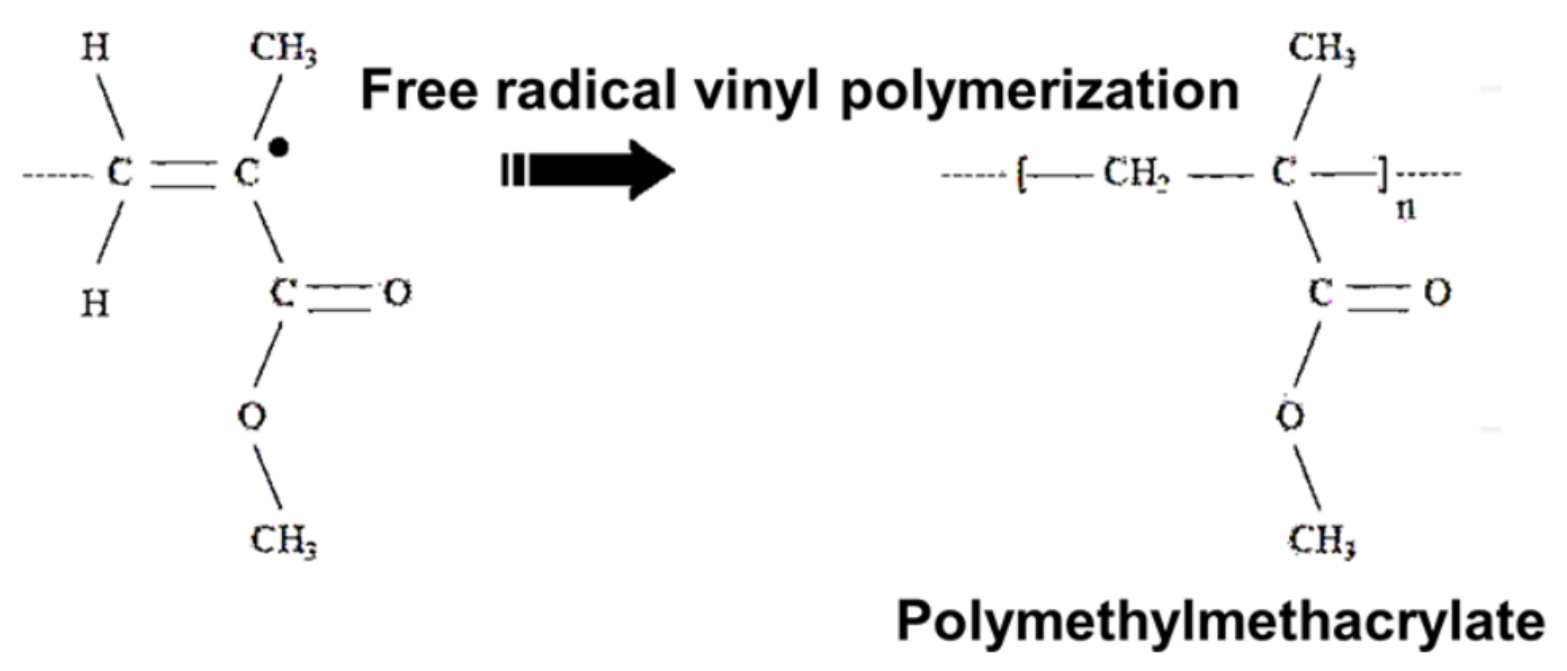
3. Preparation of Bio-Based Adhesives

Based on the numerous studies that have been conducted in order to examine the bio-adhesive synthesis and preparation, it can be concluded that there are two major process used to produce a bio-adhesive, i.e., polymerization and cross-linking [12,21,23,28,124,125,126]. The cross-linking process usually utilizes some type of bonding that can happen in the reaction, including hydrogen bonding, ionic bonding, host–guest interaction, hydrophobic bonds, imine bonds, disulfide bond, Acylhydrazone bonds, Diels-Alder reaction, boronate bonds, and oxime bonds [125]. In orthopedic surgery and orthodontics, poly(methyl methacrylate)s (PMMA) has been widely used. The polymerization of methyl methacrylate (MMA) via a free radical process utilizing an azo compound or peroxide as an initiator is a method to produce PMMA. Commercially, polymerization can be conducted, i.e., in bulk, solution, suspension, or emulsion. A viscous paste will be formed after blending these constituents which solidify via monomer radicals or anionic polymerization [11,26].
Alkyl 2-cyanoacrylates are now the most researched and most widely used group of bone adhesives. By altering the length of the alkyl chain, it is possible to produce a wide range of different 2-cyanoacrylates esters. The structure of poly (alkyl cyanoacrylate) (PACA), which was investigated for potential use in bone bio-based adhesives, is presented in Figure 2 [127]. The polymerization process can be carried out at room temperature without the need of a heating step, the addition of a catalyst, or the application of pressure thanks to the profound reactivity of these materials. The reaction that must take place in order to generate these materials begins with the anionic polymerization of the monomers, which is triggered by water. The acrylate bond can be broken by a nucleophilic attack carried out by weak bases such as water or amines. In order to accomplish bio-adhesion to bone, an electron-withdrawing nitrile group polarizes the acrylate bond. Because of this, the acrylate bond is susceptible to nucleophilic attack by weak bases, such as the amines that are found in the collagenous matrix of bone tissues. Increasing the length of the alkyl chain can, in general, result in greater polymerization rates, stronger bonding strengths in bone tissues, and can form more flexible chains [25].
Mixing a solution that contains a fibrinogen source (from plasma, platelet-rich plasma), or heterologous/autologous cryoprecipitate) and factor XIII with another separate solution consisting of thrombin source (bovine, human, or recombinant), anti-fibrinolytic agent, and calcium to prevent rapid fibrinolysis is the most common method that is used to produce fibrin-based adhesive systems. When brought together, these substances cause the formation of a clot that is devoid of cells. During this process, thrombin cleaves fibrinogen, which results in the production of soluble fibrin monomers. These monomers then self-assemble into loosely aggregated fibrils via hydrogen bonding, and then into a more robust cross-linked fibrin polymer via covalent bonding. Thrombin also activates factor XIII, which, in the presence of calcium, provides for the formation of covalent bonds between fibrin polymer chains. However, a considerable amount of preparation is required before employing this adhesive made from biomaterials [11,39].
Starch was oxidized with periodic acid in order to produce aldehyde side groups, and chitosan was used as the amino-group carrier throughout this process. In the bio-based adhesives system, amino groups that are present in the surrounding tissues will react with aldehyde groups in a manner analogous to that of chitosan. After being mixed together in water, the two components produce a Schiff’s base, which results in a covalent cross-linking that allows for a strong adhesion to tissue. This is accomplished by the production of covalent bonds. The bio-based glue had the potential to form bonds with any other exposed amino groups, such as those that are present in shattered bone for example. In addition, increasing the bio-adhesion strength to bone can be accomplished by conjugating starch or dextran compounds with 3,4-dihydroxy—phenylalanine (DOPA) [21,42,46,47,48]. A study reported that free radical copolymerization of monoacryloxyethyl phosphate (MAEP), dopamine methacrylate (DMA), and acrylamide (Aam) are used to produce bio-mimetic adhesive complex. This bio-based adhesive has the capability to bond wet bones together either in vitro and in vivo, demonstrating suitability for utilizing in the reconstruction of craniofacial fractures, and showed good degradability and osteoconductivity [11].
Biocompatible in situ-gelling Schiff’s base reaction and ionic interactions was conducted to produce carboxymethyl cellulose (CMC)-glycol chitosan (GC) hydrogel, a potential three-dimensional (3D) printing biomaterial ink for tissue engineering applications, the probable reaction is shown at Figure 3 [15]. A successful strategy to address cell-behavior on biomaterials was also presented by the plasma enhanced–chemical vapor deposition (PE-CVD) of polyethylene oxide-like (PEO)-like coatings [128]. Moreover, Tris(trimethylsiloxy)silyl (M3T) containing methacrylate copolymers with low surface energy were designed and synthesized [91].
Chitosan thiomer derivatives are utilized in order to produce a novel three-dimensional (3D) scaffold with potential soft tissue repair applications. A covalent coupling reaction was conducted to synthesize amino acid-grafted chitosan (cysteine, CHICys) and N-acylated chitosan (11- mercaptoundecanoic acid, CHIMerc) derivatives, and hydrogel scaffolds were produced by freeze-drying process. They were comprehensively characterized by swelling and degradation behaviors, NMR, FTIR, and Raman spectroscopy, SEM, and X-ray microcomputed tomography [14].
A series of chitosan-graft- polypeptides were synthesized by ring-opening polymerization of three N-carboxyanhydrides (NCAs)—3,4-di- hydroxyphenylalanine-N-carboxyanhydride (DOPA-NCA), cysteine-NCA (Cys-NCA) and arginine-NCA (Arg- NCA)—using partial-NH2-protected chitosan as an initiator since inspired by the mussel foot protein and chitosan-based macromolecular adhesives. Based on the result, these copolymers demonstrated good biodegradability and low cytotoxicity for application in orthopedic implant and scaffold [89].
A research also reported utilizing the 3,4-dihydroxyphenylalanine (DOPA), l-3,4-dihydroxyphenylalanine methyl ester (l-DOPAME), and Candida antartica fraction B (CAL-B) lipase in order to produce bio-based tissue adhesive by conducting direct conjugation of DOPA at the C-terminus on the surface of the protein and protein conjugation with tailor-made glycopolymers (DOPA-hyaluronic acid (HA) polymers) at the N-terminus [47]. The above are some examples of preparation process of bio-based adhesive production.

4. Characterization of Bio-Adhesives

4.1. In Vitro Methods

4.1.1. Shear Strength Measurement

The strength of bio-adhesion is commonly characterized by using mechanical testing, including crack growth assessment, peel test, and shear strength test. In the case of mucoadhesive assessment, shear strength measurements are commonly utilized to measure the forces within the mucus layer that slides each other in a parallel direction to the contact plane. Another method that can be utilized to measure the mucoadhesive strength is the flow channel method. The method assesses the shear strength by measuring the force needed to get the particle of adhesive from the mucin gel surface using forced humid air via flow cell. Furthermore, in order to assess the development of crack yielding from the dental implant, the bending tests were also conducted in the application of bio-based adhesive in orthodontic. The cracks are usually produced as a result of polymerization due to the shrinkage of the composite materials used in the implant. Characterization and interpretation of the bending test results is conducted using Griffith’s energy balance model. For example, the teeth elastic energy (usually the average elastic energy of tooth and the dental implant material) and the crack surface energy is set up using this balancing model. The experimental crack development assessment will decide the strain energy release rate or the stress intensity while the Poisson’s ration and modulus of the implant material will calculate the fracture energy [9,11,12,19,23].

4.1.2. Peel Strength Evaluation

Fractographic techniques, e.g., transmission electron microscopy (TEM) or scanning electron microscopy (SEM), are used in order to assess the quality of the dental implants-surface after performing the tensile test. American Standards for Testing and Materials (ASTM) with various tests are conducted on the interface of adhesion and the substrates. In order to obtain better shear strength, peel strength, and adhesion failure temperature, a pressure-sensitive adhesive (PSA) is formed as a composite material by supplementing it with montmorillonite, an organo-clay based element [14].

4.1.3. Flow through Experiment and Plate Method

The flow through channel method, which is the macro-scale measure of flow rate that can yield the depletion of bio-adhesive coated over the substrate sphere, is conducted to measure the mucoadhesion of DDS. The biophysical assessment method is conducted to measure the fluctuation in sedimentation coefficient that emerges due to the molecular weight change through an analytical centrifuge. The Wilhelm plate method is used for surface tension evaluation by utilizing natural or synthetic mucus rather than a conventional water medium. This method is known for use as macro-scale bio-adhesion assessment method. This method is conducted by coating a plate with any polymer material before the changes in interfacial properties and the bio-adhesion property are measured with respect to time [11,19].

4.2. Ex Vitro Methods

4.2.1. Adhesion Weight Method

A specific test method is developed in order to determine the weight of adherent particles that emerged in the interior mucous layers of guinea pig digestive tract due to the ion exchange. The particle size effect and adhesion charge after 5 min of time with the pig’s digestive tract was determined using this method. Based on the result it is recommended that the weight of the digestive tract increased due to bio-adhesion. However, when a larger change within the biological tissue emerged due to regeneration or degeneration of the digestive tract tissues, this method will posture a diminished reproducibility of the data [11,19].

4.2.2. Fluorescent Probe Methods

Fluorescent probe methods could determine the relationship between the polymer molecules and epithelial cell membranes. The formulation of an orally utilized bio-based adhesive polymer can actually be improved by knowing its structural requirements. The investigated bio-based adhesives can be tagged on to the cell membrane which consists of proteins and the lipid bi-layer membranes and the variations in fluorescent spectrum are noted. Excimer and monomer bands are two different Pyrene bands shown by these materials, and environmental viscosity will administer the ratio of these bands. Because of that, by assessing the bands ratio, the viscosity changes can be noted. Based on this result, it can be concluded that the adhesion strength is directly related to the viscosity change. The bond between polymer and protein membrane can be observed using a quantitative method (fluorescence depolarization), while the interactions of soluble polymers can be compared with that of peel of the cell [11,19].

5. Challenges

Based on the findings, presented in numerous research articles, it can be seen that bio-based adhesives have demonstrated their superiority in various medical applications. At present, the application of bio-based adhesives in the medical sector, including orthopedics, is an area of great scientific interest due to the recent advances in their formulations.
Bio-based adhesives consist of synthetic materials and natural materials. A number of studies have shown that synthetic materials have advantages from several aspects, especially with regards to mechanical properties [129]. Synthetic materials have adhesive strength, shear strength, and tensile strength which are much higher than natural materials, but these synthetic materials have major weaknesses in terms of biocompatibility and cytotoxicity, as it is known that synthetic materials have elements that are toxic to the human body. Meanwhile, natural materials have advantages in terms of biocompatibility and cytotoxicity because they are acceptable for use in the human body, but these materials still have weaknesses in terms of mechanical properties. Several studies have shown that the adhesive strength produced from bio-based adhesives derived from natural raw materials is still very low and is only sufficient to meet the need for adhesives for soft tissue or surgical sutures. Therefore, it is necessary to conduct future research to optimize the material properties of bio-based adhesives in order to obtain materials that have both satisfactory mechanical properties and good biocompatibility properties.
Bio-based adhesives for orthopedic applications, which are produced from utilization of natural resource polymers, are less studied. The main challenge in using bio-based adhesives for orthopedic applications is meeting the stringent requirements for high bond strength in a demanding clinical environment [116]. Markedly, bio-based adhesives have better biodegradability and biocompatibility properties, although bio-based adhesives from synthetic or chemical preparation have higher adhesion strength to bone. Polymers known as hydrogels are more suitable to be used in bio-based adhesive applications for soft tissue than bio-based adhesives for bone adhesive due to the low cohesive strength produced by these materials. A more rational design should be executed to develop bio-based adhesive materials with particular bonding strength to bone tissue. Bone type, age of the patients, fracture location, samples storage, and treatment can affect the physicochemical properties of bone, so a particular method is required to characterize the properties of bio-based bone adhesives. Various distinctive assessment methods have been conducted, including the foremost common butt tensile strength tests, which compromise coordinate comparison between various tests. In this respect, the establishment of standardized and reproducible assessment protocols for the adhesion strength of bio-based adhesives to bone is of utmost importance.
Another obstacle in this field is lowering the cost of bio-based adhesive materials [117]. Several researchers have emphasized the growing need to convert available agricultural wastes into biocompatible and biodegradable products [118,119]. Nonetheless, research on the subject is still lacking, necessitating ongoing investigations.

6. Future Perspectives

Polymers with biodegradability properties, prepared using synthetic or chemical methods, can be designed by tuning the chemical groups, the degree of cross-linking, viscosity, and surface tension. They represent a promising approach for bone fracture healing. The chemical composition of bone must be the main consideration for future bone adhesive design. A matrix consisting of organic collagen (≈30 wt.%), reinforced by nanocrystals of calcium phosphate mineral (≈70 wt.%), is the main constituent of bone tissue. Biodegradable polymers, such as PEG or alginate, can be outlined to connect firmly to bone tissue through functionalization with the addition of chemical groups, which have high bonding strength with bone components. N-hydroxysuccinimide esters (NHS esters) are known to build solid covalent amide bonds with primary amines found within the collagenous extracellular bone matrix. On the other hand, a few functional groups are known to bind to Ca2+ as present within the mineral phase of bone tissue, i.e., hydroxyapatite. Bisphosphonates (BP) are anti-osteoporotic particles that are known for their uncommonly strong affinity for hydroxyapatite. Moreover, in this manner, BP-functionalized polymers might strongly adhere to bone by forming ionic bonds between pendant BP groups and Ca2+. Organic compounds, consisting of carboxyl groups with strong adhesion for Ca2+, generally include proteins, sequences of peptide, amino acids in single form, and carboxyl groups, such as sulfate groups, hydroxyl, and catechol. Moreover, the adhesion to bone can be improved by expanding the amount of functional (side) groups with high affinity to bone in the polymer.
The use of bio-based adhesives can reduce the complicated invasive techniques currently used in orthopedics, which in turn will result in improved patient treatment protocols and quality of life. It can be expected that, in the future, the bio-based bone adhesive materials with more judicious design will permit bone fracture fixation utilizing internal fixation.
Future research should be focused on comprehensive studies of the suitability of various bio-based adhesives for bone tissue scaffolds, development of innovative injectable adhesives for fracture treatment, and mechanical testing of bio-based adhesives intended for orthopedic applications. The existing studies in the field remain rather limited, and the results will be of great importance for the development of bio-based adhesives for orthopedic applications with optimal performance.

Author Contributions

Conceptualization, N.F.N.,M.A.R.L. and P.A.; methodology, N.F.N. and M.A.R.L.; validation, N.F.N., M.A.R.L. and P.A.; investigation, N.F.N. and M.A.R.L.; resources, N.F.N.,M.A.R.L., D.J., E.M., P.A., L.K. and L.S.H.; data curation, N.F.N.; writing—original draft preparation, N.F.N.; writing—review and editing, N.F.N.,M.A.R.L., P.A., L.K. and L.S.H.; visualization, D.J. and E.M.; supervision, M.A.R.L. and P.A.; project administration, M.A.R.L., D.J. and P.A. All authors have read and agreed to the published version of the manuscript.

Funding

This research received no external funding.

Institutional Review Board Statement

The study did not require ethical approval.

Informed Consent Statement

Not applicable.

Data Availability Statement

Not applicable.

Acknowledgments

This study was supported by Research Collaboration Center for Biomass and Biorefinery between BRIN and Universitas Padjadjaran, and Research Collaboration Center for Biomedical Scaffold between BRIN and Universitas Gadjah Mada.

Conflicts of Interest

The authors declare no conflict of interest.

References

  1. Tsiridis, E.; Upadhyay, N.; Giannoudis, P. Molecular Aspects of Fracture Healing: Which Are the Important Molecules? Int. J. Care Inj. 2007, 38S1, S11–S25. [Google Scholar] [CrossRef] [PubMed]
  2. Dimitriou, R.; Tsiridis, E.; Giannoudis, P.V. Current Concepts of Molecular Aspects of Bone Healing. Injury 2005, 36, 1392–1404. [Google Scholar] [CrossRef] [PubMed]
  3. Marsell, R.; Einhorn, T.A. The Biology of Fracture Healing. Injury 2011, 42, 551–555. [Google Scholar] [CrossRef] [PubMed] [Green Version]
  4. Barnes, G.L.; Kostenuik, P.J.; Gerstenfeld, L.C.; Einhorn, T.A. Growth Factor Regulation of Fracture Repair. J. Bone Miner. Res. 1999, 14, 1805–1815. [Google Scholar] [CrossRef]
  5. Al-Aql, Z.S.; Alagl, A.S.; Graves, D.T.; Gerstenfeld, L.C.; Einhorn, T.A. Molecular Mechanisms Controlling Bone Formation During Fracture Healing and Distraction Osteogenesis. J. Dent. Res. 2008, 87, 107–118. [Google Scholar] [CrossRef] [PubMed]
  6. Yu, Y.; Brió Pérez, M.; Cao, C.; de Beer, S. Switching (Bio-) Adhesion and Friction in Liquid by Stimulus Responsive Polymer Coatings. Eur. Polym. J. 2021, 147, 110298. [Google Scholar] [CrossRef]
  7. Kumar, M.; Tomar, M.; Punia, S.; Dhakane-Lad, J.; Dhumal, S.; Changan, S.; Senapathy, M.; Berwal, M.K.; Sampathrajan, V.; Sayed, A.A.S.; et al. Plant-Based Proteins and Their Multifaceted Industrial Applications. Lwt 2022, 154, 112620. [Google Scholar] [CrossRef]
  8. Sardella, E.; Gristina, R.; Fanelli, F.; Veronico, V.; Da Ponte, G.; Kroth, J.; Fracassi, F.; Favia, P. How to Confer a Permanent Bio-Repelling and Bio-Adhesive Character to Biomedical Materials through Cold Plasmas. Appl. Sci. 2020, 10, 9101. [Google Scholar] [CrossRef]
  9. Majeed, H.; Rehman, K.; Ali, A.; Khalid, M.F.; Akash, M.S.H. Wound Healing Adhesives. In Green Adhesives; John Wiley & Sons, Inc.: Hoboken, NJ, USA, 2020; pp. 181–204. [Google Scholar]
  10. Xiang, L. Molecular Interaction and Adhesion Mechanisms of Mussel-Inspired Adhesive Coatings; University of Alberta: Alberta, Canada, 2020. [Google Scholar]
  11. Ramesh, M.; Kumar, L.R. Bioadhesives. In Green Adhesives: Preparation, Properties and Applications; John Wiley & Sons: Inc Hoboken, NJ, USA, 2020; pp. 145–163. [Google Scholar]
  12. Rathi, S.; Saka, R.; Domb, A.J.; Khan, W. Protein-Based Bioadhesives and Bioglues. Polym. Adv. Technol. 2019, 30, 217–234. [Google Scholar] [CrossRef]
  13. Bolghari, N.; Shahsavarani, H.; Anvari, M.; Habibollahi, H. A Novel Recombinant Chimeric Bio-Adhesive Protein Consisting of Mussel Foot Protein 3, 5, Gas Vesicle Protein A, and CsgA Curli Protein Expressed in Pichia Pastoris. AMB Express 2022, 12, 1–19. [Google Scholar] [CrossRef]
  14. Medeiros Borsagli, F.G.L.; Carvalho, I.C.; Mansur, H.S. Amino Acid-Grafted and N-Acylated Chitosan Thiomers: Construction of 3D Bio-Scaffolds for Potential Cartilage Repair Applications. Int. J. Biol. Macromol. 2018, 114, 270–282. [Google Scholar] [CrossRef] [PubMed]
  15. Janarthanan, G.; Tran, H.N.; Cha, E.; Lee, C.; Das, D.; Noh, I. 3D Printable and Injectable Lactoferrin-Loaded Carboxymethyl Cellulose-Glycol Chitosan Hydrogels for Tissue Engineering Applications. Mater. Sci. Eng. C 2020, 113, 111008. [Google Scholar] [CrossRef] [PubMed]
  16. Shahryarimorad, K.; Alipour, A.; Honar, Y.S.; Abtahi, B. In Silico Prediction and in Vitro Validation of the Effect of PH on Adhesive Behaviour of the Fused CsgA - MFP3 Protein. AMB Express 2022, 12, 1–13. [Google Scholar] [CrossRef] [PubMed]
  17. Kazemi, S.; Pourmadadi, M.; Yazdian, F.; Ghadami, A. The Synthesis and Characterization of Targeted Delivery Curcumin Using Chitosan-Magnetite-Reduced Graphene Oxide as Nano-Carrier. Int. J. Biol. Macromol. 2021, 186, 554–562. [Google Scholar] [CrossRef] [PubMed]
  18. Alqosaibi, A.I. Nanocarriers for Anticancer Drugs: Challenges and Perspectives. Saudi J. Biol. Sci. 2022, 29, 103298. [Google Scholar] [CrossRef] [PubMed]
  19. Chopra, H.; Kumar, S.; Singh, I. Bioadhesive Hydrogels and Their Applications. In Bioadhesives in Drug Delivery; John Wiley & Sons: Inc Hoboken, NJ, USA, 2020; pp. 147–170. ISBN 9781119640240. [Google Scholar]
  20. Lyu, Q.; Peng, L.; Hong, X.; Fan, T.; Li, J.; Cui, Y.; Zhang, H.; Zhao, J. Smart Nano-Micro Platforms for Ophthalmological Applications: The State-of-the-Art and Future Perspectives. Biomaterials 2021, 270, 120682. [Google Scholar] [CrossRef] [PubMed]
  21. Li, D.; Chen, J.; Wang, X.; Zhang, M.; Li, C.; Zhou, J. Recent Advances on Synthetic and Polysaccharide Adhesives for Biological Hemostatic Applications. Front. Bioeng. Biotechnol. 2020, 8, 926. [Google Scholar] [CrossRef] [PubMed]
  22. Guo, Q.; Chen, J.; Wang, J.; Zeng, H.; Yu, J. Recent Progress in Synthesis and Application of Mussel-Inspired Adhesives. Nanoscale 2020, 12, 1307–1324. [Google Scholar] [CrossRef]
  23. Bu, Y.; Pandit, A. Cohesion Mechanisms for Bioadhesives. Bioact. Mater. 2022, 13, 105–118. [Google Scholar] [CrossRef]
  24. Patil, N.A.; Kandasubramanian, B. Functionalized Polylysine Biomaterials for Advanced Medical Applications: A Review. Eur. Polym. J. 2021, 146, 110248. [Google Scholar] [CrossRef]
  25. Korde, J.M.; Kandasubramanian, B. Biocompatible Alkyl Cyanoacrylates and Their Derivatives as Bio-Adhesives. Biomater. Sci. 2018, 6, 1691–1711. [Google Scholar] [CrossRef] [PubMed]
  26. Al-Abassi, A.; Papini, M.; Towler, M. Review of Biomechanical Studies and Finite Element Modeling of Sternal Closure Using Bio-Active Adhesives. Bioengineering 2022, 9, 198. [Google Scholar] [CrossRef] [PubMed]
  27. Yousefi, A.M. A Review of Calcium Phosphate Cements and Acrylic Bone Cements as Injectable Materials for Bone Repair and Implant Fixation. J. Appl. Biomater. Funct. Mater. 2019, 17, 2280800019872594. [Google Scholar] [CrossRef]
  28. Chen, S.; Gil, C.J.; Ning, L.; Jin, L.; Perez, L.; Kabboul, G.; Tomov, M.L.; Serpooshan, V. Adhesive Tissue Engineered Scaffolds: Mechanisms and Applications. Front. Bioeng. Biotechnol. 2021, 9, 683079. [Google Scholar] [CrossRef] [PubMed]
  29. Venturella, F. Development of Nontoxic Bio–Adhesives for Wet Environments; Università degli Studi di Palermo: Palermo, Italy, 2021. [Google Scholar]
  30. Gillman, N.; Lloyd, D.; Bindra, R.; Ruan, R.; Zheng, M. Surgical Applications of Intracorporal Tissue Adhesive Agents: Current Evidence and Future Development. Expert Rev. Med. Devices 2020, 17, 443–460. [Google Scholar] [CrossRef]
  31. Park, K.; Kim, S.; Jo, Y.; Park, J.; Kim, I.; Hwang, S.; Lee, Y.; Kim, S.Y.; Seo, J. Lubricant Skin on Diverse Biomaterials with Complex Shapes via Polydopamine-Mediated Surface Functionalization for Biomedical Applications. Bioact. Mater. 2022. [Google Scholar] [CrossRef]
  32. Zheng, D.; Ruan, H.; Chen, W.; Zhang, Y.; Cui, W.; Chen, H.; Shen, H. Advances in Extracellular Vesicle Functionalization Strategies for Tissue Regeneration. Bioact. Mater. 2022. [Google Scholar] [CrossRef]
  33. Liu, C.; Yu, Q.; Yuan, Z.; Guo, Q.; Liao, X.; Han, F.; Feng, T.; Liu, G.; Zhao, R.; Zhu, Z.; et al. Engineering the Viscoelasticity of Gelatin Methacryloyl (GelMA) Hydrogels via Small “Dynamic Bridges” to Regulate BMSC Behaviors for Osteochondral Regeneration. Bioact. Mater. 2022. [Google Scholar] [CrossRef]
  34. Shokri, M.; Dalili, F.; Kharaziha, M.; Baghaban Eslaminejad, M.; Ahmadi Tafti, H. Strong and Bioactive Bioinspired Biomaterials, next Generation of Bone Adhesives. Adv. Colloid Interface Sci. 2022, 305, 102706. [Google Scholar] [CrossRef]
  35. Wang, X.; Fang, X.; Gao, X.; Wang, H.; Li, S.; Li, C.; Qing, Y.; Qin, Y. Strong Adhesive and Drug-Loaded Hydrogels for Enhancing Bone–Implant Interface Fixation and Anti-Infection Properties. Colloids Surf. B Biointerfaces 2022, 219, 112817. [Google Scholar] [CrossRef]
  36. Du, J.; Zhou, Y.; Bao, X.; Kang, Z.; Huang, J.; Xu, G.; Yi, C.; Li, D. Surface Polydopamine Modification of Bone Defect Repair Materials: Characteristics and Applications. Front. Bioeng. Biotechnol. 2022, 10, 1–18. [Google Scholar] [CrossRef] [PubMed]
  37. Abourehab, M.A.S.; Rajendran, R.R.; Singh, A.; Pramanik, S.; Shrivastav, P.; Ansari, M.J.; Manne, R.; Amaral, L.S.; Deepak, A. Alginate as a Promising Biopolymer in Drug Delivery and Wound Healing: A Review of the State-of-the-Art. Int. J. Mol. Sci. 2022, 23, 9035. [Google Scholar] [CrossRef] [PubMed]
  38. Bohara, S.; Suthakorn, J. Surface Coating of Orthopedic Implant to Enhance the Osseointegration and Reduction of Bacterial Colonization: A Review. Biomater. Res. 2022, 26, 1–17. [Google Scholar] [CrossRef] [PubMed]
  39. Pei, X.; Wang, J.; Cong, Y.; Fu, J. Recent Progress in Polymer Hydrogel Bioadhesives. J. Polym. Sci. 2021, 59, 1312–1337. [Google Scholar] [CrossRef]
  40. Espinoza-Ramirez, A.; Fuentes-Rodriguez, H.; Hernandez-Herrera, E.; Mora-Sandi, A.; Vega-Baudrit, J.R. Nanobiodiversity and Biomimetic Adhesives Development: From Nature to Production and Application. J. Biomater. Nanobiotechnol. 2019, 10, 78–101. [Google Scholar] [CrossRef] [Green Version]
  41. Zheng, G.; Cui, Y.; Lu, L.; Guo, M.; Hu, X.; Wang, L.; Yu, S.; Sun, S.; Li, Y.; Zhang, X.; et al. Microfluidic Chemostatic Bioreactor for High-Throughput Screening and Sustainable Co-Harvesting of Biomass and Biodiesel in Microalgae. Bioact. Mater. 2022, 1–11. [Google Scholar] [CrossRef]
  42. Samyn, P. A Platform for Functionalization of Cellulose, Chitin/Chitosan, Alginate with Polydopamine: A Review on Fundamentals and Technical Applications. Int. J. Biol. Macromol. 2021, 178, 71–93. [Google Scholar] [CrossRef]
  43. Dongre, R.S. Marine Polysaccharides in Pharmaceutical Uses; Springer: Berlin/Heidelberg, Germany, 2022; ISBN 9783030357344. [Google Scholar]
  44. Bashir, S.M.; Rather, G.A.; Patrício, A.; Haq, Z.; Sheikh, A.A.; Zahoor, M.; Singh, H.; Khan, A.A.; Imtiyaz, S.; Ahmad, S.B. Chitosan Nanoparticles: A Versatile Platform for Biomedical Applications. Materials 2022, 15, 6521. [Google Scholar] [CrossRef]
  45. Abourehab, M.A.S.; Pramanik, S.; Abdelgawad, M.A.; Abualsoud, B.M.; Kadi, A.; Ansari, M.J.; Deepak, A. Recent Advances of Chitosan Formulations in Biomedical Applications. Int. J. Mol. Sci. 2022, 23, 10975. [Google Scholar] [CrossRef] [PubMed]
  46. Chen, Y.; Cui, G.; Dan, N.; Huang, Y.; Bai, Z.; Yang, C.; Dan, W. Preparation and Characterization of Dopamine–Sodium Carboxymethyl Cellulose Hydrogel. SN Appl. Sci. 2019, 1, 1–10. [Google Scholar] [CrossRef]
  47. Jaramillo, J.; Rodriguez-Oliva, I.; Abian, O.; Palomo, J.M. Specific Chemical Incorporation of L-DOPA and Functionalized l-DOPA-Hyaluronic Acid in Candida Antarctica Lipase: Creating Potential Mussel-Inspired Bioadhesives. SN Appl. Sci. 2020, 2, 1–12. [Google Scholar] [CrossRef]
  48. Lutz, T.M.; Kimna, C.; Casini, A.; Lieleg, O. Bio-Based and Bio-Inspired Adhesives from Animals and Plants for Biomedical Applications. Mater. Today Bio. 2022, 13, 100203. [Google Scholar] [CrossRef] [PubMed]
  49. Sun, J. Fabrication and Mechanical Properties of Supercharged Polypeptides Based Biomaterials: From Adhesives to Fibers; University of Groningen: The Netherlands, 2020. [Google Scholar]
  50. Pandey, N.; Soto-Garcia, L.F.; Liao, J.; Zimmern, P.; Nguyen, K.T.; Hong, Y. Mussel-Inspired Bioadhesives in Healthcare: Design Parameters, Current Trends, and Future Perspectives. Biomater. Sci. 2020, 8, 1240–1255. [Google Scholar] [CrossRef] [PubMed]
  51. Kang, V.; Lengerer, B.; Wattiez, R.; Flammang, P. Molecular Insights into the Powerful Mucus-Based Adhesion of Limpets (Patella Vulgata L.): Molecular Insights into Limpets Adhesion. Open Biol. 2020, 10, 200019. [Google Scholar] [CrossRef]
  52. Wang, H.; Wang, L.; Zhang, S.; Zhang, W.; Li, J.; Han, Y. Mussel-Inspired Polymer Materials Derived from Nonphytogenic and Phytogenic Catechol Derivatives and Their Applications. Polym. Int. 2021, 70, 1209–1224. [Google Scholar] [CrossRef]
  53. Shi, C.; Chen, X.; Zhang, Z.; Chen, Q.; Shi, D.; Kaneko, D. Mussel Inspired Bio-Adhesive with Multi-Interactions for Tissue Repair. J. Biomater. Sci. Polym. Ed. 2020, 31, 491–503. [Google Scholar] [CrossRef]
  54. Park, M.K.; Li, M.X.; Yeo, I.; Jung, J.; Yoon, B.I.L.; Joung, Y.K. Balanced Adhesion and Cohesion of Chitosan Matrices by Conjugation and Oxidation of Catechol for High-Performance Surgical Adhesives. Carbohydr. Polym. 2020, 248, 116760. [Google Scholar] [CrossRef] [PubMed]
  55. Zhou, D.; Li, S.; Pei, M.; Yang, H.; Gu, S.; Tao, Y.; Ye, D.; Zhou, Y.; Xu, W.; Xiao, P. Dopamine-Modified Hyaluronic Acid Hydrogel Adhesives with Fast-Forming and High Tissue Adhesion. ACS Appl. Mater. Interfaces 2020, 12, 18225–18234. [Google Scholar] [CrossRef] [PubMed]
  56. Xiang, L.; Zhang, J.; Wang, W.; Gong, L.; Zhang, L.; Yan, B.; Zeng, H. Nanomechanics of π-Cation-π Interaction with Implications for Bio-Inspired Wet Adhesion. Acta Biomater. 2020, 117, 294–301. [Google Scholar] [CrossRef] [PubMed]
  57. Ventura, I.V.P. Characterization of Glycoproteins Involved in Sea Urchin Adhesion; University of Coimbra: Portugal, 2020. [Google Scholar]
  58. Capitain, C.; Wagner, S.; Hummel, J.; Tippkötter, N. Investigation of C–N Formation Between Catechols and Chitosan for the Formation of a Strong, Novel Adhesive Mimicking Mussel Adhesion. Waste Biomass Valorization 2021, 12, 1761–1779. [Google Scholar] [CrossRef]
  59. Kwon, Y.; Yang, D.H.; Lee, D. A Titanium Surface-Modified with Nano-Sized Hydroxyapatite and Simvastatin Enhances Bone Formation and Osseointegration. J. Biomed. Nanotechnol. 2015, 11, 1007–1015. [Google Scholar] [CrossRef] [PubMed]
  60. Cheng, Z.; Guo, C.; Dong, W.; He, F.; Zhao, S.; Yang, G. Effect of Thin Nano-Hydroxyapatite Coating on Implant Osseointegration in Ovariectomized Rats. Oral Maxillofac. Surg. 2012, 113, 48–53. [Google Scholar] [CrossRef]
  61. Pang, K.-M.; Lee, J.-K.; Seo, Y.-K.; Kim, S.-M.; Kim, M.-J.; Lee, J.-H. Biologic Properties of Nano-Hydroxyapatite: An in Vivo Study of Calvarial Defects, Ectopic Bone Formation and Bone Implantation. Biomed. Mater. Eng. 2015, 25, 25–38. [Google Scholar] [CrossRef] [PubMed]
  62. Juliadmi, D.; Nuswantoro, N.F.; Fajri, H.; Indriyani, I.Y.; Affi, J.; Manjas, M.; Tjong, D.H.; Gunawarman. The Coating of Bovine-Source Hydroxyapatite on Titanium Alloy (Ti-6Al-4V ELI) Using Electrophoretic Deposition for Biomedical Application. Mater. Sci. Forum 2020, 1000, 97–106. [Google Scholar] [CrossRef]
  63. Kusrini, E.; Sontang, M. Characterization of X-Ray Diffraction and Electron Spin Resonance: Effects of Sintering Time and Temperature on Bovine Hydroxyapatite. Radiat. Phys. Chem. 2012, 81, 118–125. [Google Scholar] [CrossRef]
  64. Khandan, A.; Abdellahi, M.; Ozada, N.; Ghayour, H. Study of the Bioactivity, Wettability and Hardness Behaviour of the Bovine Hydroxyapatite-Diopside Bio-Nanocomposite Coating. J. Taiwan Inst. Chem. Eng. 2016, 60, 538–546. [Google Scholar] [CrossRef]
  65. Mihailescu, N.; Stan, G.E.; Duta, L.; Chifiriuc, C.M.; Bleotu, C.; Sopronyi, M.; Luculescu, C.; Oktar, F.N.; Mihailescu, I.N. Structural, Compositional, Mechanical Characterization and Biological Assessment of Bovine-Derived Hydroxyapatite Coatings Reinforced with MgF 2 or MgO for Implants Functionalization. Mater. Sci. Eng. C 2016, 59, 863–874. [Google Scholar] [CrossRef]
  66. Gunawarman; Mulyadi, I.H.; Arif, Z.; Nuswantoro, N.F.; Affi, J.; Niinomi, M. Effect of Particle Size on Adhesion Strength of Bovine Hydroxyapatite Layer on Ti-12Cr Coated by Using Electrophoretic Deposition (EPD) Method. In Proceedings of the 2nd Conference on Innovation in Technology (CITES 2020), Padang, Indonesia, 4–5 November 2020; pp. 1–8. [Google Scholar]
  67. Fajri, H.; Ramadhan, F.; Nuswantoro, N.F.; Juliadmi, D.; Tjong, D.H.; Manjas, M.; Affi, J.; Yetri, Y.; Gunawarman. Electrophoretic Deposition (EPD) of Natural Hydroxyapatite Coatings on Titanium Ti-29Nb-13Ta-4. 6Zr Substrates for Implant Material. Mater. Sci. Forum 2020, 1000, 123–131. [Google Scholar] [CrossRef]
  68. Gunawarman; Affi, J.; Yetri, Y.; Ilhamdi; Juliadmi, D.; Nuswantoro, N.F.; Fajri, H.; Ahli, A.; Gundini, R.; Nur, H. Synthesis and Characterization of Calcium Precursor for Hydroxyapatite Synthesis from Blood Clam Shell (Anadara Antiquata) Using Planetary Ball Mill Process. In Proceedings of the IOP Conference Series: Materials Science and Engineering, Padang, Indonesia, 8–9 November 2018; pp. 1–6. [Google Scholar]
  69. Khalili, V.; Khalil-allafi, J.; Xia, W.; Parsa, A.B.; Frenzel, J.; Somsen, C.; Eggeler, G. Preparing Hydroxyapatite-Silicon Composite Suspensions with Homogeneous Distribution of Multi-Walled Carbon Nano-Tubes for Electrophoretic Coating of NiTi Bone Implant and Their Effect on the Surface Morphology. Appl. Surf. Sci. 2016, 366, 158–165. [Google Scholar] [CrossRef]
  70. Gunawarman; Nuswantoro, N.F.; Juliadmi, D.; Fajri, H.; Budiman, A.; Djong, H.T.; Manjas, M. Hydroxyapatite Coatings on Titanium Alloy TNTZ Using Electrophoretic Deposition. In Proceedings of the IOP Conference Series: Materials Science and Engineering, Padang, Indonesia, 8–9 November 2018; pp. 1–11. [Google Scholar]
  71. Juliadmi, D.; Fauzi, V.R.; Gunawarman; Nur, H.; Idris, M.H. Hydroxyapatite Coating on Titanium Alloy Ti-6Al-4V with Electrophoretic Deposition (EPD) for Dental Root Application. Int. J. Adv. Sci. Eng. Informational Technol. 2017, 7, 2152–2158. [Google Scholar] [CrossRef]
  72. Łukaszewska-Kuska, M.; Krawczyk, P.; Martyla, A.; Hędzelek, W.; Dorocka-bobkowska, B.; Dorocka-bobkowska, B. Hydroxyapatite Coating on Titanium Endosseous Implants for Improved Osseointegration: Physical and Chemical Considerations Address for Correspondence. Adv. Clin. Exper. Med. 2018, 27, 1055–1059. [Google Scholar] [CrossRef] [PubMed]
  73. Liang, H.; Xu, X.; Feng, X.; Deng, X.; Wu, S.; Liu, X.; Yang, C. Gold Nanoparticles-Loaded Hydroxyapatite Composites Guide Osteogenic Differentiation of Human Mesenchymal Stem Cells through Wnt/β–Catenin Signaling Pathway. Int. J. Nanomed. 2019, 14, 6151–6163. [Google Scholar] [CrossRef] [PubMed] [Green Version]
  74. Nuswantoro, N.F.; Juliadmi, D.; Fajri, H.; Manjas, M.; Suharti, N.; Tjong, D.H.; Affi, J.; Gunawarman. Electrophoretic Deposition Performance of Hydroxyapatite Coating on Titanium Alloys for Orthopedic Implant Application. Mater. Sci. Forum 2020, 1000, 69–81. [Google Scholar] [CrossRef]
  75. Nuswantoro, N.F.; Manjas, M.; Suharti, N.; Juliadmi, D.; Fajri, H.; Tjong, D.H.; Affi, J.; Niinomi, M.; Gunawarman. Hydroxyapatite Coating on Titanium Alloy TNTZ for Increasing Osseointegration and Reducing Inflammatory Response in Vivo on Rattus Norvegicus Wistar Rats. Ceram. Int. 2021, 47, 16094–16100. [Google Scholar] [CrossRef]
  76. Nuswantoro, N.F.; Gunawarman; Saputra, M.R.; Nanda, I.P.; Idris, M.H.; Arafat, A. Microstructure Analysis of Hydroxyapatite Coating on Stainless Steel 316L Using Investment Casting Technique for Implant Application. Int. J. Adv. Sci. Eng. Inform. Technol. 2018, 8, 2168–2174. [Google Scholar] [CrossRef] [Green Version]
  77. Beig, B.; Liaqat, U.; Niazi, M.F.K.; Douna, I.; Zahoor, M.; Niazi, M.B.K. Current Challenges and Innovative Developments in Hydroxyapatite-Based Coatings on Metallic Materials for Bone Implantation: A Review. Coatings 2020, 10, 1249. [Google Scholar] [CrossRef]
  78. Bose, S.; Tarafder, S.; Bandyopadhyay, A. Hydroxyapatite Coatings for Metallic Implants. In Hydroxyapatite (HAP) for Biomedical Applications. Elsevier Ltd.: Amsterdam, The Netherlands, 2015; Volume 7, pp. 143–157. ISBN 9781782420330. [Google Scholar]
  79. Nuswantoro, N.F.; Maulana, I.; Djong, H.T.; Manjas, M.; Gunawarman. Hydroxyapatite Coating on New Type Titanium, TNTZ, Using Electrophoretic Deposition. J. Ocean. Mech. Aerosp. 2018, 56, 1–4. [Google Scholar]
  80. Gunawarman; Affi, J.; Sutanto, A.; Putri, D.M.; Juliadmi, D.; Nuswantoro, N.F.; Fajri, H.; Tjong, D.H.; Manjas, M. Adhesion Strength of Hydroxyapatite Coating on Titanium Alloy (Ti-6Al- 4V ELI) for Biomedical Application. In Proceedings of the International Colloquium on Computational and Experimental Mechanics (ICCEM 2020), Selangor, Malaysia, 25–26 June 2020; pp. 1–9. [Google Scholar]
  81. Araghi, A.; Hadianfard, M.J. Fabrication and Characterization of Functionally Graded Hydroxyapatite/TiO2 Multilayer Coating on Ti-6Al-4V Titanium Alloy for Biomedical Applications. Ceram. Int. 2015, 41, 12668–12679. [Google Scholar] [CrossRef]
  82. Nuswantoro, N.F.; Budiman, I.; Septiawarman, A.; Djong, H.T.; Manjas, M.; Gunawarman. Effect of Applied Voltage and Coating Time on Nano Hydroxyapatite Coating on Titanium Alloy Ti6Al4V Using Electrophoretic Deposition for Orthopaedic Implant Application. In Proceedings of the IOP Conference Series: Materials Science and Engineering, Bandung, Indonesia, 6–7 September 2018; Volume 547, pp. 1–11. [Google Scholar]
  83. Bovand, D.; Allazadeh, M.R.; Rasouli, S.; Khodadad, E.; Borhani, E. Studying the Effect of Hydroxyapatite Particles in Osteoconductivity of Ti-HA Bioceramic. J. Aust. Ceram. Soc. 2019, 55, 395–403. [Google Scholar] [CrossRef] [Green Version]
  84. Han, Y.; Zhao, W.; Zheng, Y.; Wang, H.; Sun, Y.; Zhang, Y.; Luo, J.; Zhang, H. Self-Adhesive Lubricated Coating for Enhanced Bacterial Resistance. Bioact. Mater. 2021, 6, 2535–2545. [Google Scholar] [CrossRef] [PubMed]
  85. Alinavaz, S.; Mahdavinia, G.R.; Jafari, H.; Hazrati, M.; Akbari, A. Hydroxyapatite (HA)-Based Hybrid Bionanocomposite Hydrogels: Ciprofloxacin Delivery, Release Kinetics and Antibacterial Activity. J. Mol. Struct. 2021, 1225, 129095. [Google Scholar] [CrossRef]
  86. Albu, A.M.; Draghicescu, W.; Munteanu, T.; Ion, R.; Mitran, V.; Cimpean, A.; Popescu, S.; Pirvu, C. Nitrodopamine vs Dopamine as an Intermediate Layer for Bone Regeneration Applications. Mater. Sci. Eng. C 2019, 98, 461–471. [Google Scholar] [CrossRef]
  87. Wang, Y.; Guo, J.; Li, B.; Li, D.; Meng, Z.; Sun, S.K. Biocompatible Therapeutic Albumin/Genipin Bioglue for Postoperative Wound Adhesion and Residual Tumor Ablation. Biomaterials 2021, 279, 121179. [Google Scholar] [CrossRef] [PubMed]
  88. Vargas Villanueva, J.G.; Sarmiento Huertas, P.A.; Galan, F.S.; Esteban Rueda, R.J.; Briceño Triana, J.C.; Casas Rodriguez, J.P. Bio-Adhesion Evaluation of a Chitosan-Based Bone Bio-Adhesive. Int. J. Adhes. Adhes. 2019, 92, 80–88. [Google Scholar] [CrossRef]
  89. Lu, D.; Wang, H.; Wang, X.; Li, Y.; Guo, H.; Sun, S.; Zhao, X.; Yang, Z.; Lei, Z. Biomimetic Chitosan-Graft-Polypeptides for Improved Adhesion in Tissue and Metal. Carbohydr. Polym. 2019, 215, 20–28. [Google Scholar] [CrossRef] [PubMed]
  90. Singh, G.; Nayal, A.; Malhotra, S.; Koul, V. Dual Functionalized Chitosan Based Composite Hydrogel for Haemostatic Efficacy and Adhesive Property. Carbohydr. Polym. 2020, 247, 116757. [Google Scholar] [CrossRef]
  91. Lei, H.; Xiong, M.; Xiao, J.; Zheng, L.; Zhuang, Q. Fluorine-Free Coating with Low Surface Energy and Anti-Biofouling Properties. Prog. Org. Coatings 2018, 124, 158–164. [Google Scholar] [CrossRef]
  92. Ma, L.; Cheng, S.; Ji, X.; Zhou, Y.; Zhang, Y.; Li, Q.; Tan, C.; Peng, F.; Zhang, Y.; Huang, W. Immobilizing Magnesium Ions on 3D Printed Porous Tantalum Scaffolds with Polydopamine for Improved Vascularization and Osteogenesis. Mater. Sci. Eng. C 2020, 117, 111303. [Google Scholar] [CrossRef]
  93. Smith, J.A.; Mele, E.; Rimington, R.P.; Capel, A.J.; Lewis, M.P.; Silberschmidt, V.V.; Li, S. Polydimethylsiloxane and Poly(Ether) Ether Ketone Functionally Graded Composites for Biomedical Applications. J. Mech. Behav. Biomed. Mater. 2019, 93, 130–142. [Google Scholar] [CrossRef] [Green Version]
  94. Kazi, G.A.S.; Yamamoto, O. Effectiveness of the Sodium Alginate as Surgical Sealant Materials. Wound Med. 2019, 24, 18–23. [Google Scholar] [CrossRef]
  95. Shirzaei Sani, E.; Portillo Lara, R.; Aldawood, Z.; Bassir, S.H.; Nguyen, D.; Kantarci, A.; Intini, G.; Annabi, N. An Antimicrobial Dental Light Curable Bioadhesive Hydrogel for Treatment of Peri-Implant Diseases. Matter 2019, 1, 926–944. [Google Scholar] [CrossRef] [PubMed]
  96. Fernandez, M.J.S. Development of Poly (2-Oxazoline) -Based Bone-Adhesive Biomaterials. J. Biomed. Mater. Res. Part B Appl. Biomater. 2021. [Google Scholar]
  97. Wales, D.J.; Keshavarz, M.; Howe, C.; Yeatman, E. 3D Printability Assessment of Poly(Octamethylene Maleate (Anhydride) Citrate) and Poly(Ethylene Glycol) Diacrylate Copolymers for Biomedical Applications. Appl. Polym. Mater. 2022. [Google Scholar] [CrossRef]
  98. Feng, C.; Wang, F.; Xu, Z.; Sui, H.; Fang, Y.; Tang, X.; Shen, X. Characterization of Soybean Protein Adhesives Modified by Xanthan Gum. Coatings 2018, 8, 342. [Google Scholar] [CrossRef] [Green Version]
  99. Xue, B.; Gu, J.; Li, L.; Yu, W.; Yin, S.; Qin, M.; Jiang, Q.; Wang, W.; Cao, Y. Hydrogel Tapes for Fault-Tolerant Strong Wet Adhesion. Nat. Commun. 2021, 12, 1–12. [Google Scholar] [CrossRef] [PubMed]
  100. Moon, Y.J.; Yoon, S.J.; Koo, J.H.; Yoon, Y.; Byun, H.J.; Kim, H.S.; Khang, G.; Chun, H.J.; Yang, D.H. β-Cyclodextrin/Triclosan Complex-Grafted Methacrylated Glycol Chitosan Hydorgel by Photocrosslinking via Visible Light Irradiation for a Tissue Bio-Adhesive. Int. J. Mol. Sci. 2021, 22, 700. [Google Scholar] [CrossRef]
  101. Negrescu, A.M.; Mitran, V.; Draghicescu, W.; Popescu, S.; Pirvu, C.; Ionascu, I.; Soare, T.; Uzun, S.; Croitoru, S.M.; Cimpean, A. TiO2 Nanotubes Functionalized with Icariin for an Attenuated In Vitro Immune Response and Improved In Vivo Osseointegration. J. Funct. Biomater. 2022, 13, 43. [Google Scholar] [CrossRef]
  102. Scalzone, A.; Bonifacio, M.A.; Cometa, S.; Cucinotta, F.; De Giglio, E.; Ferreira, A.M.; Gentile, P. PH-Triggered Adhesiveness and Cohesiveness of Chondroitin Sulfate-Catechol Biopolymer for Biomedical Applications. Front. Bioeng. Biotechnol. 2020, 8, 1–14. [Google Scholar] [CrossRef]
  103. Zou, C.Y.; Lei, X.X.; Hu, J.J.; Jiang, Y.L.; Li, Q.J.; Song, Y.T.; Zhang, Q.Y.; Li-Ling, J.; Xie, H.Q. Multi-Crosslinking Hydrogels with Robust Bio-Adhesion and pro-Coagulant Activity for First-Aid Hemostasis and Infected Wound Healing. Bioact. Mater. 2022, 16, 388–402. [Google Scholar] [CrossRef]
  104. Yu, J.; Qin, Y.; Yang, Y.; Zhao, X.; Zhang, Z.; Zhang, Q.; Su, Y.; Zhang, Y.; Cheng, Y. Robust Hydrogel Adhesives for Emergency Rescue and Gastric Perforation Repair. Bioact. Mater. 2022, 19, 703–716. [Google Scholar] [CrossRef]
  105. Tabatabaei, F.; Rasoulianboroujeni, M.; Yadegari, A.; Tajik, S.; Moharamzadeh, K.; Tayebi, L. Osteo-Mucosal Engineered Construct: In Situ Adhesion of Hard-Soft Tissues. Mater. Sci. Eng. C 2021, 128, 112255. [Google Scholar] [CrossRef] [PubMed]
  106. Hou, M.; Wang, X.; Yue, O.; Zheng, M.; Zhang, H.; Liu, X. Development of a Multifunctional Injectable Temperature-Sensitive Gelatin-Based Adhesive Double-Network Hydrogel. Mater. Sci. Eng. C 2021, 134, 112556. [Google Scholar] [CrossRef] [PubMed]
  107. Zhou, H.; Liang, C.; Wei, Z.; Bai, Y.; Bhaduri, S.B.; Webster, T.J.; Bian, L.; Yang, L. Injectable Biomaterials for Translational Medicine. Mater. Today 2019, 28, 81–97. [Google Scholar] [CrossRef]
  108. Xu, R.; Yu, F.; Huang, L.; Zhou, W.; Wang, Y.; Wang, F.; Sun, X.; Chang, G.; Fang, M.; Zhang, L.; et al. Isocyanate-Terminated Urethane-Based Dental Adhesive Bridges Dentinal Matrix Collagen with Adhesive Resin. Acta Biomater. 2019, 83, 140–152. [Google Scholar] [CrossRef]
  109. Kim, M.H.; Lee, J.N.; Lee, J.; Lee, H.; Park, W.H. Enzymatically Cross-Linked Poly(γ-Glutamic Acid) Hydrogel with Enhanced Tissue Adhesive Property. ACS Biomater. Sci. Eng. 2020, 6, 3103–3113. [Google Scholar] [CrossRef]
  110. Tao, C.; Jin, M.; Yao, H.; Wang, D.A. Dopamine Based Adhesive Nano-Coatings on Extracellular Matrix (ECM) Based Grafts for Enhanced Host-Graft Interfacing Affinity. Nanoscale 2021, 13, 18148–18159. [Google Scholar] [CrossRef]
  111. Zhang, Y.; Debenedictis, E.P.; Keten, S. Cohesive and Adhesive Properties of Crosslinked Semiflexible Biopolymer Networks. Soft Matter 2019, 15, 3807–3816. [Google Scholar] [CrossRef]
  112. Wang, A. Molecular Mechanisms Governing the Mechanics of Polymeric and Protein-Based Materials. Adv. Drug Del. Rev. 2021. [Google Scholar]
  113. Liu, H.; Yuan, M.; Sonamuthu, J.; Yan, S.; Huang, W.; Cai, Y.; Yao, J. A Dopamine-Functionalized Aqueous-Based Silk Protein Hydrogel Bioadhesive for Biomedical Wound Closure. New J. Chem. 2020, 44, 884–891. [Google Scholar] [CrossRef]
  114. Wang, H.; Su, X.; Chai, Z.; Tian, Z.; Xie, W.; Wang, Y.; Wan, Z.; Deng, M.; Yuan, Z.; Huang, J. A Hydra Tentacle-Inspired Hydrogel with Underwater Ultra-Stretchability for Adhering Adipose Surfaces. Chem. Eng. J. 2022, 428, 131049. [Google Scholar] [CrossRef]
  115. Zussma, M.; Giladi, S.; Zilberman, M. In Vitro Characterization of Injectable Chlorhexidine-Eluting Gelatin Hydrogels for Local Treatment of Periodontal Infections. Polym. Adv. Technol. 2022, 1–12. [Google Scholar]
  116. Eshkol-Yogev, I.; Tobias, T.; Keren, A.; Gilhar, A.; Gilboa, E.; Furer, A.; Ullman, Y.; Zilberman, M. Dual Composite Bioadhesives for Wound Closure Applications: An in Vitro and in Vivo Study. Polym. Adv. Technol. 2022, 1–16. [Google Scholar] [CrossRef]
  117. Karami, P. Design and Development of Injectable, Tough and Intrinsically-Adhesive Hydrogels for Biomedical Applications. EPFL 2021. [Google Scholar]
  118. Lu, X.; Shi, S.; Li, H.; Gerhard, E.; Lu, Z.; Tan, X.; Li, W.; Rahn, K.M.; Xie, D.; Xu, G.; et al. Magnesium Oxide-Crosslinked Low-Swelling Citrate-Based Mussel-Inspired Tissue Adhesives. Biomaterials 2020, 232, 119719. [Google Scholar] [CrossRef]
  119. Li, Q.; Zhang, P.; Yang, C.; Duan, H.; Hong, W. Switchable Adhesion between Hydrogels by Wrinkling. Extrem. Mech. Lett. 2021, 43, 101193. [Google Scholar] [CrossRef]
  120. Li, A.; Xu, Z.; Sun, N.; Si, Z.; Xu, Y.; Guo, X. Cellulose-Reinforced Catechol-Modified Polyacrylic Acid-Zn2+ Coacervate as Strong Composite Adhesive. J. Appl. Polym. Sci. 2019, 136, 3–9. [Google Scholar] [CrossRef]
  121. Moini, N.; Khaghanipour, M.; Kabiri, K.; Salimi, A.; Zohuriaan-Mehr, M.J.; Jahandideh, A. Engineered Green Adhesives Based on Demands: Star-Shaped Glycerol-Lactic Acid Oligomers in Anaerobic Adhesives. ACS Sustain. Chem. Eng. 2019, 7, 16247–16256. [Google Scholar] [CrossRef]
  122. Xu, X.; Sui, B.; Liu, X.; Sun, J. A Bioinspired and High-Strengthed Hydrogel for Regeneration of Perforated Temporomandibular Joint Disc: Construction and Pleiotropic Immunomodulatory Effects. Bioact. Mater. 2022. [Google Scholar] [CrossRef]
  123. Yi, B.; Zhou, B.; Song, Z.; Yu, L.; Wang, W.; Liu, W. Step-Wise CAG@PLys@PDA-Cu2+ Modification on Micropatterned Nanofibers for Programmed Endothelial Healing. Bioact. Mater. 2022. [Google Scholar] [CrossRef]
  124. Chávez-Villarreal, A.; de los Ángeles Andrea Carvajal-Montes de Oca, M.; Garza-Enríquez, M.; Elizondo-Cantú, O. The Use of Cyanoacrylate in Surgical Procedure in Periodontics: A Literature Review. Int. J. Appl. Dent. Sci. 2019, 5, 330–332. [Google Scholar]
  125. Anupama Devi, V.K.; Shyam, R.; Palaniappan, A.; Jaiswal, A.K.; Oh, T.H.; Nathanael, A.J. Self-Healing Hydrogels: Preparation, Mechanism and Advancement in Biomedical Applications. Polymers 2021, 13, 3782. [Google Scholar] [CrossRef] [PubMed]
  126. Khadem, E.; Kharaziha, M.; Bakhsheshi-Rad, H.R.; Das, O.; Berto, F. Cutting-Edge Progress in Stimuli-Responsive Bioadhesives: From Synthesis to Clinical Applications . Polymers 2022, 14, 1709. [Google Scholar] [CrossRef] [PubMed]
  127. Hyldbakk, A.; Mørch, Y.; Snipstad, S.; Åslund, A.K.O.; Klinkenberg, G.; Nakstad, V.T.; Wågbø, A.M.; Schmid, R.; Molesworth, P.P. Identification of Novel Cyanoacrylate Monomers for Use in Nanoparticle Drug Delivery Systems Prepared by Miniemulsion Polymerisation—A Multistep Screening Approach. Int. J. Pharm. X 2022, 4, 100124. [Google Scholar] [CrossRef] [PubMed]
  128. Li, D.; Zhuang, B.; Wang, X.; Wu, Z.; Wei, W.; Aladejana, J.T.; Hou, X.; Yves, K.G.; Xie, Y.; Liu, J. Chitosan Used as a Specific Coupling Agent to Modify Starch in Preparation of Adhesive Film. J. Clean. Prod. 2020, 277, 123210. [Google Scholar] [CrossRef]
  129. Salama, A.H.; Abdelkhalek, A.A.; Elkasabgy, N.A. Etoricoxib-Loaded Bio-Adhesive Hybridized Polylactic Acid-Based Nanoparticles as an Intra-Articular Injection for the Treatment of Osteoarthritis. Int. J. Pharm. 2020, 578, 119081. [Google Scholar] [CrossRef] [PubMed]
Figure 1. Formation of a polymer chain of PMMA cement. Reused from an Open Access article [27].
Designs 06 00096 g001
Figure 2. Structure of Poly (alkyl cyanoacrylate) (PACA). Reused from an open access article [127].
Designs 06 00096 g002
Figure 3. Probable mechanism of gel formation using Schiff’s base reaction method. Reused/adapted with permission from Ref. [15]. 2020, Elsevier, License Number 5401720806257.
Designs 06 00096 g003
Table 1. Summary of Bio-Adhesive Source, Materials, Preparation, and Applications.
No.MaterialsMechanical PropertiesPreparationApplicationsReference
1.
  • Dopamine methacrylamide (DMA)
  • Methacrylic anhydride (MPC)
  • Surface Roughness (Ra) = 14.5 nm
  • Shear strength 54.6 MPa
Polymerization
  • Implant coating
  • Self-lubrication
[84]
2.
  • Hidroksiapatit (HA)
  • Polyvinyl Alcohol/K-carrageenan (PVACar)
  • Cumulative Release (CR) 200% in 200 h (pH 7.4) and 60% in 250 h (pH 3.0)
PolymerizationBone Scaffold[85]
3.
  • Poly(dopamine) (DP)
  • Nitrodopamine (NDP)
  • Titanium oxide nanotubes (NT-TiO2/Ti)
  • Surface Roughness 128 nm (Poly(dopamine)) and 220 nm (Nitrodopamine)
  • Surface Energy 56.98 mJ/mm2 (Poly(dopamine) and 69.05 mJ/mm2 (Nitrodopamine)
  • Bending resistance 8.64 MPa (Poly(dopamine)) and 5.32 MPa (Nitrodopamine)
Polymerization Implant Coating[86]
4.
  • Albumin/genipinbioglue
  • Bovine serum albumin (BSA)
  • Genipin (GP)
  • Adhesion strength 0.98 N
PolymerizationTissue glue[87]
5.
  • Chitosan
  • Tensile bond strength up to 0.024 ± 0.0036 MPa
  • Shear bond strength up to 0.031 ± 0.0069 MPa
  • Fracture toughness of 2.38 ± 0.54 J/m2
Cross-linkingBone glue[88]
6.
  • Chitosan-graft-polypeptide
  • N-carboxyanhydrides (NCAs)—3,4-di-hydroxyphenylalanine-N-carboxyanhydride (DOPA-NCA)
  • Cysteine-NCA (Cys-NCA)
  • Aginine-NCA (Arg-NCA)
  • Lap-shear adhesion strength 195.97 ± 21.1 kPa and 3080 ± 320 kPa
  • Tensile adhesion strength 642.70 ± 61.1 kPa
Ring opening polymerizationBone glue[89]
7.
  • Chitosan
  • Tensile strength 0.082 ± 0.03 MPa
  • Elasticity 19.42 ± 6.9% elongation
Cross-linkingBone Glue[90]
8.
  • Tris(trimethylsiloxy)silyl (M3T)
  • Trimethoxysilane propyl methacrylate (TMOSPMA)
  • Propyl methacrylate (PMA)
  • Terpolymer (M3T-co-PMA-co-TMOSPMA)
  • Water Contact Angle 98 +/− 0.4°
  • Pencil hardness B
PolymerizationAnti-bacterial implant coating [91]
9.
  • Tantalum
  • Magnesium
  • Polydopamine (Ta-PDA-Mg)
  • Compression strength 116.46 ± 1.01 MPa
  • Elastic modulus 4.85 ± 0.11 GPa
3D PrintingScaffold and drug release[92]
10.
  • Polydimethylsiloxane and poly(ether) ether ketone (PDMS-PEEK)
  • Elastic modulus 3.68 MPa
  • Ultimate tensile strength1.57 MPa
  • Elongation at break180.74%
Polymerization
  • Orthodontic prosthetic
  • Artificial vein
  • Cartilage Scaffold
[93]
11.
  • Sodium alginate hydrogel
  • Elastic modulus4–21 kPa
Cross-linkingTissue engineering[94]
12.
  • Visible-light-acti- vated naturally derived polymer (gelatin)
  • Antimicrobial peptide (AMP)
  • Adhesive strength55.3 G 6.7 kPa
  • Lap shear strength 60 kPa
  • Burst pressure37.7 G 6.5 kPa
Cross-linkingScaffold for teeth[95]
13.
  • Poly(2-oxazoline)
  • Tensile strength 1–4 MPa
  • Tensile Modulus 20–80 MPa
PolymerizationBone implant[96]
14.
  • Poly(octamethylene maleate (anhydride) citrate) (POMaC)
  • Poly(ethylene glycol) diacrylate (PEGDA)
  • Young’s modulus 1.22 ± 0.01 MPa
  • Tensile strength 0.163 ± 0.010 MPa
  • Elongation 15.44 ± 0.05
  • adhesive strengths 190 g.cm−2
  • 3D Printing
  • Polymerization
Scaffold[97]
15.
  • Soybean
  • Porcine Bone
  • Xanthan Gum
  • Calcium Chloride
  • Phosphate buffer saline (PBS)
  • Ethyl ether
  • Adhesion strength 361 kPa
PolymerizationBone adhesive[98]
16.
  • Bovine serum albumin (BSA)
  • Electro-oxidized alginate-dopa
  • Polyacrylic acid (PAA)
  • Shear strength on vessel 80 kPa, stomach 30 kPa, liver 30 kPa, intestine 32 kPa, and heart 40 kPa.
  • Adhesion strength on vessel 0.25 MPa, stomach 0.13 MPa, liver 0.15 MPa, intestine 0.1 MPa, and heart 0.15 MPa
Cross-linking
  • Closing wound in surgeries
  • Fixing implantable devices
  • Haemostasis
[99]
17.
  • Hydrogel system (MGC-g-CD-ic-TCS) composedbytriclosan (TCS)-complexed beta-cyclodextrin (β-CD)-conjugated methacrylated glycol chitosan (MGC)
  • Lap shear strength 40 kPa
Photo-cross-linking via Visible Light IrradiationTissue bio-adhesive and anti-bacterial[100]
18.
  • TiO2 nanotube (TNT)
  • Icariin (Ica)
  • Polydopamine (DP)
  • Surface roughness159 nm
  • Surface Energy59.27 mJ/m2
Electrochemical AnodizationBone implant osseointegration[101]
19.
  • Sulfate-Catechol Biopolymer
  • Compresive strength 7.8 ± 1.0 kPa
Carbodiimide coupling reactionSoft tissue engineering[102]
20.
  • Bio-adhesive polysaccharide-based hydrogels
  • Carboxymethyl chitosan
  • Modified sodium alginate
  • Tannic acid
  • Adhesion strength 162.6 kPa
  • Dynamic covalent bonds
  • Photo-triggered covalent bonds
  • Hydrogen bonds
  • Multi-cross-linking
  • Wound healing
  • Hemostatic
  • Anti-bacterial
[103]
21.
  • Polymerization N-acryloyl aspartic acid (PAASP)
  • Adhesion strength120 kPa
Polymerization
  • Tissue and organ repair
  • Wound healing
[104]
22.
  • Nitrodopamine (NDP)
  • Poly-Dopamine (DP)
  • Surface roughness 220 ± 9 nm
  • Surface energy 56.98 mJ/m2
  • Elongation 60 N
  • Adhesive strength 8.64 MPa
Melt graftingImplant Coating[86]
23.
  • Polycaprolactane (PCL, Mw 45,000)
  • Beta-tricalcium phosphate (βTCP)
  • Shear strength157.6 ± 25.1 kDa
Cross-linkingBone scaffold[105]
24.
  • Multifunctional injectable temperature-sensitive gelatin-based adhesive double-network hydrogel (DNGel)
  • Adhesive strength 3.75 MPa
  • Cross-linking
  • Facile dual-syringe methodology
Wound healing[106]
25.
  • PMMA
  • CaP
  • BG
  • Collagen
  • ECM
  • BcP
  • Alginate
  • Chitosan
  • HA
  • Compressive strength 15 MPa
  • Polimerization
  • Cross-linking
  • 3D Printing
  • Bone Adhesive
  • Bone Scaffold
  • Bone Graft
  • Bone Cement
[107]
26.
  • Isocyanate-terminated urethane methacrylate precursors (UMP)
  • Tensile strength 34 ± 4 MPa
PolymerizationOrthodontic[108]
27.
  • Polycatechol (PC)
  • Pyrocatechol (PC), lithium chloride (LiCl), sodium chloride (NaCl), potassium chloride (KCl), tetramethylammonium chloride (NMe4Cl), potassium nitrate (KNO3) and N, N-bis(2- hydroxyethyl) glycine (bicine)
  • Adhesion Fad/R ~ 27.36 mN/m
Polymerization
  • Implant coating
  • Tissue engineering
[56]
28.
  • Poly(γ-glutamicacid) (γ-PGA)-dopamine (PGADA)
  • Adhesive strength 260 kPa
Cross-linkingTissue adhesive[109]
29.
  • Dopamine modified chondroitin sulfate (CSD)
  • N-(3-dimethylaminopropyl)-N′-ethylcarbodiimide hydrochloride/N-hydroxysuccinimide(EDC/NHS)
  • Lap shear strength 163.3 ± 9.1 kPa
Coupling reaction
  • Bone graft
  • Implant coating
[110]
30.
  • Semiflexible biopolymers (modeling)
  • Adhesive strength range (εA ≥ 2.5 kBT)
  • Cross-linking
  • Tissue engineering
[111]
31.
  • Ethylene propylene diene monomer rubber (EPDM)
  • Tensile strength 378 ± 17 MPa
PolymerizationTissue engineering[112]
32.
  • Dopamine-conjugated dialdehyde−HA (DAHA) hydrogels
  • Adhesive strength of 90.0 ± 6.7 kPa
Cross-linkingWound healing[55]
33.
  • Silk fibroin (SF)
  • Poly(ethylene glycol) (PEG)
  • Tensile strength 503.32 ? 16.54 kPa
Cross-linkingWound healing[113]
34.
  • 3,4-dihydroxyphenyalanine (DOPA)
  • dopamine (DA)
  • 3,4-dihydroxybenzaldehyde(DBA)
  • 3-(3,4-dihydroxyphenyl) propionic acid (DPPA)
  • Adhesive strength 57 kPa
  • Polymerization
  • Cross-linking
Tissue adhesive[52]
35.
  • IPAM
  • BPAM
  • SAM
  • Adhesive strength 5.7 kPa
Genetic engineeringTissue adhesive[51]
36.
  • P-D-C/A/W hydrogel
  • Adhesive strength 5.5 kPa
Cross-linking
  • Biomedicine
  • Flexible electronic
[114]
37.
  • Novel gelatin-based hydrogel system crosslinked using a carbodiimide
  • Chlorhexidine (CHX)
  • Burst strength (sealing ability) 233–357 mmHg
  • Tensile modulus 47–69 kPa
  • Compressive modulus 58–104 kPa
  • Tensile strain 42–113%
Cross-linking
  • Local treatment for periodontal infections
[115]
38.
  • Chitosan-based Adhesive
  • Tensile strength 0.024 ± 0.0036 MPa,
  • Shear strength 0.031 ± 0.0069 MPa
  • Fracture toughness 2.38 ± 0.54 J/m2
Cross-linkingBone Bio-Adhesive[88]
39.
  • Catechol-conjugated chitosan (CCs)
  • Adhesive shear strength64.8 ± 5.7 kPa
  • Chemical Conjugation
  • Chemical Oxidation
Surgical Adhesive[54]
40.
  • 3,4-dihydroxyphenyl propionic acid (DPA)
  • Dopamine (DA)
  • Chitosan (CS)
  • y-polyglutamic acid yPGA)
  • Adhesive strength150 kPa
Cross-linking
  • Bone adhesive
  • Wound healing
[53]
41.
  • 3,4-dihydroxyphenylalanine (DOPA)
  • l-3,4-dihydroxyphenylalanine methyl ester (l-DOPAME)
  • Candida antartica fraction B (CAL-B) lipase
  • Covalent adhesion 100% after 90 min
  • Direct conjugation of DOPA at the C-terminus on the surface of the protein
  • Protein conjugation with tailor-made glycopolymers (DOPA-hyaluronic acid (HA) polymers) at the N-terminus
Tissue adhesive[47]
42.
  • Polypetide-based adhesive
  • Covalent cross-linked adhesives 110 mN with F∙w-1 = 22 N∙m-1
  • Recombinant protein fusion
  • DOPA modified polymers or peptide
  • Polymerization
  • Cross-linking
  • Tissue adhesive
[49]
43.
  • Coldwater fish skin “type A” gelatin (G7041)
  • Alginic acid sodium salt (A1112)
  • Crosslinking agent: N-(3-dimethy laminopropyl)-N-ethylcarbodiimide hydrochloride (EDC, E7750)
  • Fillers: Sodium montmorillonite (Cloisite Na+)
  • Kaolin (K1512)
  • Cellulose fibers TECHNOCEL® 300 (fiber length 500 μm)
  • Bonding strength 400 and 485 KPa
  • Burst strength 605 and 562
  • Tensile strength 90 kPa
  • Young’s Modulus 150 kPa
Cross-linkingWound healing[116]
44.
  • PEGDMA
  • Poly(ethylene glycol)
  • Adhesion strength 150 kPa
Cross-linkingTissue engineering[117]
45.
  • Magnesium oxide (MgO)
  • Poly(ethylene glycol)-block-poly(propylene glycol)-block-poly(ethylene glycol) (PEG-PPG-PEG, Pluronic® L-31)
  • Dopamine
  • Tensile strength ≤ 4.5 MPa
  • Adhesion strength 125 kPa
Cross-linkingTissue engineering[118]
46.
  • Acrylamide
  • Acrylic acid
  • N, N’-Methylenebis (acrylamide)
  • 3-(trimethoxysilyl) propyl methacrylate (TMSPMA, Aladdin, S111153)
  • Hydrogel ini- tiators include α-Ketoglutaric acid (Aladdin, K105571) and α, α’-Azodiisobutyramidinedihydrochloride (V50, ShangHaiD&B Biological Science and Technology Co. Ltd., Shanghai, China)
  • Work of debonding 129 J/m2
Cross-linkingTissue engineering[119]
47.
  • Polycaprolactane (PCL, Mw 45,000), and beta-tricalcium phosphate (βTCP)
  • Shear strength157.6 kDa ± 25.1
3D printing
  • Scaffold
  • Tissue engineering
[105]
48.
  • Cellulose-reinforced catechol-modified polyacrylic acid-Zn2+
  • PAA, N,N0- dicyclohexylcarbodiimide (DCC) and sodium tetraborate (Na2B4O7)
  • Cellulose fibers
  • Dopamine hydrochloride (DOPA HCl), N-hydroxysuccinimide (NHS)and zinc chloride (ZnCl2)
  • Cellulose nanocrystals
  • Polyvinyl alcohol glue, polyurethane glue, epoxy glue and cyanoacrylate glue
  • Bonding strength 10 MPa
Amidation reactionImplant coating[120]
49.
  • (L)-Lactic acid, anhydrous glycerol, and methacrylic anhydride
  • p-Toluenesulfonic acid (≥99.0%; Merck)
  • Toluene (≥99.8%; Merck)
  • 2-Hydroxyethylmethacrylate (HEMA)
  • MMA (≥99.0%, Merck)
  • Benzoylperoxide (BP) (A75%; Merck)
  • ASA and N,N,N′,N′- tetramethylethylenediamine
  • Thermal−mechanical properties (LSS: 8.62 MPa, Tdeg: 370 °C)
Condensation reaction of glycerin and LABiomedical application[121]
50.
  • PEG crosslinked by trilysine amine
  • Polyethylene glycol (PEG)—polyethylenimi ne (PEI) copolymer
  • Polyurethane polymer
  • Ultimate Tensile Strength 21.6 ± 8.4 MPa
  • Elastic Modulus 83.3 ± 34.9 MPa
Cross-linkingSpinal sealant[30]
51.
  • Dopamine (DA)
  • Sodium carboxymethyl cellulose (CMC)
  • Catechol-modified CMC–DA
  • Adhesion strength 11.37 ± 2.62 kPa
  • Carbodiimide chemistry method
  • Cross-linking
Wound healing[46]
52.
  • Chitosan
  • Protocatechuic acid (PCA)
  • Adhesion strength of 4.56 ± 0.54 MPa
Michael-type additionBio-glue[58]
53.
  • Poly (ethylene glycol) (PEG) based sealants
  • Albumin and glutaraldehyde
  • Adhesion strength 0.31 MPa
Cross-linking
  • Sealing graft
  • Bone graft
[50]
54.
  • TGI/HA-CS (tilapia type I gelatin/hyaluronic acid-chondroitin sulfate)
  • Compressive strength 11.34 ± 1.18 MPa
Cross-linkingtemporomandibular joint disc[122]
55.
  • CAG@PLys@PDA-Cu2+
  • Tensile stress 5.5 MPa (Strain 400%)
  • Tensile zzzstrangth 5.3 MPa
  • Young′s Modulus 1 Mpa
step-wise modifi- cation of parallel-microgroove-patternedEndothelial healing[123]
Publisher’s Note: MDPI stays neutral with regard to jurisdictional claims in published maps and institutional affiliations.

