
Corneal Confocal Microscopy in the Diagnosis of Small Fiber Neuropathy: Faster, Easier, and More Efficient Than Skin Biopsy?

 , ,
, ,  , and
, and 
Abstract
:1. Introduction
2. Etiology and Manifestations of Small Fiber Neuropathy
3. Corneal Confocal Microscopy: Discovery and Method
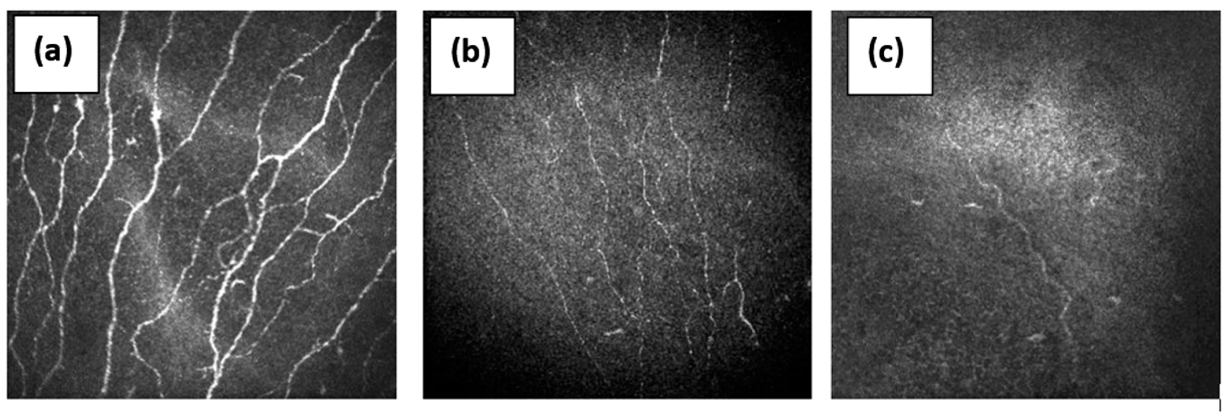
4. Corneal Confocal Microscopy in Small Fiber Neuropathy
5. Conclusions
Funding
Institutional Review Board Statement
Informed Consent Statement
Conflicts of Interest
References
- Brouwer, B.A.; Bakkers, M.; Hoeijmakers, J.G.; Faber, C.G.; Merkies, I.S. Improving assessment in small fiber neuropathy. J. Peripher. Nerv. Syst. 2015, 20, 333–340. [Google Scholar] [CrossRef] [PubMed]
- Brines, M.; Culver, D.A.; Ferdousi, M.; Tannemaat, M.R.; Velzen van, M.; Dahan, A.; Malik, A. Corneal nerve fiber size adds utility to the diagnosis and assessment of therapeutic response in patients with small fiber neuropathy. Sci. Rep. 2018, 8, 4734. [Google Scholar] [CrossRef] [Green Version]
- Hoitsma, E.; Reulen, J.P.; de Baets, M.; Drent, M.; Spaans, F.; Faber, C.G. Small fiber neuropathy: A common and important clinical disorder. J. Neurol. Sci. 2004, 227, 119–130. [Google Scholar] [CrossRef]
- Basantsova, N.Y.; Zinchenko, Y.U.S.; Starshinova, A.A.; Yablonsky, P.K. Features of the diagnosis of small fiber neuropathies in various diseases (literature review). Pediatrician 2018, 9, 101–110. [Google Scholar] [CrossRef]
- Johnson, S.A.; Shouman, K.; Shelly, S.; Sandroni, P.; Berini, S.E.; Dyck, P.J.B.; Hoffman, E.M.; Mandrekar, J.; Niu, Z.; Lamb, C.J.; et al. Small Fiber Neuropathy Incidence, Prevalence, Longitudinal Impairments, and Disability. Neurology 2021, 97, e2236–e2247. [Google Scholar] [CrossRef]
- Peters, M.J.; Bakkers, M.; Merkies, I.S.; Hoeijmakers, J.G.; van Raak, E.P.; Faber, C.G. Incidence and prevalence of small-fiber neuropathy: A survey in the Netherlands. Neurology 2013, 81, 1356–1360. [Google Scholar] [CrossRef]
- Freeman, R.; Gewandter, J.S.; Faber, C.G.; Gibbons, C.; Haroutounian, S.; Lauria, G.; Levine, T.; Malik, R.A.; Singleton, J.R.; Smith, A.G.; et al. Idiopathic distal sensory polyneuropathy: ACTTION diagnostic criteria. Neurology 2020, 95, 1005–1014. [Google Scholar] [CrossRef] [PubMed]
- Sun, B.; LiZhi, L.; Liu, L.; Chen, Z.; Ling, L.; Yang, F.; Liu, J.; Liu, H.; Huang, X. SFN-SIQ, SFNSL, and skin biopsy of 55 cases with small fiber involvement. Int. J. Neurosci. 2018, 128, 442–448. [Google Scholar] [CrossRef]
- Provitera, V.; Gibbons, C.H.; Wendelschafer-Crabb, G.; Donadio, V.; Vitale, D.F.; Stancanelli, A.; Caporaso, G.; Liguori, R.; Wang, N.; Santoro, L.; et al. A multi-center, multinational age- and genderadjusted normative dataset for immunofluorescent intraepidermal nerve fiber density at the distal leg. Eur. J. Neurol. 2016, 23, 333–338. [Google Scholar] [CrossRef] [PubMed]
- Lang, M.; Treister, R.; Oaklande, A.L. Diagnostic value of blood tests for occult causes of initially idiopathic small fiber polyneuropathy. J. Neurol. 2016, 263, 2515–2527. [Google Scholar] [CrossRef] [Green Version]
- Malik, R.A.; Kallinikos, P.; Abbott, C.A.; van Schie, C.H.; Morgan, P.; Efron, N.; Boulton, A.J. Corneal confocal microscopy: A noninvasive surrogate of nerve fibre damage and repair in diabetic patients. Diabetologia 2003, 46, 683–688. [Google Scholar] [CrossRef]
- Terkelsen, A.J.; Karlsson, P.; Lauria, G.; Freeman, R.; Finnerup, N.; Jensen, T.S. The diagnostic challenge of small fibre neuropathy: Clinical presentations, evaluations, and causes. Lancet Neurol. 2017, 16, 934–944. [Google Scholar] [CrossRef]
- Kalteniece, A.; Ferdousi, M.; Azmi, S.; Mubita, W.M.; Marshall, A.; Lauria, G.; Faber, C.G.; Soran, H.; Malik, R.A. Corneal confocal microscopy detects small nerve fbre damage in patients with painful diabetic neuropathy. Sci. Rep. 2020, 10, 3371. [Google Scholar] [CrossRef] [PubMed]
- Alam, U.; Jeziorska, M.; Petropoulos, I.N.; Asghar, O.; Fadavi, H.; Ponirakis, G.; Marshall, A.; Tavakoli, M.; Boulton, A.J.M.; Efron, N.; et al. Diagnostic utility of corneal confocal microscopy and intra-epidermal nerve fibre density in diabetic neuropathy. PLoS ONE 2017, 12, e0180175. [Google Scholar] [CrossRef] [Green Version]
- Moulton, E.A.; Borsook, D. C-Fiber Assays in the Cornea vs. Skin. Brain Sci. 2019, 9, 320. [Google Scholar] [CrossRef] [Green Version]
- Bucher, F.; Schneider, C.; Blau, T.; Cursiefen, C.; Fink, G.R.; Lehmann, H.C.; Heindl, L.M. Small-Fiber Neuropathy Is Associated With Corneal Nerve and Dendritic Cell Alterations: An In Vivo Confocal Microscopy Study. Cornea 2015, 34, 1114–1119. [Google Scholar] [CrossRef]
- Varela, N.; Chorny, A.; Gonzalez-Rey, E.; Delgado, M. Tuning inflammation with anti-inflammatory neuropeptides. Expert Opin. Biol. Ther. 2007, 7, 461–478. [Google Scholar] [CrossRef]
- Lauria, G.; Merkies, I.S.G.; Faber, C.G. Small fiber neuropathy. Curr. Opin. Neur. 2012, 25, 542–549. [Google Scholar] [CrossRef] [PubMed]
- Sene, D. Small fiber neuropathy: Diagnosis, causes and treatment. Jt. Bone Spine 2018, 85, 553–559. [Google Scholar] [CrossRef]
- Farhad, K.; Traub, R.; Ruzhansky, K.M.; Brannagan, T.H. Causes of neuropathy in patients referred as “idiopathic neuropathy”. Muscle Nerve 2016, 53, 856–861. [Google Scholar] [CrossRef] [PubMed]
- Devigili, G.; Tugnoli, V.; Penza, P.; Camozzi, F.; Lombardi, R.; Melli, G.; Broglio, L.; Granieri, E.; Lauria, G. The diagnostic criteria for small fibre neuropathy: From symptoms to neuropathology. Brain 2008, 131, 1912–1925. [Google Scholar] [CrossRef] [Green Version]
- Lefaucheur, J.-P.; Wahab, A.; Planté-Bordeneuve, V.; Sène, D.; Ménard-Lefaucheur, I.; Rouie, D.; Tebbal, D.; Salhi, H.; Créange, A.; Zouari, H.; et al. Diagnosis of small fiber neuropathy: A comparative study of five neurophysiological tests. Neurophysiol. Clin. 2015, 45, 445–455. [Google Scholar] [CrossRef] [PubMed]
- Gemignani, F.; Brindani, F.; Vitetta, F.; Marbini, A.; Calzetti, S. Restless legs syndrome in diabetic neuropathy: A frequent manifestation of small fiber neuropathy. J. Peripher. Nerv. Syst. 2007, 12, 50–53. [Google Scholar] [CrossRef] [PubMed]
- Sène, D.; Cacoub, P.; Authier, F.-J.; Haroche, J.; Créange, A.; Saadoun, D.; Amoura, Z.; Guillausseau, P.-J.; Lefaucheur, J.-P. Small fibre neuropathy: Diagnostic approach and therapeutic issues, and its association with primary Sjögren’s syndrome. Med. Baltim. 2013, 92, 10–18. [Google Scholar] [CrossRef] [PubMed] [Green Version]
- Fromy, B.; Lingueglia, E.; Sigaudo-Roussel, D.; Saumet, J.L.; Lazdunski, M. Asic3 is a neuronal mechanosensor for pressure-induced vasodilation that protects against pressure ulcers. Nat. Med. 2012, 18, 1205–1207. [Google Scholar] [CrossRef]
- Grundmann, A. Microspectrophotometry of the cell within the range of the visible spectrum. Dtsch. Med. J. 1963, 88, 98–103. [Google Scholar] [CrossRef]
- Egger, M.D.; Petran, M. New Reflected-Light Microscope for Viewing Unstained Brain and Ganglion Cells. Science 1967, 157, 305–307. [Google Scholar] [CrossRef]
- Masters, B.R. Confocal Microscopy and Multiphoton Excitation Microscopy: The Genesis of Live Cell Imaging; SPIE Press: Washington, DC, USA, 2006; p. 234. [Google Scholar]
- Wang, E.F.; Misra, S.L.; Patel, D.V. In Vivo Confocal Microscopy of the Human Cornea in the Assessment of Peripheral Neuropathy and Systemic Diseases. Biomed Res. Int. 2015, 2015, 951081. [Google Scholar] [CrossRef] [Green Version]
- Dabbah, M.A.; Graham, J.; Petropoulos, I.N.; Tavakoli, M.; Malik, R.A. Automatic analysis of diabetic peripheral neuropathy using multi-scale quantitative morphology of nerve fibres in corneal confocal microscopy imaging. Med. Image Anal. 2011, 15, 738–747. [Google Scholar] [CrossRef]
- Chen, X.; Graham, J.; Dabbah, M.A.; Petropoulos, I.N.; Tavakoli, M.; Malik, R.A. An Automatic Tool for Quantification of Nerve Fibers in Corneal Confocal Microscopy Images. IEEE Trans. Biomed. Eng. 2017, 64, 786–794. [Google Scholar] [CrossRef] [Green Version]
- Tavakoli, M.; Ferdousi, M.; Petropoulos, I.N.; Morris, J.; Pritchard, N.; Zhivov, A.; Ziegler, D.; Pacaud, D.; Romanchuk, K.; Perkins, B.A.; et al. Normative values for corneal nerve morphology assessed using corneal confocal microscopy: A multinational normative data set. Diabetes Care 2015, 38, 838–843. [Google Scholar] [CrossRef] [Green Version]
- Williams, B.M.; Borroni, D.; Liu, R.; Zhao, Y.; Zhang, J.; Lim, J.; Ma, B.; Romano, V.; Qi, H.; Ferdousi, M.; et al. An artificial intelligence-based deep learning algorithm for the diagnosis of diabetic neuropathy using corneal confocal microscopy: A development and validation study. Diabetologia 2020, 63, 419–430. [Google Scholar] [CrossRef] [Green Version]
- Salahouddin, T.; Petropoulos, I.N.; Ferdousi, M.; Ponirakis, G.; Asghar, O.; Alam, U.; Kamran, S.; Mahfoud, Z.R.; Efron, N.; Malik, R.A.; et al. Artificial Intelligence-Based Classification of Diabetic Peripheral Neuropathy From Corneal Confocal Microscopy Images. Diabetes Care 2021, 44, e151–e153. [Google Scholar] [CrossRef]
- Cazzato, D.; Lauria, G. Small fiber neuropathy. Curr. Opin. Neur. 2017, 30, 490–499. [Google Scholar] [CrossRef]
- Gemignani, F.; Ferrari, G.; Vitetta, F.; Giovanelli, M.; Macaluso, C.; Marbini, A. Non-length-dependent small fibre neuropathy. Confocal microscopy study of the corneal innervation. J. Neurol. Neurosurg. Psychiatry 2010, 81, 731–733. [Google Scholar] [CrossRef] [PubMed]
- Egenolf, N.; Zu Altenschildesche, C.M.; Kreß, L.; Eggermann, K.; Namer, B.; Gross, F.; Klitsch, A.; Malzacher, T.; Kampik, D.; Malik, R.A.; et al. Diagnosing small fiber neuropathy in clinical practice: A deep phenotyping study. Ther. Adv. Neurol. Disord. 2021, 14, 17562864211004318. [Google Scholar] [CrossRef] [PubMed]
- Azmi, S.; Ferdousi, M.; Petropoulos, I.N.; Ponirakis, G.; Alam, U.; Fadavi, H.; Asghar, O.; Marshall, A.; Atkinson, A.J.; Jones, W.; et al. Corneal Confocal Microscopy Identifies Small-Fiber Neuropathy in Subjects With Impaired Glucose Tolerance Who Develop Type 2 Diabetes. Diabetes Care 2015, 38, 1502–1508. [Google Scholar] [CrossRef] [Green Version]
- Ferdousi, M.; Azmi, S.; Kalteniece, A.; Petropoulos, I.N.; Ponirakis, G.; Asghar, O.; Alam, U.; Marshall, A.; Boulton, A.J.M.; Efron, N.; et al. Greater small nerve fibre damage in the skin and cornea of type 1 diabetic patients with painful compared to painless diabetic neuropathy. Eur. J. Neurol. 2021, 28, 1745–1751. [Google Scholar] [CrossRef]
- Kalteniece, A.; Ferdousi, M.; Azmi, S.; Khan, S.U.; Worthington, A.; Marshall, A.; Faber, C.G.; Lauria, G.; Boulton, A.J.M.; Soran, H.; et al. Corneal nerve loss is related to the severity of painful diabetic neuropathy. Eur. J. Neurol. 2021, 27. [Google Scholar] [CrossRef]
- Culver, D.A.; Dahan, A.; Bajorunas, D.; Jeziorska, M.; van Velzen, M.; Aarts, L.P.H.J.; Tavee, J.; Tannemaat, M.R.; Dunne, A.N.; Kirk, R.I.; et al. Cibinetide Improves Corneal Nerve Fiber Abundance in Patients With Sarcoidosis-Associated Small Nerve Fiber Loss and Neuropathic Pain. Investig. Ophthalmol. Vis. Sci. 2017, 58, BIO52–BIO60. [Google Scholar] [CrossRef] [PubMed] [Green Version]
- Kemp, H.I.; Petropoulos, I.N.; Rice, A.S.C.; Vollert, J.; Maier, C.; Strum, D.; Schargus, M.; Peto, T.; Hau, S.; Chopra, R.; et al. Use of Corneal Confocal Microscopy to Evaluate Small Nerve Fibers in Patients With Human Immunodeficiency Virus. JAMA Ophthalmol. 2017, 135, 795–800. [Google Scholar] [CrossRef]
- Burgess, J.; Ferdousi, M.; Gosal, D.; Boon, C.; Matsumoto, K.; Marshall, A.; Mak, T.; Marshall, A.; Frank, B.; Malik, R.A.; et al. Chemotherapy-Induced Peripheral Neuropathy: Epidemiology, Pathomechanisms and Treatment. Oncol. Ther. 2021, 9, 385–450. [Google Scholar] [CrossRef] [PubMed]
- Evdokimov, D.; Frank, J.; Klitsch, A.; Unterecker, S.; Warrings, B.; Serra, J.; Papagianni, A.; Saffer, N.; Meyer Zu Altenschildesche, C.; Kampik, D.; et al. Reduction of skin innervation is associated with a severe fibromyalgia phenotype. Ann. Neurol. 2019, 86, 504–516. [Google Scholar] [CrossRef] [Green Version]
- Ferdousi, M.; Kalteniece, A.; Azmi, S.; Petropoulos, I.N.; Ponirakis, G.; Alam, U.; Asghar, O.; Marshall, A.; Fullwood, C.; Jeziorska, M.; et al. Diagnosis of Neuropathy and Risk Factors for Corneal Nerve Loss in Type 1 and Type 2 Diabetes: A Corneal Confocal Microscopy Study. Diabetes Care 2021, 44, 150–156. [Google Scholar] [CrossRef] [PubMed]
- Perkins, B.A.; Lovblom, L.E.; Lewis, E.J.H.; Bril, V.; Ferdousi, M.; Orszag, A.; Edwards, K.; Pritchard, N.; Russell, A.; Dehghani, C.; et al. Corneal Confocal Microscopy Predicts the Development of Diabetic Neuropathy: A Longitudinal Diagnostic Multinational Consortium Study. Diabetes Care 2021, 44, 2107–2114. [Google Scholar] [CrossRef]
- Gad, H.; Petropoulos, I.N.; Khan, A.; Ponirakis, G.; MacDonald, R.; Alam, U.; Malik, R.A. Corneal confocal microscopy for the diagnosis of diabetic peripheral neuropathy: A systematic review and meta-analysis. J. Diabetes Investig. 2021, 6, 567–570. [Google Scholar] [CrossRef]
- Oudejans, L.C.; Niesters, M.; Brines, M.; Dahan, A.; van Velzen, M. Quantification of small fiber pathology in patients with sarcoidosis and chronic pain using cornea confocal microscopy and skin biopsies. J. Pain Res. 2017, 10, 2057–2065. [Google Scholar] [CrossRef] [Green Version]
- Oudejans, L.; He, X.; Niesters, M.; Dahan, A.; Brines, M.; van Velzen, M. Cornea nerve fiber quantification and construction of phenotypes in patients with fibromyalgia. Sci. Rep. 2016, 6, 23573. [Google Scholar] [CrossRef] [PubMed] [Green Version]
- Petropoulos, I.N.; Ponirakis, G.; Khan, A.; Gad, H.; Almuhannadi, H.; Brines, M.; Cerami, A.; Malik, R.A. Corneal confocal microscopy: Ready for prime time. Clin. Exp. Optom. 2020, 103, 265–277. [Google Scholar] [CrossRef]


| Causes | Diseases |
|---|---|
| Metabolic | Diabetes mellitus Impaired glucose tolerance Vitamin B12 and B6 deficiency Dyslipidemia Chronic hypothyroidism Chronic kidney disease |
| Infectious | HIV Hepatitis C Lyme Disease Leprosy |
| Toxic | Alcohol Antibiotics (metronidazole, linezolid) Statins |
| Autoimmune/inflammatory | ASIA-syndrome Sjogren’s syndrome Sarcoidosis Celiac disease Rheumatoid arthritis Systemic lupus erythematosus Amyloidosis |
| Hereditary | Fabry disease Tangier’s disease Friedreich Ataxia Familial amyloidosis (mutation of transthyretin) Sodium channels mutations Nav1.8, 1.7 |
Publisher’s Note: MDPI stays neutral with regard to jurisdictional claims in published maps and institutional affiliations. |
© 2021 by the authors. Licensee MDPI, Basel, Switzerland. This article is an open access article distributed under the terms and conditions of the Creative Commons Attribution (CC BY) license (https://creativecommons.org/licenses/by/4.0/).
Share and Cite
Lukashenko, M.V.; Gavrilova, N.Y.; Bregovskaya, A.V.; Soprun, L.A.; Churilov, L.P.; Petropoulos, I.N.; Malik, R.A.; Shoenfeld, Y. Corneal Confocal Microscopy in the Diagnosis of Small Fiber Neuropathy: Faster, Easier, and More Efficient Than Skin Biopsy? Pathophysiology 2022, 29, 1-8. https://doi.org/10.3390/pathophysiology29010001
Lukashenko MV, Gavrilova NY, Bregovskaya AV, Soprun LA, Churilov LP, Petropoulos IN, Malik RA, Shoenfeld Y. Corneal Confocal Microscopy in the Diagnosis of Small Fiber Neuropathy: Faster, Easier, and More Efficient Than Skin Biopsy? Pathophysiology. 2022; 29(1):1-8. https://doi.org/10.3390/pathophysiology29010001
Chicago/Turabian StyleLukashenko, Mariia V., Natalia Y. Gavrilova, Anna V. Bregovskaya, Lidiia A. Soprun, Leonid P. Churilov, Ioannis N. Petropoulos, Rayaz A Malik, and Yehuda Shoenfeld. 2022. "Corneal Confocal Microscopy in the Diagnosis of Small Fiber Neuropathy: Faster, Easier, and More Efficient Than Skin Biopsy?" Pathophysiology 29, no. 1: 1-8. https://doi.org/10.3390/pathophysiology29010001
APA StyleLukashenko, M. V., Gavrilova, N. Y., Bregovskaya, A. V., Soprun, L. A., Churilov, L. P., Petropoulos, I. N., Malik, R. A., & Shoenfeld, Y. (2022). Corneal Confocal Microscopy in the Diagnosis of Small Fiber Neuropathy: Faster, Easier, and More Efficient Than Skin Biopsy? Pathophysiology, 29(1), 1-8. https://doi.org/10.3390/pathophysiology29010001