Intraoperative Margin Control in Eyelid Tumor Surgery: Current Standards, Imaging Advances, and Emerging Techniques

Simple Summary
Abstract
1. Introduction
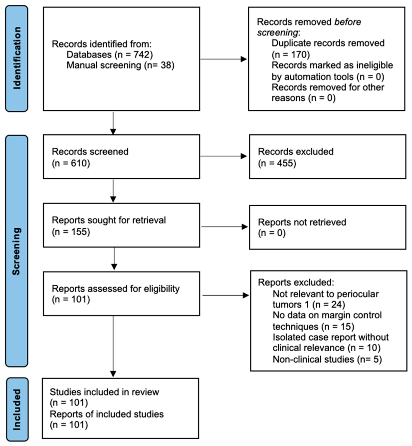
2. Materials and Methods
3. Results
3.1. Overview of Eyelid Malignancies
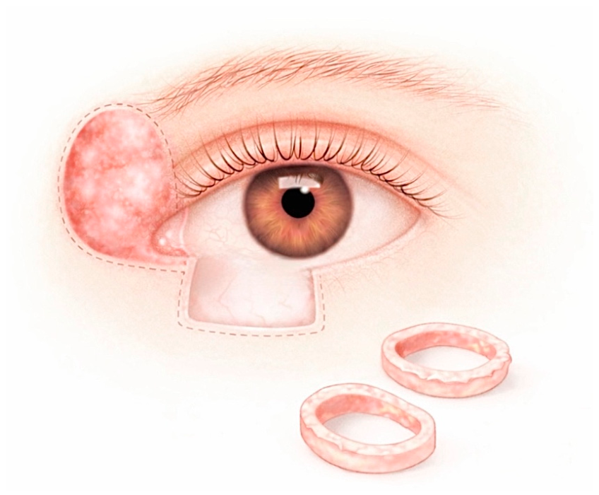
3.2. Mohs Micrographic Surgery
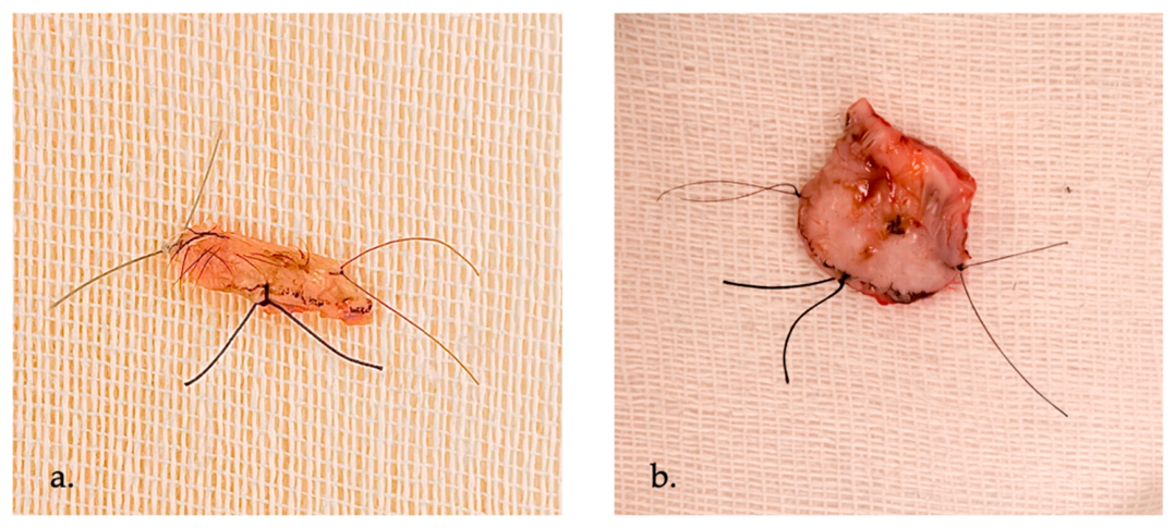
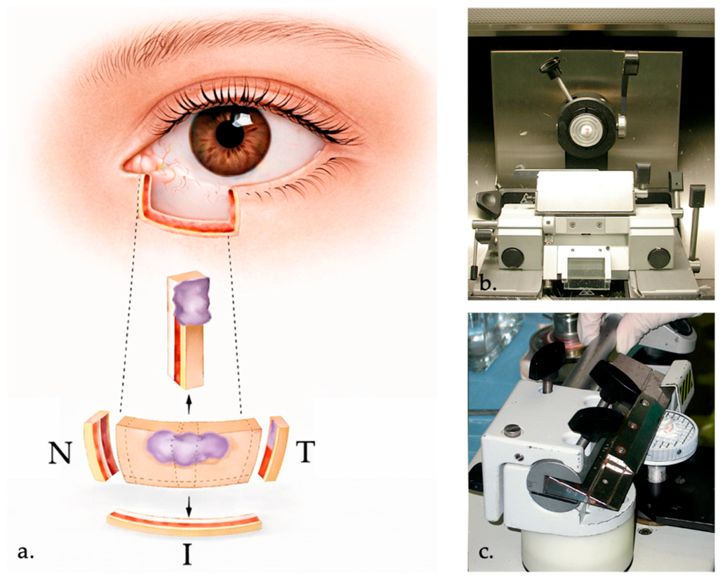
3.3. Frozen Sections-Controlled Surgical Excision
3.4. Emerging Imaging Modalities
3.4.1. Fluorescence Confocal Microscopy
3.4.2. Reflectance Confocal Microscopy
3.4.3. Optical Coherence Tomography
3.4.4. Combined RCM-OCT
3.4.5. Line-Field Confocal OCT
3.4.6. Photoacoustic Imaging
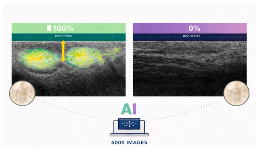
3.5. Artificial Intelligence–Based Approaches
4. Conclusions
Author Contributions
Funding
Data Availability Statement
Acknowledgments
Conflicts of Interest
References
- Cook, B. Epidemiologic characteristics and clinical course of patients with malignant eyelid tumors in an incidence cohort in Olmsted county, Minnesota. Ophthalmology 1999, 106, 746–750. [Google Scholar] [CrossRef]
- Gąsiorowski, K.; Gontarz, M.; Bargiel, J.; Marecik, T.; Szczurowski, P.; Wyszyńska-Pawelec, G. Reconstructive Techniques Following Malignant Eyelid Tumour Excision—Our Experience. J. Clin. Med. 2024, 13, 6120. [Google Scholar] [CrossRef]
- Silverman, N.; Shinder, R. What’s New in Eyelid Tumors. Asia-Pac. J. Ophthalmol. 2017, 6, 143–152. [Google Scholar] [CrossRef]
- Desiato, V.M.; Byun, Y.J.; Nguyen, S.A.; Thiers, B.H.; Day, T.A. Sebaceous Carcinoma of the Eyelid: A Systematic Review and Meta-Analysis. Dermatol. Surg. 2021, 47, 104–110. [Google Scholar] [CrossRef]
- Balasubramanian, A.; Kannan, N.S. Eyelid Malignancies- Always Quite Challenging. J. Clin. Diagn. Res. JCDR 2017, 11, XR01–XR04. [Google Scholar] [CrossRef]
- Sundar, G.; Manjandavida, F.P. Excision of Eyelid Tumors: Principles and Techniques. In Surgical Ophthalmic Oncology; Chaugule, S.S., Honavar, S.G., Finger, P.T., Eds.; Springer International Publishing: Cham, Switzerland, 2019; pp. 15–32. [Google Scholar] [CrossRef]
- Moul, D.K.; Chern, P.L.; Shumaker, P.R.; Zelac, D.E.; Greenway, H.T. Mohs Micrographic Surgery for Eyelid and Periorbital Skin Cancer. Int. Ophthalmol. Clin. 2009, 49, 111–127. [Google Scholar] [CrossRef] [PubMed]
- Van Lee, C.B.; Ip Vai Ching, E.E.F.; Nasserinejad, K.; Neumann, H.A.M.; Bol, M.G.W.; Dikrama, P.K.; Kelleners-Smeets, N.W.J.; Koljenović, S.; Munte, K.; Noordhoek Hegt, V.; et al. Reliability of diagnosis from Mohs slides: Interpersonal and intrapersonal agreement on basal cell carcinoma presence and histological subtype. Br. J. Dermatol. 2016, 175, 549–554. [Google Scholar] [CrossRef]
- Resti, A.G.; Sacconi, R.; Baccelli, N.; Bandello, F. Outcome of 110 Basal Cell Carcinomas of the Eyelid Treated with Frozen Section–Controlled Excision: Mean Follow-up over 5 Years. Eur. J. Ophthalmol. 2014, 24, 476–482. [Google Scholar] [CrossRef] [PubMed]
- Kose, K.; Fox, C.A.; Rossi, A.; Jain, M.; Cordova, M.; Dusza, S.W.; Ragazzi, M.; Gardini, S.; Moscarella, E.; Diaz, A.; et al. An international 3-center training and reading study to assess basal cell carcinoma surgical margins with ex vivo fluorescence confocal microscopy. J. Cutan. Pathol. 2021, 48, 1010–1019. [Google Scholar] [CrossRef] [PubMed]
- Venturini, M.; Gualdi, G.; Zanca, A.; Lorenzi, L.; Pellacani, G.; Calzavara-Pinton, P.G. A new approach for presurgical margin assessment by reflectance confocal microscopy of basal cell carcinoma. Br. J. Dermatol. 2016, 174, 380–385. [Google Scholar] [CrossRef]
- Bergeron, S.; Arthurs, B.; Sanft, D.-M.; Mastromonaco, C.; Burnier, M.N. Optical Coherence Tomography of Peri-Ocular Skin Cancers: An Optical Biopsy. Ocul. Oncol. Pathol. 2021, 7, 149–158. [Google Scholar] [CrossRef] [PubMed]
- Di Stefani, A.; Cappilli, S.; Cuffaro, G.; Fionda, B.; Pagliara, M.M.; Paradisi, A.; Ricci, C.; Rossi, E.; Sammarco, M.G.; Schinzari, G.; et al. Line-Field Confocal Optical Coherence Tomography Evaluation of Eyelid Skin Lesions. Diagnostics 2023, 13, 3590. [Google Scholar] [CrossRef] [PubMed]
- Dahlstrand, U.; Sheikh, R.; Malmsjö, M. Photoacoustic imaging for intraoperative micrographic control of the surgical margins of eyelid tumours. Acta Ophthalmol. 2020, 98, E264–E265. [Google Scholar] [CrossRef] [PubMed]
- Esteva, A.; Kuprel, B.; Novoa, R.A.; Ko, J.; Swetter, S.M.; Blau, H.M.; Thrun, S. Dermatologist-level classification of skin cancer with deep neural networks. Nature 2017, 542, 115–118. [Google Scholar] [CrossRef]
- Furdova, A.; Kapitanova, K.; Kollarova, A.; Sekac, J. Periocular basal cell carcinoma-clinical perspectives. Oncol. Rev. 2020, 14, 36–42. [Google Scholar] [CrossRef]
- Rosner, M. Basal cell carcinoma. In Clinical Ophthalmic Oncology; Elsevier: Amsterdam, The Netherlands, 2007; pp. 76–80. [Google Scholar] [CrossRef]
- Leibovitch, I.; Huilgol, S.C.; Selva, D.; Richards, S.; Paver, R. Basal cell carcinoma treated with Mohs surgery in Australia III. Perineural invasion. J. Am. Acad. Dermatol. 2005, 53, 458–463. [Google Scholar] [CrossRef]
- Xu, S.; Sagiv, O.; Rubin, M.L.; Sa, H.-S.; Tetzlaff, M.T.; Nagarajan, P.; Ning, J.; Esmaeli, B. Validation Study of the AJCC Cancer Staging Manual, Eighth Edition, Staging System for Eyelid and Periocular Squamous Cell Carcinoma. JAMA Ophthalmol. 2019, 137, 537–542. [Google Scholar] [CrossRef]
- Limawararut, V.; Leibovitch, I.; Sullivan, T.; Selva, D. Periocular squamous cell carcinoma. Clin. Exp. Ophthalmol. 2007, 35, 174–185. [Google Scholar] [CrossRef]
- Sun, M.T.; Andrew, N.H.; O’Donnell, B.; McNab, A.; Huilgol, S.C.; Selva, D. Periocular Squamous Cell Carcinoma: TNM Staging and Recurrence. Ophthalmology 2015, 122, 1512–1516. [Google Scholar] [CrossRef]
- Lu, T.J.; Fan, J.; Guimaraes De Sousa, L.; Sagiv, O.; Hernandez, S.; Bolanos Gomez, L.; Akhave, N.; Ferrarotto, R.; Esmaeli, B. PD-1 Inhibitors for Periocular Squamous Cell Carcinoma With Perineural Spread to the Orbit and Skull Base. Ophthalmic Plast. Reconstr. Surg. 2025, 41, 664–670. [Google Scholar] [CrossRef]
- Buitrago, W.; Joseph, A.K. Sebaceous carcinoma: The great masquerader. Dermatol. Ther. 2008, 21, 459–466. [Google Scholar] [CrossRef] [PubMed]
- Chao, A.N.; Shields, C.L.; Krema, H.; Shields, J.A. Outcome of patients with periocular sebaceous gland carcinoma with and without conjunctival intraepithelial invasion. Ophthalmology 2001, 108, 1877–1883. [Google Scholar] [CrossRef]
- McGrath, L.A.; Currie, Z.I.; Mudhar, H.S.; Tan, J.H.Y.; Salvi, S.M. Management of recurrent sebaceous gland carcinoma. Eye 2020, 34, 1685–1692. [Google Scholar] [CrossRef]
- Jacobs, D.; Huang, H.; Olino, K.; Weiss, S.; Kluger, H.; Judson, B.L.; Zhang, Y. Assessment of Age, Period, and Birth Cohort Effects and Trends in Merkel Cell Carcinoma Incidence in the United States. JAMA Dermatol. 2021, 157, 59–65. [Google Scholar] [CrossRef]
- Nayak, A.L.; Pickett, A.T.; Delisle, M.; Dingley, B.; Mallick, R.; Hamilton, T.; Stuart, H.; Talbot, M.; McKinnon, G.; Jost, E.; et al. Survival of Patients With Head and Neck Merkel Cell Cancer: Findings From the Pan-Canadian Merkel Cell Cancer Collaborative. JAMA Netw. Open 2023, 6, e2344127. [Google Scholar] [CrossRef]
- Heath, M.; Jaimes, N.; Lemos, B.; Mostaghimi, A.; Wang, L.C.; Peñas, P.F.; Nghiem, P. Clinical characteristics of Merkel cell carcinoma at diagnosis in 195 patients: The AEIOU features. J. Am. Acad. Dermatol. 2008, 58, 375–381. [Google Scholar] [CrossRef]
- Miller, R.; Walker, S.; Shui, I.; Brandtmüller, A.; Cadwell, K.; Scherrer, E. Epidemiology and survival outcomes in stages II and III cutaneous melanoma: A systematic review. Melanoma Manag. 2020, 7, MMT39. [Google Scholar] [CrossRef]
- Chan, F.M.; O’Donnell, B.A.; Whitehead, K.; Ryman, W.; Sullivan, T.J. Treatment and Outcomes of Malignant Melanoma of the Eyelid. Ophthalmology 2007, 114, 187–192. [Google Scholar] [CrossRef] [PubMed]
- Gąsiorowski, K.; Iwulska, K.; Zapała, J.; Wyszyńska-Pawelec, G. Periocular basal cell carcinoma: Recurrence risk factors/when to reoperate? Postep. Dermatol. Alergol. 2020, 37, 927–931. [Google Scholar] [CrossRef] [PubMed]
- Strianese, D.; Iuliano, A.; Quaranta Leoni, F.M. Secondary Orbital Tumours and Orbital Metastases. In Oculoplastic, Lacrimal and Orbital Surgery; Quaranta Leoni, F.M., Verity, D.H., Paridaens, D., Eds.; Springer Nature: Cham, Switzerland, 2024; pp. 351–369. [Google Scholar] [CrossRef]
- Gąsiorowski, K.; Gontarz, M.; Marecik, T.; Szczurowski, P.; Bargiel, J.; Zapała, J.; Wyszyńska-Pawelec, G. Risk Factors for Orbital Invasion in Malignant Eyelid Tumors, Is Orbital Exenteration Still Necessary? J. Clin. Med. 2024, 13, 726. [Google Scholar] [CrossRef]
- Hernandez-Herrera, G.A.; Calcano, G.A.; Nagelschneider, A.A.; Routman, D.M.; Van Abel, K.M. Imaging Modalities for Head and Neck Cancer. Surg. Oncol. Clin. N. Am. 2024, 33, 617–649. [Google Scholar] [CrossRef]
- Mohs, F.E. Micrographic Surgery for the Microscopically Controlled Excision of Eyelid Cancers. Arch. Ophthalmol. 1986, 104, 901–909. [Google Scholar] [CrossRef] [PubMed]
- Blumenthal, S.R.; Swick, M.; Bayan, C.-A.; Ramanathan, D.; Maher, I. Complex Eyelid Reconstruction: A Practical Guide for the Mohs Surgeon. Dermatol. Surg. 2022, 48, 916–923. [Google Scholar] [CrossRef]
- Malhotra, R. The Australian Mohs database, part II*1Periocular basal cell carcinoma outcome at 5-year follow-up. Ophthalmology 2004, 111, 631–636. [Google Scholar] [CrossRef]
- Nemet, A.Y.; Deckel, Y.; Martin, P.A.; Kourt, G.; Chilov, M.; Sharma, V.; Benger, R. Management of Periocular Basal and Squamous Cell Carcinoma: A Series of 485 Cases. Am. J. Ophthalmol. 2006, 142, 293–297. [Google Scholar] [CrossRef]
- Phan, K.; Oh, L.J.; Goyal, S.; Rutherford, T.; Yazdabadi, A. Recurrence rates following surgical excision of periocular basal cell carcinomas: Systematic review and meta-analysis. J. Dermatol. Treat. 2020, 31, 597–601. [Google Scholar] [CrossRef]
- Hartley, M.J.; Meredith, P.R.; Oliphant, T. Outcomes of Mohs micrographic surgery for periocular squamous cell carcinoma. Eur. J. Ophthalmol. 2024, 34, 1506–1510. [Google Scholar] [CrossRef]
- Malhotra, R. The Australian Mohs database*1Periocular squamous cell carcinoma. Ophthalmology 2004, 111, 617–623. [Google Scholar] [CrossRef] [PubMed]
- Shi, K.; Wang, L.; Srivastava, D.; Nijhawan, R.I. Mohs micrographic surgery for periocular skin cancer: A single-institution experience. Arch. Dermatol. Res. 2022, 315, 1181–1186. [Google Scholar] [CrossRef]
- Shumaker, P.R.; Kelley, B.; Swann, M.H.; Greenway, H.T. Modified Mohs Micrographic Surgery for Periocular Melanoma and Melanoma In Situ: Long-Term Experience at Scripps Clinic. Dermatol. Surg. 2009, 35, 1263–1270. [Google Scholar] [CrossRef] [PubMed]
- Zhou, C.; Wu, F.; Chai, P.; Shi, Y.; Ye, J.; Shi, X.; Tan, J.; Ding, Y.; Luo, Y.; Esmaeli, B.; et al. Mohs micrographic surgery for eyelid sebaceous carcinoma: A multicenter cohort of 360 patients. J. Am. Acad. Dermatol. 2019, 80, 1608–1617.e1. [Google Scholar] [CrossRef]
- McInnis-Smith, K.M.; Asamoah, E.M.; Demer, A.M.; Sharma, K.; Yu, C.Y.; Bradley, E.A.; Tooley, A.A.; Wagner, L.H. Mohs Micrographic Surgery With Immunohistochemistry for the Treatment of Periocular Melanoma In Situ. Ophthalmic Plast. Reconstr. Surg. 2025, 41, 78–83. [Google Scholar] [CrossRef]
- Beaulieu, D.; Fathi, R.; Srivastava, D.; Nijhawan, R.I. Current perspectives on Mohs micrographic surgery for melanoma. Clin. Cosmet. Investig. Dermatol. 2018, 11, 309–320. [Google Scholar] [CrossRef]
- Paridaens, D.; Verdijk, R.; Giordano Resti, A.; Quaranta Leoni, F.M. Eyelid Tumour Excision: Frozen Sections Versus Mohs’ surgery. In Oculoplastic, Lacrimal and Orbital Surgery; Quaranta Leoni, F.M., Verity, D.H., Paridaens, D., Eds.; Springer Nature: Cham, Switzerland, 2024; pp. 267–274. [Google Scholar] [CrossRef]
- Morris, D.S.; Elzaridi, E.; Clarke, L.; Dickinson, A.J.; Lawrence, C.M. Periocular basal cell carcinoma: 5-year outcome following Slow Mohs surgery with formalin-fixed paraffin-embedded sections and delayed closure. Br. J. Ophthalmol. 2009, 93, 474–476. [Google Scholar] [CrossRef]
- Şahan, B.; Çiftçi, F.; Özkan, F.; Öztürk, V. The Importance of Frozen Section-Controlled Excision in Recurrent Basal Cell Carcinoma of the Eyelids. Türk Oftalmol. Derg. 2016, 46, 277–281. [Google Scholar] [CrossRef] [PubMed]
- McCabe, G.A.; Mulcahy, L.T.; Mulligan, N.; Fulcher, T. Recurrence rates of periocular basal cell carcinoma following intra-operative en-face frozen section margin controlled excision. Ir. J. Med. Sci. 2024, 193, 1209–1213. [Google Scholar] [CrossRef] [PubMed]
- Conway, R.M. Surgery for primary basal cell carcinoma including the eyelid margins with intraoperative frozen section control: Comparative interventional study with a minimum clinical follow up of 5 years. Br. J. Ophthalmol. 2004, 88, 236–238. [Google Scholar] [CrossRef]
- Dailey, J.R.; Kennedy, R.H.; Flaharty, P.M.; Eagle, R.C.; Flanagan, J.C. Squamous Cell Carcinoma of the Eyelid. Ophthalmic Plast. Reconstr. Surg. 1994, 10, 153–159. [Google Scholar] [CrossRef]
- Gayre, G.S.; Hybarger, C.P.; Mannor, G.; Meecham, W.; Delfanti, J.B.; Mizono, G.S.; Guerry, T.L.; Chien, J.S.; Sooy, C.D.; Anooshian, R.; et al. Outcomes of excision of 1750 eyelid and periocular skin basal cell and squamous cell carcinomas by modified en face frozen section margin-controlled technique. Int. Ophthalmol. Clin. 2009, 49, 97–110. [Google Scholar] [CrossRef]
- Donaldson, M.J. Squamous cell carcinoma of the eyelids. Br. J. Ophthalmol. 2002, 86, 1161–1165. [Google Scholar] [CrossRef] [PubMed]
- Cheung, J.J.C.; Esmaeli, B.; Lam, S.C.; Kwok, T.; Yuen, H.K.L. The practice patterns in the management of sebaceous carcinoma of the eyelid in the Asia Pacific region. Eye 2019, 33, 1433–1442. [Google Scholar] [CrossRef]
- Otsuka, A.C.V.G.; Bertolli, E.; de Macedo, M.P.; Pinto, C.A.L.; Duprat Neto, J.P. Intraoperative assessment of surgical margins using “en face” frozen sections in the management of cutaneous carcinomas. An. Bras. Dermatol. 2022, 97, 583–591. [Google Scholar] [CrossRef]
- Parashar, K.; Torres, A.E.; Boothby-Shoemaker, W.; Kohli, I.; Veenstra, J.; Neel, V.; Ozog, D.M. Imaging technologies for presurgical margin assessment of basal cell carcinoma. J. Am. Acad. Dermatol. 2023, 88, 144–151. [Google Scholar] [CrossRef] [PubMed]
- Boostani, M.; Bozsányi, S.; Suppa, M.; Cantisani, C.; Lőrincz, K.; Bánvölgyi, A.; Holló, P.; Wikonkál, N.M.; Huss, W.J.; Brady, K.L.; et al. Novel imaging techniques for tumor margin detection in basal cell carcinoma: A systematic scoping review of FDA and EMA-approved imaging modalities. Int. J. Dermatol. 2025, 64, 287–301. [Google Scholar] [CrossRef]
- Longo, C.; Ragazzi, M.; Rajadhyaksha, M.; Nehal, K.; Bennassar, A.; Pellacani, G.; Malvehy Guilera, J. In Vivo and Ex Vivo Confocal Microscopy for Dermatologic and Mohs Surgeons. Dermatol. Clin. 2016, 34, 497–504. [Google Scholar] [CrossRef]
- Gareau, D.S.; Karen, J.K.; Dusza, S.W.; Tudisco, M.; Nehal, K.S.; Rajadhyaksha, M. Sensitivity and specificity for detecting basal cell carcinomas in Mohs excisions with confocal fluorescence mosaicing microscopy. J. Biomed. Opt. 2009, 14, 034012. [Google Scholar] [CrossRef] [PubMed]
- Bennàssar, A.; Vilata, A.; Puig, S.; Malvehy, J. Ex vivo fluorescence confocal microscopy for fast evaluation of tumour margins during Mohs surgery. Br. J. Dermatol. 2014, 170, 360–365. [Google Scholar] [CrossRef] [PubMed]
- Longo, C.; Rajadhyaksha, M.; Ragazzi, M.; Nehal, K.; Gardini, S.; Moscarella, E.; Lallas, A.; Zalaudek, I.; Piana, S.; Argenziano, G.; et al. Evaluating ex vivo fluorescence confocal microscopy images of basal cell carcinomas in M ohs excised tissue. Br. J. Dermatol. 2014, 171, 561–570. [Google Scholar] [CrossRef]
- Longo, C.; Ragazzi, M.; Gardini, S.; Piana, S.; Moscarella, E.; Lallas, A.; Raucci, M.; Argenziano, G.; Pellacani, G. Ex vivo fluorescence confocal microscopy in conjunction with Mohs micrographic surgery for cutaneous squamous cell carcinoma. J. Am. Acad. Dermatol. 2015, 73, 321–322. [Google Scholar] [CrossRef]
- Jain, M.; Rajadhyaksha, M.; Nehal, K. Implementation of fluorescence confocal mosaicking microscopy by “early adopter” Mohs surgeons and dermatologists: Recent progress. J. Biomed. Opt. 2017, 22, 024002. [Google Scholar] [CrossRef]
- Que, S.K.T.; Fraga-Braghiroli, N.; Grant-Kels, J.M.; Rabinovitz, H.S.; Oliviero, M.; Scope, A. Through the looking glass: Basics and principles of reflectance confocal microscopy. J. Am. Acad. Dermatol. 2015, 73, 276–284. [Google Scholar] [CrossRef]
- Guitera, P.; Menzies, S.W.; Longo, C.; Cesinaro, A.M.; Scolyer, R.A.; Pellacani, G. In Vivo Confocal Microscopy for Diagnosis of Melanoma and Basal Cell Carcinoma Using a Two-Step Method: Analysis of 710 Consecutive Clinically Equivocal Cases. J. Investig. Dermatol. 2012, 132, 2386–2394. [Google Scholar] [CrossRef]
- Shavlokhova, V.; Vollmer, M.; Vollmer, A.; Gholam, P.; Saravi, B.; Hoffmann, J.; Engel, M.; Elsner, J.; Neumeier, F.; Freudlsperger, C. In vivo reflectance confocal microscopy of wounds: Feasibility of intraoperative basal cell carcinoma margin assessment. Ann. Transl. Med. 2021, 9, 1716. [Google Scholar] [CrossRef]
- Espinasse, M.; Cinotti, E.; Grivet, D.; Labeille, B.; Prade, V.; Douchet, C.; Cambazard, F.; Thuret, G.; Gain, P.; Perrot, J.L. ‘En face’ ex vivo reflectance confocal microscopy to help the surgery of basal cell carcinoma of the eyelid. Clin. Exp. Ophthalmol. 2017, 45, 442–447. [Google Scholar] [CrossRef] [PubMed]
- Pellacani, G.; Guitera, P.; Longo, C.; Avramidis, M.; Seidenari, S.; Menzies, S. The Impact of In Vivo Reflectance Confocal Microscopy for the Diagnostic Accuracy of Melanoma and Equivocal Melanocytic Lesions. J. Investig. Dermatol. 2007, 127, 2759–2765. [Google Scholar] [CrossRef]
- Gambichler, T.; Jaedicke, V.; Terras, S. Optical coherence tomography in dermatology: Technical and clinical aspects. Arch. Dermatol. Res. 2011, 303, 457–473. [Google Scholar] [CrossRef]
- Welzel, J.; Lankenau, E.; Birngruber, R.; Engelhardt, R. Optical coherence tomography of the human skin. J. Am. Acad. Dermatol. 1997, 37, 958–963. [Google Scholar] [CrossRef]
- Jain, M.; Chang, S.-W.; Singh, K.; Kurtansky, N.R.; Huang, S.-L.; Chen, H.H.; Chen, C.-S.J. High-resolution full-field optical coherence tomography microscope for the evaluation of freshly excised skin specimens during Mohs surgery: A feasibility study. J. Biophotonics 2024, 17, e202300275. [Google Scholar] [CrossRef] [PubMed]
- Ulrich, M.; Braunmuehl, T.; Kurzen, H.; Dirschka, T.; Kellner, C.; Sattler, E.; Berking, C.; Welzel, J.; Reinhold, U. The sensitivity and specificity of optical coherence tomography for the assisted diagnosis of nonpigmented basal cell carcinoma: An observational study. Br. J. Dermatol. 2015, 173, 428–435. [Google Scholar] [CrossRef]
- Adan, F.; Mosterd, K.; Kelleners-Smeets, N.W.J.; Nelemans, P.J. Diagnostic Value of Optical Coherence Tomography Image Features for Diagnosis of Basal Cell Carcinoma. Acta Derm. Venereol. 2021, 101, adv00607. [Google Scholar] [CrossRef] [PubMed]
- Meekings, A.; Utz, S.; Ulrich, M.; Bienenfeld, A.; Nandanan, N.; Fisher, J.; McKenzie, G.; Siegel, D.M.; Feldman, E.; Markowitz, O. Differentiation of Basal Cell Carcinoma Subtypes in Multi-Beam Swept Source Optical Coherence Tomography (MSS-OCT). J. Drugs Dermatol. 2016, 15, 545–550. [Google Scholar]
- De Carvalho, N.; Schuh, S.; Kindermann, N.; Kästle, R.; Holmes, J.; Welzel, J. Optical coherence tomography for margin definition of basal cell carcinoma before micrographic surgery—Recommendations regarding the marking and scanning technique. Ski. Res. Technol. 2018, 24, 145–151. [Google Scholar] [CrossRef]
- Alawi, S.A.; Kuck, M.; Wahrlich, C.; Batz, S.; McKenzie, G.; Fluhr, J.W.; Lademann, J.; Ulrich, M. Optical coherence tomography for presurgical margin assessment of non-melanoma skin cancer—A practical approach. Exp. Dermatol. 2013, 22, 547–551. [Google Scholar] [CrossRef] [PubMed]
- Wang, K.X.; Meekings, A.; Fluhr, J.W.; McKenzie, G.; Lee, D.A.; Fisher, J.; Markowitz, O.; Siegel, D.M. Optical Coherence Tomography–Based Optimization of Mohs Micrographic Surgery of Basal Cell Carcinoma: A Pilot Study. Dermatol. Surg. 2013, 39, 627–633. [Google Scholar] [CrossRef]
- Akella, S.S.; Lee, J.; May, J.R.; Puyana, C.; Kravets, S.; Dimitropolous, V.; Tsoukas, M.; Manwar, R.; Avanaki, K. Using optical coherence tomography to optimize Mohs micrographic surgery. Sci. Rep. 2024, 14, 8900. [Google Scholar] [CrossRef] [PubMed]
- Rashed, D.; Shah, D.; Freeman, A.; Cook, R.J.; Hopper, C.; Perrett, C.M. Rapid ex vivo examination of Mohs specimens using optical coherence tomography. Photodiagnosis Photodyn. Ther. 2017, 19, 243–248. [Google Scholar] [CrossRef] [PubMed]
- Levine, A.; Siegel, D.; Markowitz, O. Imaging in cutaneous surgery. Future Oncol. 2017, 13, 2329–2340. [Google Scholar] [CrossRef]
- Aleissa, S.; Navarrete-Dechent, C.; Cordova, M.; Sahu, A.; Dusza, S.W.; Phillips, W.; Rossi, A.; Lee, E.; Nehal, K.S. Presurgical evaluation of basal cell carcinoma using combined reflectance confocal microscopy-optical coherence tomography: A prospective study. J. Am. Acad. Dermatol. 2020, 82, 962–968. [Google Scholar] [CrossRef]
- Sahu, A.; Yélamos, O.; Iftimia, N.; Cordova, M.; Alessi-Fox, C.; Gill, M.; Maguluri, G.; Dusza, S.W.; Navarrete-Dechent, C.; González, S.; et al. Evaluation of a Combined Reflectance Confocal Microscopy–Optical Coherence Tomography Device for Detection and Depth Assessment of Basal Cell Carcinoma. JAMA Dermatol. 2018, 154, 1175. [Google Scholar] [CrossRef]
- Suppa, M.; Palmisano, G.; Tognetti, L.; Lenoir, C.; Cappilli, S.; Fontaine, M.; Orte Cano, C.; Diet, G.; Perez-Anker, J.; Schuh, S.; et al. Line-field confocal optical coherence tomography in melanocytic and non-melanocytic skin tumors. Ital. J. Dermatol. Venereol. 2023, 158, 180–189. [Google Scholar] [CrossRef]
- Gust, C.; Schuh, S.; Welzel, J.; Daxenberger, F.; Hartmann, D.; French, L.E.; Ruini, C.; Sattler, E.C. Line-Field Confocal Optical Coherence Tomography Increases the Diagnostic Accuracy and Confidence for Basal Cell Carcinoma in Equivocal Lesions: A Prospective Study. Cancers 2022, 14, 1082. [Google Scholar] [CrossRef] [PubMed]
- Boostani, M.; Pellacani, G.; Wortsman, X.; Suppa, M.; Goldust, M.; Cantisani, C.; Pietkiewicz, P.; Lőrincz, K.; Bánvölgyi, A.; Wikonkál, N.M.; et al. FDA and EMA-approved noninvasive imaging techniques for basal cell carcinoma subtyping: A systematic review. JAAD Int. 2025, 21, 73–86. [Google Scholar] [CrossRef]
- Fischman, S.; Viel, T.; Perrot, J.; Pérez-Anker, J.; Suppa, M.; Cinotti, E.; Lenoir, C.; Orte Cano, C.; Welzel, J.; Schuh, S.; et al. AI-assisted basal cell carcinoma diagnosis with LC-OCT: A multicentric retrospective study. Acad. Dermatol. Venereol. 2025; Epub ahead of print.
- Mozaffari, M.; Tavernier, C.; Ogien, J.; Godet, P.; Fünfer, K.; Wirsching, H.; Deußing, M.; Sattler, E.; Welzel, J.; Schuh, S. Co-Localized Dermoscopy and LC-OCT for AI-Assisted Margin Assessment of Basal Cell Carcinoma: Development of a “BCC-One-Stop-Shop” Workflow. Diagnostics 2026, 16, 750. [Google Scholar] [CrossRef]
- Battilotti, C.; Ambrosio, L.; Di Lella, G.; Zalaudek, I.; Pellacani, G.; Conforti, C. Line-field Confocal Optical Coherence Tomography (LC-OCT) for Monitoring Tumor Clearance in Basal Cell Carcinoma After Cryotherapy. Dermatol. Pract. Concept. 2026, 16, 5895. [Google Scholar] [CrossRef]
- Paradisi, A.; Cornacchia, L.; Cappilli, S.; Abeni, D.; Federico, F.; Di Stefani, A.; Mannino, M.; Peris, K. Preoperative evaluation of high-risk basal cell carcinoma with line-field confocal optical coherence tomography (LC-OCT) reduces Mohs micrographic surgery stage number: A case-control study. EJC Ski. Cancer 2024, 2, 100015. [Google Scholar] [CrossRef]
- Jacobsen, K.; Wenande, E.; Glud, M.; Haedersdal, M.; Ortner, V.K. Line-field Confocal Optical Coherence Tomography Image Markers of Basal Cell Carcinoma in Excised Mohs Micrographic Surgery Tissue: A Case Series. Dermatol. Pract. Concept. 2025, 15, 5031. [Google Scholar] [CrossRef]
- Verzì, A.E.; Russo, A.; Castellino, N.; Caltabiano, R.; Fallico, M.; Cappellani, F.; Micali, G.; Lacarrubba, F. Line-field confocal optical coherence tomography of eyelid margin growths: A case series. Ski. Res. Technol. 2024, 30, e13559. [Google Scholar] [CrossRef]
- Fakhoury, J.W.; Lara, J.B.; Manwar, R.; Zafar, M.; Xu, Q.; Engel, R.; Tsoukas, M.M.; Daveluy, S.; Mehregan, D.; Avanaki, K. Photoacoustic imaging for cutaneous melanoma assessment: A comprehensive review. J. Biomed. Opt. 2024, 29, S11518. [Google Scholar] [CrossRef]
- Stridh, M.T.; Hult, J.; Merdasa, A.; Albinsson, J.; Pekar-Lukacs, A.; Gesslein, B.; Dahlstrand, U.; Engelsberg, K.; Berggren, J.; Cinthio, M.; et al. Photoacoustic imaging of periorbital skin cancer ex vivo: Unique spectral signatures of malignant melanoma, basal, and squamous cell carcinoma. Biomed. Opt. Express 2022, 13, 410–425. [Google Scholar] [CrossRef] [PubMed]
- Dahlstrand, U.; Sheikh, R.; Berggren, J.; Hult, J.; Albinsson, J.; Cinthio, M.; Malmsjö, M. Spectral Signatures in the Different Layers of the Human Eyelid by Photoacoustic Imaging. Lasers Surg. Med. 2020, 52, 341–346. [Google Scholar] [CrossRef]
- Attia, A.B.E.; Balasundaram, G.; Moothanchery, M.; Dinish, U.S.; Bi, R.; Ntziachristos, V.; Olivo, M. A review of clinical photoacoustic imaging: Current and future trends. Photoacoustics 2019, 16, 100144. [Google Scholar] [CrossRef]
- Sohn, G.K.; Sohn, J.H.; Yeh, J.; Chen, Y.; Brian Jiang, S.I. A deep learning algorithm to detect the presence of basal cell carcinoma on Mohs micrographic surgery frozen sections. J. Am. Acad. Dermatol. 2021, 84, 1437–1438. [Google Scholar] [CrossRef]
- Gorman, B.G.; Lifson, M.A.; Vidal, N.Y. Artificial intelligence and frozen section histopathology: A systematic review. J. Cutan. Pathol. 2023, 50, 852–859. [Google Scholar] [CrossRef] [PubMed]
- Mirza, F.N.; Haq, Z.; Abdi, P.; Diaz, M.J.; Libby, T.J. Artificial Intelligence for Mohs and Dermatologic Surgery: A Systematic Review and Meta-Analysis. Dermatol. Surg. 2024, 50, 799–806. [Google Scholar] [CrossRef]
- Tan, E.; Lim, S.; Lamont, D.; Epstein, R.; Lim, D.; Lin, F.P.Y. Development and validation of a deep learning model for improving detection of nonmelanoma skin cancers treated with Mohs micrographic surgery. JAAD Int. 2024, 14, 39–47. [Google Scholar] [CrossRef] [PubMed]
- Essers, B.A.B.; Dirksen, C.D.; Nieman, F.H.M.; Smeets, N.W.J.; Krekels, G.A.M.; Prins, M.H.; Neumann, H.A.M. Cost-effectiveness of Mohs Micrographic Surgery vs Surgical Excision for Basal Cell Carcinoma of the Face. Arch. Dermatol. 2006, 142, 187–194. [Google Scholar] [CrossRef] [PubMed]



| Tumor Type | Epidemiology | Cell of Origin | Typical Clinical Features | Biological Behavior |
|---|---|---|---|---|
| Basal cell carcinoma | ~90% of eyelid malignancies | Basal keratinocytes of the epidermis | Pearly papule with telangiectasia, possible ulceration; may cause eyelash loss when involving the eyelid margin | Locally invasive with low metastatic potential; may show subclinical extension [16] |
| Squamous cell carcinoma | ~5–10% | Keratinocytes of the epidermis | Hyperkeratotic plaque or ulcerated lesion; may arise from actinic keratosis | More aggressive than BCC; potential for PNI and regional metastasis [19,20,21] |
| Sebaceous gland carcinoma | <5% | Sebaceous glands (mainly Meibomian glands) | Yellowish or nodular lesion, often mimicking chalazion; may show pagetoid spread along conjunctiva | Aggressive tumor with risk of local recurrence and regional or distant metastasis [23] |
| Merkel cell carcinoma | Rare | Neuroendocrine cells of the skin | Rapidly growing, painless reddish-violet nodule on sun-exposed skin | Highly aggressive tumor with early lymphatic spread and poor prognosis [26,27] |
| Malignant melanoma | Rare | Melanocytes at the dermo-epidermal junction | Pigmented lesion, irregular borders; may arise from nevus or lentigo maligna | Potentially life-threatening with high metastatic potential; management guided by Breslow thickness [29,30] |
| Feature | Mohs Micrographic Surgery (MMS) | Frozen Section–Controlled Excision (FSC) |
|---|---|---|
| Margin assessment | Complete circumferential peripheral and deep margin control (en face sections) | Sampling-based margin assessment (vertical bread-loaf sections or en face depending on technique) |
| Tissue preservation | Maximal tissue preservation | Good tissue preservation, but less precise than MMS |
| Reconstruction | Often delayed until margin clearance; sometimes staged | Performed during the same surgical session |
| Number of surgical stages | Multiple stages often required | Single-stage excision with possible additional resections |
| Recurrence rates | Very low (≈1–3% for periocular BCC) | Low (≈1–2% in reported periocular series) |
| Histologic processing | Frozen sections with en face mapping; no formalin-fixed paraffin-embedded (FFPE) tissue | Intraoperative frozen sections + postoperative FFPE sections for final diagnosis and quality assurance |
| Anesthesia | Usually local anesthesia in an outpatient dermatologic setting | Local anesthesia with sedation or general anesthesia depending on the surgical context |
| Availability | Limited to specialized centers | More widely available |
| Operative time | Longer, due to multiple stages | Shorter |
| Cost | Higher overall cost | Lower cost compared with MMS |
| Technical expertise required | Mohs-trained dermatologist performing both surgery and histologic interpretation | Surgeon (often oculoplastics-trained) with intraoperative support from a pathologist |
| Accuracy of margin control | Highest accuracy | High accuracy but sampling-dependent |
| Indications | High-risk tumors, recurrent lesions, aggressive histologic subtypes, medial canthus tumors | Primary tumors, well-defined lesions, centers without MMS availability |
| Limitations | Time-consuming; resource-intensive; limited availability; lower histologic detail due to frozen sections; not recommended for invasive malignant melanoma | Sampling error; lower histologic detail due to frozen sections (although FFPE improves final assessment) |
| Alternative approach | Slow Mohs (paraffin-embedded sections) | Fast paraffin section control and delayed reconstruction |
| Imaging Modality | Principle | Resolution/Penetration | Main Applications | Advantages | Limitations | Evidence in Periocular Tumors |
|---|---|---|---|---|---|---|
| Fluorescence Confocal Microscopy (FCM) | Laser scanning confocal microscopy with fluorescent dye (e.g., acridine orange) | Near-histologic resolution; ex vivo imaging | Intraoperative margin assessment, Mohs surgery adjunct | Rapid imaging, digital staining, large mosaics, high diagnostic accuracy for BCC | Requires tissue excision, training required, limited periocular data | Limited evidence; promising for eyelid BCC margin assessment [55,56,57,58,59,60] |
| Reflectance Confocal Microscopy (RCM) | Near-infrared laser; reflectance from intrinsic tissue structures | ~200–300 µm penetration; cellular resolution | Noninvasive tumor mapping, superficial margin evaluation | Real-time in vivo imaging, high sensitivity | Limited depth, operator dependent, small field of view | Limited but promising for preoperative mapping [61,62,63,64,65] |
| Optical Coherence Tomography (OCT) | Low-coherence infrared light; cross-sectional imaging | 1–1.5 mm penetration; micrometer resolution | Tumor characterization, margin delineation, presurgical mapping | Noninvasive, deeper penetration than RCM, real-time imaging | Lower accuracy in infiltrative subtypes, motion artifacts, operator dependence | Increasing periocular evidence; diagnostic accuracy ~95% in BCC [11,66,67,68,69,70,71,72,73,74,75,76] |
| Combined RCM–OCT | Single probe combining RCM (en face) and OCT (cross-sectional) | High resolution + moderate penetration | 3D tumor mapping, presurgical planning | Complementary structural information, improved diagnostic performance | Limited availability, minimal periocular-specific studies | No dedicated eyelid tumor studies yet [78,79] |
| Line-field Confocal OCT (LC-OCT) | Combines confocal microscopy with OCT using line-field illumination | ~1 µm resolution; ~500 µm penetration | Tumor diagnosis, margin mapping, therapy monitoring | Near-histologic resolution, 3D imaging, AI integration possible | Optical artifacts in ex vivo imaging, limited penetration depth | Strong periocular data; ~92% concordance with histopathology [12,80,81,82,83,84,85,86,87,88] |
| Photoacoustic Imaging (PI) | Laser-induced ultrasound based on chromophore absorption | Deeper penetration with molecular contrast | Tumor detection, intraoperative margin assessment | Molecular specificity, 3D imaging, differentiates tumor types | Limited availability, mostly ex vivo data, lower resolution at depth | Early evidence in eyelid tumors; promising but preliminary [13,89,90,91,92] |
Disclaimer/Publisher’s Note: The statements, opinions and data contained in all publications are solely those of the individual author(s) and contributor(s) and not of MDPI and/or the editor(s). MDPI and/or the editor(s) disclaim responsibility for any injury to people or property resulting from any ideas, methods, instructions or products referred to in the content. |
© 2026 by the authors. Licensee MDPI, Basel, Switzerland. This article is an open access article distributed under the terms and conditions of the Creative Commons Attribution (CC BY) license.
Share and Cite
Nardella, M.; Argentesi, A.; Pirro, C.; Leoni, C.Q.; Leoni, F.M.Q. Intraoperative Margin Control in Eyelid Tumor Surgery: Current Standards, Imaging Advances, and Emerging Techniques. Curr. Oncol. 2026, 33, 273. https://doi.org/10.3390/curroncol33050273
Nardella M, Argentesi A, Pirro C, Leoni CQ, Leoni FMQ. Intraoperative Margin Control in Eyelid Tumor Surgery: Current Standards, Imaging Advances, and Emerging Techniques. Current Oncology. 2026; 33(5):273. https://doi.org/10.3390/curroncol33050273
Chicago/Turabian StyleNardella, Michele, Anna Argentesi, Claudia Pirro, Claudia Quaranta Leoni, and Francesco M. Quaranta Leoni. 2026. "Intraoperative Margin Control in Eyelid Tumor Surgery: Current Standards, Imaging Advances, and Emerging Techniques" Current Oncology 33, no. 5: 273. https://doi.org/10.3390/curroncol33050273
APA StyleNardella, M., Argentesi, A., Pirro, C., Leoni, C. Q., & Leoni, F. M. Q. (2026). Intraoperative Margin Control in Eyelid Tumor Surgery: Current Standards, Imaging Advances, and Emerging Techniques. Current Oncology, 33(5), 273. https://doi.org/10.3390/curroncol33050273