Endoscopic Treatment of Superficial Gastric Cancer: Present Status and Future

 , , , ,
Abstract
:1. Introduction
2. Endoscopic Screening and Diagnosis
2.1. Endoscopic Screening for Discovering Early Gastric Cancer
2.2. Indications of Endoscopic Resection for Gastric Cancer
2.3. Assessment of Horizontal Extent and Depth Diagnosis of Gastric Cancer
2.4. History of Endoscopic Resection for Gastric Cancer
2.5. Methods of Endoscopic Resection for Gastric Cancer
ESD (How to Perform)
2.6. Curability of Endoscopic Resection for Gastric Cancer
2.6.1. eCuraA
2.6.2. eCuraB
2.6.3. eCuraC
3. Complication of ESD
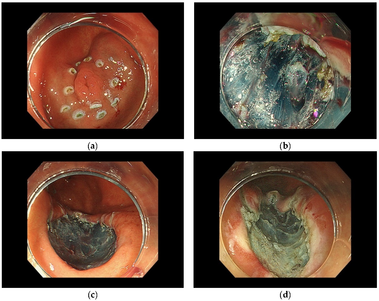
3.1. Postoperative Hemorrhage
3.2. Perforation
4. Future Perspectives
5. Conclusions
Author Contributions
Funding
Institutional Review Board Statement
Informed Consent Statement
Data Availability Statement
Conflicts of Interest
References
- Sung, H.; Ferlay, J.; Siegel, R.L.; Laversanne, M.; Soerjomataram, I.; Jemal, A.; Bray, F. Global Cancer Statistics 2020: GLOBOCAN Estimates of Incidence and Mortality Worldwide for 36 Cancers in 185 Countries. CA Cancer J. Clin. 2021, 71, 209–249. [Google Scholar] [CrossRef] [PubMed]
- Inoue, M.; Sawada, N.; Matsuda, T.; Iwasaki, M.; Sasazuki, S.; Shimazu, T.; Shibuya, K.; Tsugane, S. Attributable causes of cancer in Japan in 2005—Systematic assessment to estimate current burden of cancer attributable to known preventable risk factors in Japan. Ann. Oncol. 2012, 23, 1362–1369. [Google Scholar] [CrossRef] [PubMed]
- Clinton, S.K.; Giovannucci, E.L.; Hursting, S.D. The World Cancer Research Fund/American Institute for Cancer Research Third Expert Report on Diet, Nutrition, Physical Activity, and Cancer: Impact and Future Directions. J. Nutr. 2020, 150, 663–671. [Google Scholar] [CrossRef] [PubMed]
- Takachi, R.; Inoue, M.; Shimazu, T.; Sasazuki, S.; Ishihara, J.; Sawada, N.; Yamaji, T.; Iwasaki, M.; Iso, H.; Tsubono, Y.; et al. Consumption of sodium and salted foods in relation to cancer and cardiovascular disease: The Japan Public Health Center-based Prospective Study. Am. J. Clin. Nutr. 2010, 91, 456–464. [Google Scholar] [CrossRef] [Green Version]
- Hamashima, C. Update version of the Japanese Guidelines for Gastric Cancer Screening. Jpn J. Clin. Oncol. 2018, 48, 673–683. [Google Scholar] [CrossRef] [Green Version]
- Matsumoto, S.; Ishikawa, S.; Yoshida, Y. Reduction of gastric cancer mortality by endoscopic and radiographic screening in an isolated island: A retrospective cohort study. Aust. J. Rural Health 2013, 21, 319–324. [Google Scholar] [CrossRef]
- Hamashima, C.; Ogoshi, K.; Okamoto, M.; Shabana, M.; Kishimoto, T.; Fukao, A. A community-based, case-control study evaluating mortality reduction from gastric cancer by endoscopic screening in Japan. PLoS ONE 2013, 8, e79088. [Google Scholar] [CrossRef] [Green Version]
- Ono, H.; Yao, K.; Fujishiro, M.; Oda, I.; Uedo, N.; Nimura, S.; Yahagi, N.; Iishi, H.; Oka, M.; Ajioka, Y.; et al. Guidelines for endoscopic submucosal dissection and endoscopic mucosal resection for early gastric cancer (second edition). Dig. Endosc. Off. J. Jpn. Gastroenterol. Endosc. Soc. 2021, 33, 4–20. [Google Scholar] [CrossRef]
- Pimentel-Nunes, P.; Dinis-Ribeiro, M.; Ponchon, T.; Repici, A.; Vieth, M.; De Ceglie, A.; Amato, A.; Berr, F.; Bhandari, P.; Bialek, A.; et al. Endoscopic submucosal dissection: European Society of Gastrointestinal Endoscopy (ESGE) Guideline. Endoscopy 2015, 47, 829–854. [Google Scholar] [CrossRef] [Green Version]
- Hosokawa, O.; Miyanaga, T.; Kaizaki, Y.; Hattori, M.; Dohden, K.; Ohta, K.; Itou, Y.; Aoyagi, H. Decreased death from gastric cancer by endoscopic screening: Association with a population-based cancer registry. Scand. J. Gastroenterol. 2008, 43, 1112–1115. [Google Scholar] [CrossRef]
- Matsumoto, S.; Yamasaki, K.; Tsuji, K.; Shirahama, S. Results of mass endoscopic examination for gastric cancer in Kamigoto Hospital, Nagasaki Prefecture. World J. Gastroenterol. 2007, 13, 4316–4320. [Google Scholar] [CrossRef] [PubMed]
- Choi, K.S.; Jun, J.K.; Park, E.C.; Park, S.; Jung, K.W.; Han, M.A.; Choi, I.J.; Lee, H.Y. Performance of different gastric cancer screening methods in Korea: A population-based study. PLoS ONE 2012, 7, e50041. [Google Scholar] [CrossRef] [PubMed] [Green Version]
- Lee, K.S.; Oh, D.K.; Han, M.A.; Lee, H.; Jun, J.K.; Choi, K.S.; Park, E.U. The Korean guideline for gastric cancer screening. J. Korean Med. Assoc. 2015, 58, 373–384. [Google Scholar]
- Kawamura, T.; Wada, H.; Sakiyama, N.; Ueda, Y.; Shirakawa, A.; Okada, Y.; Sanada, K.; Nakase, K.; Mandai, K.; Suzuki, A.; et al. Examination time as a quality indicator of screening upper gastrointestinal endoscopy for asymptomatic examinees. Dig. Endosc. Off. J. Jpn. Gastroenterol. Endosc. Soc. 2017, 29, 569–575. [Google Scholar] [CrossRef]
- Ryu, J.E.; Choi, E.; Lee, K.; Jun, J.K.; Suh, M.; Jung, K.W.; Choi, K.S. Trends in the Performance of the Korean National Cancer Screening Program for Gastric Cancer from 2007 to 2016. Cancer Res. Treat. 2021. [Google Scholar] [CrossRef]
- Gotoda, T.; Yanagisawa, A.; Sasako, M.; Ono, H.; Nakanishi, Y.; Shimoda, T.; Kato, Y. Incidence of lymph node metastasis from early gastric cancer: Estimation with a large number of cases at two large centers. Gastric Cancer 2000, 3, 219–225. [Google Scholar] [CrossRef] [Green Version]
- Hirasawa, T.; Gotoda, T.; Miyata, S.; Kato, Y.; Shimoda, T.; Taniguchi, H.; Fujisaki, J.; Sano, T.; Yamaguchi, T. Incidence of lymph node metastasis and the feasibility of endoscopic resection for undifferentiated-type early gastric cancer. Gastric Cancer 2009, 12, 148–152. [Google Scholar] [CrossRef] [Green Version]
- Ono, H.; Yao, K.; Fujishiro, M.; Oda, I.; Nimura, S.; Yahagi, N.; Iishi, H.; Oka, M.; Ajioka, Y.; Ichinose, M.; et al. Guidelines for endoscopic submucosal dissection and endoscopic mucosal resection for early gastric cancer. Dig. Endosc. 2016, 28, 3–15. [Google Scholar] [CrossRef] [Green Version]
- Hasuike, N.; Ono, H.; Boku, N.; Mizusawa, J.; Takizawa, K.; Fukuda, H.; Oda, I.; Doyama, H.; Kaneko, K.; Hori, S.; et al. A non-randomized confirmatory trial of an expanded indication for endoscopic submucosal dissection for intestinal-type gastric cancer (cT1a): The Japan Clinical Oncology Group study (JCOG0607). Gastric Cancer 2018, 21, 114–123. [Google Scholar] [CrossRef] [Green Version]
- Takizawa, K.; Ono, H.; Hasuike, N.; Takashima, A.; Minashi, K.; Boku, N.; Kushima, R.; Katayama, H.; Ogawa, G.; Fukuda, H.; et al. A nonrandomized, single-arm confirmatory trial of expanded endoscopic submucosal dissection indication for undifferentiated early gastric cancer: Japan Clinical Oncology Group study (JCOG1009/1010). Gastric Cancer 2021, 24, 479–491. [Google Scholar] [CrossRef]
- Japamese Gastric Cancer Association; Japanese gastric cancer treatment guidelines 2018 (5th edition). Gastric Cancer 2021, 24, 1–21. [CrossRef] [PubMed] [Green Version]
- Nagahama, T.; Yao, K.; Maki, S.; Yasaka, M.; Takaki, Y.; Matsui, T.; Tanabe, H.; Iwashita, A.; Ota, A. Usefulness of magnifying endoscopy with narrow-band imaging for determining the horizontal extent of early gastric cancer when there is an unclear margin by chromoendoscopy (with video). Gastrointest. Endosc. 2011, 74, 1259–1267. [Google Scholar] [CrossRef] [PubMed]
- Muto, M.; Yao, K.; Kaise, M.; Kato, M.; Uedo, N.; Yagi, K.; Tajiri, H. Magnifying endoscopy simple diagnostic algorithm for early gastric cancer (MESDA-G). Dig. Endosc. Off. J. Jpn. Gastroenterol. Endosc. Soc. 2016, 28, 379–393. [Google Scholar] [CrossRef] [PubMed] [Green Version]
- Yao, K.; Anagnostopoulos, G.K.; Ragunath, K. Magnifying endoscopy for diagnosing and delineating early gastric cancer. Endoscopy 2009, 41, 462–467. [Google Scholar] [CrossRef] [PubMed] [Green Version]
- Yao, K.; Doyama, H.; Gotoda, T.; Ishikawa, H.; Nagahama, T.; Yokoi, C.; Oda, I.; Machida, H.; Uchita, K.; Tabuchi, M. Diagnostic performance and limitations of magnifying narrow-band imaging in screening endoscopy of early gastric cancer: A prospective multicenter feasibility study. Gastric Cancer 2014, 17, 669–679. [Google Scholar] [CrossRef]
- Abe, S.; Oda, I.; Shimazu, T.; Kinjo, T.; Tada, K.; Sakamoto, T.; Kusano, C.; Gotoda, T. Depth-predicting score for differentiated early gastric cancer. Gastric Cancer 2011, 14, 35–40. [Google Scholar] [CrossRef]
- Tsujii, Y.; Kato, M.; Inoue, T.; Yoshii, S.; Nagai, K.; Fujinaga, T.; Maekawa, A.; Hayashi, Y.; Akasaka, T.; Shinzaki, S.; et al. Integrated diagnostic strategy for the invasion depth of early gastric cancer by conventional endoscopy and EUS. Gastrointest. Endosc. 2015, 82, 452–459. [Google Scholar] [CrossRef]
- Nagahama, T.; Yao, K.; Imamura, K.; Kojima, T.; Ohtsu, K.; Chuman, K.; Tanabe, H.; Yamaoka, R.; Iwashita, A. Diagnostic performance of conventional endoscopy in the identification of submucosal invasion by early gastric cancer: The “non-extension sign” as a simple diagnostic marker. Gastric Cancer 2017, 20, 304–313. [Google Scholar] [CrossRef] [Green Version]
- Yanai, H.; Noguchi, T.; Mizumachi, S.; Tokiyama, H.; Nakamura, H.; Tada, M.; Okita, K. A blind comparison of the effectiveness of endoscopic ultrasonography and endoscopy in staging early gastric cancer. Gut 1999, 44, 361–365. [Google Scholar] [CrossRef]
- Choi, J.; Kim, S.G.; Im, J.P.; Kim, J.S.; Jung, H.C.; Song, I.S. Comparison of endoscopic ultrasonography and conventional endoscopy for prediction of depth of tumor invasion in early gastric cancer. Endoscopy 2010, 42, 705–713. [Google Scholar] [CrossRef]
- Pei, Q.; Wang, L.; Pan, J.; Ling, T.; Lv, Y.; Zou, X. Endoscopic ultrasonography for staging depth of invasion in early gastric cancer: A meta-analysis. J. Gastroenterol. Hepatol. 2015, 30, 1566–1573. [Google Scholar] [CrossRef] [PubMed]
- Oguro, Y.; Hirashima, T.; Tajiri, H.; Yoshida, S.; Yamaguchi, H.; Yoshimori, M.; Itabashi, M.; Hirota, T. Endoscopic treatment of early gastric cancer: Polypectomy and laser treatment. Jpn J. Clin. Oncol. 1984, 14, 271–282. [Google Scholar]
- Tada, M.; Murakami, A.; Karita, M.; Yanai, H.; Okita, K. Endoscopic resection of early gastric cancer. Endoscopy 1993, 25, 445–450. [Google Scholar] [CrossRef] [PubMed]
- Hosokawa, K.; Yoshida, S. Recent advances in endoscopic mucosal resection for early gastric cancer. Gan Kagaku Ryoho 1998, 25, 476–483. [Google Scholar] [PubMed]
- Gotoda, T.; Kondo, H.; Ono, H.; Saito, Y.; Yamaguchi, H.; Saito, D.; Yokota, T. A new endoscopic mucosal resection procedure using an insulation-tipped electrosurgical knife for rectal flat lesions: Report of two cases. Gastrointest. Endosc. 1999, 50, 560–563. [Google Scholar] [CrossRef]
- Ono, H.; Kondo, H.; Gotoda, T.; Shirao, K.; Yamaguchi, H.; Saito, D.; Hosokawa, K.; Shimoda, T.; Yoshida, S. Endoscopic mucosal resection for treatment of early gastric cancer. Gut 2001, 48, 225–229. [Google Scholar] [CrossRef] [PubMed] [Green Version]
- Park, Y.M.; Cho, E.; Kang, H.Y.; Kim, J.M. The effectiveness and safety of endoscopic submucosal dissection compared with endoscopic mucosal resection for early gastric cancer: A systematic review and metaanalysis. Surg. Endosc. 2011, 25, 2666–2677. [Google Scholar] [CrossRef]
- Lian, J.; Chen, S.; Zhang, Y.; Qiu, F. A meta-analysis of endoscopic submucosal dissection and EMR for early gastric cancer. Gastrointest. Endosc. 2012, 76, 763–770. [Google Scholar] [CrossRef]
- Hatta, W.; Gotoda, T.; Oyama, T.; Kawata, N.; Takahashi, A.; Yoshifuku, Y.; Hoteya, S.; Nakamura, K.; Hirano, M.; Esaki, M.; et al. Is radical surgery necessary in all patients who do not meet the curative criteria for endoscopic submucosal dissection in early gastric cancer? A multi-center retrospective study in Japan. J. Gastroenterol. 2017, 52, 175–184. [Google Scholar] [CrossRef]
- Hatta, W.; Gotoda, T.; Oyama, T.; Kawata, N.; Takahashi, A.; Yoshifuku, Y.; Hoteya, S.; Nakagawa, M.; Hirano, M.; Esaki, M.; et al. A Scoring System to Stratify Curability after Endoscopic Submucosal Dissection for Early Gastric Cancer: “eCura system”. Am. J. Gastroenterol. 2017, 112, 874–881. [Google Scholar] [CrossRef]
- Suzuki, H.; Takizawa, K.; Hirasawa, T.; Takeuchi, Y.; Ishido, K.; Hoteya, S.; Yano, T.; Tanaka, S.; Endo, M.; Nakagawa, M.; et al. Short-term outcomes of multicenter prospective cohort study of gastric endoscopic resection: ‘Real-world evidence’ in Japan. Dig. Endosc. Off. J. Jpn. Gastroenterol. Endosc. Soc. 2019, 31, 30–39. [Google Scholar] [CrossRef] [PubMed] [Green Version]
- Pimentel-Nunes, P.; Pioche, M.; Albéniz, E.; Berr, F.; Deprez, P.; Ebigbo, A.; Dewint, P.; Haji, A.; Panarese, A.; Weusten, B.; et al. Curriculum for endoscopic submucosal dissection training in Europe: European Society of Gastrointestinal Endoscopy (ESGE) Position Statement. Endoscopy 2019, 51, 980–992. [Google Scholar] [CrossRef] [Green Version]
- Kakushima, N.; Fujishiro, M.; Kodashima, S.; Muraki, Y.; Tateishi, A.; Omata, M. A learning curve for endoscopic submucosal dissection of gastric epithelial neoplasms. Endoscopy 2006, 38, 991–995. [Google Scholar] [CrossRef] [PubMed]
- Tsuji, Y.; Ohata, K.; Sekiguchi, M.; Ito, T.; Chiba, H.; Gunji, T.; Yamamichi, N.; Fujishiro, M.; Matsuhashi, N.; Koike, K. An effective training system for endoscopic submucosal dissection of gastric neoplasm. Endoscopy 2011, 43, 1033–1038. [Google Scholar] [CrossRef] [PubMed]
- Yoshida, M.; Kakushima, N.; Mori, K.; Igarashi, K.; Kawata, N.; Tanaka, M.; Takizawa, K.; Ito, S.; Imai, K.; Hotta, K.; et al. Learning curve and clinical outcome of gastric endoscopic submucosal dissection performed by trainee operators. Surg. Endosc. 2017, 31, 3614–3622. [Google Scholar] [CrossRef]
- Goto, O.; Fujishiro, M.; Kodashima, S.; Ono, S.; Niimi, K.; Hirano, K.; Yamamichi, N.; Koike, K. A second-look endoscopy after endoscopic submucosal dissection for gastric epithelial neoplasm may be unnecessary: A retrospective analysis of postendoscopic submucosal dissection bleeding. Gastrointest. Endosc. 2010, 71, 241–248. [Google Scholar] [CrossRef]
- Libânio, D.; Costa, M.N.; Pimentel-Nunes, P.; Dinis-Ribeiro, M. Risk factors for bleeding after gastric endoscopic submucosal dissection: A systematic review and meta-analysis. Gastrointest. Endosc. 2016, 84, 572–586. [Google Scholar] [CrossRef] [Green Version]
- Takizawa, K.; Oda, I.; Gotoda, T.; Yokoi, C.; Matsuda, T.; Saito, Y.; Saito, D.; Ono, H. Routine coagulation of visible vessels may prevent delayed bleeding after endoscopic submucosal dissection--an analysis of risk factors. Endoscopy 2008, 40, 179–183. [Google Scholar] [CrossRef]
- Mochizuki, S.; Uedo, N.; Oda, I.; Kaneko, K.; Yamamoto, Y.; Yamashina, T.; Suzuki, H.; Kodashima, S.; Yano, T.; Yamamichi, N.; et al. Scheduled second-look endoscopy is not recommended after endoscopic submucosal dissection for gastric neoplasms (the SAFE trial): A multicentre prospective randomised controlled non-inferiority trial. Gut 2015, 64, 397–405. [Google Scholar] [CrossRef]
- Nishizawa, T.; Suzuki, H.; Kinoshita, S.; Goto, O.; Kanai, T.; Yahagi, N. Second-look endoscopy after endoscopic submucosal dissection for gastric neoplasms. Dig. Endosc. Off. J. Jpn. Gastroenterol. Endosc. Soc. 2015, 27, 279–284. [Google Scholar] [CrossRef]
- Lee, B.I.; Kim, B.W.; Kim, H.K.; Choi, H.; Ji, J.S.; Hwang, S.M.; Cho, Y.S.; Chae, H.S.; Choi, K.Y. Routine mucosal closure with a detachable snare and clips after endoscopic submucosal dissection for gastric epithelial neoplasms: A randomized controlled trial. Gut Liver 2011, 5, 454–459. [Google Scholar] [CrossRef] [PubMed] [Green Version]
- Maekawa, S.; Nomura, R.; Murase, T.; Ann, Y.; Harada, M. Complete closure of artificial gastric ulcer after endoscopic submucosal dissection by combined use of a single over-the-scope clip and through-the-scope clips (with videos). Surg. Endosc. 2015, 29, 500–504. [Google Scholar] [CrossRef] [Green Version]
- Goto, O.; Oyama, T.; Ono, H.; Takahashi, A.; Fujishiro, M.; Saito, Y.; Abe, S.; Kaise, M.; Iwakiri, K.; Yahagi, N. Endoscopic hand-suturing is feasible, safe, and may reduce bleeding risk after gastric endoscopic submucosal dissection: A multicenter pilot study (with video). Gastrointest. Endosc. 2020, 91, 1195–1202. [Google Scholar] [CrossRef] [PubMed]
- Ohta, T.; Ishihara, R.; Uedo, N.; Takeuchi, Y.; Nagai, K.; Matsui, F.; Kawada, N.; Yamashina, T.; Kanzaki, H.; Hanafusa, M.; et al. Factors predicting perforation during endoscopic submucosal dissection for gastric cancer. Gastrointest. Endosc. 2012, 75, 1159–1165. [Google Scholar] [CrossRef] [PubMed]
- Mannen, K.; Tsunada, S.; Hara, M.; Yamaguchi, K.; Sakata, Y.; Fujise, T.; Noda, T.; Shimoda, R.; Sakata, H.; Ogata, S.; et al. Risk factors for complications of endoscopic submucosal dissection in gastric tumors: Analysis of 478 lesions. J. Gastroenterol. 2010, 45, 30–36. [Google Scholar] [CrossRef]
- Minami, S.; Gotoda, T.; Ono, H.; Oda, I.; Hamanaka, H. Complete endoscopic closure of gastric perforation induced by endoscopic resection of early gastric cancer using endoclips can prevent surgery (with video). Gastrointest. Endosc. 2006, 63, 596–601. [Google Scholar] [CrossRef]
- Hirao, M.; Yamada, T.; Michida, T.; Nishikawa, K.; Hamakawa, T.; Mita, E.; Mano, M.; Sekimoto, M. Peritoneal Seeding after Gastric Perforation during Endoscopic Submucosal Dissection for Gastric Cancer. Dig. Surg. 2018, 35, 457–460. [Google Scholar] [CrossRef]
- Fernández-Esparrach, G.; Marín-Gabriel, J.C.; de Tejada, A.H.; Albéniz, E.; Nogales, O.; Del Pozo-García, A.J.; Rosón, P.J.; Goicotxea, U.; Uchima, H.; Terán, A.; et al. Implementation of endoscopic submucosal dissection in a country with a low incidence of gastric cancer: Results from a prospective national registry. United Eur. Gastroenterol. J. 2021, 9, 718–726. [Google Scholar] [CrossRef]
- Ribeiro-Mourão, F.; Pimentel-Nunes, P.; Dinis-Ribeiro, M. Endoscopic submucosal dissection for gastric lesions: Results of an European inquiry. Endoscopy 2010, 42, 814–819. [Google Scholar] [CrossRef]
- Tsuda, M.; Asaka, M.; Kato, M.; Matsushima, R.; Fujimori, K.; Akino, K.; Kikuchi, S.; Lin, Y.; Sakamoto, N. Effect on Helicobacter pylori eradication therapy against gastric cancer in Japan. Helicobacter 2017, 22. [Google Scholar] [CrossRef] [Green Version]
- Kamada, T.; Hata, J.; Sugiu, K.; Kusunoki, H.; Ito, M.; Tanaka, S.; Inoue, K.; Kawamura, Y.; Chayama, K.; Haruma, K. Clinical features of gastric cancer discovered after successful eradication of Helicobacter pylori: Results from a 9-year prospective follow-up study in Japan. Aliment Pharmacol. Ther. 2005, 21, 1121–1126. [Google Scholar] [CrossRef] [PubMed]
- Yamamoto, K.; Kato, M.; Takahashi, M.; Haneda, M.; Shinada, K.; Nishida, U.; Yoshida, T.; Sonoda, N.; Ono, S.; Nakagawa, M.; et al. Clinicopathological analysis of early-stage gastric cancers detected after successful eradication of Helicobacter pylori. Helicobacter 2011, 16, 210–216. [Google Scholar] [CrossRef] [PubMed]
- Hata, K.; Ito, M.; Boda, T.; Kotachi, T.; Kiso, M.; Masuda, K.; Kurihara, M.; Kuroki, K.; Yorita, N.; Nagasaki, N.; et al. Gastric Cancer with Submucosal Invasion after Successful Helicobacter pylori Eradication: A Propensity Score-Matched Analysis of Patients with Annual Patient Endoscopic Survey. Digestion 2019, 99, 59–65. [Google Scholar] [CrossRef]
- Ito, M.; Tanaka, S.; Chayama, K. Characteristics and Early Diagnosis of Gastric Cancer Discovered after Helicobacter pylori Eradication. Gut Liver 2021, 15, 338–345. [Google Scholar] [CrossRef] [PubMed] [Green Version]
- Pelc, Z.; Skórzewska, M.; Rawicz-Pruszyński, K.; Polkowski, W.P. Lymph Node Involvement in Advanced Gastric Cancer in the Era of Multimodal Treatment-Oncological and Surgical Perspective. Cancers 2021, 13, 2509. [Google Scholar] [CrossRef] [PubMed]
- Choi, J.H.; Kim, E.S.; Lee, Y.J.; Cho, K.B.; Park, K.S.; Jang, B.K.; Chung, W.J.; Hwang, J.S.; Ryu, S.W. Comparison of quality of life and worry of cancer recurrence between endoscopic and surgical treatment for early gastric cancer. Gastrointest. Endosc. 2015, 82, 299–307. [Google Scholar] [CrossRef]
- Nunobe, S.; Oda, I.; Ishikawa, T.; Akazawa, K.; Katai, H.; Isobe, Y.; Miyashiro, I.; Tsujitani, S.; Ono, H.; Tanabe, S.; et al. Surgical outcomes of elderly patients with Stage I gastric cancer from the nationwide registry of the Japanese Gastric Cancer Association. Gastric Cancer 2020, 23, 328–338. [Google Scholar] [CrossRef]

| Depth of Invasion | Ulceration | Differentiated Type | Undifferentiated Type | ||
|---|---|---|---|---|---|
| cT1a(M) | UL0 | ≤20 mm diameter absolute indications for EMR/ESD *** | >20 mm diameter absolute indications for ESD | ≤20 mm diameter absolute indications for ESD | >20 mm diameter Relative indications |
| UL1 | ≤30 mm diameter absolute indications for ESD | >30 mm diameter Relative indications | Relative indications | ||
| cT1b(SM) | Relative indications | Relative indications | |||
| Depth of Invasion | Ulceration | Differentiated Type | Undifferentiated Type | ||
|---|---|---|---|---|---|
| cT1a(M) | UL0 | eCuraA *** | ≤20 mm diameter eCuraA | >20 mm diametere CuraB | |
| UL1 | ≤30 mm diameter eCuraA | >30 mm diameter eCuraC-2 | eCuraC-2 | ||
| cT1b(SM1) | ≤30 mm diameter eCuraB | >30 mm diameter eCuraC-2 | eCuraC-2 | ||
| cT1b2(SM2) | eCuraC-2 | eCuraC-2 | |||
Publisher’s Note: MDPI stays neutral with regard to jurisdictional claims in published maps and institutional affiliations. |
© 2022 by the authors. Licensee MDPI, Basel, Switzerland. This article is an open access article distributed under the terms and conditions of the Creative Commons Attribution (CC BY) license (https://creativecommons.org/licenses/by/4.0/).
Share and Cite
Hisada, H.; Sakaguchi, Y.; Oshio, K.; Mizutani, S.; Nakagawa, H.; Sato, J.; Kubota, D.; Obata, M.; Cho, R.; Nagao, S.; et al. Endoscopic Treatment of Superficial Gastric Cancer: Present Status and Future. Curr. Oncol. 2022, 29, 4678-4688. https://doi.org/10.3390/curroncol29070371
Hisada H, Sakaguchi Y, Oshio K, Mizutani S, Nakagawa H, Sato J, Kubota D, Obata M, Cho R, Nagao S, et al. Endoscopic Treatment of Superficial Gastric Cancer: Present Status and Future. Current Oncology. 2022; 29(7):4678-4688. https://doi.org/10.3390/curroncol29070371
Chicago/Turabian StyleHisada, Hiroyuki, Yoshiki Sakaguchi, Kaori Oshio, Satoru Mizutani, Hideki Nakagawa, Junichi Sato, Dai Kubota, Miho Obata, Rina Cho, Sayaka Nagao, and et al. 2022. "Endoscopic Treatment of Superficial Gastric Cancer: Present Status and Future" Current Oncology 29, no. 7: 4678-4688. https://doi.org/10.3390/curroncol29070371
APA StyleHisada, H., Sakaguchi, Y., Oshio, K., Mizutani, S., Nakagawa, H., Sato, J., Kubota, D., Obata, M., Cho, R., Nagao, S., Miura, Y., Mizutani, H., Ohki, D., Yakabi, S., Takahashi, Y., Kakushima, N., Tsuji, Y., Yamamichi, N., & Fujishiro, M. (2022). Endoscopic Treatment of Superficial Gastric Cancer: Present Status and Future. Current Oncology, 29(7), 4678-4688. https://doi.org/10.3390/curroncol29070371