Next Article in Journal
Stammzelltherapie in der Kardiologie: Der Ersehnte Jungbrunnen Oder Falsche Hoffnung?
Previous Article in Journal
Kardiale Resynchronisationstherapie (CRT): Mit Oder Ohne ICD-Back-Up?
 
 
Cardiovascular Medicine is published by MDPI from Volume 28 Issue 1 (2025). Previous articles were published by another publisher in Open Access under a CC-BY (or CC-BY-NC-ND) licence, and they are hosted by MDPI on mdpi.com as a courtesy and upon agreement with Editores Medicorum Helveticorum (EMH).
Article

Utility of Echocardiography for Tailoring Cardiac Resynchronisation Therapy

Cardiology Service, University Hospital of Geneva, 1205 Geneva, Switzerland
*
Author to whom correspondence should be addressed.
Cardiovasc. Med. 2006, 9(5), 188; https://doi.org/10.4414/cvm.2006.01175
Submission received: 28 February 2006 / Revised: 31 March 2006 / Accepted: 30 April 2006 / Published: 31 May 2006

Summary

Cardiac resynchronisation therapy may lead to remarkable improvement in clinical status in selected patients with heart failure. However, approximately 20% of patients may not respond to this treatment. A major challenge lies in reducing the number of non-responders. Echocardiography has been the focus of growing interest for improving patient selection as well as for device optimisation. This article reviews the current state of the art of assessment of dyssynchrony by echocardiography, and overviews practical aspects of the technique.

Resumé

La stimulation biventriculaire a fait ses preuves dans le traitement de l’insuffisance cardiaque chez des patients sélectionnés. Toutefois, environ 20% de ces patients ne répondent pas à cette thérapie. Un défi majeur consiste à diminuer le taux des non-répondeurs. L’échocardiographie fait l’objet d’un intérêt croissant pour l’amélioration de la sélection des patients, ainsi que pour l’optimalisation du réglage des paramètres de stimulation. Cet article passe en revue les données de la litérature ainsi que les aspects pratiques des techniques d’échocardiographies dans cette application.
Mot-clefs: thérapie par resynchronisation cardiaque; stimulation biventriculaire; échocardiographie; insuffisance cardiaque.

Introduction

It is now well established that cardiac resynchronisation therapy (CRT) is an effective treatment for advanced drug-refractory heart failure in selected patients. Large multicenter trials have shown that biventricular pacing improves symptoms and functional capacity, reduces admission for heart failure [1,2,3] and prolongs survival [3]. Nevertheless, about 20% of patients in these trials do not respond clinically. This may be due to a variety of reasons, such as non-delivery of left ventricular pacing (due to high pacing thresholds, or atrial arrhythmias with uncontrolled ventricular rate), inappropriate lead positioning, suboptimal device programming, or inappropriate patient selection. A major challenge lies in reducing the number of non-responders of this invasive and expensive therapy. Imaging techniques may play a role in selecting patients, targeting ideal pacing sites, and optimising device settings. Magnetic resonance imaging may be used to measure ventricular volumes and function, as well as for the quantification of ventricular dyssynchrony [4]. However, this technique is expensive and time-consuming, and may not be used for studying the patient once a device is implanted. Nuclear ventriculography with phase analysis is a promising technique for assessing ventricular function and dyssynchrony [5], but has limited time resolution and does not yield information on mitral regurgitation (which may also be improved by biventricular pacing). Echocardiography on the other hand, may be used for evaluating all of these parameters, has excellent time resolution, and is widely available. It is therefore not surprising that it has been the focus of growing interest for studying CRT. This article provides an overview of the various echocardiographic techniques that are being used for this application.

Evaluation of cardiac dyssynchrony for patient selection

Heart failure may be associated with dyssynchrony at various levels. Atrioventricular dyssynchrony may be present due to long PR intervals, to interatrial conduction delay, or to delayed relaxation of the left ventricle associated with heart failure, resulting in fusion of the E and A waves of mitral inflow (and reduction in left ventricular filling time). Interventricular dyssynchrony may result from delayed ejection of one of the ventricles, this usually being the left ventricle in the setting of left bundle branch block. Finally, intraventricular dyssynchrony refers to delay in contraction of segments within the left ventricle, usually of the basal lateral wall [6,7].
There is growing evidence to suggest that presence of baseline ventricular dyssynchrony may be a marker for predicting favorable response to biventricular pacing. Even though QRS duration has been used as an inclusion criterion in all major trials, it has been shown that electrical dyssynchrony is not automatically associated with mechanical dyssynchrony, and vice-versa [8]. Mechanical dyssynchrony of the left ventricle may be absent in about 30% of patients with congestive heart failure and intra-ventricular conduction delay [8,9], and present in about 50% of patients with a QRS duration of ≤120 ms [8,9,10]. In a small single-center study [11], biventricular pacing in patients with a narrow QRS and echographic evidence of dyssynchrony was shown to have comparable efficacy as in patients with bundle-branch block.
A variety of echocardiographic imaging modes have been used for evaluating dyssynchrony (Figure 1). In addition, several measurement techniques have been used to assess dyssynchrony with each imaging mode, especially as regards tissue Doppler imaging (TDI). When evaluating dyssynchrony by the various echocardiographic methods, the reference timepoint for the different measurements is QRS onset. It is therefore mandatory to have a reference ECG of good quality. If dyssynchrony is evaluated during ventricular pacing, it is useful to program a unipolar pacing mode in order to use the pacing artifact as a reference point. Sweep speed should be set to at least 100 mm/s for reducing measurement error. Measurements should be averaged over 3–5 cycles. Recordings are particularly difficult to interpret during irregular heart rhythm, such as during atrial fibrillation, and have not been validated in this setting.

M-mode echocardiography

Pitzalis et al. [12] measured the “septal to posterior wall motion delay” (SPWMD) using M-mode on a parasternal short-axis view of the left ventricle at the papillary muscle level. The shortest delay between the maximal inward displacement of the septal wall and the posterior wall was measured, and a value of ≥130 ms found to have 85% accuracy for predicting reduction in left ventricular endsystolic volume (LVESV) index at follow-up after initiation of CRT. Recently, the same investigators have shown that this index is also predictive of clinical events [13]. However, this measurement may be difficult to interpret due to ill defined maximal wall excursion (especially in patients with large anteroseptal myocardial infarction), and was not shown to predict clinical or echocardiographic improvement by other investigators [14].
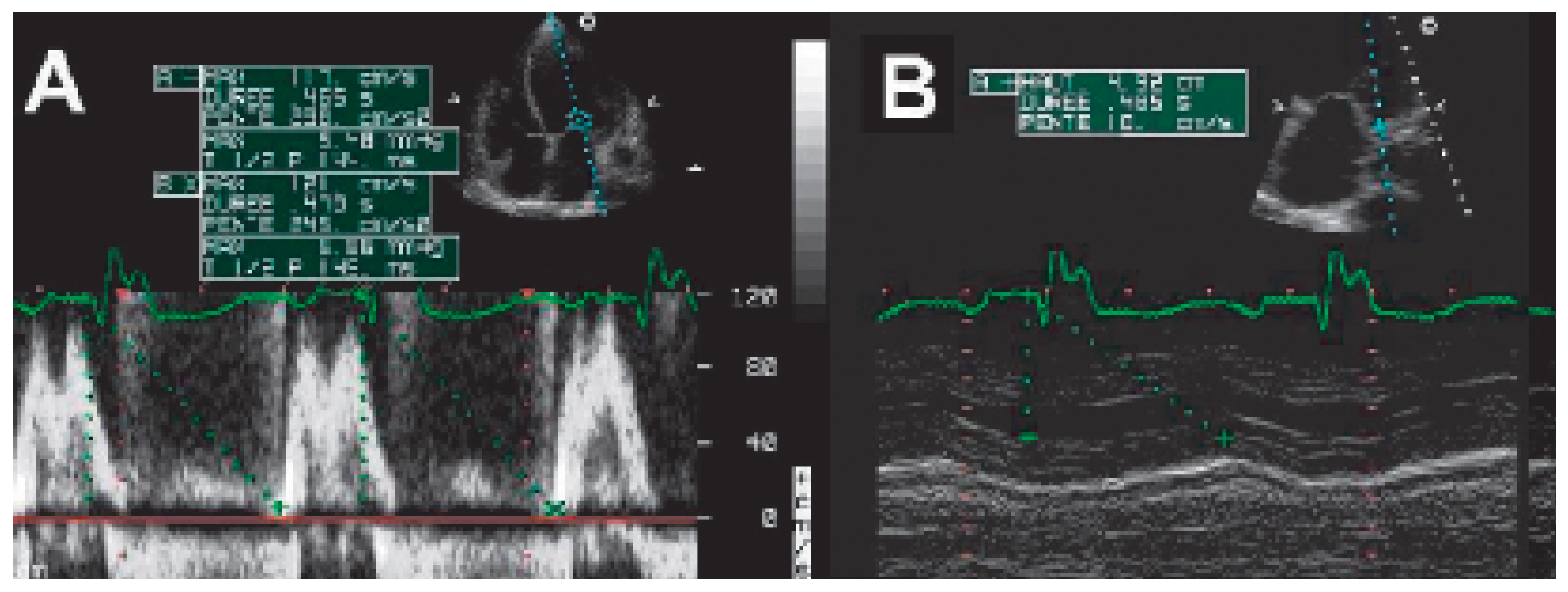
In the CARE-HF study [3], M-mode imaging was used for evaluating inward displacement of the postero-lateral wall (in the parasternal long-axis view or the apical fourchamber view) after onset of left ventricular filling (E wave onset). An abnormal result would be an interval from QRS to E-wave onset that is shorter than to maximal displacement with M-mode (Figure 2). We have found this criterion to be far less likely to show intraventricular dyssynchrony than pulsed-wave TDI in the same patient [15].

Conventional Doppler echography

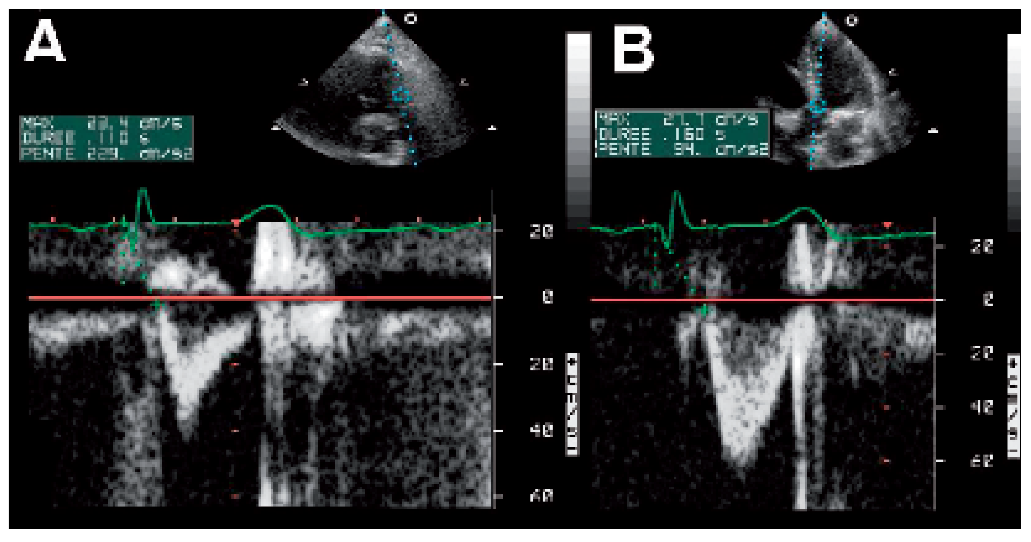
Pre-ejection intervals from QRS onset to the beginning of ventricular ejection may be easily measured at the pulmonary and aortic valve levels using pulsed-wave Doppler (Figure 3). It is useful to place the Doppler volume at the leaflet tips to obtain a valve opening and closure artifact, as this will help to better define the measurement points. Interventricular dyssynchrony is calculated as the difference between these two measurements, otherwise known as the interventricular mechanical delay (IVMD). Normal delays have been defined as being <20 ms [11] or <40 ms [3]. Unpublished data from the MIRACLE study have shown that patients with a baseline IVMD of >40 ms had greater improvement in echographic parameters in response to biventricular pacing as compared to those with ≤40 ms [16]. In the same study, patients with an aortic pre-ejection interval of >160 ms were found in addition to have greater improvement in clinical outcome such as quality of life scores, six-minute walking distance and peak VO as compared to those with an interval of ≤160 ms [17]. In the recently reported CARE-HF study [3], patients with a baseline IVMD of >50 ms showed a trend in better outcome (death or admission for heart failure) compared to those with <50 ms.
This is the imaging mode that has attracted most attention for studying CRT. Data may be acquired using either pulsed-wave TDI samples (Figure 4A), or using color TDI velocity curves (Figure 4B) available on some models (for example Vivid Seven/General Electric-Vingmed™ and iE33/Philips Medical Systems™). The latter technique allows offline measurement of myocardial velocities in different regions of the heart during the same cardiac cycle. This accelerates acquisition time considerably, and avoids variations in measurements at different cycles that are due to respiration and heart rate. The digitally-acquired data may also be processed for strain* and strain-rate** analysis. * Strain represents the extent of deformation of a tissue segment over time,  and  is  expressed  as the percent change from the original dimension. ** Strain-rate measures the rate of deformation of a tissue segment. These measurements allow differentiation of contraction from passive motion.
Pulsed-wave TDI recordings have been measured to the onset of sustained systolic motion (Sm) [10,18,19,20] (Figure 4A) or alternatively to peak Sm [21,22]. When using color TDI velocity curves, measurements have been most often performed to the peak of sustained systolic velocity [6,9,23,24,25,26], but also to maximal velocity occurring anytime after isovolumic contraction [27]. Numbers of left ventricular segments analysed have varied from 2 (basal septal and lateral) to 12 (all basal and mid segments).
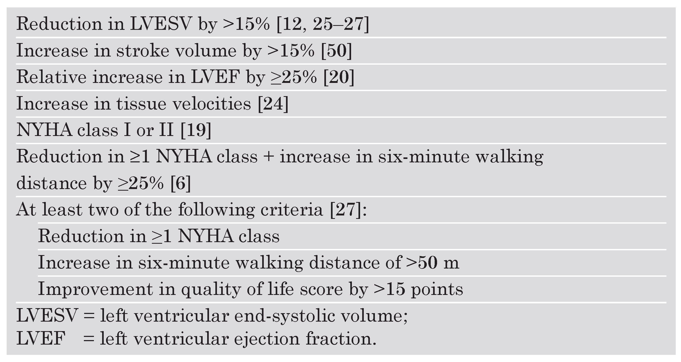
Table 1. Endpoints used in echocardiographic studies for assessing response to biventricular pacing.
Cardiovascmed 09 00188 i001
As ventricular contraction has longitudinal, radial, and circumferential vector components, the signal obtained by TDI will depend on proper alignment of the Doppler beam with wall motion. TDI measurements should not be performed on akinetic segments. The lowvelocity signals recorded are difficult to interpret, and most likely reflect passive motion rather than contraction.
Interventricular dyssynchrony has been assessed by differences in delays between the lateral wall of the right ventricle and of the interventrical septum [19,26], or of the lateral wall of the left ventricle [6], or of the most delayed segment of the left ventricle [20]. Normal values were assessed by Yu et al. in 106 control subjects [28]. Color TDI was used for measuring delays to peak Sm in 12 segments of the left ventricle and in the free wall of the right ventricle. Right ventricular activation was delayed as compared to the left ventricle by about 30 ms.
Intraventricular dyssynchrony of the left ventricle has been defined as the difference in delays between the septal and lateral walls [19,23,26], between maximal and minimal delays [6,9,10,20,21,27], or as the standard deviation of delays in the 12-segment model [9,21,22,25,26]. In normal subjects, there is no significant difference in delay of activation between the different segments of the left ventricle, either as assessed using color TDI to peak Sm [28], or using pulsed-wave TDI to Sm onset [29].
Intraventricular dyssynchrony may also be assessed by presence of delayed longitudinal contraction of basal segments occurring during diastole (after closure of the aortic valve) [24]. Investigators have also used strain and strain rate for evaluating dyssynchrony [23,24,26,30,31]. This technique allows differentiation of passive wall motion from true contraction, but has not been shown to be superior than TDI velocity for predicting response to therapy [26].
The confusion created by the multitude of TDI measurement techniques is amplified by use of different endpoints for defining response to biventricular pacing (table 1). These include echographic parameters such as changes in systolic function (left ventricular ejection fraction [20,24] and myocardial velocities [24]) and ventricular remodeling (defined as a reduction in LVESV by >15%) [25,26,27]. More useful are clinical endpoints such as improvement in NYHA class, used either alone or in combination with improvement in 6 minute walking distance and quality of life scores [6,19,27].
Bax et al. [6] have recently reported data on the utility of TDI for predicting response to biventricular pacing in 85 patients. Intraventricular dyssynchrony with >65 ms delay between 4 basal segments predicted clinical response at 6 months (improvement by 01 NYHA score and 025% six-minute walking distance) with 80% sensitivity and specificity. The same cutoff predicted reverse remodeling, defined as a 015% reduction in LVESV, with 92% sensitivity and specificity. Prognosis at one year (cardiac mortality and admission for heart failure) was better in patients with baseline intraventricular dyssynchrony than in those without this criterion (6% vs. 50%; p <0.001). However, this single-center experience has yet to be confirmed on a more widespread basis, before being adopted universally for patient selection.
Yu et al. [32] have reported that a left ventricular dyssynchrony index of >32.6 ms (the standard deviation of time to peak systolic velocity measured on 12 basal and mid segments) was 100% sensitive and specific for identifying responders with reverse remodelling. In another series, they also evaluated 16 different parameters of intraventricular dyssynchrony, and found that the dyssynchrony index was the only independent predictor for response [26]. However, the very same parameter was not predictive of clinical or echographic response when evaluated by other investigators [33], most probably due to differences in measurement technique. This underlines the necessity of validating parameters in multicenter studies before applying them widely for patient selection.
In order to be adopted in the clinical setting, it would be preferable that time-consuming parameters be processed semi-automatically. A step in this direction is “tissue synchronicity imaging” (TSI) which codes regions in different colors according to local delays of peak tissue velocity. This technique has been shown to predict acute increase in stroke volume in response to CRT, but was not indicative of remodeling during follow-up [34]. TDI velocity curves can be sometimes difficult to interpret, even for an experienced practitioner. Therefore automated methods will have to undergo careful validation before being used in current practice.

Real-time 3D echocardiography

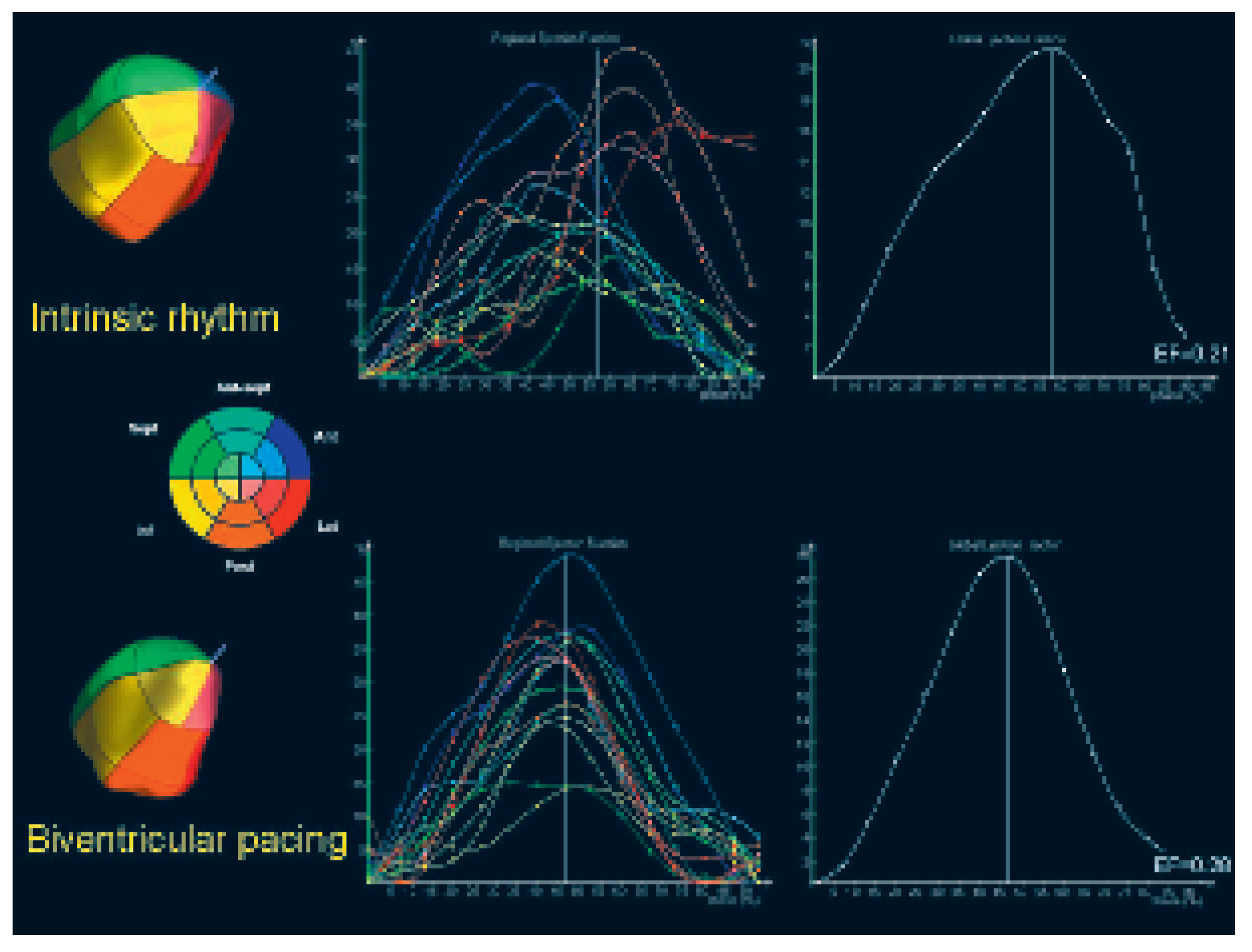
Since the advent of real-time 3D imaging, the full volume of the left ventricle is captured over 4 cardiac cycles during a breath-hold, and then reconstructed offline on an external workstation using semi-automated endocardial border detection. This tool provides an easily appreciable display of resynchronisation by biventricular pacing with analysis of regional volumes and ejection fraction (Figure 5). Kapetenakis et al. [35] have reported that clinical responders had greater baseline dyssynchrony than nonresponders. The drawbacks of this method are incomplete visualisation of the entire left ventricle (especially of the apical segments of dilated hearts due to truncation by the pyramidal imaging sector), need for specialised equipment, and time-consuming post-processing.

Other imaging modes

Breithardt et al. [36] have assessed left ventricular dyssynchrony using phase analysis of septal and lateral wall motion in the apical four-chamber view in 34 patients in the PATHCHF study. Dyssynchrony evaluated by this method predicted acute increase in dP/dt by biventricular pacing. Kawaguchi et al. [37] evaluated 10 patients using contrast-enhanced images in the four-chamber view for deriving a parameter referred to as cardiac variability imaging. Endocardial borders were traced manually to obtain regional fractional area changes plotted against time, yielding displacement maps. Both methods, however, have restricted availability, and assess dyssynchrony in a single plane.

Utility of echocardiography for the implant procedure and for targeting lead placement

Preoperative echocardiography may yield valuable information for the implanting physician. Right atrial size may influence choice of guide catheters to cannulate the coronary sinus. The coronary sinus is easily visualised in the apical four-chamber view with posterior tilt of the transducer. A persistent left superior vena cava may be identified by visualising a dilated coronary sinus and injecting microbubbles via a peripheral vein in the left arm. This anatomical variant results in the inability to perform a coronary sinus venogram for identifying target veins at implantation, and also results in unstable lead positions, making it often necessary to implant a left epicardial lead surgically. Peroperative transoesophageal echocardiography has been used by some operators to locate the coronary sinus ostium. This however requires general anaesthesia for airway protection, which limits its use for routine cases.
Echocardiography may also play a role in positioning of the left ventricular lead. Walls which have suffered from myocardial infarction and show evidence of non-viability (thinning, hyperechogenicity and dyskinesia) may be identified. It is nevertheless unlikely to implant the left ventricular lead in a zone of nonviable myocardium, as this will result in unacceptably high pacing thresholds.
It is not yet well defined whether myocardial walls that show latest mechanical activation should be targeted as pacing sites. Ansalone et al. [38] studied 31 patients who had been implanted with a biventricular pacemaker. Patients in whom the lead was positioned in proximity to segments that showed latest mechanical activation in sinus rhythm had greater improvement in echographic endpoints (ejection fraction and LVESV) on the day following device implantation. However, there were no significant differences between groups in clinical parameters such as NYHA class and six-minute walking distance after a week. An unanswered issue is whether the left ventricular site with latest activation should be measured in intrinsic rhythm, or during right ventricular pacing.

Device optimisation by echocardiography

Haemodynamic response to pacing may be affected by timing of the atrioventricular (AV) interval, affecting synchronicity of atrial and ventricular contraction. In addition, current biventricular devices have separate right and left ventricular channels that allow programming of an interventricular (VV) interval with right or left ventricular preexcitation. This may affect interas well as intraventricular synchrony, as the septal wall is usually activated by the right ventricular lead, and the lateral wall by the coronary sinus lead.

AV interval optimisation

Patients with heart failure may have E-A fusion assessed by pulsed wave Doppler at the mitral valve leaflet tips. Shortening the AV interval by pacing the ventricle will result in anticipation of the E wave of the following cycle, leading to dissociation of the E and A waves and prolongation of diastolic filling time. However, the AV interval should not be too short, as A wave truncation will result, compromising ventricular filling. Thus the optimal AV interval corresponds to the shortest interval that does not interrupt the end of the A wave. Several techniques have been described for optimising the AV interval:

The Ritter method (Figure 6)

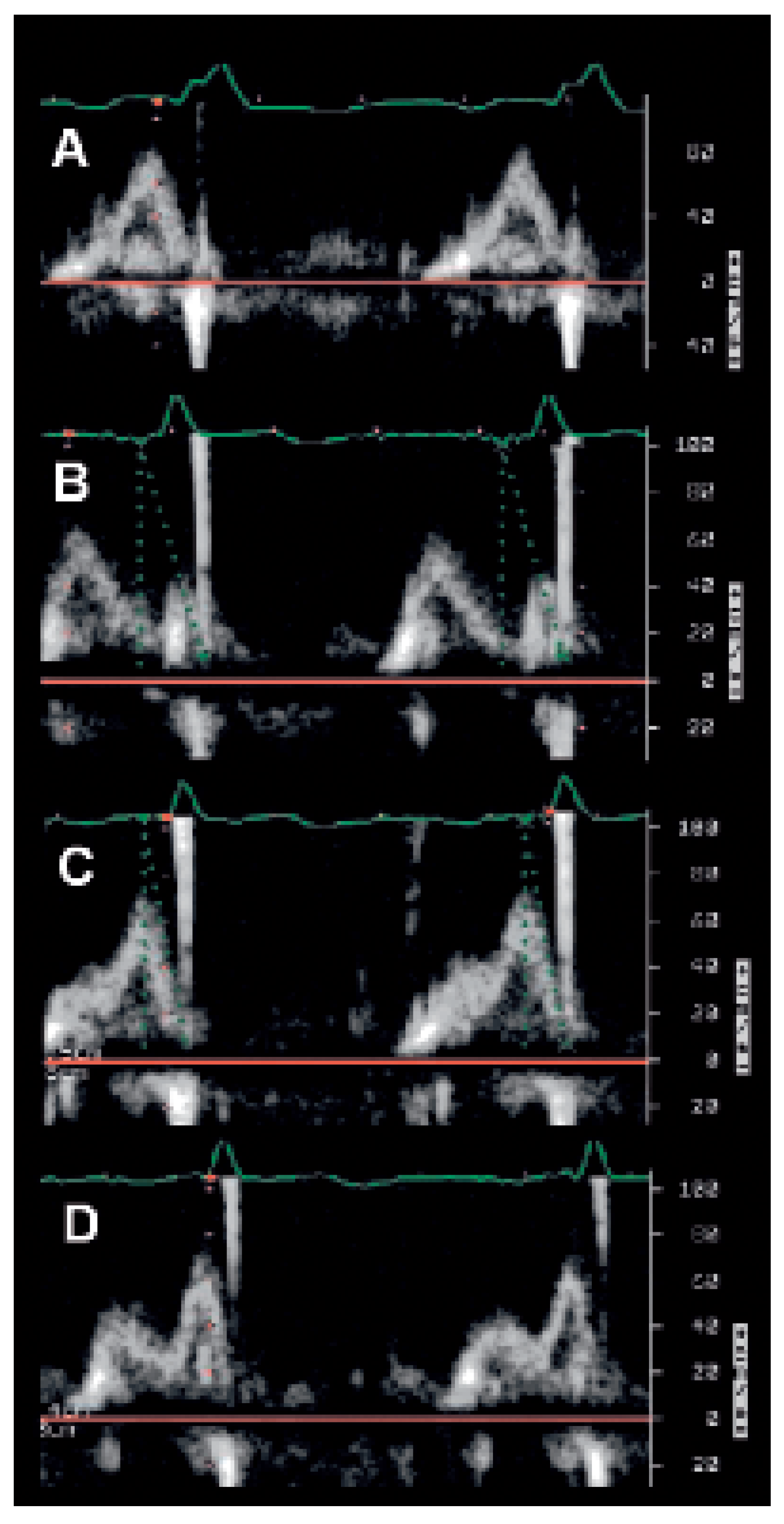
This method has been validated in patients with atrioventricular block implanted with standard pacemakers [39,40], but not as yet for biventricular pacing. AV intervals are programmed to a short and to a long value (eg 50 ms and 150 ms), and the delay between QRS onset and the end of the A-wave (QA interval) measured at each setting. The following formula is then used to calculate the optimal delay: AVopt = AVshort + [(AVlong + QAlong) – (AVshort + QAshort)].
This may be simplified to: AVopt = AVlong – (QAshort – QAlong).

The iterative method

A long AV interval (eg 200 ms) is programmed and the AV interval decremented in 20 ms steps, until A-wave truncation is observed. The AV interval may then be incremented in 10 ms steps to obtain the optimal setting.

The aortic velocity-time integral (VTI) method

Measurement of aortic VTI by pulsed or continuous-wave Doppler is a surrogate of stroke volume, and may be measured at different programmed AV intervals. This method was recently evaluated in a study of 40 patients implanted with a biventricular pacemaker randomised to either an empiric AV interval of 120 ms or to individually optimised AV intervals [41]. Patients with optimised settings had greater improvement in NYHA class and quality of life at 3 months, although there was no difference in six-minute walking distance.
Limitations with all these methods are that the AV intervals are optimised in a supine, resting patient and at a given heart rate. These conditions obviously do not reflect haemodynamics in a patient who is active. Recently, Scharf et al. reported that the optimal AV interval is longer during exercise than at rest [42].

VV interval optimisation

A simple method for optimising VV intervals would be to program settings that would result in least interventricular mechanical delay (IVMD). However, we and others have previously shown that reduced interventricular dyssynchrony is not correlated with improved left ventricular systolic function [21,43]. Sogaard et al. [44] optimised settings by measuring averaged tissue Doppler imaging (TDI) velocities of the 16 left ventricular segments, and defined the optimal VV interval as that which resulted in the highest mean global TDI velocity. However, clinical impact of this technique for device optimisation has not been assessed. The few other studies that have evaluated sequential biventricular pacing by echocardiography have used pulsed-wave Doppler measurement of aortic outflow velocity-time integral (VTI) at different VV intervals [21,45,46]. The sample volume should be placed at exactly the same point in the aortic outflow tract at each setting. It is therefore important that a second person assists the examinator by manipulating the pacemaker programmer for changing device settings. Recordings should be taken at each setting and VTI measured offline. Impact of different settings will usually be of small magnitude, and measurement error may play a significant part in observed differences. Alternatively, continuous-wave Doppler VTI may be used (although this has only been so far reported for AV interval optimisation [41]). This may result in less variability due to positioning of the Doppler sample.
However, the contours of the signal may be less well defined compared to pulsed-wave Doppler, making it more difficult to obtain reproducible measurements.
It is best to start with AV interval optimisation during simultaneous biventricular pacing. VV intervals may be optimised next. If the left ventricle is preexcited, the AV interval does not need to be modified. However, if the right ventricle is activated first, the AV interval must be reprogrammed by subtracting the VV delay from the optimal AV interval measured during simultaneous biventricular pacing.
Whether individually optimised VV intervals improves outcome in patients with biventricular pacing remains to be proven. Prospective randomised studies with clinical endpoints are still needed to establish the role of optimised VV intervals in CRT. A single nonrandomised study with 34 patients showed no difference in NYHA class and six-minute hall walk distance after three months of optimised sequential pacing, as compared to patients with simultaneous pacing [45].

Conclusion

Echocardiography will probably play an increasing role in patient selection for CRT. A multitude of techniques are being evaluated, some of which are simple and rapid to perform, whilst others are more complicated and timeconsuming. However, accuracy of proposed indices for identifying responders to CRT is still equivocal. Prospective multicenter trials addressing these questions are currently underway and will hopefully clarify these issues. In the meantime, it is inappropriate to deny CRT to patients who fulfill currently accepted criteria [47,48] simply because dyssynchrony may not be demonstrable by echocardiography.
The second major application of echocardiography in CRT is device optimisation, which may be helpful in a subset of patients. The technique is, however, relatively time-consuming. With the current growth in number of CRT implantations, many centers will not have the means to optimise devices in all their patients. A pragmatic approach would be to program nominal settings that are most likely to result in improvement, and focus tests on non-responders at follow-up.

References

  1. Abraham, W.T.; Fisher, W.G.; Smith, A.L.; Delurgio, D.B.; Leon, A.R.; Loh, E.; et al. Cardiac resynchronization in chronic heart failure. N Engl J Med 2002, 346, 1845–1853. [Google Scholar] [CrossRef]
  2. Bristow, M.R.; Saxon, L.A.; Boehmer, J.; Krueger, S.; Kass, D.A.; De Marco, T.; et al. Cardiac-resynchronization therapy with or without an implantable defibrillator in advanced chronic heart failure. N Engl J Med 2004, 350, 2140–2150. [Google Scholar] [CrossRef] [PubMed]
  3. Cleland, J.G.; Daubert, J.C.; Erdmann, E.; Freemantle, N.; Gras, D.; Kappenberger, L.; et al. The effect of cardiac resynchronization on morbidity and mortality in heart failure. N Engl J Med 2005, 352, 1539–1549. [Google Scholar] [CrossRef] [PubMed]
  4. Nelson, G.S.; Curry, C.W.; Wyman, B.T.; Kramer, A.; Declerck, J.; Talbot, M.; et al. Predictors of systolic augmentation from left ventricular preexcitation in patients with dilated cardiomyopathy and intraventricular conduction delay. Circulation 2000, 101, 2703–2709. [Google Scholar] [CrossRef] [PubMed]
  5. Fauchier, L.; Marie, O.; Casset-Senon, D.; Babuty, D.; Cosnay, P.; Fauchier, J.P. Interventricular and intraventricular dyssynchrony in idiopathic dilated cardiomyopathy: A prognostic study with fourier phase analysis of radionuclide angioscintigraphy. J Am Coll Cardiol 2002, 40, 2022–2030. [Google Scholar] [CrossRef]
  6. Bax, J.J.; Bleeker, G.B.; Marwick, T.H.; Molhoek, S.G.; Boersma, E.; Steendijk, P.; et al. Left ventricular dyssynchrony predicts response and prognosis after cardiac resynchronization therapy. J Am Coll Cardiol 2004, 44, 1834–1840. [Google Scholar] [CrossRef]
  7. Yu, C.M.; Chau, E.; Sanderson, J.E.; Fan, K.; Tang, M.O.; Fung, W.H.; et al. Tissue Doppler echocardiographic evidence of reverse remodeling and improved synchronicity by simultaneously delaying regional contraction after biventricular pacing therapy in heart failure. Circulation 2002, 105, 438–445. [Google Scholar] [CrossRef]
  8. Fauchier, L.; Marie, O.; Casset-Senon, D.; Babuty, D.; Cosnay, P.; Fauchier, J.P. Reliability of QRS duration and morphology on surface electrocardiogram to identify ventricular dyssynchrony in patients with idiopathic dilated cardiomyopathy. Am J Cardiol 2003, 92, 341–344. [Google Scholar] [CrossRef]
  9. Yu, C.M.; Lin, H.; Zhang, Q.; Sanderson, J.E. High prevalence of left ventricular systolic and diastolic asynchrony in patients with congestive heart failure and normal QRS duration. Heart 2003, 89, 54–60. [Google Scholar] [CrossRef]
  10. Bader, H.; Garrigue, S.; Lafitte, S.; Reuter, S.; Jais, P.; Haissaguerre, M.; et al. Intra-left ventricular electromechanical asynchrony. A new independent predictor of severe cardiac events in heart failure patients. J Am Coll Cardiol 2004, 43, 248–256. [Google Scholar] [CrossRef]
  11. Achilli, A.; Sassara, M.; Ficili, S.; Pontillo, D.; Achilli, P.; Alessi, C.; et al. Long-term effectiveness of cardiac resynchronization therapy in patients with refractory heart failure and “narrow” QRS. J Am Coll Cardiol 2003, 42, 2117–2124. [Google Scholar] [CrossRef]
  12. Pitzalis, M.V.; Iacoviello, M.; Romito, R.; Massari, F.; Rizzon, B.; Luzzi, G.; et al. Cardiac resynchronization therapy tailored by echocardiographic evaluation of ventricular asynchrony. J Am Coll Cardiol 2002, 40, 1615–1622. [Google Scholar] [CrossRef]
  13. Pitzalis, M.V.; Iacoviello, M.; Romito, R.; Guida, P.; De Tommasi, E.; Luzzi, G.; et al. Ventricular asynchrony predicts a better outcome in patients with chronic heart failure receiving cardiac resynchronization therapy. J Am Coll Cardiol 2005, 45, 65–69. [Google Scholar] [CrossRef]
  14. Diaz-Infante, E.; Mont Girbau, J.; Vidal, B.; Sitges, M.; Tamborero, D.; Roig, E.; et al. Is septal-to-posterior wall delay measured by M-mode a good predictor of response to resynchronisation therapy? (abstract). Europace 2005, 7, 145. [Google Scholar]
  15. Burri, H.; Müller, H.; Lerch, R. Measuring ventricular asynchrony using standard echocardiography or by tissue Doppler imaging: Do the results agree? (abstract). Eur Heart J 2005, 26, S211. [Google Scholar]
  16. Adamson, P.B.; St John Sutton, M.; Plappert, T.; Abraham, W. Echo-defined ventricular dysynchrony predicts magnitude of response to cardiac resynchronization (abstract). Circulation 2002, 106, 381. [Google Scholar]
  17. St John Sutton, M.; Plappert, T.; Hilpisch, K.E.; Chinchoy, E. Baseline aortic pre-ejection interval (bAPEI) as a predictor of response to cardiac resynchronization therapy (abstract). Circulation 2002, 106, 380. [Google Scholar]
  18. Bordachar, P.; Garrigue, S.; Lafitte, S.; Reuter, S.; Jais, P.; Haissaguerre, M.; et al. Interventricular and intra-left ventricular electromechanical delays in right ventricular paced patients with heart failure: Implications for upgrading to biventricular stimulation. Heart 2003, 89, 1401–1405. [Google Scholar] [CrossRef][Green Version]
  19. Garrigue, S.; Reuter, S.; Labeque, J.N.; Jais, P.; Hocini, M.; Shah, D.C.; et al. Usefulness of biventricular pacing in patients with congestive heart failure and right bundle branch block. Am J Cardiol 2001, 88, 1436–1441. [Google Scholar] [CrossRef]
  20. Penicka, M.; Bartunek, J.; De Bruyne, B.; Vanderheyden, M.; Goethals, M.; De Zutter, M.; et al. Improvement of left ventricular function after cardiac resynchronization therapy is predicted by tissue Doppler imaging echocardiography. Circulation 2004, 109, 978–983. [Google Scholar] [CrossRef]
  21. Bordachar, P.; Lafitte, S.; Reuter, S.; Sanders, P.; Jaïs, P.; Haïssaguerre, M.; et al. Echocardiographic parameters of ventricular dyssynchrony validation in patients with heart failure using sequential biventricular pacing. J Am Coll Cardiol 2004, 44, 2157–2165. [Google Scholar] [CrossRef]
  22. Bordachar, P.; Lafitte, S.; Reuter, S.; Garrigue, S.; Sanders, P.; Roudaut, R.; et al. Biventricular pacing and left ventricular pacing in heart failure: Similar hemodynamic improvement despite marked electromechanical differences. J Cardiovasc Electrophysiol 2004, 15, 1342–1347. [Google Scholar] [CrossRef]
  23. Tada, H.; Toide, H.; Naito, S.; Kurosaki, K.; Ito, S.; Miyaji, K.; et al. Tissue Doppler imaging and strain Doppler imaging as modalities for predicting clinical improvement in patients receiving biventricular pacing. Circ J 2005, 69, 194–200. [Google Scholar] [CrossRef] [PubMed][Green Version]
  24. Sogaard, P.; Egeblad, H.; Kim, W.Y.; Jensen, H.K.; Pedersen, A.K.; Kristensen, B.O.; et al. Tissue Doppler imaging predicts improved systolic performance and reversed left ventricular remodeling during long-term cardiac resynchronization therapy. J Am Coll Cardiol 2002, 40, 723–730. [Google Scholar] [CrossRef] [PubMed]
  25. Yu, C.M.; Fung, W.H.; Lin, H.; Zhang, Q.; Sanderson, J.E.; Lau, C.P. Predictors of left ventricular reverse remodeling after cardiac resynchronization therapy for heart failure secondary to idiopathic dilated or ischemic cardiomyopathy. Am J Cardiol 2003, 91, 684–688. [Google Scholar] [CrossRef] [PubMed]
  26. Yu, C.M.; Fung, J.W.H.; Zhang, Q.; Chan, C.K.; Chan, Y.S.; Lin, H.; et al. Tissue Doppler imaging is superior to strain rate imaging and postsystolic shortening on the prediction of reverse remodeling in both ischemic and nonischemic heart failure after cardiac resynchronization therapy. Circulation 2004, 110, 66–73. [Google Scholar] [CrossRef]
  27. Notabartolo, D.; Merlino, J.D.; Smith, A.L.; DeLurgio, D.B.; Vera, F.V.; Easley, K.A.; et al. Usefulness of the peak velocity difference by tissue Doppler imaging technique as an effective predictor of response to cardiac resynchronization therapy. Am J Cardiol 2004, 94, 817–820. [Google Scholar] [CrossRef]
  28. Yu, C.M.; Lin, H.; Ho, P.C.; Yang, H. Assessment of left and right ventricular systolic and diastolic synchronicity in normal subjects by tissue Doppler echocardiography and the effects of age and heart rate. Echocardiography 2003, 20, 19–27. [Google Scholar] [CrossRef]
  29. Pai, R.G.; Gill, K.S. Amplitudes, durations, and timings of apically directed left ventricular myocardial velocities: I. Their normal pattern and coupling to ventricular filling and ejection. J Am Soc Echocardiogr 1998, 11, 105–111. [Google Scholar] [CrossRef]
  30. Breithardt, O.A.; Stellbrink, C.; Herbots, L.; Claus, P.; Sinha, A.M.; Bijnens, B.; et al. Cardiac resynchronization therapy can reverse abnormal myocardial strain distribution in patients with heart failure and left bundle branch block. J Am Coll Cardiol 2003, 42, 486–494. [Google Scholar] [CrossRef]
  31. Popovic, Z.B.; Grimm, R.A.; Perlic, G.; Chinchoy, E.; Geraci, M.; Sun, J.P.; et al. Noninvasive assessment of cardiac resynchronization therapy for congestive heart failure using myocardial strain and left ventricular peak power as parameters of myocardial synchrony and function. J Cardiovasc Electrophysiol 2002, 13, 1203–1208. [Google Scholar] [CrossRef]
  32. Yu, C.M.; Fung, W.H.; Lin, H.; Zhang, Q.; Sanderson, J.E.; Lau, C.P. Predictors of left ventricular reverse remodeling after cardiac resynchronization therapy for heart failure secondary to idiopathic dilated or ischemic cardiomyopathy. Am J Cardiol 2003, 91, 684–688. [Google Scholar] [CrossRef]
  33. El-Chami. Assessment of the dyssynchrony index as a predictor of response to resynchronization therapy. (abstract). Circulation 2004, 110, 724.
  34. Gorcsan, J., 3rd; Kanzaki, H.; Bazaz, R.; Dohi, K.; Schwartzman, D. Usefulness of echocardiographic tissue synchronization imaging to predict acute response to cardiac resynchronization therapy. Am J Cardiol 2004, 93, 1178–1181. [Google Scholar] [CrossRef] [PubMed]
  35. Kapetanakis, S.; Kearney, M.; Siva, A.; Gall, N.; Cooklin, M.; Monaghan, M. Real-time three-dimensional echocardiography: A novel technique to quantify global left ventricular mechanical dyssynchrony. Circulation 2005, 112, 992–1000. [Google Scholar] [CrossRef] [PubMed]
  36. Breithardt, O.A.; Stellbrink, C.; Kramer, A.P.; Sinha, A.M.; Franke, A.; Salo, R.; et al. Echocardiographic quantification of left ventricular asynchrony predicts an acute hemodynamic benefit of cardiac resynchronization therapy. J Am Coll Cardiol 2002, 40, 536–545. [Google Scholar] [CrossRef]
  37. Kawaguchi, M.; Murabayashi, T.; Fetics, B.J.; Nelson, G.S.; Samejima, H.; Nevo, E.; et al. Quantitation of basal dyssynchrony and acute resynchronization from left or biventricular pacing by novel echo-contrast variability imaging. J Am Coll Cardiol 2002, 39, 2052–2058. [Google Scholar] [CrossRef][Green Version]
  38. Ansalone, G.; Giannantoni, P.; Ricci, R.; Trambaiolo, P.; Fedele, F.; Santini, M. Doppler myocardial imaging to evaluate the effectiveness of pacing sites in patients receiving biventricular pacing. J Am Coll Cardiol 2002, 39, 489–499. [Google Scholar] [CrossRef]
  39. Kindermann, M.; Frohlig, G.; Doerr, T.; Schieffer, H. Optimizing the AV delay in DDD pacemaker patients with high degree AV block: Mitral valve Doppler versus impedance cardiography. Pacing Clin Electrophysiol 1997, 20, 2453–2462. [Google Scholar] [CrossRef]
  40. Dupuis, J.M.; Kobeissi, A.; Vitali, L.; Gaggini, G.; Merheb, M.; Rouleau, F.; et al. Programming optimal atrioventricular delay in dual chamber pacing using peak endocardial acceleration: Comparison with a standard echocardiographic procedure. Pacing Clin Electrophysiol 2003, 26, 210–213. [Google Scholar] [CrossRef]
  41. Sawhney, N.; Waggoner, A.; Garhwal, S.; Chawla, M.; J; Faddis, M. Randomized prospective trial of atrioventricular delay programming for cardiac resynchronization therapy. Heart Rhythm 2004, 1, 526–567. [Google Scholar] [CrossRef]
  42. Scharf, C.; Li, P.; Muntwyler, J.; Chugh, A.; Oral, H.; Pelosi, F.; et al. Rate-dependent AV delay optimization in cardiac resynchronization therapy. Pacing Clin Electro 2005, 28, 279–284. [Google Scholar] [CrossRef] [PubMed]
  43. Burri, H.; Sunthorn, H.; Zaza, S.; Somsen, A.; Fleury, E.; Righetti, A.; et al. Effect of sequential biventricular pacing on interventricular asynchrony (abstract). Circulation 2004, 110, 724. [Google Scholar]
  44. Sogaard, P.; Egeblad, H.; Pedersen, A.K.; Kim, W.Y.; Kristensen, B.O.; Hansen, P.S.; et al. Sequential versus simultaneous biventricular resynchronization for severe heart failure: Evaluation by tissue Doppler imaging. Circulation 2002, 106, 2078–2084. [Google Scholar] [CrossRef]
  45. Mortensen, P.T.; Sogaard, P.; Mansour, H.; Ponsaille, J.; Gras, D.; Lazarus, A.; et al. Sequential biventricular pacing: Evaluation of safety and efficacy. Pacing Clin Electrophysiol 2004, 27, 339–345. [Google Scholar] [CrossRef]
  46. Riedlbauchova, L.; Kautzner, J.; Fridl, P. Influence of different atrioventricular and interventricular delays on cardiac output during cardiac resynchronization therapy. Pacing Clin Electrophysiol 2005, 28, S19–S23. [Google Scholar] [CrossRef]
  47. Hunt, S.A.; Abraham, W.T.; Chin, M.H.; Feldman, A.M.; Francis, G.S.; Ganiats, T.G.; et al. ACC/AHA 2005 Guideline Update for the Diagnosis and Management of Chronic Heart Failure in the Adult: A Report of the American College of Cardiology/American Heart Association Task Force on Practice Guidelines. Circulation 2005, 112, 1825–1852. [Google Scholar]
  48. Swedberg, K.; Cleland, J.; Dargie, H.; Drexler, H.; Follath, F.; Komajda, M.; et al. Guidelines for the diagnosis and treatment of chronic heart failure: Executive summary (update 2005): The Task Force for the Diagnosis and Treatment of Chronic Heart Failure of the European Society of Cardiology. Eur Heart J 2005, 26, 1115–1140. [Google Scholar] [CrossRef]
  49. Burri, H.; Lerch, R. Visualization of cardiac resynchronization using real-time three-dimensional echocardiography. Heart Rhythm 2005, 2, 447–448. [Google Scholar] [CrossRef]
  50. Gorcsan, J.; 3rd Kanzaki, H.; Bazaz, R.; Dohi, K.; Schwartzman, D. Usefulness of echocardiographic tissue synchronization imaging to predict acute response to cardiac resynchronization therapy. Am J Cardiol 2004, 93, 1178–1181. [Google Scholar] [CrossRef]
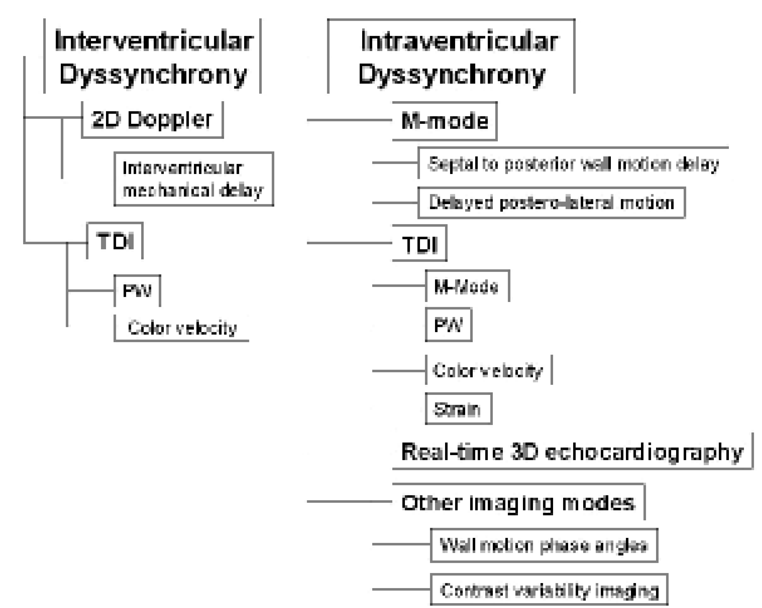
Figure 1. Overview of the different echocardiographic imaging modes that may be used for evaluating interventricular and left intraventricular dyssynchrony. TDI = tissue Doppler imaging; PW = pulsed-wave Doppler.
Cardiovascmed 09 00188 g001
Figure 2. Evaluation of intraventricular dyssynchrony using conventional echocardiography. Delay from QRS onset to mitral E-wave onset is measured (A) and compared with delay from QRS onset. (B) to maximal excursion of the lateral wall in M-mode. This example shows evidence of intraventricular dyssynchrony (interval of [B] >[A]).
Cardiovascmed 09 00188 g002
Figure 3. Evaluation of interventricular dyssynchrony using pulsed-wave Doppler samples placed consecutively at the pulmonary (A) and aortic valves (B) to measure pre-ejection intervals (110 ms and 160 ms respectively), with QRS onset being used as a reference point. Interventricular delay is calculated as the difference between the two measurements, being 50 ms in this example (N <40 ms). Notice that this patient has a narrow QRS (100 ms).
Cardiovascmed 09 00188 g003
Figure 4. A Pulsed-wave TDI measurement at the interventricular septum measured to Sm onset (dotted line). Alternatively, the delay may be measured to Sm peak. B Color TDI velocity curves recorded simultaneously at the basal segments of the left ventricular lateral wall (in yellow) and the interventricular septum (in green), showing motion in opposite directions (courtesy of Prof. G. Derumeaux, Hôpital Louis-Pradel, Lyon, France). TDI = tissue Doppler imaging; Sm = systolic motion.
Cardiovascmed 09 00188 g004
Figure 5. Real-time 3D reconstruction of the left ventricle during intrinsic rhythm and during biventricular pacing. Regional dyssynchrony is reduced by biventricular pacing, and global ejection fraction increased from 21 to 28%. (adapted from [49]). EF = ejection fraction.
Cardiovascmed 09 00188 g005
Figure 6. AV interval optimisation using the Ritter method. A Intrinsic rhythm with EA fusion. B Biventricular pacing with short AV interval (50 ms). QA = 120 ms (dotted line). Note the truncated A wave. C Long AV interval. QA = 80 ms. D Optimal AV interval = 150–(120–80) = 110 ms. AV = atrioventricular.
Cardiovascmed 09 00188 g006