Next Article in Journal
Moderne Evaluation der Mitralinsuffizienz mit Echo-Methoden: Echokardiographische Abklärung Einer Begleitenden Koronaren Herzkrankheit
Previous Article in Journal
Controversies in cardiology: Die Chlamydien-lnfektion ist ein wichtiger Faktor in der Ätiologie der koronaren Herzkrankheit
 
 
Cardiovascular Medicine is published by MDPI from Volume 28 Issue 1 (2025). Previous articles were published by another publisher in Open Access under a CC-BY (or CC-BY-NC-ND) licence, and they are hosted by MDPI on mdpi.com as a courtesy and upon agreement with Editores Medicorum Helveticorum (EMH).
Proceeding Paper

Ischaemic mitral regurgitation

Department of Cardiology, University of Wales College of Medicine, Heath Park, GB-Cardiff CF4 4XN, UK
*
Author to whom correspondence should be addressed.
Cardiovasc. Med. 1999, 2(3), 170-180; https://doi.org/10.3390/cardiovascmed2030032 (registering DOI)
Published: 31 May 1999

Abstract

There are still controversies regarding the mechanisms and optimal treatment of ischaemic mitral regurgitation. Coronary heart disease is the third most important cause of mitral regurgitation (10%), after myxomatous degeneration (45%), and rheumatic heart disease (40%). Ischaemic mitral regurgitation is a strong adverse risk factor, and it is more common in patients with triple vessel disease. Recent experimental and echocardiographic studies have shown that changes in the shape of the left ventricle, the development of akinetic segments, and asymmetrical mitral annular dilatation are the most important mechanisms. There is no evidence to support the concept of papillary muscle dysfunction as a clinical cause of mitral regurgitation. Dynamic mitral regurgitation may be the cause of dyspnoea as the predominant symptom in a patient with myocardial ischaemia. Regurgitation occurs when there is asymmetrical apposition of the mitral valve leaflets, and/or loss of coaptation. Echocardiography is the investigation of choice for identifying the mechanisms and severity of mitral regurgitation, and for planning optimal treatment.

Introduction

Mitral regurgitation is described as ischaemic when it occurs during or after an acute myocardial infarction, or during episodes of myocardial ischaemia, in the absence of previous disease of the mitral apparatus [1,2,3,4].
Based on our experience of studying mitral valve function, we review the most recent studies regarding the mechanisms, evaluation, and main principles of treatment of ischaemic mitral regurgitation. A new classification of this heterogeneous concept is proposed (Table 1), and some directions for future research are highlighted.

Incidence and prognosis

The reported incidence of ischaemic mitral regurgitation depends on the method used for its diagnosis, which may be echocardiography or angiography. Detailed studies are available only concerning the incidence of post-infarction mitral regurgitation. Angiographic studies published before the thrombolytic era, showed an incidence of 9–19% of post-infarction mitral regurgitation, with severe regurgitation being present in 0.8–3.4% of cases. Mitral regurgitation is more common with anterior (43–74%) than with inferior myocardial infarction (22–45%) [5,6,7,8,9,10,11].
The effects of thrombolysis on mitral regurgitation after infarction are controversial. The TIMI study showed no statistical difference in arterial patency after thrombolysis between those patients who had mitral regurgitation and those who did not; the overall incidence of regurgitation was 13%. Moreover, vessel patency did not predict progression or regression of the severity of mitral regurgitation [8,9]. Tcheng et al. showed that severity of mitral regurgitation is not changed by successful reperfusion, achieved by either thrombolysis or angioplasty [10]. However, other relatively small studies suggested that successful reperfusion may reduce the incidence of significant mitral regurgitation associated with inferior myocardial infarction (from 16% to 4% at 24 hours, and from 15% to 7% at 30 days, comparing control subjects to patients receiving thrombolysis, respectively) [12,13].
Echocardiographic studies demonstrate a higher prevalence of mitral regurgitation after myocardial infarction, up to 74%, probably because this method is more sensitive than angiography for the diagnosis of mitral regurgitation [14].
Mortality after heart attack is strongly related to the presence and severity of mitral regurgitation. Post-infarction mitral regurgitation is an independent predictor of mortality at one year [8,9,11]. For example, in the TIMI study, post-infarction mortality was 1.7% at 10 days and 2.8% at 1 year in patients who had no mitral regurgitation, 9.1% and 18.2% respectively, in patients with mild mitral regurgitation, and 20% and 60% in patients with moderate or severe mitral regurgitation [8,9]. Tcheng et al. reported mortality at one year after myocardial infarction of 52% in patients with severe mitral regurgitation, compared with only 11% in patients without mitral regurgitation [10]. Survival and Ventricular Enlargement investigators recently showed that patients with even mild mitral regurgitation have excess cardiovascular mortality compared with patients with no mitral regurgitation (29% versus 12%, respectively) [11].

Echocardiographic diagnosis

The severity of mitral regurgitation is now best assessed by echocardiography. The different approaches and modalities available allow not only assessment of the severity of mitral regurgitation (Table 2), but detailed identification of the underlying mechanism(s) of regurgitation. Simple colour Doppler methods have been applied in order to assess the severity of mitral regurgitation. Extension of the regurgitant jet into the left atrium is still used widely for grading mitral regurgitation, although it is at best a semi-quantitative guide [15]. Maximal regurgitant jet area correlates reasonably (r = 0.76–0.83) with angiography. A maximal jet area of more than 8 cm2 has been reported to have a sensitivity of 82% and a specificity of 94% for the diagnosis of severe mitral regurgitation, whereas an area of less than 4 cm2 has a sensitivity of 85% and a specificity of 75% for the diagnosis of mild mitral regurgitation [16,17]. The ratio of maximal jet area to the area of the left atrium has been reported to correlate even better with angiography, with a ratio of more than 40% implying severe mitral regurgitation (sensitivity 93%, specificity 96%), and a ratio of less than 20% implying mild mitral regurgitation (sensitivity 94%, specificity 100%) [16]. However, left ventriculography is not a true “gold standard” for such comparisons, and echocardiographic methods based on colour flow mapping have multiple limitations, related for example to haemodynamic effects on the jet size of the colour flow or to technical settings of the colour Doppler.
A major limitation of simple colour Doppler methods, particularly in patients with ischaemic mitral regurgitation, is that jets which are eccentric and adherent to the atrial wall are underestimated in severity on colour flow mapping, due to the Coanda phenomenon: the area of an adherent jet on the colour flow map will be only 40% of that of a free standing jet of equal volumetric flow [18,19,20]. A newer colour Doppler indicator of severity of regurgitation, which seems to be independent of haemodynamic effects on the jet size, is the proximal width of the jet at its narrowest point (“vena contracta”). A diameter of more than 6.5 mm has a sensitivity of 94% and a specificity of 91% for the diagnosis of severe mitral regurgitation [21,22].
Calculation of regurgitant volume by Doppler estimation and then comparison of the transmitral and aortic or pulmonary flows is timeconsuming and prone to multiple errors. Recently available technology, which allows automatic calculation of flow, may reduce the limitations of this method [23].
Retrograde systolic flow in the pulmonary vein is a semi-quantitative index of severe mitral regurgitation (sensitivity 87%, specificity 93%). However, this method can be used mainly cluring transoesophageal echocardiography, since the pulmonary veins can be visualised from the precordium in fewer than 60% of adult patients [24,25]. Confounding factors which reduce the utility of pulmonary venous Doppler for grading mitral regurgitation, include the reduction in systolic forward flow which is present normally in patients in atrial fibrillation. Newer echocardiographic methods for the assessment of severity of mitral regurgitation are being evaluated but appear promising. The “convergence zone” method has been reported from different centres to correlate well with angiography (r >0.90), but it remains impractical in patients with very diseased valves and/or multiple jets, and still quite time-consuming [26,27,28]. The continuity equation applied to jet momentum may be a good method in the future, when digitisation of the colour flow map will be improved [29]. Three-dimensional reconstruction of the regurgitant jet allows more accurate calculation of regurgitant volume, but further prospective studies are needed [30].
The mechanism of mitral regurgitation is determined by detailed analysis of the pattern of closure of the mitral valve leaflets. For the valve to be closed normally and competent during systole, both normal coaptation and normal apposition are required. The mitral leaflets are said to coapt when their edges touch during the whole of systole. On echocardiographic images, the zone of coaptation is composed of echoes from those segments of the leaflets which are in contact with each other (the “rough zones”); the normal width of the rough zone is 3–5 mm. Normal apposition means that these edges of the leaflets are symmetrically aligned opposite each other during systole. Particularly in patients with ischaemic mitral regurgitation, even quite subtle abnormalities of apposition and coaptation may cause significant regurgitation, and so detailed morphological analysis of the whole valve is required. Often, this is achieved best by a transoesophageal study. Identifying the pattern of closure of the mitral leaflets is important because this in turn determines the appropriate surgical technique for mitral valve repair [3,4,31,32].

Acute ischaemic mitral regurgitation

Ruptured papillary muscle is a rare complication of acute myocardial infarction, with an incidence found on autopsy of patients with myocardial infarction of 1–5% [33,34]. Usually, it occurs between days 3 and 5 after infarction. It may cause torrential regurgitation with clinically severe pulmonary oedema and/or cardiogenic shock. It carries a high mortality, of about 50% in the first 24 hours [35]. Rupture of the posteromedial papillary muscle is 2.5 times more frequent than rupture of the anterolateral one [34,36]. In about 50% of cases the blood supply of the posteromedial papillary muscle is from only one artery, either a branch of the right coronary artery or an obtuse marginal branch of the circumflex. Rupture can involve either the head or the body of the papillary muscle.
Precordial echocardiography may show the ruptured papillary muscle attached to the mitral cords, freely moving with the mitral leaflets, or a flail mitral valve, with severe mitral regurgitation due to loss of coaptation and apposition of the mitral leaflets [37,38]. Sometimes, the rupture may be only partial, or a rupture may be partially contained by attachments of the ruptured portion of the papillary muscle to the adjacent left ventricular wall by false tendons. In these cases, some coaptation may be preserved, and careful investigation of the papillary muscles is needed [39]. If the patient is very ill or has required artificial ventilation, precordial echocardiography may give inadequate images and a transoesophageal study may be needed [38,40,41]. Differential diagnosis from rupture of the ventricular septum is usually easy to make using cross-sectional imaging and colour flow mapping together [42].
If the rupture involves the head of the papillary muscle, it may be possible to repair the mitral valve, but if the rupture involves the body of the muscle or if both papillary muscles are infarcted, mitral valve replacement is usually needed. Peri-operative mortality is high, at about 20-50% [32,35,43,44].
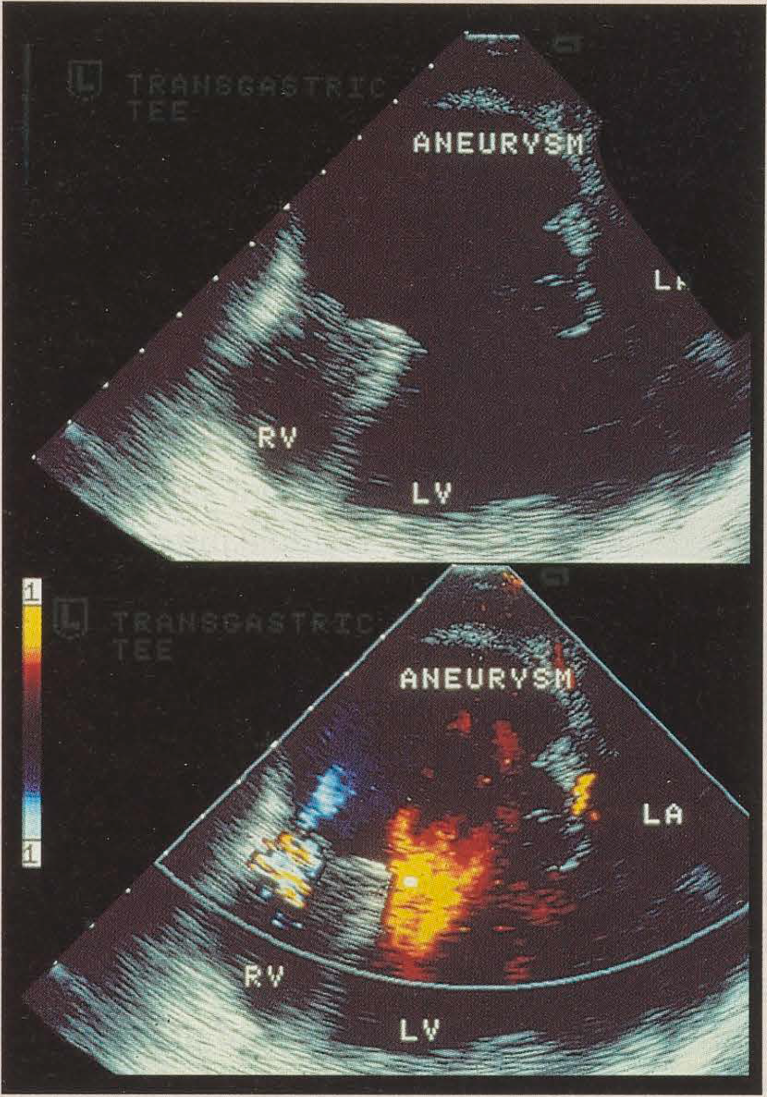
Pseudoaneurysm of the left ventricle with malposition of the papillary muscle and asymmetrical subvalvar traction, producing abnormal apposition, has been described as a cause of acute mitral regurgitation (Figure 1) [45,46].

Chronic ischaemic mitral regurgitation

Post-infarction mitral regurgitation has different mechanisms, depending on the site of myocardial infarction [47].
Restricted motion of the posterior mitral leaflet and increased sphericity of the left ventricular cavity represent the main mechanisms of mitral regurgitation associated with posterobasal myocardial infarction [48,49,50]. Since the posterobasal myocardial wall and the posteromedial papillary muscle are often both supplied by branches arising from the right coronary artery, posterobasal infarction may be associated with necrosis of the posteromedial papillary muscle. Ischaemic injury with consequent fibrosis results in loss of the contractile function of the papillary muscle. In addition, hypokinesis or akinesis of the posterobasal wall, secondary to the infarction, causes asymmetrical dilatation and modified long-axis torsion of the left ventricle. Thus, during systole, the posteromedial papillary muscle is displaced posteriorly, resulting in increased subvalvar traction on the tendinous cords with apical systolic displacement of the posterior mitral leaflet, and abnormal apposition [49,50]. Sometimes, frank aneurysmal dilatation of the posterobasal wall may develop, causing more severe subvalvar traction.
Another important mechanism of chronic ischaemic regurgitation is increased sphericity of the left ventricle. Kaul et al. demonstrated experimentally that mitral regurgitation occurs during ischaemia only when global left ventricular function is affected [48]. Van Dantzig et al., in an echocardiographic study, demonstrated that only infero-postero-lateral asynergy and an abnormal left ventricular sphericity index are independently associated with significant mitral regurgitation late after infarction [50]. Another possible mechanism may be asymmetrical dilatation of the mitral annulus, mainly in its posterior segment. Gorman et al. recently showed in sheep that posterior wall infarction caused by circumflex arterial occlusion was associated with papillary muscle “discoordination” [51]. Systolic shortening of the posteromedial papillary muscle was decreased while the normally contracting anterolateral papillary muscle was displaced posteriorly because of hypokinesis of the adjacent myocardial wall. In this study, mitral regurgitation was caused by a combination of relative atrial displacement of leaflet tissue attached to the posteromedial papillary muscle, and restriction of the leaflet tissue attached to the anterolateral papillary muscle.
Anterior myocardial infarction causes mitral regurgitation either by global dilatation of the left ventricle, or by subvalvar traction secondary to an anteroapical aneurysm.
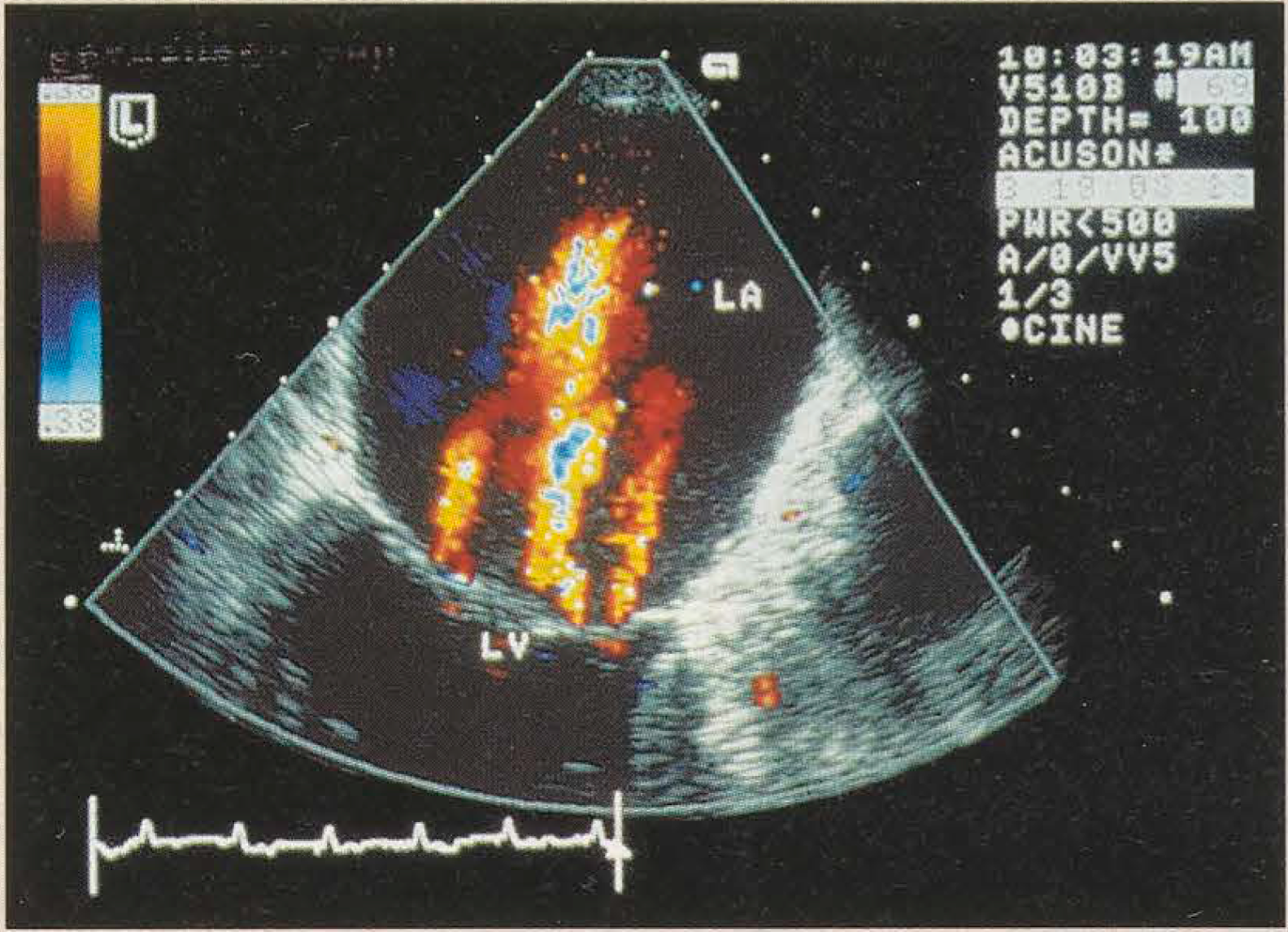
Precordial or transoesophageal echocardiography may show: (1) hypokinesis or akinesis of the posterobasal wall of the left ventricle, or else a posterobasal aneurysm; (2) apical systolic displacement of the tip of one or both papillary muscles, relative to the plane of the mitral annulus; (3) abnormal apposition of the mitral valve leaflets; (4) an eccentric jet of mitral regurgitation when apposition is abnormal, in which case the jet is directed away from the abnormal leaflet; (5) multiple jets arising along the whole of the zone of coaptation of the leaflets in systole, directed centrally into the left atrium, when regurgitation is caused by increased subvalvar traction of the tendinous cords (Figure 2); and/or (6) increased left ventricular sphericity compared with the normal more ellipsoid shape in systole. Mitral annular dilatation may also be present [50,52].
Mild mitral regurgitation after myocardial infarction often improves after coronary arterial bypass surgery, without any need for repair. Treatment of severe post-infarction mitral regurgitation consists of mitral valve repair associated with coronary arterial bypass surgery. It remains a difficult and controversial decision whether or not to perform mitral valve repair for moderate mitral regurgitation, during coronary arterial surgery, but this is an important problem since it has already been proved that residual mitral regurgitation is an adverse longterm prognostic factor. Czer et al. [53] reported that moderate mitral regurgitation which is left untreated when patients have coronary arterial bypass surgery, does not usually resolve, whereas Christenson et al. [54] reported an average improvement of at least one grade in mitral regurgitation after revascularisation. Intraoperative echocardiography may have an important role, with mitral valve repair being indicated in moderate or severe residual mitral regurgitation (more than grade II) [53], but further prospective studies are required. New echocardiographic methods such as the analysis of regional function using Doppler myocardial imaging or of perfusion using contrast echocardiography, may be useful if post-bypass efficacy of coronary revascularisation is shown to correlate with late clinical outcome. If so, then surgeons could operate for mitral regurgitation in those patients in whom any doubts remain that left ventricular function will improve and regurgitation diminish after arterial surgery alone.
Typically, repair will involve only mitral annuloplasty, and peri-operative mortality is about 5–15% [55,56]. Sometimes, mitral valve replacement may be needed, but if at all possible the subvalvar mitral apparatus should be preserved. After valve replacement, the peri-operative mortality is slightly higher, at about 10–15% [57]. Patients with poor left ventricular function (ejection fraction <30%) have a very high mortality after either mitral valve repair (43%) or mitral valve replacement (50%). Therefore, heart transplantation may need to be considered in such patients [58]. Mitral regurgitation secondary to a large anteroapical aneurysm usually resolves after aneurysmectomy [59].

Dynamic mitral regurgitation

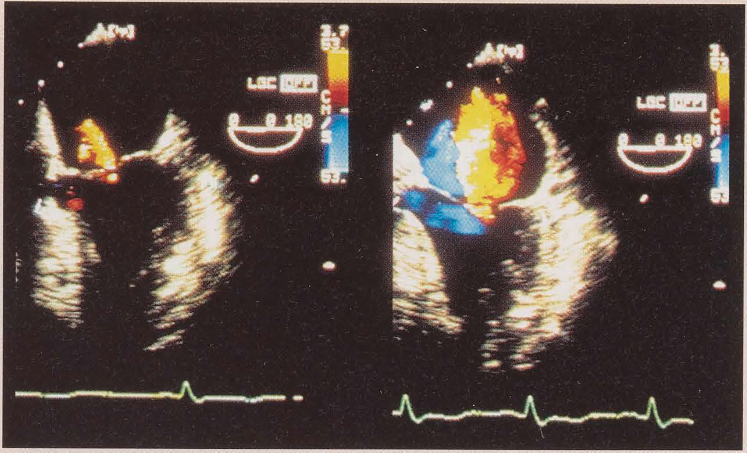
Dynamic mitral regurgitation is defined as mitral insufficiency which occurs “de novo” or is significantly exacerbated during transient episodes of myocardial ischaemia (Figure 3).
It was Burch et al. in 1963 who first introduced the concept of “papillary muscle dysfunction”, in order to explain the mechanism of dynamic mitral regurgitation [60]. Further studies have not proved this hypothesis, however, since ischaemia of either papillary muscle does not cause regurgitation [48,61,62]. This is not surprising since both papillary muscles and their cords support both mitral leaflets: any dysfunction would produce symmetrical displacement of the rough zones. Moreover, animal studies using selective embolisation of the coronary arteries showed that neither papillary muscle dysfunction nor dysfunction of the adjacent myocardial wall can cause mitral regurgitation. Dynamic mitral regurgitation occurs only in the presence of global left ventricular dysfunction and its severity is directly related to the degree of the left ventricular dysfunction [48]. Indeed, this phenomenon is more common in patients with triple rather than single vessel disease, and in patients with stenosis of the left anterior descending artery (60% of the cases) rather than of the right coronary artery or the circumflex artery [63].
“Incomplete mitral closure” was proposed in echocardiographic studies as the direct mechanism of dynamic mitral regurgitation [61], but the term is tautological in the context of mitral regurgitation and thus uninformative. It probably means loss of coaptation yet with normal apposition, caused by geometric changes of the left ventricle and global left ventricular dysfunction [48,61,62,63,64].
Recently, the concept of “mitral valve reserve” has been introduced to describe the degree of change in mitral valve geometry which can occur without causing mitral regurgitation [62,64,65]. Mitral valve reserve is surprisingly limited: dynamic mitral regurgitation has been proved to occur if the coaptation point is displaced apically by more than 3 mm and/or the mitral annulus dilates by more than 5 mm. Displacement of the coaptation point seems to be the more important mechanism. It occurs because of apical traction of the mitral leaflets secondary to a transient more spherical shape of the left ventricle during ischaemia with changing of the spatial orientation of the papillary muscles. It may also be due in part to subendocardial dysfunction resulting in reduced longitudinal shortening of the left ventricle, and thus failure of the annulus to develop its normal systolic shape which brings the leaflets together. The geometric change of the left ventricle is related to diffuse and usually severe myocardial ischaemia. Acute dilatation of the mitral annulus seems to be very rarely involved as a mechanism of dynamic mitral regurgitation [63,66].
The diagnosis of dynamic mitral regurgitation is important because it may aggravate the consequences of myocardial ischaemia. Left atrial and pulmonary capillary pressures are increased and an episode of pulmonary oedema may be precipitated. Dynamic mitral regurgitation is one of the common causes of breathlessness during episodes Of myocardial ischaemia, with or without associated angina. Dynamic mitral regurgitation during stress echocardiography may be an indirect marker of myocardial ischaemia.
Echocardiographic criteria of dynamic mitral regurgitation can be summarised as follows: (1) mitral regurgitation occurring “de novo” or significantly exacerbated during transient episodes of induced myocardial ischaemia; (2) apical displacement of the coaptation point of the mitral leaflets by more than 3 mm and/or mitral annular dilatation by more than 5 mm; (3) decrease in the ratio of the length to the width of the left ventricular cavity (“longaxis/short-axis ratio”) [45,46,47,48]. These criteria are valid for both precordial and transoesophageal echocardiography. However, diagnosis of ischaemic mitral regurgitation is better by transoesophageal echocardiography because of its superior resolution for detecting mitral regurgitation and for analysing the pattern of closure of the mitral leaflets [67].
The main difficulty of the echocardiographic diagnosis of dynamic mitral regurgitation is choosing the optimal stress modality, able to induce simultaneously both myocardial ischaemia and mitral regurgitation. Dynamic mitral regurgitation during exercise is a marker of severe myocardial ischaemia, typically associated with significant stenosis of the left anterior descending or the right coronary artery [63]. However, exercise echocardiography, requiring data acquisition within 2 minutes of peak exercise, may be difficult in patients with poor echo windows and/or severe breathlessness on exercise.
It has been suggested that dynamic mitral regurgitation may be assessed better by using pacing-induced ischaemia during transoesophageal echocardiography, but further large studies are required [64,68]. Dobutamine stress echocardiography is unreliable because its haemodynamic effects, including reduction of left ventricular filling pressure, are variable and unpredictable, and may influence the degree of mitral regurgitation [69]. It is important that whichever stress is used for an individual patient is able to provoke myocardial ischaemia. Revascularisation, by either PTCA or surgery, is the treatment of choice for dynamic mitral regurgitation [43,44]. In patients with this problem it is expected that mitral regurgitation will disappear when myocardial ischaemia is relieved and normal left ventricular function restored.

Ischaemic dilatation of the left ventricle

Mitral regurgitation secondary to ischaemic dilatation of the left ventricle has been described as “functional” mitral regurgitation, presumably to imply that the mitral valve apparatus appears normal on echocardiography and on surgical inspection. However, since regurgitation is not a normal function of the mitral valve and since the term does not describe how the regurgitation arises, it is perhaps best avoided.
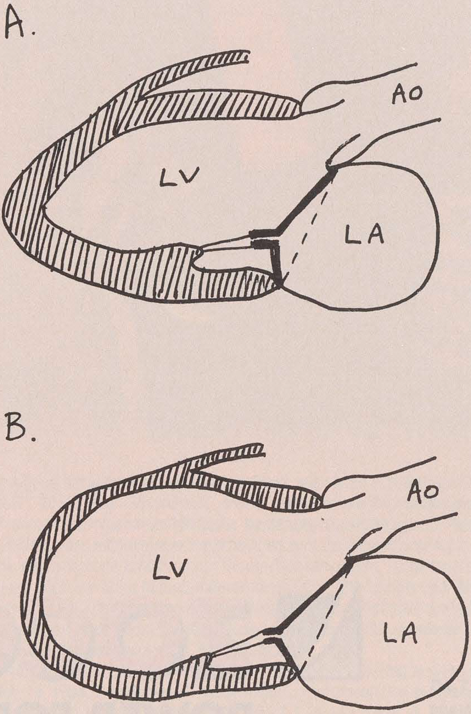
The mechanisms of mitral regurgitation secondary to dilatation of the left ventricle are actually the same for all the different aetiologies causing left ventricular dilatation: (1) change of the spatial orientation of the papillary muscles secondary to an enlarged and a more spherical than normal systolic shape of the cavity of the left ventricle, which causes increased subvalvar traction, and apical displacement of the zone of coaptation; (2) mitral annular dilatation; (3) poor contraction of the left ventricle, with a slowed rate of rise of intraventricular pressure and slow closure of the leaflets [70,71]. The net result of these morphological changes is to reduce the overlap of the rough zones of the mitral leaflets, until regurgitation starts to arise from multiple sites where they no longer coapt completely (Figure 4). On echocardiography, the regurgitant jets are usually multiple and centrally directed. The left ventricle is dilated and diffusely hypokinetic. The mitral annulus is often dilated; its diameter in a parasternal view, measured from the hinge point of the aortic valve leaflet with the mitral curtain anteriorly, to the hinge point of the posterior leaflet with the mitral annulus, should not exceed 35 mm in a typical adult.
Moderate mitral regurgitation does not need surgery, but may be diminished by reducing afterload, for example by giving an ACE inhibitor [72]. Severe mitral regurgitation is usually associated with marked left ventricular dilatation and poor global function and, therefore, partial ventriculectomy or heart transplantation may be the only therapeutic options [43,44].

Conclusion

Ischaemic mitral regurgitation is a common disease, with multiple mechanisms, which may influence the prognosis of patients with different forms of coronary heart disease. Diagnosis of its mechanism is very important for optimising treatment. Echocardiography, either precordial or transoesophageal, is the only method that can evaluate the morphology of ischaemic mitral regurgitation and, together with angiography, represents an important method for assessing its severity. New echocardiographic modalities may allow better identification of patients with moderate mitral regurgitation who need valve repair when they undergo coronary arterial surgical revascularisation.

References

  1. Fenster, M.S.; Feldman, M.D. Mitral regurgitation: an overview. Curr. Probl. Cardiol. 1995, 28, 193–280. [Google Scholar] [CrossRef]
  2. Oury, J.H.; Cleveland, J.C.; Duran, C.G. Ischemic mitral valve disease: classification and systemic approach to management. J. Card. Surg. 1994, 9, 262–273. [Google Scholar] [CrossRef]
  3. Fraser, A.G. Disease of mitral and tricuspid valves. In Transesophageal echocardiography in clinical practice; Sutherland, G.R., Roelandt, J., Fraser, A.G., Anderson, R., Eds.; Gower Medical Publishing: London, 1991; pp. 6.1–6.20. [Google Scholar]
  4. Fraser, A.G. Atrioventricular valve repair. In Transesophageal echocardiography in congenital heart disease; Stümper, O., Sutherland, G.R., Eds.; Edward Arnold: London, 1994; pp. 215–236. [Google Scholar]
  5. Hickey, M.S.; Smith, L.R.; Muhlbaier, L.H.; Harrel FEJr Reves, J.C.; Hinohara, T.; et al. Current prognosis of ischemic mitral regurgitation. Implications for future management. Circulation 1998, 78, 151–159. [Google Scholar]
  6. Barzilai, B.; Davis, V.G.; Stone, P.H.; Jaffe, A.S. Prognostic significance of mitral regurgitation in acute myocardial infarction. The MILIS study group. Am. J. Cardiol. 1990, 65, 1169–1175. [Google Scholar] [CrossRef]
  7. De Servi, S.; Vaccari, L.; Assandri, J.; Poma, E.; Cioffi, P.; Scire, A.; et al. Clinical significance of mitral regurgitation in patients with recent myocardial infarction. Eur. Heart J. 1988, 9, 5–9. [Google Scholar] [CrossRef] [PubMed]
  8. Lehmann, K.G.; Francis, C.K.; Sheehan, F.H.; Dodge, H.T. Effect of thrombolysis on acute mitral regurgitation during evolving myocardial infarction. Experience from the Thrombolysis in Myocardial Infarction (TIMI) Trial. J. Am. Coll. Cardiol. 1993, 22, 714–719. [Google Scholar] [CrossRef]
  9. Lehmann, K.G.; Francis, C.K.; Dodge, H.T. Mitral regurgitation in early myocardial infarction. Incidence, clinical detection, and prognostic implications. TIMI Study Group. Ann. Intern Med. 1992, 117, 10–17. [Google Scholar] [CrossRef] [PubMed]
  10. Tcheng, J.E.; Jackman JDJr Nelson, C.L.; Gardner, L.H.; Smith, L.R.; Rankin, J.S.; et al. Outcome of patients sustaining acute ischemic mitral regurgitation during myocardial infarction. Ann. Intern Med. 1992, 117, 18–24. [Google Scholar] [CrossRef] [PubMed]
  11. Lamas, G.A.; Mitchell, G.F.; Flaker, G.C.; Smith, SCJr; Gersh, B.J.; Basta, L.; et al. Clinical significance of mitral regurgitation after acute myocardial infarction. Survival and Ventricular Enlargement Investigators. Circulation 1997, 96, 827–833. [Google Scholar] [CrossRef]
  12. Leor, J.; Feinberg, M.S.; Vered, Z.; Hod, H.; Kaplinsky, E.; Goldbourt, U.; et al. Effect of thrombolytic therapy on the evolution of significant mitral regurgitation in patients with a first inferior myocardial infarction. J. Am. Coll. Cardiol. 1993, 21, 1661–1666. [Google Scholar] [CrossRef][Green Version]
  13. Tenenbaum, A.; Leor, J.; Motro, M.; Hod, H.; Kaplinsky, E.; Rabinowitz, B.; et al. Improved posterobasal segment function after thrombolysis is associated with decreased incidence of significant mitral regurgitation in a first inferior myocardial infarction. J. Am. Coll. Cardiol. 1995, 25, 1558–1563. [Google Scholar] [CrossRef][Green Version]
  14. Alam, M.; Thorstrand, C.; Rosenhamer, G. Mitral regurgitation following first-time acute myocardial infarction—early and late findings by Doppler echocardiography. Clin. Cardiol. 1993, 16, 30–34. [Google Scholar] [CrossRef] [PubMed]
  15. Miyatake, K.; Izumi, S.; Okamoto, M.; Kinoshita, N.; Asonuma, H.; Nakagava, H.; et al. Semiquantitative grading of severity of mitral regurgitation by real-time two-dimensional Doppler flow imaging. J. Am. Coll. Cardiol. 1986, 7, 82–88. [Google Scholar]
  16. Helmcke, F.; Nanda, N.C.; Hsiung, M.C.; Soto, B.; Adey, C.K.; Goyal, R.G.; et al. Color Doppler assessment of mitral regurgitation with orthogonal planes. Circulation 1987, 75, 175–183. [Google Scholar] [CrossRef] [PubMed]
  17. Spain, M.G.; Smith, M.D.; Grayburn, P.A.; Harlamert, E.A.; DeMaria, A.N. Quantitative assessment of mitral regurgitation by Doppler color flow imaging: angiographic and hemodynamic correlations. J. Am. Coll. Cardiol. 1989, 13, 585–590. [Google Scholar]
  18. Chen, C.G.; Thomas, J.D.; Anconina, J.; Harrigan, P.; Mueller, L.; Picard, M.H.; et al. Impact of impinging wall jet on color Doppler quantification of mitral regurgitation. Circulation 1991, 84, 712–720. [Google Scholar] [CrossRef]
  19. Enriquez-Sarano, M.; Tajik, A.J.; Bailey, K.R.; Seward, J.B. Color flow imaging compared with quantitative Doppler assessment of severity of mitral regurgitation: influence of eccentricity of jet and mechanism of regurgitation. J. Am. Coll. Cardiol. 1993, 21, 1211–1219. [Google Scholar] [CrossRef] [PubMed]
  20. McCully, R.B.; Enriquez-Sarano, M.; Tajik, A.J.; Seward, J.B. Overestimation of severity of ischemic/functional mitral regurgitation by color Doppler jet area. Am. J. Cardiol. 1994, 74, 790–793. [Google Scholar] [CrossRef]
  21. Fehske, W.; Omran, H.; Manz, M.; Kohler, J.; Hagendorff, A.; Luderitz, B. Color-coded Doppler imaging of the vena contracta as a basis for quantification of pure mitral regurgitation. Am. J. Cardiol. 1994, 73, 268–274. [Google Scholar] [CrossRef]
  22. Hall, S.A.; Brickner, E.; Willett, D.L.; Irani, W.N.; Afridi, I.; Grayburn, P.A. Assessment of mitral regurgitation severity by Doppler color flow mapping of the vena contracta. Circulation 1997, 95, 636–642. [Google Scholar] [CrossRef]
  23. Hozumi, T.; Yoshida, K.; Akasaka, T.; Takagi, K.; Yamamuro, A.; Yagi, T.; et al. Automated assessment of mitral regurgitant volume and regurgitant fraction by a newly developed digital color Doppler velocity profile integration method. Am. J. Cardiol. 1997, 80, 1325–1330. [Google Scholar] [CrossRef]
  24. Klein, A.L.; Obarski, T.P.; Stewart, W.J.; Casale, P.N.; Pearce, G.L.; Husbands, K.; et al. Transesophageal Doppler echocardiography of pulmonary venous flow: a new marker of mitral regurgitation severity. J. Am. Coll. Cardiol. 1991, 18, 518–526. [Google Scholar] [CrossRef] [PubMed]
  25. Pieper, E.P.; Hellemans, 1M.; Hamer, H.P.M.; Ravelli, A.C.J.; Cheriex, E.C.; Tijssen, J.G.P.; et al. Value of systolic pulmonary venous flow reversal and color Doppler jet measurements assessed with transesophageal echocardiography in recognizing severe pure mitral regurgitation. Am. J. Cardiol. 1996, 78, 444–450. [Google Scholar] [CrossRef] [PubMed]
  26. Recusani, F.; Bargiggia, G.S.; Yoganathan, A.P.; Raisaro, A.; VadesCruz, L.M.; Sung, H.W.; et al. A new method for quantification of regurgitant flow rate using color Doppler flow imaging of the flow convergence region proximal to a discrete orifice. An in vitro study. Circulation 1991, 83, 594–604. [Google Scholar] [CrossRef]
  27. Chen, C.; Koschyk, D.; Brockhoff, C.; Heik, S.; Hamm, C.; Bleifeld, W.; et al. Noninvasive estimation of regurgitant flow rate and volume in patients with mitral regurgitation by Doppler color mapping of accelerating flow field. J. Am. Coll. Cardiol. 1993, 21, 374–383. [Google Scholar] [CrossRef] [PubMed]
  28. Dujardin, K.S.; Enriquez-Sarano, M.; Bailey, K.R.; Nishimura, R.A.; Seward, J.B.; Tajik, A.J. Grading of mitral regurgitation by quantitative Doppler echocardiography: calibration by left ventricular angiography in routine clinical practice. Circulation 1997, 96, 3409–3415. [Google Scholar] [CrossRef]
  29. Thomas, J.D.; Liu, C.M.; Flachskampf, F.A.; O’Shea, J.P.; Davidoff, R.; Weyman, A.E. Quantification of jet flow by momentum analysis. An in vitro color Doppler flow study. Circulation 1990, 81, 247–259. [Google Scholar] [CrossRef]
  30. Shiota, T.; Sinclair, B.; Ishii, M.; Zhou, X.; Ge, S.; Teien, D.E.; et al. Three-dimensional reconstruction of color Doppler convergence regions and regurgitants jets: an in vitro quantitative study. J. Am. Coli Cardiol. 1996, 27, 1511–1518. [Google Scholar] [CrossRef][Green Version]
  31. Fraser, A.G.; van Herwerden, L.A. Surgical echocardiography of the native and reconstructed mitral valve. In Mitral Valve Disease; Wells, F.C., Shapiro, L.M., Eds.; Butterworth Heinemann: Oxford, 1996; pp. 51–70. [Google Scholar]
  32. Lee, E.M.; Shapiro, L.M.; Wells, F.C. Echocardiography in mitral valve repair for mitral regurgitation: the surgeon’s needs. J. Heart Valve Dis. 1997, 6, 228–233. [Google Scholar]
  33. Barbour, D.J.; Roberts, W.C. Rupture of a left ventricular papillary muscle during acute myocardial infarction: analysis of 22 necropsy patients. J. Am. Coll. Cardiol. 1986, 8, 558–565. [Google Scholar] [CrossRef]
  34. Sharma, S.K.; Seckler, J.; Israel, D.H.; Borrico, S.; Ambrose, J.A. Clinical, angiographic and anatomic findings in acute severe ischemic mitral regurgitation. Am. J. Cardiol. 1992, 70, 277–280. [Google Scholar] [CrossRef]
  35. Calvo, F.E.; Figueras, J.; Cortadellas, J.; Soler-Soler, J. Severe mitral regurgitation complicating acute myocardial infarction. Clinical and angiographic differences between patients with and without papillary muscle rupture. Eur. Heart J. 1997, 18, 1606–1610. [Google Scholar] [CrossRef] [PubMed]
  36. Voci, P.; Bilotta, F.; Caretta, Q.; Mercanti, C.; Marino, B. Papillary muscle perfusion pattern. A hypothesis for ischemic papillary muscle dysfunction. Circulation 1995, 91, 1714–1718. [Google Scholar] [CrossRef] [PubMed]
  37. Erbel, R.; Schweizer, P.; Bardos, P.; Meyer, J. Two-dimensional echocardiographic diagnosis of papillary muscle rupture. Chest 1981, 79, 595–598. [Google Scholar] [CrossRef] [PubMed]
  38. Zotz, R.J.; Dohmen, G.; Genth, S.; Erbel, R.; Meyer, J. Diagnosis of papillary muscle rupture after acute myocardial infarction by transthoracic and transesophageal echocardiography. Clin. Cardiol. 1993, 16, 665–670. [Google Scholar] [CrossRef]
  39. Nishimura, R.A.; Shub, C.; Tajik, A.J. Two-dimensional echocardiographic diagnosis of partial papillary muscle rupture. Br. Heart J. 1982, 48, 598–600. [Google Scholar] [CrossRef]
  40. Herrera, C.J.; Gurevicius, J.; Stecy, P.; Dahodwala, M.; Tummala, A.; Nemickas, R. The clinical utility of transesophageal echocardiography in ischemic papillary muscle rupture. Am. J. Card. Imaging 1995, 9, 226–228. [Google Scholar]
  41. Kranidis, A.; Koulouris, S.; Filippatos, G.; Sideris, A.; Anthopoulos, L. Mitral regurgitation from papillary muscle rupture: role of transesophageal echocardiography. J. Heart Valve Dis. 1993, 2, 529–532. [Google Scholar]
  42. Smyllie, J.H.; Sutherland, G.R.; Geuskens, R.; Dawkins, K.D.; Conway, N.; Roelandt, J.R.T.C. Doppler color flow mapping in the diagnosis of ventricular septal rupture and acute mitral regurgitation after myocardial infarction. J. Am. Coll. Cardiol. 1990, 15, 1449–1455. [Google Scholar] [CrossRef]
  43. David, T.E. Techniques and results of mitral valve repair for ischemic mitral regurgitation. J. Card. Surg. 1994, 9, 274–277. [Google Scholar] [CrossRef]
  44. Radermecker, M.A.; Limet, R. Les insuffisances mitrales ischémiques: principes de leur traitement chirurgical. Rev. Med. Liege 1995, 50, 295–297. [Google Scholar]
  45. Ezzat, M.A.; Abdelmeguid, I.; Leclerc, D.; Nguyen, A.; Piwnica, A. Left ventricular pseudoaneurysm associated with mitral regurgitaion. Ann. Thorac. Surg. 1992, 53, 504–506. [Google Scholar] [CrossRef] [PubMed]
  46. Komeda, M.; David, T.E. Surgical treatment of postinfarction false aneurysm of the left ventricle. J. Thorac. Cardiovasc Surg. 1993, 106, 1189–1191. [Google Scholar] [CrossRef]
  47. Gorman JH3rd Gorman, R.C.; Plappert, T.; Jackson, B.M.; Hiramatsu, Y.; St John Sutton, M.G.; et al. Infarct size and location determine development of mitral regurgitation in the sheep model. J. Thorac. Cardiovasc Surg. 1998, 115, 615–622. [Google Scholar] [CrossRef]
  48. Kaul, S.; Sponitz, W.D.; Glasheen, W.P.; Touchstone, D.A. Mechanism of ischemic mitral regurgitation. An experimental evaluation. Circulation 1991, 84, 2167–2180. [Google Scholar] [CrossRef]
  49. Llaneras, M.R.; Nance, M.L.; Streicher, J.T.; Lima, J.A.; Savino, J.S.; Bogen, D.K.; et al. Large animal model of ischemic mitral regurgitation. Ann. Thorac. Surg. 1994, 57, 432–439. [Google Scholar] [CrossRef]
  50. Van Dantzig, J.M.; Delemarre, B.J.; Koster, R.W.; Bot, H.; Visser, C.A. Pathogenesis of mitral regurgitation in acute myocardial infarction: importance in changes in left ventricular shape and regional function. Am. Heart J. 1996, 131, 865–871. [Google Scholar] [CrossRef] [PubMed]
  51. Gorman, J.H., 3rd; Jackson, B.M.; Gorman, R.C.; Kelley, S.T.; Gikakis, N.; Edmunds, L.H., Jr. Papillary muscle discoordination rather than increased annular area facilitates mitral regurgitation after acute posterior myocardial infarction. Circulation 1997, 96, II124–7. [Google Scholar]
  52. Kisanuki, A.; Otsuji, Y.; Kuroiwa, R.; Murayama, T.; Matsushita, R.; Shibata, K.; et al. Two-dimensional echocardiographic assessment of papillary muscle contractility in patients with prior myocardial infarction. J. Am. Coll. Cardiol. 1993, 21, 932–938. [Google Scholar] [CrossRef]
  53. Czer, L.S.; Maurer, G.; Bolger, A.F.; DeRobertis, M.; Chaux, A.; Matloff, J.M. Revascularisation alone or combined with suture annuloplasty for ischemic mitral regurgitation. Evaluation by color Doppler echocardiography. Tex. Heart Inst. J. 1996, 23, 270–278. [Google Scholar] [PubMed]
  54. Christenson, J.T.; Simonet, F.; Bloch, A.; Maurice, J.; Velebit, V.; Schmuziger, M. Should a mild to moderate ischemic mitral valve regurgitation in patients with poor left ventricular function be repaired or not? J. Heart Valve Dis. 1995, 4, 484–488. [Google Scholar] [PubMed]
  55. Kay, G.L.; Kay, J.H.; Mendez, M.A.; Zubiate, P.; Yokoyama, T. Long-term results of operations for mitral regurgitation secondary to coronary artery disease. Cardiovasc Clin. 1987, 17, 41–51. [Google Scholar]
  56. Galloway, A.C.; Grossi, E.A.; Spencer, F.C.; Colvin, S.B. Operative therapy for mitral insufficiency from coronary artery disease. Semin Thorac. Cardiovasc Surg. 1995, 7, 227–232. [Google Scholar]
  57. Dion, R.; Benetis, R.; Elias, B.; Guennaoui, T.; Raphael, D.; Van Dyck, M.; et al. Mitral valve procedures in ischemic regurgitation. J. Heart Valve Dis. 1995, 4, S124–S129. [Google Scholar]
  58. Hausmann, H.; Siniawski, H.; Hotz, H.; Hofmeister, J.; Chavez, T.; Schmidt, G.; et al. Mitral valve reconstruction and mitral valve replacement for ischemic mitral insufficiency. J. Card. Surg. 1997, 12, 8–14. [Google Scholar] [CrossRef] [PubMed]
  59. Mickleborough, L.L. Surgical management of left ventricular aneurysm. Semin Thorac. Cardiovasc Surg. 1995, 7, 233–239. [Google Scholar]
  60. Burch, G.E.; DePasquale, N.P.; Phillips, J.H. The syndrome of papillary muscle dysfunction. Am. Heart J. 1968, 75, 399–415. [Google Scholar] [CrossRef]
  61. Kono, T.; Sabbah, H.N.; Rosman, H.; Alam, M.; Jafri, S.; Stein, P.D.; et al. Mechanism of functional mitral regurgitation during acute myocardial ischemia. J. Am. Coll. Cardiol. 1992, 19, 1101–1105. [Google Scholar] [CrossRef] [PubMed]
  62. Jones, R.A.; Masani, N.D.; Fraser, A.G.; Hall, R.J.C. Papillary muscle ischaemia is not the only cause of dynamic mitral regurgitation. J. Am. Coll. Cardiol. Special Issue. 1994, 447A. [Google Scholar]
  63. Fehrenbacher, G.; Schmidt, D.H.; Bommer, W.J. Evaluation of transient mitral regurgitation in coronary artery disease. Am. J. Cardiol. 1991, 68, 868–873. [Google Scholar] [CrossRef]
  64. Kamp, O.; de Cock, C.C.; van Eenige, M.J.; Visser, C.A. Influence of pacing-induced myocardial ischemia on left atrial regurgitant jet: a transesophageal echocardiographic study. J. Am. Coli Cardiol. 1994, 23, 1584–1591. [Google Scholar] [CrossRef]
  65. Jones, R.A.; Masani, N.D.; Fraser, A.G.; Hall, R.J.C. Left ventricular shape during ischemia: an important determinant of dynamic mitral regurgitation. Br. Heart J. 1995, 73, P56. [Google Scholar]
  66. Komeda, M.; Glasson, J.R.; Bolger, A.F.; Daughters, G.T., 2nd; Maclsaac, A.; Oesterle, S.N.; et al. Geometric determinants of ischemic mitral regurgitation. Circulation 1997, 96, II128–33. [Google Scholar]
  67. Flachskampf, F.A.; Hanrath, P. Multiplane transesophageal imaging in mitral regurgitation due to coronary artery disease. Coron. Artery Dis. 1996, 7, 202–205. [Google Scholar]
  68. Gonzales, A.; Naqvi, S.E.; Tak, T.; Choudhary, R.S.; Rahimtoola, S.H.; Chandraratna, P.A. Normalization of Doppler indices of diastolic dysfunction during pacing is a sign of ischemic mitral regurgitation. Am. Heart J. 1991, 121, 118–125. [Google Scholar] [CrossRef]
  69. Heinle, S.K.; Tice, F.D.; Kisslo, J. Effect of dobutamine stress echocardiography on mitral regurgitation. J. Am. Coll. Cardiol. 1995, 25, 122–127. [Google Scholar] [CrossRef][Green Version]
  70. He, S.; Fontaine, A.A.; Schwammenthal, E.; Yoganathan, A.P.; Levine, R.A. Integrated mechanism for functional mitral regurgitation: leaflet restriction versus coapting forces: in vitro studies. Circulation 1997, 96, 1826–1834. [Google Scholar] [CrossRef]
  71. Otsuji, Y.; Handschumacher, M.D.; Schwammenthal, E.; Jiang, L.; Song, J.K.; Guerrero, J.L.; et al. Insights from three-dimensional echocardiography into the mechanism of functional mitral regurgitation: direct in vivo demonstration of altered leaflet tethering geometry. Circulation 1997, 96, 1999–2008. [Google Scholar] [CrossRef]
  72. Gaasch, W.H.; John, R.M.; Aurigemma, G.P. Managing asymptomatic patients with chronic mitral regurgitation. Chest 1995, 108, 842–847. [Google Scholar] [CrossRef]
Figure 1. Transoesophageal transgastric images in the longitudinal axis in a patient with acute mechanical complications of myocardial infarction. There is a pseudoaneurysm with a large cavity inferior to the basal inferior left ventricular surface, as well as a ventricular septal defect, and some mitral regurgitation caused by asymmetrical closure (“abnormal apposition”) of the leaflets.
Cardiovascmed 02 00170 g001
Figure 2. Transoesophageal colour flow mapping in the longitudinal axis demonstrating multiple small regurgitant jets, as found in a patient with an enlarged left ventricle and increased subvalvar traction on the tendinous cords.
Cardiovascmed 02 00170 g002
Figure 3. Transoesophageal images of the mitral valve before and during dipyridamole stress echocardiography in a patient with ischaemic heart disease and dynamic mitral regurgitation. While the systolic blood pressure did not change significantly, stress caused a marked increase in the area of the regurgitant jet demonstrated by colour flow mapping. (Courtesy of Dr R. Jones).
Cardiovascmed 02 00170 g003
Figure 4. Diagrams comparing mitral valvar anatomy and left ventricular morphology during systole in a normal heart (a), and after anterior myocardial infarction with chronic ischaemic mitral regurgitation (b). Note the more spherical shape of the left ventricle, and apical displacement of the tip of the papillary muscle and the zone of coaptation of the mitral leaflets. Displace ment of the edges of the leaflets away from the plane of the mitral annulus results in reduced overlap of the rough zones, and multiple regurgitant jets between the edges of the leaflets (not shown). LA = left atrium; LV = left ventricle; Ao = aorta.
Cardiovascmed 02 00170 g004
Table 1. Classification of ischaemic mitral regurgitation.
Cardiovascmed 02 00170 i001
Table 2. Echocardiographic assessment of severity of mitral regurgitation. Numbers in parenthesis are relevant references.
Cardiovascmed 02 00170 i002