Next Article in Journal
Pulmonary Hypertension Associated with Left Heart and Lung Diseases
Previous Article in Journal
Pulmonary Hypertension: Classification and Pathobiology
 
 
Cardiovascular Medicine is published by MDPI from Volume 28 Issue 1 (2025). Previous articles were published by another publisher in Open Access under a CC-BY (or CC-BY-NC-ND) licence, and they are hosted by MDPI on mdpi.com as a courtesy and upon agreement with Editores Medicorum Helveticorum (EMH).
Review

Chronic Thromboembolic Pulmonary Hypertension

by
Séverine Müller-Mottet
1,
Elisabeth Hasler
1,
Isabelle Opitz
2,
Walter Weder
2,
Reto A. Schuepbach
3,
Rudolf Speich
1 and
Silvia Ulrich
1,*
1
Clinic for Pulmonology, University Hospital, Zürich, Switzerland
2
Division of Thoracic Surgery, University Hospital, Zürich, Switzerland
3
Division of Surgical Intensive Care Medicine, University Hospital, Zürich, Switzerland
*
Author to whom correspondence should be addressed.
Cardiovasc. Med. 2014, 17(11), 328; https://doi.org/10.4414/cvm.2014.00285
Submission received: 19 August 2014 / Revised: 19 September 2014 / Accepted: 19 October 2014 / Published: 19 November 2014

Abstract

Chronic thromboembolic pulmonary hypertension (CTEPH) is a rare and potentially fatal disease which occasionally occurs as a complication of acute pulmonary embolism. The mechanisms leading to failure of thrombus resolution are not completely understood with only some risk factors identified. Vascular alterations are present in the large and small vessel compartment. Signs and symptoms of CTEPH are nonspecific, rendering diagnosis challenging. A VQ scan followed by computed tomography, magnetic resonance or conventional pulmonary angiography is mandatory to confirm diagnosis and assess operability by a multidisciplinary team. Pulmonary endarterectomy remains the treatment of choice in operable patients and results in significantly improved haemodynamics and functional capacity. In inoperable patients medical treatment is well defined, and mainly includes the recently introduced pharmacological substances with impact on haemodynamics and functional performance.

Introduction

Chronic thromboembolic pulmonary hypertension (CTEPH) is defined as symptomatic PH with persistent pulmonary perfusion defects despite 3–6 months of adequate anticoagulation [1]. CTEPH is a rare complication following an acute pulmonary embolism (PE) with an incidence reported up to 5% [2]. However, previous PE is not necessary for CTEPH diagnosis. Obstruction is composed of a central mechanical obstruction with potential for resection and a small vessel component of variable importance. In operable patients, CTEPH might be curable by surgical pulmonary endarterectomy [3]. In surgically inaccessible patients, medical therapy improves exercise capacity and haemodynamics [4]. CTEPH has been recently comprehensively summarised by the last WHO conference on PH held in Nice [5]. We review CTEPH from epidemiology and pathogenesis to clinical presentation, diagnosis and treatment.

Epidemiology

The prevalence of CTEPH varies between 3 to 30/1 million in the population [6]. Among newly diagnosed cases only 60% to 75% have a history of acute PE. Thus, pathophysiological mechanisms other than PE might lead to CTEPH. The incidence of CTEPH following acute PE is unknown and varies greatly according to the setting. Numbers from 0.6% to 8.8% have been reported [2,7,8]. In one study, the cumulative incidence of CTEPH was 1% at 6 months, 3.1% at 1 year and 3.8% at 2 years after acute PE [2]. Interestingly, no new CTEPH cases were noted after 2 years in the follow-up period up to 10 years.
Predictors for CTEPH after acute PE are young age, large perfusion defects, multiple PE episodes and elevated pulmonary pressure at the time of PE, especially a right ventricular systolic pressure ≥50 mm Hg [9]. Additional risk factors for CTEPH include splenectomy, thyroid replacement therapy, infected pacemaker lines, infected tunnelled catheter systems, chronic osteomyelitis, a history of malignancy, ventriculoatrial shunt, antiphospholipid antibodies/lupus anticoagulant, inflammatory bowel disease, HLA polymorphism, non-O blood groups and fibrinogen variants resistant to fibrinolysis [10,11].
The development of CTEPH due to prothrombotic states is supported by the following observations: a strong association with venous thromboembolism and high levels of factor VIII [12]. Interestingly, even after a successful surgical pulmonary endarterectomy (PEA), the factor VIII level remains elevated [13]. In contrast, Factor V Leiden mutations, protein C and S deficiency and antithrombin III were not associated with CTEPH [14,15].
Haemodynamic parameters at CTEPH diagnosis predict survival. A mean pulmonary arterial pressure (mPAP) of ≥50 mm Hg is associated with a 2-year mortality of >80%, a mPAP >30 mm Hg with a 3-year mortality of 90% [16].
Echocardiography screening in patients after high- and intermediate risk acute PE should be considered up to 2 years after the acute event [17]. In order to allocate resources to the patient who will profit most, it is suggested to focus surveillance programmes after acute PE on patients with mainly central vessel embolisation and evidence of right ventricle dysfunction [18].

Pathogenesis

In CTEPH patients, pulmonary vascular changes are observed at two levels.
The large vessel compartment is mainly affected by obstruction and remodelling of the pulmonary vascular bed by fibrous material, which may completely occlude or obstruct the lumen by webs and bands of scaring thrombus [19,20]. The normal pathophysiological reaction, driven by vascular endothelial growth factor and basic fibroblastic growth factor [21], involves an endothelial activation of the affected vessel with penetration of the thrombus in order to finally form vascular channels within the occluding clot [22]. In CTEPH patients, a combination of inappropriate angiogenesis [23] and inflammatory state (presence of proinflammatory cytokine macrophage chemoattractant protein-1 [24] impairs the thrombus resolution. Abnormal fibrinogen types, failure to cleave fibrinogen by plasminogen [12] and fibrotic changes are also involved.
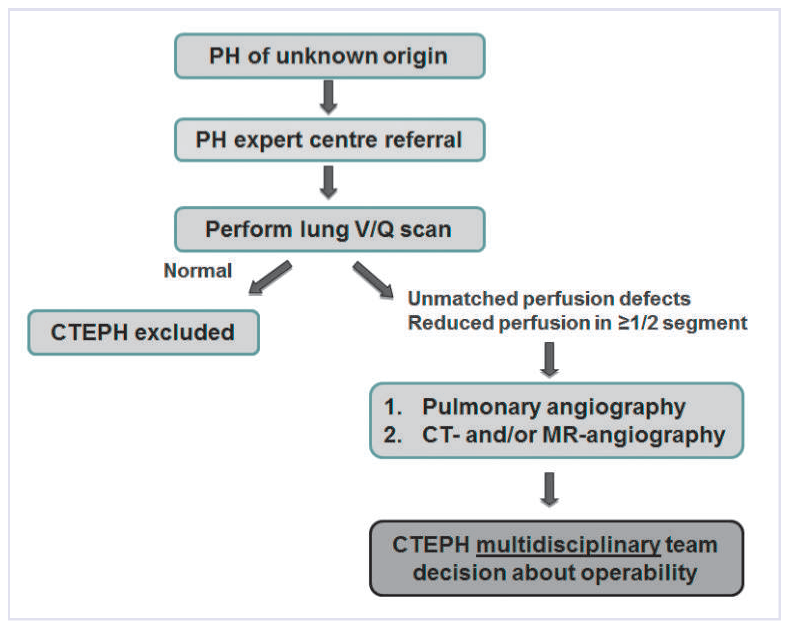
Figure 1. CTEPH diagnostic algorithm.
Cardiovascmed 17 00328 g001
Within the small vessel compartment, changes similar to pulmonary arterial hypertension (PAH) are observed in CTEPH [12]. Abnormal endothelial function, excessive proliferation of smooth muscle cells, migration of fibroblasts and inhibition of apoptosis in vascular smooth muscle cells lead to endothelial dysfunction, vascular remodelling and microthrombosis [25]. As in PAH, a persistent vasoconstrictive state, characterised by high levels of plasma endothelin-1 and overexpression of type B endothelin receptors, may be present in CTEPH patients [26].
The combination of proximal thrombus occlusion and vascular remodelling in nonoccluded vessels leads to elevated pulmonary vascular pressure and resistance (PVR) with consecutive right heart hypertrophy and progressive right heart failure.

Clinical presentation

The main symptoms associated with CTEPH are nonspecific, such as progressive dyspnoea and exercise intolerance. They are often primarily considered to be associated to other states, like obstructive lung disease, deconditioning and obesity [5]. Haemodynamically, the cardiac output is limited by the high PVR. Minute ventilation is augmented due to an increase of dead space [27]. Considering that 35% of patients do not have a history of acute PE, the diagnosis is challenging. In clinical examination, a fixed split or accentuated pulmonic component of the S2 are common findings. Exertional syncope and signs of RV failure are found in progressive disease. If a right-to-left shunt is present through a patent foramen ovale, cyanosis might be observed.

CTEPH diagnosis

A ventilation perfusion (VQ) scan is the screening goal of choice to distinguish CTEPH from PH. With 96% sensitivity, a normal VQ scan allows CTEPH to be ruled out. VQ scans only require a limited radiation dose, do not depend on intravenous contrast agents and are of low cost. Despite these advantages, VQ scans seem to be underutilised in the evaluation of CTEPH. In one study, 43% of patients with precapillary PH did not have a VQ scan according to a quality report and thus might have been misclassified [28].
In case of an abnormal VQ scan, pulmonary digital subtraction angiography is still considered as the golden standard to diagnose CTEPH. Pulmonary angiography accurately depicts the extension of the thromboembolic disease and, together with pulmonary haemodynamics, allows evaluation of operability for pulmonary endarterectomy (PEA).
Computed tomography pulmonary angiogram (CTPA) is performed in many centres to diagnose CTEPH in addition to VQ scans and pulmonary angiography. Some specialist teams even rely on this technique to base their decision on surgical accessibility of CTEPH. CTPA has the advantage of providing additional data on lung parenchyma, mediastinal lymph nodes, bronchial arteries and differential diagnosis. However, the diagnostic accuracy of this imaging technique depends on the experience of the radiologist and other interpreters, as various pathologies might present similarly as CTEPH, such as pulmonary artery sarcoma, tumour emboli into the pulmonary artery (from renal, thyroid, testicular and uterine cancer), embolisation of hydatic cysts of the liver, pulmonary arteriitis (Takayashu, Behçet) or fibrous mediastinitis. Proximal lining thrombi associated with pulmonary arterial hypertension can also mimic chronic thromboembolic disease. On the other hand, distal disease in segmental or subsegmental arteries may be missed in CTPA. However, in the latest generation of CT scanners, the resolution of the images allows a better evaluation of vascular wall thickness and surrounding structures. In comparison to conventional angiography, no need for catheter access makes CTPA more comfortable. Emerging techniques, such as dual energy CT and lung perfusion MRI are additional promising tools in the diagnosis and operability assessment of CTEPH. It is important to state that both pulmonary angiography and CTPA should be performed and interpreted in experienced PH-centres with access to surgical pulmonary endarterectomy.
In summary, according to the NICE PH conference 2013:
VQ scan is recommended as screening tool for chronic thromboembolic disease in all patients with no obvious reason for precapillary PH.
Pulmonary angiography remains the gold standard for confirmation of chronic thromboembolic disease and evaluation of operability.
High-quality multidetector CTPA may be a suitable alternative to pulmonary angiography in centres with experience in CTEPH.

Surgical therapy

Pulmonary endarterectomy (PEA) has transformed CTEPH from a fatal disease to a potentially curable form of PH. PEA is classically performed via a median sternotomy to have access to both lungs and to install a cardiopulmonary bypass. Cooling to 20 °C allows deep hypothermic circulatory arrest (DHCA) to provide a clear operating field. The endarterectomy dissection plane should be circumferential in order to be able to dissect until the level of segmental and even subsegmental branches, a simple thrombectomy or embolectomy without a true endarterectomy will not help to reduce the PVR (Figure 2) [29]. In experienced centres, the procedure is safe with a mortality below 5% [30,31]. Successful outcomes for patients undergoing PEA have been reported in improvement of exercise capacity, haemodynamics (improving PVR by up to 80%) quality of life and life expectancy (90% survival rate at 5 years) [32].
Recently, a prospective, controlled trial (PEACOG) showed that DHCA compared with cerebral perfusion during pulmonary endarterectomy surgery resulted in similar performance at 3 months and 1 year after surgery in the DHCA group [33]. Surprisingly, cognitive function was even improved after the PEA, likely due to the improved cardiac output. 1-year survival was 96%. The authors of the trial recommended DHCA as the best procedure, as 9 patients (from 74) had to cross over from antegrade cerebral perfusion to DHCA in order to keep the operating field clear.
Despite favourable outcomes in most patients, there are two major well-known complications in the postoperative course of PEA: residual PH and reperfusion lung injury [5]. Persistently elevated pulmonary pressure often occurs in combination with reperfusion lung injury. The emerging and improved technology of extracorporeal membrane oxygenation offers good salvage solution for both situations: venoarterial extracorporeal membrane oxygenation (ECMO) is used in case of haemodynamic instability. The right ventricle can be offloaded as well as PA pressures being reduced and therefore cardiac output and gas exchange are improved. In case of reperfusion injury alone, conservative therapy or veno-venous ECMO can be used [34]. Survival rates of these PEA-complications up to 57% are reported.
Since 2001, reports mainly from Japan emerged about innovative percutaneous pulmonary angioplasty done in patients which were noneligible for surgery due to comorbidities [35,36,37,38,39]. Multiple angioplasty procedures were necessary to achieve a significant reduction of the pulmonary vascular resistance and the studies were neither randomised nor controlled. Nevertheless, haemodynamic improvements were impressive as well as improvements in the 6-minute walking distance and WHO functional class, and the procedure was safe, even in elderly patients [36]. Thus, this novel treatment option might be considered in CTEPH-patients who are not eligible for surgery due to comorbidities. Despite these promising reports, further controlled trials and long-term data will be required in order to define the role of pulmonary angioplasty in the CTEPH-treatment algorithm and to address risks such as bleeding or vessel rupture and complications such as the occurrence of restenosis.
In conclusion, CTEPH is a surgically curable disease for some patients affected and we therefore underline the importance of the operability assessment through a multidisciplinary CTEPH specialised team.

Medical therapy

Lifelong anticoagulation is the foundation of treatment in all patients with CTEPH regardless of therapy and interventions [5]. Mostly, vitamin-K antagonists are used in this indication. Although there are no trials specifically performed in CTEPH, it is reasonable to assume that other anticoagulants such as heparines or direct inhibitors of FXa (e.g. rivaroxaban, apixaban) or thrombin (dabigatran) are similarly effective [40]. The former might more be used for short-time bridging (e.g. perioperatively) the latters have the advantage that they can be given in fixed daily doses, do not need monitoring, have less drug-drug and drug-food interactions and have demonstrated favourable bleeding-risk-tobenefit ratio as demonstrated in several trials in patients with venous thromboembolism [40,41].
CTEPH patients are considered to be inoperable in up to 50% of cases. Reasons include distal disease, comorbidities (COPD, severe left ventricular dysfunction) and excessively high PVR [19]. For patients considered unsuitable for PEA or with postoperative persistent PH, pharmacological treatment with PH-targeted therapies can be offered [5]. CTEPH and pulmonary arterial hypertension (PAH) share many pathogenetic features [42]. Thus, medical therapy as used for PAH (PAH-target therapy) can be prescribed. Benefits of pulmonary vasodilators have been reported in various trials. BENEFiT was the first large RCT supporting medication with Bosentan in patients with inoperable forms of CTEPH. Bosentan given for 6 months reduced PVR by 24% compared to placebo but could not improve the 6-minute walking distance [43].
More recently, clinically relevant primary endpoints have been reported by the large CHEST-1 trial assessing a new drug class, the soluble guanylate cyclase stimulator, Riociguat [4]. After a cautious uptitration schema in order to avoid systemic vasodilatory effects and hypotension, patients receiving Riociguat had a significant improvement of 46 m in the 6-minute walk distance and a reduction of their PVR by –31%. However, there was no effect on time to clinical worsening and the benefits were less pronounced among patients with persistent PH after PEA.
In summary, PEA remains the first therapeutic choice for patients with surgically accessible CTEPH. The evaluation for PEA must be made by an experienced CTEPH team. Inoperable patients with distal disease, comorbidities or patients with persisting symptomatic PH after PEA should be treated with medical therapy and/or considered for pulmonary angioplasty.

Funding/potential competing interests:

No financial support and no other potential conflict of interest relevant to this article were reported.

References

  1. Fedullo, P.F.; Auger, W.R.; Kerr, K.M.; Rubin, L.J. Chronic thromboembolic pulmonary hypertension. N Engl J Med. 2001, 345, 1465–1472. [Google Scholar] [CrossRef] [PubMed]
  2. Pengo, V.; Lensing, A.W.; Prins, M.H.; Marchiori, A.; Davidson, B.L.; Tiozzo, F.; et al. Incidence of chronic thromboembolic pulmonary hypertension after pulmonary embolism. N Engl J Med. 2004, 350, 2257–2264. [Google Scholar] [CrossRef] [PubMed]
  3. Jenkins, D.P.; Madani, M.; Mayer, E.; Kerr, K.; Kim, N.; Klepetko, W.; et al. Surgical treatment of chronic thromboembolic pulmonary hypertension. Eur Respir J. 2013, 41, 735–742. [Google Scholar] [CrossRef] [PubMed]
  4. Ghofrani, H.A.; D’Armini, A.M.; Grimminger, F.; Hoeper, M.M.; Jansa, P.; Kim, N.H.; et al. Riociguat for the treatment of chronic thromboembolic pulmonary hypertension. N Engl J Med. 2013, 369, 319–329. [Google Scholar] [CrossRef] [PubMed]
  5. Kim, N.H.; Delcroix, M.; Jenkins, D.P.; Channick, R.; Dartevelle, P.; Jansa, P.; et al. Chronic thromboembolic pulmonary hypertension. J Am Coll Cardiol. 2013, 62 (Suppl. 25), D92–D99. [Google Scholar] [CrossRef] [PubMed]
  6. Lang, I.M.; Pesavento, R.; Bonderman, D.; Yuan, J.X. Risk factors and basic mechanisms of chronic thromboembolic pulmonary hypertension: A current understanding. Eur Respir J. 2013, 41, 462–468. [Google Scholar] [CrossRef] [PubMed]
  7. Miniati, M.; Monti, S.; Bottai, M.; Scoscia, E.; Bauleo, C.; Tonelli, L.; et al. Survival and restoration of pulmonary perfusion in a long-term follow-up of patients after acute pulmonary embolism. Medicine 2006, 85, 253–262. [Google Scholar] [CrossRef] [PubMed]
  8. Dentali, F.; Donadini, M.; Gianni, M.; Bertolini, A.; Squizzato, A.; Venco, A.; et al. Incidence of chronic pulmonary hypertension in patients with previous pulmonary embolism. Thromb Res. 2009, 124, 256–258. [Google Scholar] [CrossRef] [PubMed]
  9. Ribeiro, A.; Lindmarker, P.; Johnsson, H.; Juhlin-Dannfelt, A.; Jorfeldt, L. Pulmonary embolism: One-year follow-up with echocardiography doppler and five-year survival analysis. Circulation 1999, 99, 1325–1330. [Google Scholar] [CrossRef] [PubMed]
  10. Bonderman, D.; Jakowitsch, J.; Adlbrecht, C.; Schemper, M.; Kyrle, P.A.; Schonauer, V.; et al. Medical conditions increasing the risk of chronic thromboembolic pulmonary hypertension. Thromb Haemost. 2005, 93, 512–516. [Google Scholar] [CrossRef] [PubMed]
  11. Bonderman, D.; Skoro-Sajer, N.; Jakowitsch, J.; Adlbrecht, C.; Dunkler, D.; Taghavi, S.; et al. Predictors of outcome in chronic thromboembolic pulmonary hypertension. Circulation 2007, 115, 2153–2158. [Google Scholar] [CrossRef] [PubMed]
  12. Morris, T.A.; Marsh, J.J.; Chiles, P.G.; Auger, W.R.; Fedullo, P.F.; Woods, V.L., Jr. Fibrin derived from patients with chronic thromboembolic pulmonary hypertension is resistant to lysis. Am J Respir Crit Care Med. 2006, 173, 1270–1275. [Google Scholar] [CrossRef] [PubMed] [PubMed Central]
  13. Bonderman, D.; Turecek, P.L.; Jakowitsch, J.; Weltermann, A.; Adlbrecht, C.; Schneider, B.; et al. High prevalence of elevated clotting factor VIII in chronic thromboembolic pulmonary hypertension. Thromb Haemost. 2003, 90, 372–376. [Google Scholar] [CrossRef] [PubMed]
  14. Friedman, R.; Mears, J.G.; Barst, R.J. Continuous infusion of prostacyclin normalizes plasma markers of endothelial cell injury and platelet aggregation in primary pulmonary hypertension. Circulation 1997, 96, 2782–2784. [Google Scholar] [CrossRef] [PubMed]
  15. Wong, C.L.; Szydlo, R.; Gibbs, S.; Laffan, M. Hereditary and acquired thrombotic risk factors for chronic thromboembolic pulmonary hypertension. Blood Coagul Fibrinolysis 2010, 21, 201–206. [Google Scholar] [CrossRef] [PubMed]
  16. Lewczuk, J.; Piszko, P.; Jagas, J.; Porada, A.; Wojciak, S.; Sobkowicz, B.; et al. Prognostic factors in medically treated patients with chronic pulmonary embolism. Chest 2001, 119, 818–823. [Google Scholar] [CrossRef] [PubMed]
  17. Jaff, M.R.; McMurtry, M.S.; Archer, S.L.; Cushman, M.; Goldenberg, N.; Goldhaber, S.Z.; et al. Management of massive and submassive pulmonary embolism, iliofemoral deep vein thrombosis, and chronic thromboembolic pulmonary hypertension: A scientific statement from the American Heart Association. Circulation 2011, 123, 1788–1830. [Google Scholar] [CrossRef] [PubMed]
  18. McNeil, K. Assessment of patients with pulmonary hypertension. Respiration 2008, 76, 373–374. [Google Scholar] [CrossRef] [PubMed]
  19. Mayer, E. Surgical and post-operative treatment of chronic thromboembolic pulmonary hypertension. Eur Respir Rev. 2010, 19, 64–67. [Google Scholar] [CrossRef] [PubMed]
  20. Humbert, M.; Simonneau, G. Vasodilators in patients with chronic obstructive pulmonary disease and pulmonary hypertension: Not ready for prime time! Am J Respir Crit Care Med. 2010, 181, 202–203. [Google Scholar] [CrossRef] [PubMed]
  21. Waltham, M.; Burnand, K.G.; Collins, M.; Smith, A. Vascular endothelial growth factor and basic fibroblast growth factor are found in resolving venous thrombi. Journal of vascular surgery 2000, 32, 988–996. [Google Scholar] [CrossRef] [PubMed]
  22. Distler, J.H.; Hirth, A.; Kurowska-Stolarska, M.; Gay, R.E.; Gay, S.; Distler, O. Angiogenic and angiostatic factors in the molecular control of angiogenesis. Q. J. Nucl. Med. Off. Publ. Ital. Assoc. Nucl. Med. 2003, 47, 149–161. [Google Scholar] [PubMed]
  23. Sakao, S.; Hao, H.; Tanabe, N.; Kasahara, Y.; Kurosu, K.; Tatsumi, K. Endothelial-like cells in chronic thromboembolic pulmonary hypertension: Crosstalk with myofibroblast-like cells. Respir Res. 2011, 12, 109. [Google Scholar] [CrossRef] [PubMed] [PubMed Central]
  24. Kimura, H.; Okada, O.; Tanabe, N.; Tanaka, Y.; Terai, M.; Takiguchi, Y.; et al. Plasma monocyte chemoattractant protein-1 and pulmonary vascular resistance in chronic thromboembolic pulmonary hypertension. Am J Respir Crit Care Med. 2001, 164, 319–324. [Google Scholar] [CrossRef] [PubMed]
  25. Moser, K.M.; Auger, W.R.; Fedullo, P.F. Chronic major-vessel thromboembolic pulmonary hypertension. Circulation 1990, 81, 1735–1743. [Google Scholar] [CrossRef] [PubMed]
  26. Wallis, J.D.; Anderson, K.C.; Miller, E.K. Single neurons in prefrontal cortex encode abstract rules. Nature 2001, 411, 953–956. [Google Scholar] [CrossRef] [PubMed]
  27. Kapitan, K.S.; Buchbinder, M.; Wagner, P.D.; Moser, K.M. Mechanisms of hypoxemia in chronic thromboembolic pulmonary hypertension. Am Rev Respir Dis. 1989, 139, 1149–1154. [Google Scholar] [CrossRef] [PubMed]
  28. McLaughlin, V.V.; Langer, A.; Tan, M.; Clements, P.J.; Oudiz, R.J.; Tapson, V.F.; et al. Contemporary trends in the diagnosis and management of pulmonary arterial hypertension: An initiative to close the care gap. Chest 2013, 143, 324–332. [Google Scholar] [CrossRef] [PubMed]
  29. Opitz, I.; de Perrot, M. Technique of pulmonary thromboendarterectomy. Oper. Tech. Thorac. Cardiovasc. Surg. 2012, 17, 168–180. [Google Scholar] [CrossRef]
  30. Mayer, E.; Jenkins, D.; Lindner, J.; D’Armini, A.; Kloek, J.; Meyns, B.; et al. Surgical management and outcome of patients with chronic thromboembolic pulmonary hypertension: Results from an international prospective registry. J Thorac Cardiovasc Surg. 2011, 141, 702–710. [Google Scholar] [CrossRef] [PubMed]
  31. Madani, M.M.; Auger, W.R.; Pretorius, V.; Sakakibara, N.; Kerr, K.M.; Kim, N.H.; et al. Pulmonary endarterectomy: Recent changes in a single institution’s experience of more than 2,700 patients. Ann Thorac Surg. 2012, 94, 97–103. [Google Scholar] [CrossRef] [PubMed]
  32. Pepke-Zaba, J.; Hoeper, M.M.; Humbert, M. Chronic thromboembolic pulmonary hypertension: Advances from bench to patient management. Eur Respir J. 2013, 41, 8–9. [Google Scholar] [CrossRef] [PubMed]
  33. Vuylsteke, A.; Sharples, L.; Charman, G.; Kneeshaw, J.; Tsui, S.; Dunning, J.; et al. Circulatory arrest versus cerebral perfusion during pulmonary endarterectomy surgery (PEACOG): A randomised controlled trial. Lancet 2011, 378, 1379–1387. [Google Scholar] [CrossRef] [PubMed]
  34. Mydin, M.; Berman, M.; Klein, A.; Tsui, S.; Dunning, J.; Valchanov, K.; et al. Extracorporeal membrane oxygenation as a bridge to pulmonary endarterectomy. Ann Thorac Surg. 2011, 92, e101–3. [Google Scholar] [CrossRef] [PubMed]
  35. Feinstein, J.A.; Goldhaber, S.Z.; Lock, J.E.; Ferndandes, S.M.; Landzberg, M.J. Balloon pulmonary angioplasty for treatment of chronic thromboembolic pulmonary hypertension. Circulation 2001, 103, 10–13. [Google Scholar] [CrossRef] [PubMed]
  36. Yanagisawa, R.; Kataoka, M.; Inami, T.; Shimura, N.; Ishiguro, H.; Fukuda, K.; et al. Safety and efficacy of percutaneous transluminal pulmonary angioplasty in elderly patients. Int J Cardiol. 2014, 175, 285–289. [Google Scholar] [CrossRef] [PubMed]
  37. Sugimura, K.; Fukumoto, Y.; Satoh, K.; Nochioka, K.; Miura, Y.; Aoki, T.; et al. Percutaneous transluminal pulmonary angioplasty markedly improves pulmonary hemodynamics and long-term prognosis in patients with chronic thromboembolic pulmonary hypertension. Circ J. 2012, 76, 485–488. [Google Scholar] [CrossRef] [PubMed]
  38. Kataoka, M.; Satoh, T.; Manabe, T.; Anzai, T.; Yoshikawa, T.; Mitamura, H.; et al. Oral sildenafil improves primary pulmonary hypertension refractory to epoprostenol. Circ J. 2005, 69, 461–465. [Google Scholar] [CrossRef] [PubMed]
  39. Mizoguchi, H.; Ogawa, A.; Munemasa, M.; Mikouchi, H.; Ito, H.; Matsubara, H. Refined balloon pulmonary angioplasty for inoperable patients with chronic thromboembolic pulmonary hypertension. Circ. Cardiovasc. Interv. 2012, 5, 748–755. [Google Scholar] [CrossRef] [PubMed]
  40. Rollins, B.M.; Silva, M.A.; Donovan, J.L.; Kanaan, A.O. Evaluation of Oral Anticoagulants for the Extended Treatment of Venous Thromboembolism Using a Mixed-Treatment Comparison, Meta-Analytic Approach. Clin. Ther. 2014. [CrossRef] [PubMed]
  41. Goldhaber, S.Z.; Bounameaux, H. Pulmonary embolism and deep vein thrombosis. Lancet 2012, 379, 1835–1846. [Google Scholar] [CrossRef] [PubMed]
  42. Ogino, H.; Ando, M.; Matsuda, H.; Minatoya, K.; Sasaki, H.; Nakanishi, N.; et al. Japanese single-center experience of surgery for chronic thromboembolic pulmonary hypertension. Ann Thorac Surg. 2006, 82, 630–636. [Google Scholar] [CrossRef] [PubMed]
  43. Jais, X.; D’Armini, A.M.; Jansa, P.; Torbicki, A.; Delcroix, M.; Ghofrani, H.A.; et al. Bosentan for treatment of inoperable chronic thromboembolic pulmonary hypertension: BENEFiT (Bosentan Effects in iNopErable Forms of chronIc Thromboembolic pulmonary hypertension), a randomized, placebo-controlled trial. J Am Coll Cardiol. 2008, 52, 2127–2134. [Google Scholar] [CrossRef] [PubMed]
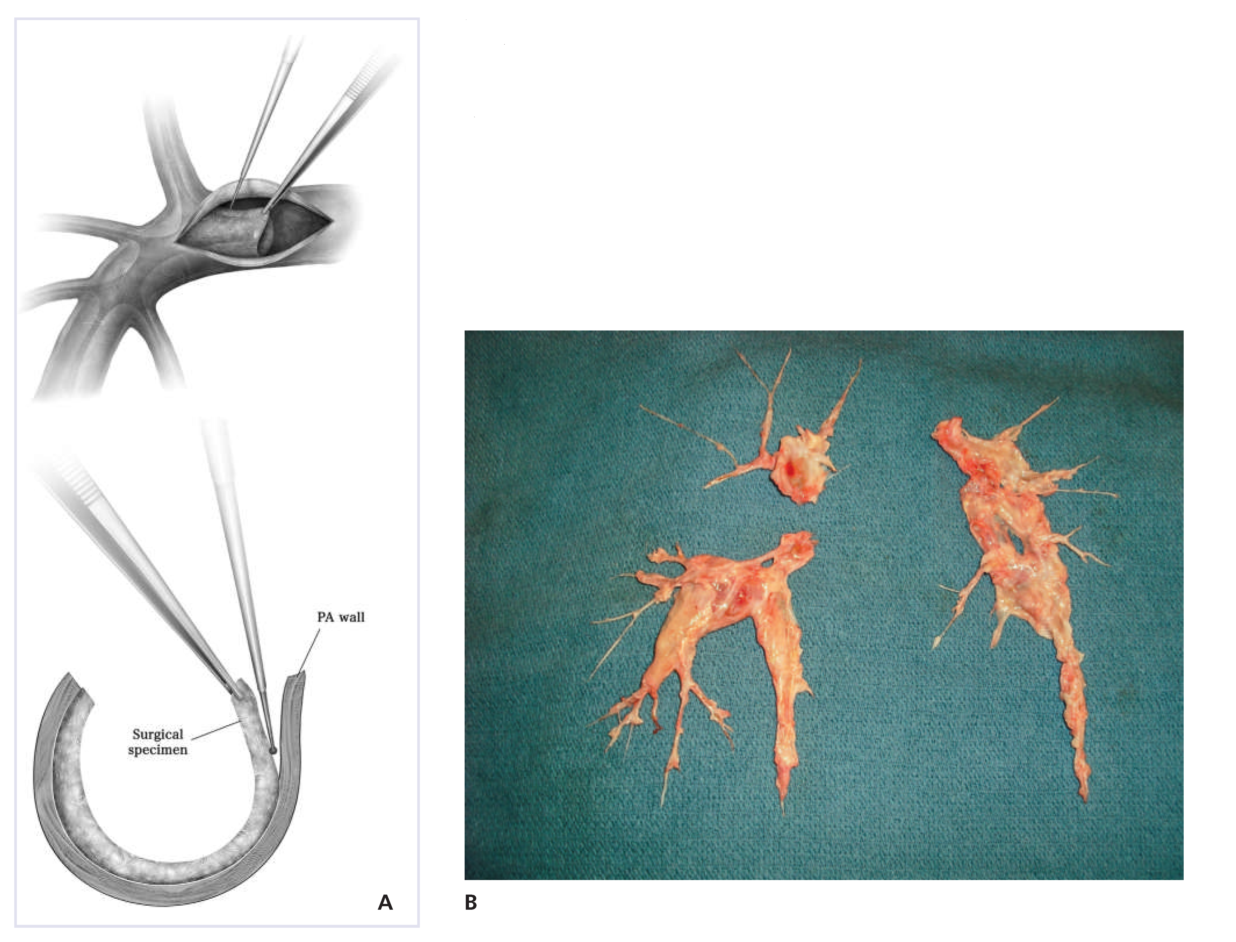
Figure 2. (A) Endarterectomy of the right pulmonary artery and dissection plane. (From: Opitz I, de Perrot M. technique of pulmonary thromboendarterectomy. Op Techniques Thorac Cardiovasc Surgery. 2012;17(3):168–80. DOI: 10.1053/j.optechstcvs.2012.07.004). (B) Endarterectomy specimen of the right and left pulmonary artery.
Cardiovascmed 17 00328 g002