Next Article in Journal
Treatment of Pulmonary Arterial Hypertension: An Update
Previous Article in Journal
Chronic Thromboembolic Pulmonary Hypertension
 
 
Cardiovascular Medicine is published by MDPI from Volume 28 Issue 1 (2025). Previous articles were published by another publisher in Open Access under a CC-BY (or CC-BY-NC-ND) licence, and they are hosted by MDPI on mdpi.com as a courtesy and upon agreement with Editores Medicorum Helveticorum (EMH).
Review

Pulmonary Hypertension Associated with Left Heart and Lung Diseases

by
Micha T. Maeder
1,2,*,
Rebekka Kleiner
2,3,
Daniel Weilenmann
1,2 and
Otto D. Schoch
2,3,*
1
Department of Cardiology, Kantonsspital St. Gallen, St. Gallen, Switzerland
2
Pulmonary Hypertension Unit, Kantonsspital St. Gallen, St. Gallen, Switzerland
3
Department of Respiratory Medicine, Kantonsspital St. Gallen, St. Gallen, Switzerland
*
Authors to whom correspondence should be addressed.
Cardiovasc. Med. 2014, 17(11), 320; https://doi.org/10.4414/cvm.2014.00278
Submission received: 19 August 2014 / Revised: 19 September 2014 / Accepted: 19 October 2014 / Published: 19 November 2014

Abstract

Pulmonary hypertension (PH) associated with left heart diseases (PH-LHD) and PH associated with lung diseases (PH-lung) are common types of PH which typically represent more advanced disease stages of left heart and lung disease, characterised by more severe symptoms and worse prognosis. The tailored work-up should include an assessment of the severity and haemodynamic constellation of PH and the mechanism of the underlying left heart or lung disease and the respective therapeutic targets. Transthoracic echocardiography is the primary noninvasive imaging tool to estimate the likelihood of PH. However, the accuracy of echocardiography is limited in patients with only moderately elevated pulmonary pressures, and acoustic windows in patients with lung disease are often poor. Therefore, right heart catheterisation is always required if exact knowledge of the haemodynamic constellation is required. Echocardiography, cardiac catheterisation, blood gas analysis, lung function testing and ergospirometry as well as additional modalities such as high-resolution computed tomography, overnight sleep studies and cardiac magnetic resonance imaging may be required to characterise the underlying left heart or lung disease and the targets for therapy. The general treatment principle for the management of PH-LHD and PH-lung is to treat the underlying left heart or lung disease in an optimal manner. Specific vasodilator therapies currently do not play a prominent role in PH-LHD and PH-lung but additional trials are underway and may define subgroups deriving benefit.

Introduction

Pulmonary hypertension (PH) associated with left heart diseases: (PH-LHD; class II PH [1]) and PH associated with lung diseases (PH lung; class III PH [1]) are the most common types of PH [2,3]. Many forms of left heart or lung diseases can be associated with PH (Table 1), and PH is typically a marker of more advanced disease, worse exercise tolerance and worse prognosis [2]. The prevalence of PH-LHD and of PH-lung varies depending on the type of disease and its severity. For PH-LHD, a prevalence ranging between 25% and 100% of the patients studied has been reported [2]. The prototype of PH-LHD is rheumatic mitral stenosis. While this type of valvular heart disease has become rare in the Western world, mitral regurgitation and aortic stenosis are now important causes of PH-LHD. Furthermore, heart failure with reduced left ventricular ejection fraction (HFrEF) and heart failure with preserved left ventricular ejection fraction (HFpEF) can be associated with PH. In addition, specific cardiomyopathies including hypertrophic cardiomyopathy or restrictive forms of cardiomyopathy can be accompanied by PH-LHD. The prevalence of PH in chronic obstructive pulmonary disease (COPD) depends on the severity of the lung disease and on the definition of PH. While 90% of the patients with COPD Global initiative for chronic Obstructive Lung Disease (GOLD) stage 4 have a mean pulmonary artery pressure (PAP) >20 mm Hg, only 3 to 5% have a mean PAP >35 mm Hg [3]. The presence of PH is a strong predictor of mortality in COPD, and a 5-year survival of less than 40% has been reported for patients with COPD and mean PAP >25 mm Hg. The severity of PH predicts mortality in COPD patients much more accurately than lung function measurements such as the forced expiratory volume within the first second or gas exchange measurements [4]. In idiopathic pulmonary fibrosis (IPF), mean PAP values >25 mm Hg were reported in 8 to 15% at initial work-up, but in advanced and end-stage disease, the prevalence increases to >60% [3]. However, very high mean PAP values (>40 mm Hg) are found in less than 10% [5]. In IPF, the correlation between PH severity and lung function impairment or computed tomography fibrosis scores is very poor. High prevalence of cigarette smoking in patients with IPF may contribute to the coexistence of obstructive ventilatory disorder and emphysema with IPF, an entity referred to as combined pulmonary fibrosis with emphysema (CPFE), a disease with characteristic findings in high-resolution computed tomography showing emphysema associated with interstitial lung disease. In lung function testing, lung volumes are usually preserved (due to the hyperinflation) although significant lung parenchymal damage is present. In CPFE, PH is common and is associated with a particularly poor prognosis [6].

Pathophysiology

PH-LHD

The primary mechanism of PH-LHD is a backward transmission of elevated left-sided filling pressures. The latter occurs because of systolic and/or diastolic left ventricular dysfunction, mitral regurgitation, decreased left atrial compliance, and increased after-load. In the majority of patients with PH-LHD, PH is a purely passive phenomenon due to venous congestion. In some patients however, there is an additional active component due to pulmonary vasoconstriction and vascular remodelling. The underlying mechanisms are thought to include decreased nitric oxide availability, increased endothelin expression, and desensitisation to natriuretic peptide-mediated vasodilatation. In these patients, the pulmonary pressure increases further and more than expected from the left ventricular filling pressure (however, the term “out of proportion” is now discouraged) [2]. Finally, there are patients with PH-LHD who had both a passive and an active component of PH, in whom the passive component is treated successfully by valve replacement or medical therapy but the active component persists even after normalisation of filling pressures. These pathophysiological principles also apply for rare forms of congenital left heart inflow/outflow tract obstructions and cardiomyopathies (class 2.4. PH). In contrast, PH in the context of congenital heart disease, typically shunts of variable complexity, belong to the PAH category (class 1.4.) and are not addressed in the present review. The pathophysiology of PH in patients with PAH associated with congenital heart disease is typically very complex and requires a case-by-case assessment.

PH-lung

For PH-lung, the mechanism is multifactorial and may vary between the different lung diseases. Pathophysiological mechanisms include hypoxia with pulmonary vasoconstriction (Euler-Liljestrand effect), rarefaction of the pulmonary vascular bed in areas of damaged lung with consecutive vascular remodelling in other areas, ventilation-perfusion mismatch, and hypoventilation with hypoxia and carbon dioxide retention. These factors lead to precapillary vasoconstriction via nitric oxide imbalance, release of vasoconstrictors and metabolic alterations [7].

Assessment and definitions of PH-LHD

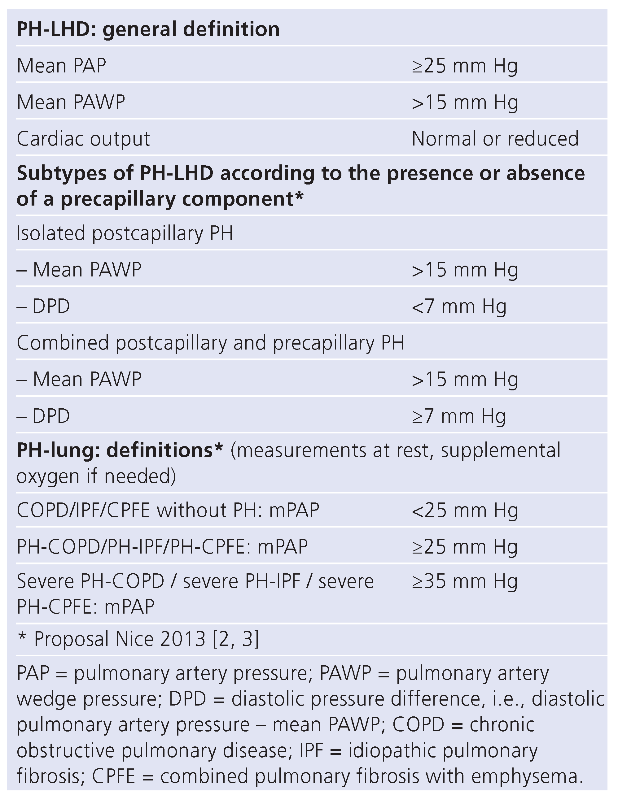
As shown in Table 2, PH-LHD is defined as a combination of a mPAP ≥25 mm Hg and a mean pulmonary artery wedge pressure (PAWP; the term “pulmonary capillary wedge pressure” is discouraged) >15 mm Hg [2]. Thus, in patients with purely pulmonary venous hypertension, the transpulmonary gradient (TPG, i.e., the difference between mean PAP and mean PAWP) is low (typically less than 10–12 mm Hg). The best way to define PH-LHD with an additional precapillary component (cf. discussion on pathobiology above) has been a matter of debate. In previous guidelines, purely passive PH-LHD was defined as mean PAP ≥25 mm Hg, mean PAWP >15 mm Hg, AND a TPG ≤12 mm Hg, whereas “reactive” or “out of proportion” PH-LHD was defined as mean PAP ≥25 mm Hg, mean PAWP >15 mm Hg, AND a TPG >12 mm Hg [8]. More recently (Nice, 2013), an alternative definition relying on the diastolic pressure difference (DPD, i.e., the difference between diastolic PAP and mean PAWP) rather than TPG has been proposed (Table 2) [2]: It has been suggested that “isolated postcapillary PH” is defined as a mean PAP ≥25 mm Hg, mean PAWP >15 mm Hg, and a DPD <7 mm Hg, whereas “combined postcapillary and precapillary PH” is defined as mean PAP ≥25 mm Hg, mean PAWP >15 mm Hg, and a DPD ≥7 mm Hg [2]. However, this definition has not been broadly applied yet, and information on clinical characteristics of patients belonging to these entities is lacking.
After a clinical assessment, transthoracic echocardiography is the primary imaging tool to assess the probability of the presence of PH-LHD and to define the underlying mechanisms of LHD and therapeutic targets. Many studies have shown very good correlations between invasively assessed systolic PAP and noninvasively estimated systolic PAP as based on the peak tricuspid regurgitation velocity (TRV; systolic PAP = 4×[peak TRV]2 according to the simplified Bernoulli equation) [9]. Thus, although these studies have typically not been performed in patients with PH-LHD [9], the likelihood of PH in the setting of suspected PHLHD is estimated based on peak TRV (peak TRV 2.9–3.4 m/s: PH possible; peak TRV >3.4 m/s: PH likely) and indirect evidence of PH including enlargement of the right cardiac chambers, deformation of the interventricular septum (“D-shape” of the left ventricle), and shortening of the pulmonary acceleration time [8]. Thus, TRV should never be looked at isolated from other echocardiographic findings. In particular, right ventricular function must always be assessed to put estimated systolic PAP into perspective. Apart from classical measures of right ventricular function including right ventricular fractional area change, tricuspid annular plane systolic excursion (as assessed by M-mode echocardiography), and right ventricular peak systolic annular velocity (as assessed by pulsed-wave tissue Doppler), novel measures indicating subtle right ventricular dysfunction such as right ventricular free wall strain may provide important information [10].
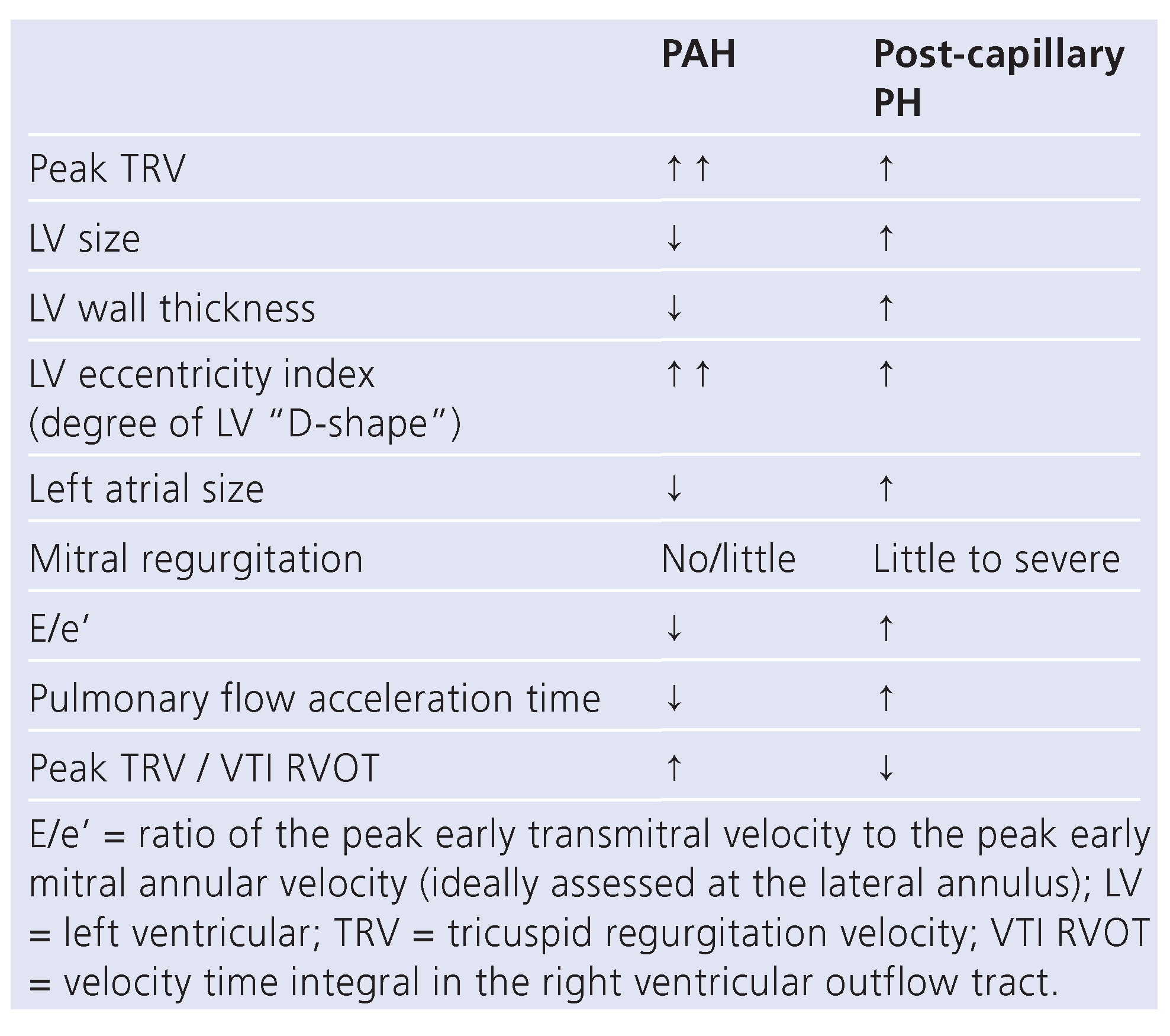
Features of PH are typically less prominent in PHLHD than in pulmonary arterial hypertension (PAH, i.e., class 1 PH) because in PH-LHD, both left-sided and right-sided filling pressures are elevated. Echocardiography can provide important information on whether PAH or PH-LHD is present. An elevated systolic PAP as estimated from the peak TRV in conjunction with a dilated left ventricle with impaired left ventricular ejection fraction, evidence of a cardiomyopathy (e.g., hypertrophic cardiomyopathy or amyloid heart disease), or severe valve disease (e.g., severe mitral regurgitation or severe aortic stenosis) is highly suggestive of PH-LHD. However, if left ventricular ejection fraction is not significantly impaired, and there is no severe valve disease, the differentiation between PAH and PH-LHD (i.e., HFpEF) is challenging. Patients with PH-LHD in the context of HFpEF are typically older and have more cardiovascular risk factors, in particular hypertension and diabetes, than those with PAH [11]. In Table 3, echocardiographic features favouring PAH or PH-LHD are summarised [11,12]. Although algorithms have been developed to differentiate between PAH and PH-LHD [12], and the noninvasive estimation of pulmonary vascular resistance (PVR) has been shown to be feasible [13], an accurate noninvasive assessment of haemodynamics is not possible in this setting [14]. Thus, right heart catheterisation will always be required if there is evidence of significant PH to confirm PH and to assess the underlying haemodynamic constellation (cf. definitions).
The role of stress echocardiography in the diagnostic assessment of patients with possible PH-LHD and PH-lung is not well defined. Stress echocardiography is generally not recommended for screening of PH in the current European guidelines [8], although one interesting study has suggested a potential role of stress echocardiography for the identification of early disease in relatives of patients with PAH [15]. However, stress echocardiography using physical exercise may have a role in certain patients with valve disease and borderline PAP at rest in whom a rapid rise in systolic PAP during exercise may suggest that the valve disease is clinically significant and that an intervention is required.
Given the well-known inaccuracies in measurements of PAWP and the inability to obtain a proper PAWP in some patients, the threshold to perform simultaneous right and left heart catheterisation should be low in subjects with a high likelihood of LHD [16]. Routine left heart catheterisation is not recommended in all patients with suspected PH but in doubt, both PAWP and left ventricular end-diastolic pressure should be measured as this will allow a reliable classification of PAH or PH-LHD [16]. A particular challenge can be present in patients with suspected PH-LHD in the context of HFpEF. Although the echocardiogram may have suggested significant PH, pulmonary pressures and PAWP may not fulfil criteria for PH-LHD during the invasive assessment at rest. This may be due to the fact that the patient has been successfully treated with diuretics and/or after-load reduction, or that haemodynamics had been influenced by fasting before cardiac catheterisation. In patients with HFpEF who typically have concentric left ventricular remodelling with a small left ventricular cavity and a steep end-diastolic pressure-volume relationship, relatively small changes in left ventricular volume may lead to significant changes in filling pressures [17]. Thus, a fluid challenge (500 ml of saline over 5 minutes) may be needed to diagnose PH-LHD in the context of HFpEF [16,18]. There is also evidence that exercise right heart catheterisation may help to gain a better definition of the haemodynamic profile of patients with HFpEF. However, both administration of a fluid bolus and exercise right heart catheterisation have not been standardised and are thus not recommended for routine use yet [16]. Additional imaging modalities including transoesophageal echocardiography, cardiac magnetic resonances imaging, and coronary angiography may be required to exactly characterise underlying left heart disease and thereby to define the therapeutic targets.

Assessment and definitions of PH-lung

Clinical symptoms and signs of PH are often similar to those of the underlying lung disease. In COPD and diffuse parenchymal lung disease with suspected PH, transthoracic echocardiography is the initial noninvasive diagnostic test of choice. To estimate the likelihood of PH the same general principles apply as discussed above in the PH-LHD section. However, the accuracy of echocardiography to estimate pulmonary pressures in patients with lung disease is limited as the acoustic window in these patients is often sub-optimal. Negative predictive values of 67 to 93% for the exclusion of significant PH by echocardiography were reported in this setting [19,20]. Furthermore, B-type natriuretic peptide values are elevated in severe PH-lung, but they lack sensitivity for moderate disease. Walking tests and ergospirometry are important tests for the assessment of lung patients, to quantify cardiopulmonary performance, to determine factors of exercise limitation and to assess exercise-induced hypoxia. Right heart catheterisation, the gold standard for PH diagnosis, is indicated if lung transplantation is considered, if clinical worsening is disproportionate to lung function impairment, if progressive gas exchange abnormalities are disproportionate to ventilatory impairment, if severe PH is suspected noninvasively, or if there is suspicion of concurrent left ventricular dysfunction, which might alter the management strategy. The term “out of proportion” PH for patients with PH-lung is discouraged and for COPD, IPF and CPFE, the definitions displayed in Table 2 are now proposed [3]. The choice of a mean PAP cut-off of 35 mm Hg for severe PH is based on the fact that only few LD patients with presumably severe vascular abnormalities have mPAP values above this threshold (approximately 1% for COPD patients). This degree of PH causes circulatory limitation and substantially reduces exercise capacity with preserved ventilatory reserve despite impairment by LD, an exercise limitation characteristic not observed in lung patients with moderate PH. Of note, class 1 PH patients (i.e., PAH) may also display mild to moderate ventilatory impairment in the absence of severe airway or parenchymal disease (Table 4). Lung diseases, particularly COPD and asthma, are common diseases and PH may not necessarily be the result of the LD, a requirement to be classified as PH class 3.

Management of PH-LHD

Treatment in patients with PH-LHD depends on the underlying left heart problem. Table 5 provides an overview of treatment modalities in different forms of PH-LHD. In patients with significant valve disease, typically severe mitral regurgitation, severe aortic stenosis, or less commonly severe mitral stenosis or severe aortic regurgitation, correction of the mechanical problem represents the primary approach. A detailed discussion of indications for and methods of valve surgery or catheter-based modalities for the treatment of valve disease is beyond the scope of the present review and can be found elsewhere [21]. In patients with isolated postcapillary PH in the context of valve disease, PAP typically normalises after correction of valvular stenosis or regurgitation. In patients with combined postcapillary and precapillary PH, the response of pulmonary pressures can vary: ideally, elimination of venous congestion is followed by normalisation of the reactive or precapillary component of PH. However, significant precapillary PH can persist after the intervention if there has been long-standing venous congestion with the development of a significant precapillary PH component as this is seen in patients with rheumatic mitral stenosis. These patients can then have the haemodynamic constellation of PAH, which means a normal PAWP and a significantly elevated TPG, DPD, and PVR even years after valve replacement.
The therapy of choice in patients with HFrEF with PH-LHD is the standard medical treatment for HFrEF which is described in detail elsewhere [22]. The presence of PH in these patients typically indicates insufficient treatment of venous congestion and the need for the intensification of treatment with diuretics, inhibitors of the renin-angiotensin system, beta-blockers, and mineralocorticoid receptor blockers as well as the use of cardiac resynchronisation therapy in selected cases. Serial echocardiograms usually allow detecting changes in central venous pressure, left ventricular end-diastolic pressure and pulmonary pressures in these patients in a qualitative manner, although an exact assessment of haemodynamic parameters by echocardiography is not possible [23]. Right heart catheterisation will be performed in severely compromised patients, particularly in those in whom cardiac transplantation will be considered. In the latter setting, a significant precapillary PH component represents a contraindication to transplantation because of a high risk of right heart failure of the allograft in the early postoperative period. Options in this setting include aggressive decongestion by diuretics and after-load reduction and/or the placement of a left ventricular assist device which has been shown to reduce both the postcapillary and the precapillary component of PH in transplant candidates. There is currently no established role of specific pulmonary vasodilators in patients with HFrEF. Studies using endothelin receptor antagonists have revealed neutral results. A small single centre study has revealed beneficial effects of the phosphodiesterase-5 inhibitor (PDE-5 inhibitor) sildenafil on haemodynamics and exercise capacity in patients with PH-LHF in the context of HFrEF [24]. In a multicentre study evaluating the effect of riociguat in patients with PH-LHF (HFrEF), no effect of riociguat on mean PAP was observed [25]. Additional trials using sildenafil and tadalafil are underway [2].
There is still no established treatment for patients with HFpEF [26]. In particular, clinical trials evaluating inhibitors of the renin-angiotensin system and the mineralocorticoid receptor blocker spironolactone have yielded neutral results, and the role of beta-blocker is not clear, although one study had suggested beneficial effects of carvedilol on measures of left ventricular diastolic function in patients with high heart rates [27]. Recent mechanistic studies in patients with HFpEF (not necessarily with PH) have revealed promising data for the selective sinus node inhibitor ivabradine [28], the selective sodium current inhibitor ranolazine [29], and aerobic exercise training [30]. Studies with clinical endpoint are not available however. Thus, management of patients with PH-LHD in this context is challenging. The primary approach should be a combination of diuretic therapy and after-load reduction, but it should be kept in mind that patients with HFpEF are characterised by small left ventricles with steep end-diastolic pressure-volume relationship, which makes them susceptible to significant changes in stroke volume and blood pressure following too aggressive preload reduction. A recent trial evaluating the use of sildenafil in patients with HFpEF (not necessarily with PH) failed to show an improvement in exercise capacity compared to placebo [31]. In contrast, a placebo-controlled haemodynamic study in patients with PH-LHF in the context of HFpEF revealed a significant reduction in mPAP and an improvement in right ventricular function by sildenafil [32]. Currently, the role of specific pulmonary vasodilators in PH-LHD in the context of HFpEF is not defined but further studies are underway.

Management of PH-lung

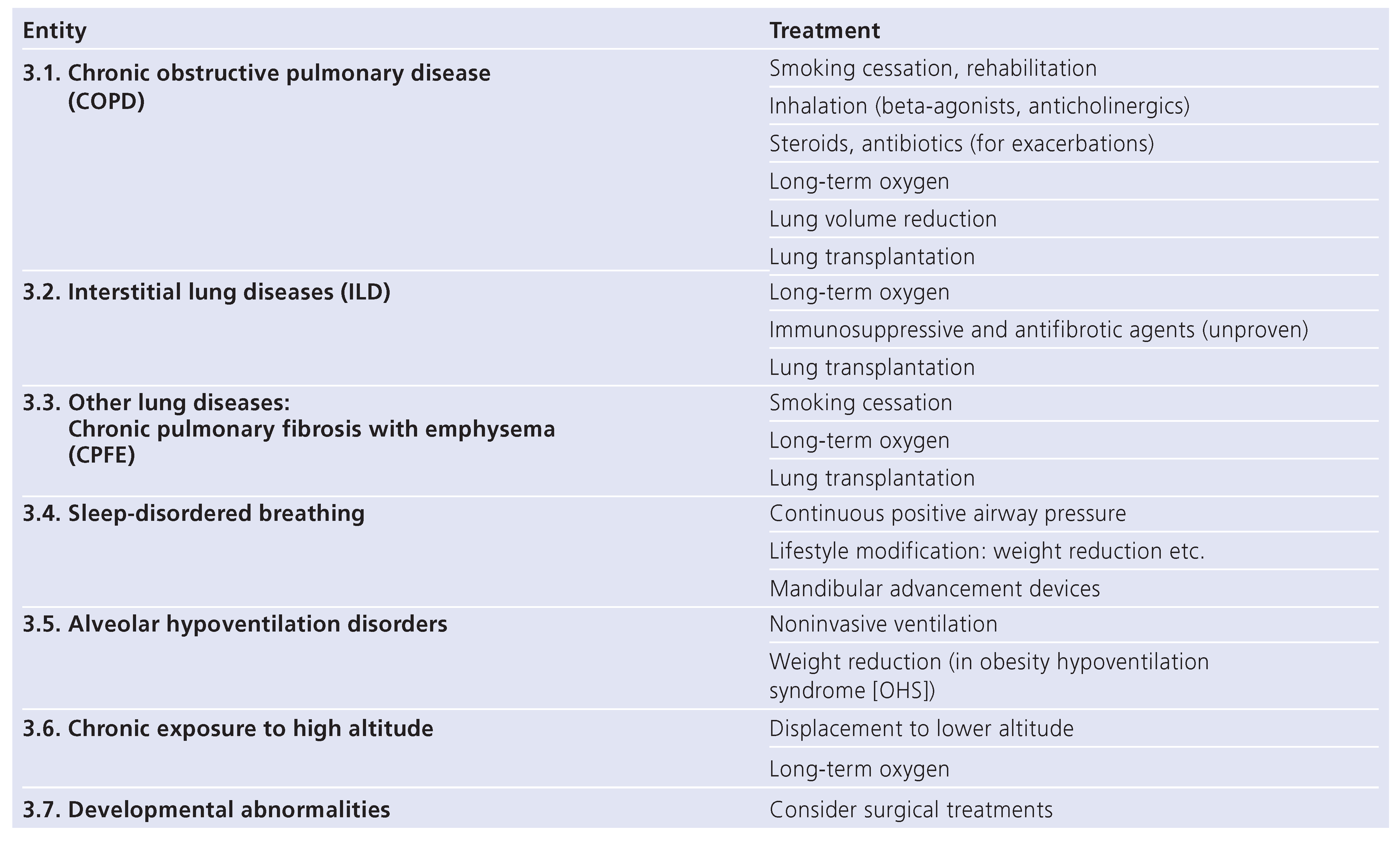
Treatment of the underlying lung diseases according to guidelines is the mainstay of therapy (Table 6). For COPD patients, long-term oxygen treatment has been shown to improve life expectancy if the partial pressure of arterial oxygen is <60 mm Hg, possibly by slowing PH development. Vasoactive therapy with pulmonary vasodilatation may worsen gas exchange in lung disease patients because it interferes with the mechanism of hypoxic vasoconstriction and diverts blood flow to poorly ventilated areas. In COPD for example, the PDE-5 inhibitor sildenafil decreases resting and exercise mean PAP, but increases ventilation/perfusion mismatch, leading to an increase in hypoxaemia with no net improvement in exercise performance and quality of life [33,34]. Trials exploring the effects of bosentan [35] and ambrisentan [36] in COPD patients yielded negative results.
In patients with diffuse interstitial lung disease or IPF, long-term oxygen to maintain arterial oxygen saturation >90% is generally recommended. Long-term studies with prostanoids for IPF are missing. The endothelin receptor antagonist bosentan was well tolerated, but failed to reduce progression of the underlying lung disease [37]. Negative results were also reported for ambrisentan and macitentan in IPF patients. Sildenafil was reported to improve 6-minute walking distance in a small open-label study [38], but a subsequent controlled trial did not meet this endpoint [39]. Interestingly, improved quality of life and echocardiographic data were reported in this trial [39]. However, taken together, the current evidence does not support the use of specific treatment for PH associated with interstitial lung disease, COPD, and CPFE.
A practical challenge in the assessment of individual patients is to decide if patients have class 1 PH (i.e., PAH) with concomitant lung disease or PH-lung (class 3). For these patients, lung function testing, high-resolution CT, exercise testing, and also sleep studies, are recommended. Table 4 may serve as guidance to classify such patients accurately. In general, mean PAP values of >35 mm Hg at rest are considered unusual for PH-lung, and the possibility of PAH with concurrent lung disease should be considered. In patients with mean PAP <35 mm Hg and concurrent severe lung disease, no data suggest the use of PAH-specific therapy.
Obstructive sleep apnoea and obesity hypoventilation syndromes are characterised by hypersomnolence and intermittent hypoxaemia during sleep. Obstructive sleep apnoea barely increases PAP, but in combination with obesity hypoventilation, elevated PAP is common. Chronic hypoxaemia leads to PH via hypoxic vasoconstriction. Standard treatment of PH in obstructive sleep apnoea and obesity hypoventilation syndromes is continuous positive airway pressure or noninvasive ventilation. Studies have shown improvement of PH after >3 months of therapy [40].

Summary

PH-LHD and PH-lung are common types of PH which typically represent more advanced disease stages of left heart and lung disease characterised by more severe symptoms and worse prognosis. The tailored work-up should include an assessment of the severity and haemodynamic constellation of PH and the mechanism of underlying left heart or lung disease and thereby the therapeutic targets. Specific vasodilator therapies currently do not play a prominent role in PH-LHD and PH-lung but additional trials are underway.

Funding/potential competing interests:

No financial support and no other potential conflict of interest relevant to this article were reported.

List of Abbreviations

COPDchronic obstructive pulmonary disease
CPFEcombined pulmonary fibrosis with
emphysema
DPDdiastolic pressure difference
HFpEFheart failure with preserved ejection fraction
HFrEFheart failure with reduced ejection fraction
IPFidiopathic pulmonary fibrosis
PAHpulmonary arterial hypertension
PAPpulmonary artery pressure
PAWPpulmonary artery wedge pressure
PDE-5 inhibitorphosphodiesterase-5 inhibitor
PHpulmonary hypertension
PH-lungpulmonary hypertension associated with
lung disease
PH-LHDpulmonary hypertension associated with left
heart disease
PVRpulmonary vascular resistance
TPGtranspulmonary gradient
TRVtricuspid regurgitation velocity

References

  1. Simonneau, G.; Gatzoulis, M.A.; Adatia, I.; Celermajer, D.; Denton, C.; Ghofrani, A.; et al. Updated clinical classification of pulmonary hypertension. J Am College Cardiol. 2013, 62, D34–D41. [Google Scholar] [CrossRef]
  2. Vachiery, J.L.; Adir, Y.; Barbera, J.A.; Champion, H.; Coghlan, J.G.; Cottin, V.; et al. Pulmonary hypertension due to left heart diseases. J Am College Cardiol. 2013, 62, D100–D108. [Google Scholar] [CrossRef]
  3. Seeger, W.; Adir, Y.; Barbera, J.A.; Champion, H.; Coghlan, J.G.; Cottin, V.; et al. Pulmonary hypertension in chronic lung diseases. J Am College Cardiol. 2013, 62, D109–D116. [Google Scholar] [CrossRef] [PubMed]
  4. Oswald-Mammosser, M.; Weitzenblum, E.; Quoix, E.; Moser, G.; Chaouat, A.; Charpentier, C.; et al. Prognostic factors in COPD patients receiving long-term oxygen therapy. Importance of pulmonary artery pressure. Chest. 1995, 107, 1193–1198. [Google Scholar] [CrossRef]
  5. Shorr, A.F.; Wainright, J.L.; Cors, C.S.; Lettieri, C.J.; Nathan, S.D. Pulmonary hypertension in patients with pulmonary fibrosis awaiting lung transplant. Eur Respir J. 2007, 30, 715–721. [Google Scholar] [CrossRef] [PubMed]
  6. Cottin, V.; Cordier, J.F. Combined pulmonary fibrosis and emphysema: An experimental and clinically relevant phenotype. Am J Respir Crit Care Med. 2005, 172, 1605–1606. [Google Scholar] [CrossRef] [PubMed]
  7. Archer, S.; Michelakis, E. The mechanism(s) of hypoxic pulmonary vasoconstriction: Potassium channels, redox O(2) sensors, and controversies. News Physiol Sci. 2002, 17, 131–137. [Google Scholar] [CrossRef][Green Version]
  8. Galie, N.; Hoeper, M.M.; Humbert, M.; Torbicki, A.; Vachiery, J.L.; Barbera, J.A.; et al. Guidelines for the diagnosis and treatment of pulmonary hypertension: The Task Force for the Diagnosis and Treatment of Pulmonary Hypertension of the European Society of Cardiology (ESC) and the European Respiratory Society (ERS), endorsed by the International Society of Heart and Lung Transplantation (ISHLT). Eur Heart J. 2009, 30, 2493–2537. [Google Scholar][Green Version]
  9. McGoon, M.; Gutterman, D.; Steen, V.; Barst, R.; McCrory, D.C.; Fortin, T.A.; et al. Screening, early detection, and diagnosis of pulmonary arterial hypertension: ACCP evidence-based clinical practice guidelines. Chest 2004, 126, 14S–34S. [Google Scholar] [CrossRef]
  10. Simon, M.A.; Rajagopalan, N.; Mathier, M.A.; Shroff, S.G.; Pinsky, M.R.; Lopez-Candales, A. Tissue Doppler imaging of right ventricular decompensation in pulmonary hypertension. Congest Heart Fail. 2009, 15, 271–276. [Google Scholar] [CrossRef]
  11. Thenappan, T.; Shah, S.J.; Gomberg-Maitland, M.; Collander, B.; Vallakati, A.; Shroff, P.; et al. Clinical characteristics of pulmonary hypertension in patients with heart failure and preserved ejection fraction. Circ Heart Fail. 2011, 4, 257–265. [Google Scholar] [CrossRef]
  12. Opotowsky, A.R.; Ojeda, J.; Rogers, F.; Prasanna, V.; Clair, M.; Moko, L.; et al. A simple echocardiographic prediction rule for hemodynamics in pulmonary hypertension. Circ Cardiovasc Imaging. 2012, 5, 765–775. [Google Scholar] [CrossRef]
  13. Abbas, A.E.; Fortuin, F.D.; Schiller, N.B.; Appleton, C.P.; Moreno, C.A.; Lester, S.J. A simple method for noninvasive estimation of pulmonary vascular resistance. J Am Coll Cardiol. 2003, 41, 1021–1027. [Google Scholar] [CrossRef] [PubMed]
  14. Maeder, M.T.; Karapanagiotidis, S.; Dewar, E.M.; Gamboni, S.E.; Htun, N.; Kaye, D.M. Accuracy of Doppler echocardiography to estimate key hemodynamic variables in subjects with normal left ventricular ejection fraction. J Card Fail. 2011, 17, 405–412. [Google Scholar] [CrossRef] [PubMed]
  15. Grunig, E.; Weissmann, S.; Ehlken, N.; Fijalkowska, A.; Fischer, C.; Fourme, T.; et al. Stress Doppler echocardiography in relatives of patients with idiopathic and familial pulmonary arterial hypertension: Results of a multicenter European analysis of pulmonary artery pressure response to exercise and hypoxia. Circulation 2009, 119, 1747–1757. [Google Scholar] [CrossRef]
  16. Hoeper, M.M.; Bogaard, H.J.; Condliffe, R.; Frantz, R.; Khanna, D.; Kurzyna, M.; et al. Definitions and diagnosis of pulmonary hypertension. J Am Coll Cardiol. 2013, 62, D42–D50. [Google Scholar] [CrossRef]
  17. Maeder, M.T.; Thompson, B.R.; Brunner-La Rocca, H.P.; Kaye, D.M. Hemodynamic basis of exercise limitation in patients with heart failure and normal ejection fraction. J Am Coll Cardiol. 2010, 56, 855–863. [Google Scholar] [CrossRef]
  18. Robbins, I.M.; Hemnes, A.R.; Pugh, M.E.; Brittain, E.L.; Zhao, D.X.; Piana, R.N.; et al. High prevalence of occult pulmonary venous hypertension revealed by fluid challenge in pulmonary hypertension. Circ Heart Fail. 2014, 7, 116–122. [Google Scholar] [CrossRef] [PubMed]
  19. Arcasoy, S.M.; Christie, J.D.; Ferrari, V.A.; Sutton, M.S.; Zisman, D.A.; Blumenthal, N.P.; et al. Echocardiographic assessment of pulmonary hypertension in patients with advanced lung disease. Am J Resp Crit Care Med. 2003, 167, 735–740. [Google Scholar] [CrossRef]
  20. Fisher, M.R.; Criner, G.J.; Fishman, A.P.; Hassoun, P.M.; Minai, O.A.; Scharf, S.M.; et al. Estimating pulmonary artery pressures by echocardiography in patients with emphysema. Eur Respir J. 2007, 30, 914–921. [Google Scholar] [CrossRef] [PubMed]
  21. Vahanian, A.; Alfieri, O.; Andreotti, F.; Antunes, M.J.; Baron-Esquivias, G.; Baumgartner, H.; et al. Guidelines on the management of valvular heart disease (version 2012). Eur Heart J. 2012, 33, 2451–2496. [Google Scholar]
  22. McMurray, J.J.; Adamopoulos, S.; Anker, S.D.; Auricchio, A.; Bohm, M.; Dickstein, K.; et al. ESC Guidelines for the diagnosis and treatment of acute and chronic heart failure 2012: The Task Force for the Diagnosis and Treatment of Acute and Chronic Heart Failure 2012 of the European Society of Cardiology. Developed in collaboration with the Heart Failure Association (HFA) of the ESC. Eur Heart J. 2012, 33, 1787–1847. [Google Scholar]
  23. Mullens, W.; Borowski, A.G.; Curtin, R.J.; Thomas, J.D.; Tang, W.H. Tissue Doppler imaging in the estimation of intracardiac filling pressure in decompensated patients with advanced systolic heart failure. Circulation 2009, 119, 62–70. [Google Scholar] [CrossRef]
  24. Lewis, G.D.; Shah, R.; Shahzad, K.; Camuso, J.M.; Pappagianopoulos, P.P.; Hung, J.; et al. Sildenafil improves exercise capacity and quality of life in patients with systolic heart failure and secondary pulmonary hypertension. Circulation 2007, 116, 1555–1562. [Google Scholar] [CrossRef] [PubMed]
  25. Bonderman, D.; Ghio, S.; Felix, S.B.; Ghofrani, H.A.; Michelakis, E.; Mitrovic, V.; et al. Riociguat for patients with pulmonary hypertension caused by systolic left ventricular dysfunction: A phase IIb double-blind, ran domized, placebo-controlled, dose-ranging hemodynamic study. Circulation 2013, 128, 502–511. [Google Scholar] [CrossRef]
  26. Maeder, M.T.; Rickli, H. [Heart failure with preserved left ventricular ejection fraction]. Praxis 2013, 102, 1299–1307. [Google Scholar] [CrossRef] [PubMed]
  27. Bergstrom, A.; Andersson, B.; Edner, M.; Nylander, E.; Persson, H.; Dahlstrom, U. Effect of carvedilol on diastolic function in patients with diastolic heart failure and preserved systolic function. Results of the Swedish Doppler-echocardiographic study (SWEDIC). Eur J Heart Fail. 2004, 6, 453–461. [Google Scholar] [CrossRef] [PubMed]
  28. Kosmala, W.; Holland, D.J.; Rojek, A.; Wright, L.; Przewlocka-Kosmala, M.; Marwick, T.H. Effect of If-channel inhibition on hemodynamic status and exercise tolerance in heart failure with preserved ejection fraction: A randomized trial. J Am Coll Cardiol. 2013, 62, 1330–1338. [Google Scholar] [CrossRef]
  29. Maier, L.S.; Layug, B.; Karwatowska-Prokopczuk, E.; Belardinelli, L.; Lee, S.; Sander, J.; et al. RAnoLazIne for the treatment of diastolic heart failure in patients with preserved ejection fraction: The RALI-DHF proof-of-concept study. JACC Heart failure 2013, 1, 115–122. [Google Scholar] [CrossRef]
  30. Edelmann, F.; Gelbrich, G.; Dungen, H.D.; Frohling, S.; Wachter, R.; Stahrenberg, R.; et al. Exercise training improves exercise capacity and diastolic function in patients with heart failure with preserved ejection fraction: Results of the Ex-DHF (Exercise training in Diastolic Heart Failure) pilot study. J Am Coll Cardiol. 2011, 58, 1780–1791. [Google Scholar] [CrossRef]
  31. Redfield, M.M.; Chen, H.H.; Borlaug, B.A.; Semigran, M.J.; Lee, K.L.; Lewis, G.; et al. Effect of phosphodiesterase-5 inhibition on exercise capacity and clinical status in heart failure with preserved ejection fraction: A randomized clinical trial. JAMA 2013, 309, 126–177. [Google Scholar] [CrossRef]
  32. Guazzi, M.; Vicenzi, M.; Arena, R.; Guazzi, M.D. Pulmonary hypertension in heart failure with preserved ejection fraction: A target of phosphodiesterase-5 inhibition in a 1–year study. Circulation 2011, 124, 164–174. [Google Scholar] [CrossRef] [PubMed]
  33. Blanco, I.; Santos, S.; Gea, J.; Guell, R.; Torres, F.; Gimeno-Santos, E.; et al. Sildenafil to improve respiratory rehabilitation outcomes in COPD: A controlled trial. Eur Respir J. 2013, 42, 982–992. [Google Scholar] [CrossRef]
  34. Blanco, I.; Gimeno, E.; Munoz, P.A.; Pizarro, S.; Gistau, C.; Rodriguez-Roisin, R.; et al. Hemodynamic and gas exchange effects of sildenafil in patients with chronic obstructive pulmonary disease and pulmonary hypertension. Am J Respir Crit Care Med. 2010, 181, 270–278. [Google Scholar] [CrossRef]
  35. Stolz, D.; Rasch, H.; Linka, A.; Di Valentino, M.; Meyer, A.; Brutsche, M.; et al. A randomised, controlled trial of bosentan in severe COPD. Eur Respir J. 2008, 32, 619–628. [Google Scholar] [CrossRef]
  36. Badesch, D.B.; Feldman, J.; Keogh, A.; Mathier, M.A.; Oudiz, R.J.; Shapiro, S.; et al. ARIES-3: Ambrisentan therapy in a diverse population of patients with pulmonary hypertension. Cardiovascular therapeutics 2012, 30, 93–99. [Google Scholar] [CrossRef]
  37. King, T.E., Jr. , Brown, K.K.; Raghu, G.; du Bois, R.M.; Lynch, D.A.; Martinez, F.; et al. BUILD-3: A randomized, controlled trial of bosentan in idiopathic pulmonary fibrosis. Am J Respir Crit Care Med. 2011, 184, 92–99. [Google Scholar] [CrossRef]
  38. Collard, H.R.; Anstrom, K.J.; Schwarz, M.I.; Zisman, D.A. Sildenafil improves walk distance in idiopathic pulmonary fibrosis. Chest 2007, 131, 897–899. [Google Scholar] [CrossRef] [PubMed]
  39. Han, M.K.; Bach, D.S.; Hagan, P.G.; Yow, E.; Flaherty, K.R.; Toews, G.B.; et al. Sildenafil preserves exercise capacity in patients with idiopathic pulmonary fibrosis and right-sided ventricular dysfunction. Chest 2013, 143, 1699–1708. [Google Scholar] [CrossRef]
  40. Arias, M.A.; Garcia-Rio, F.; Alonso-Fernandez, A.; Martinez, I.; Villamor, J. Pulmonary hypertension in obstructive sleep apnoea: Effects of continuous positive airway pressure: A randomized, controlled cross-over study. Eur Heart J. 2006, 27, 1106–1113. [Google Scholar] [CrossRef] [PubMed]
Table 1. Place of pulmonary hypertension due to left heart (class 2) and lung (class 3) diseases in the Nice 2013 pulmonary hypertension classification [1] (simplified version).
Cardiovascmed 17 00320 i001
Table 2. Haemodynamic definitions of pulmonary hypertension (PH) associated with left heart disease (PH-LHD) and PH associated with lung disease (PH-lung).
Cardiovascmed 17 00320 i002
Table 3. Echocardiographic and Doppler features favouring pulmonary arterial hypertension (PAH) or post-capillary pulmonary hypertension (PH).
Cardiovascmed 17 00320 i003
Table 4. Differential diagnosis between pulmonary arterial hypertension (PAH; group 1) and pulmonary hypertension associated with lung diseases (PH-lung; group 3) according to the Nice 2013 proposal [3].
Cardiovascmed 17 00320 i004
Table 5. Treatment of different entities of PH-LHD.
Cardiovascmed 17 00320 i005
Table 6. Treatment of different entities of PH-lung.
Cardiovascmed 17 00320 i006