Next Article in Journal
Clinical Pathophysiology of Ischemic Stroke
Previous Article in Journal
New Trends in Treatment of Coronary Artery Disease
 
 
Cardiovascular Medicine is published by MDPI from Volume 28 Issue 1 (2025). Previous articles were published by another publisher in Open Access under a CC-BY (or CC-BY-NC-ND) licence, and they are hosted by MDPI on mdpi.com as a courtesy and upon agreement with Editores Medicorum Helveticorum (EMH).
Proceeding Paper

Peripheral Artery Disease and Diabetes Mellitus

Division of Hypertension and Vascular Medicine, CHUV, 1011 Lausanne, Switzerland
Cardiovasc. Med. 1998, 1(2), 137-142; https://doi.org/10.3390/cardiovascmed1020032

Published: 30 August 1998

Abstract

Kardiovaskuläre Komplikationen Sind die weitaus schwerwiegendsten Manifestationen des Diabetes mellitus. Die Behandlung, die die Verhinderung der Progression der vaskulären Läsionen zum Ziel hat, kann Sich als aussichtslos erweisen, falls sie erst zum Zeitpunkt des Auftretens der Gefässaffektionen eingeleitet wird. Deshalb ist die Identifizierung der frühesten vaskulären Dysfunktion eine Chance, mit den entsprechenden pathogenetischen Mechanismen zu interferieren und die Progression der diabetischen Vaskulopathie zu verhindern. Im vorliegenden Artikel sollen einige der Mechanismen vorgestellt werden, welche die hämodynamische und metabolische Homöostase im Verlauf der diabetischen Erkrankung verändern können. Es werden die endotheliale Funktion (mit speziellem Augenmerk auf Stickoxid und oxidativem Stress), das Auftreten von glykosylierter Stoffwechsel-Endproduktion sowie das Renin-Angiotensin-System kurz behandelt. Neue pharmakologische Agentien, die mit obengenannten Mechanismen oder Parametern interferieren können, befinden Sich derzeit in klinischer Prüfung. Es gilt indessen weiterhin, dass die engmaschige Kontrolle der Blutzuckerwerte und die Modifikation der übrigen kardiovaskulären Risikofaktoren ein Hauptprinzip der Behandlung zur Verhinderung der fortschreitenden Vaskulopathie beim Diabetes bleibt.

Introduction

Lower extremity arterial disease is one of the dreadful vascular complications suffered by patients with diabetes mellitus. The development of vascular disease is only partly preventable by a tight glycaemic control [1]. Indeed, only a minority of diabetic patients can achieve normoglycaemia. Accordingly, late complications of diabetes will still develop in the majority of diabetic patients. Cardiovascular complications represent by far the most common and devastating manifestation and are the major cause of hospital admission in diabetic patients. In the United States 77% of hospitalisations of diabetic patients are related to cardiovascular disease and 10% to diabetic nephropathy [2]. The high incidence of cardiovascular disease is not fully explained by hyperglycaemia or by association with other known cardiovascular risk factors. Epidemiological studies have reported a 3-fold increase in the relative risk of myocardial infarction when compared with matched non-diabetic populations [3]. In patients with claudication of the lower limbs, the presence of diabetes doubles the mortality rate at 5 years to 49% as compared to the 5 year mortality rate of 25% in patients with atherosclerosis without diabetes [4]. In other words, the presence of lower extremity arterial disease in diabetic patients is a powerful predictive factor for mortality.
Sixty-seven % of diabetic patients dying from cardiovascular disease within 5 years had peripheral artery disease at baseline compared with 15% in those who survived [5].
The consequences of diabetic micro- and macroangiopathy represent the principal cause of mortality and disability in patients with diabetes mellitus. Although the pathogenic mechanisms and age distribution differ between type 1 diabetes and non-insulin-dependent diabetes mellitus (type 2), atherosclerosis, retinopathy, nephropathy and neuropathy develop in both types of diabetic populations at an accelerated rate. At the time of the diagnosis, 8% of the subjects have already lower extremity arterial disease [6]. Arterial hypertension which is present in up to 60% of the patients with type 2 diabetes as well as different forms of dyslipidemia may further amplify or accelerate the vascular disease that affects most diabetic patients, which explains the higher prevalence of large vessel lesions in this population.
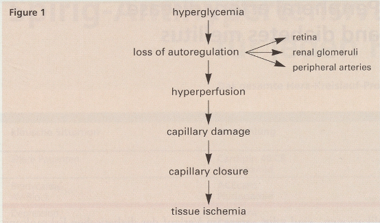
Diabetic patients have a multifocal distribution of atherosclerosis with a tendency to develop predominantly below knee lesions (calf and foot) and occlusions of the deep femoral arteries. In contrast, nondiabetic patients with lower extremity arterial disease suffer more frequently iliac and superficial femoral artery disease. From a clinical point of view, all diabetic patients with claudication or clinical and laboratory signs of lower extremity arterial disease should be controlled on an annual basis. Too many diabetic patients present with advanced vascular lesions which are not easily amenable to revascularisation. Special care towards controlling cardiovascular risk factor in addition to diabetes should be paid in these patients who are at high risk of developing critical ischaemia and amputation before the disease has progressed to point of “no return”. Are there any evidences that a common functional environment may contribute to the diabetic vasculopathy and are there therapeutic tools to stop its progression? The pathogenesis of the functional defects in diabetes is not fully understood and is likely to be multifactorial with genetically determined susceptibility. Nevertheless, there are a few potentially unifying mechanisms early in the evolution of the disease that merit to be considered. A loss of autoregulation in blood flow characterises early functional alteration in diabetic patients. It can be demonstrated in different vascular beds. Increase in glomerular filtration rate with accompanying renal hypertrophy, failure of the venoarteriolar reflex particularly manifest in the lower extremities inducing capillary hypertension and increase in retinal blood flow concomitant with arterial hypertension represent common features of diabetic haemodynamic changes that may lead to structural damage (Figure 1). A few important mechanisms that may contribute early in diabetic vasculopathy are presented in the following section.
Cardiovascmed 01 00137 g001

Endothelial dysfunction

The endothelium plays a key role in maintaining vessel wall homeostasis through the synthesis of several substances that modulate vascular tone, regulate the balance between thrombosis and fibrinolysis, control permeability and influence smooth muscle cell growth and extracellular matrix composition. It has been shown that endothelial function is already abnormal early in the development of diabetes mellitus. This is now supported both by results obtained in animal models of diabetes mellitus and by functional abnormalities in the coronary as well as in the peripheral circulation in patients with type 1 and type 2 diabetes [7,8,9]. Endothelial dysfunction may represent a common pathogenic framework that contributes in the two types of diabetes mellitus to the development of vascular lesions that affect the micro- as well as the macrocirculation.
The close association observed between albuminuria and endothelial dysfunction led Deckert et al. to postulate the Steno hypothesis, proposing that albuminuria reflects a widespread vasculopathy of the micro- and macrocirculation which is the consequence of a generalised endothelial dysfunction [10].
Several approaches have been used to assess endothelial function in diabetic patients. Among them, the most frequent test applied to quantitate endothelial function relies on the rather indirect flow and vessel diameter responsiveness to intra-arterial infusion of muscarinic receptor agonists such as acetylcholine or metacholine. Blunted endothelium-dependent vasodilatation in patients with type 1 and type 2 diabetes has been reported in the coronary and in the peripheral circulation in a fashion similar to that observed in arterial hypertension, in hypercholesterolaemia or in post-menopausal women. However, if flowmediated vasodilatation was assessed following infusion of a potent direct vasodilator distal to the site of arterial diameter measurement (nitroglycerin) or during reactive hyperaemia following release of arterial occlusion, no significant changes in vasomotion could be observed between diabetic and control subjects [8]. This is not at all surprising in the light of the seminal work performed by Zeiher and collaborators on the coronary arteries [11]. These authors showed different sensitivities between the muscarinic agonist infusions and the flow-mediated vasodilatation in patients undergoing coronary artery catheterisation with different severity of atherosclerosis. Acetylcholine induced a paradoxical vasoconstriction in angiographically smooth arteries in patients with elevated LDL cholesterol. In contrast, flow-mediated vasodilatation could still be observed in severely atherosclerotic arteries following distal infusion of papaverine. Although flow-mediated vasomotion represents a more physiologic stimulus to test the endothelial function, it has a low sensitivity to identify early endothelial dysfunction in the presence of diabetes or other cardiovascular risk factors. A series of other indicators has been used to assess endothelial function. Among them, plasma von Willebrand factor (vWF) is an interesting molecule that can easily be assayed without intra-arterial manipulations [12]. However, only patients with type 1 or type 2 diabetes and albuminuria were shown to have an elevated plasma vWF, suggesting that it reflects marked endothelial cell disruption or activation [9]. New biological indicators of incipient endothelial abnormality are needed to identify patients at risk to develop vascular complications in an attempt to prevent the progression of the disease.

The nitric oxide pathway

What are the mechanisms that may participate in endothelial dysfunction? One possibility is a reduced bioavailability of the endotheliumderived nitric oxide (NO). Different pathways can contribute to the net reduction of bioavailable NO. They include abnormalities in signal transduction, reduced synthesis of NO, enhanced inactivation of NO, release of competing vasoconstrictors. Supplementation of L-arginine, the substrate for the endothelial NO synthase does result in a beneficial effect in hypertension and hypercholesterolaemia models of endothelial dysfunction as shown previously. However, in diabetic patients it does not seem to restore the abnormal vascular response. Recently, Nitenberg and collaborators infused L-arginine or deferoxamine, a chelator that inhibits reactive oxygen species formation, into the coronary arteries of diabetic patients before challenging them with a cold pressure test or with papaverine [13]. Diabetic patients who exhibited a paradoxical vasoconstriction during a cold pressure test did not show any diameter modification of the left anterior descending (LAD) coronary artery following L-arginine infusion when challenged again with the cold pressure test. In contrast, a significant vasodilatation of the LAD was observed after infusion of deferoxamine. This supports the observation of Ting and colleagues who demonstrated a similar effect following intra-arterial infusion of vitamin C in the brachial artery of type 2 diabetic patients [14]. These findings taken together suggest that NO inactivation by reactive oxygen species contributes to some extent to the abnormal vasomotion observed in diabetic patients.

Role of the AGEs and VEGF in endothelial dysfunction and vascular permeability

The exposure of the vascular environment (proteins and lipids) to increased reducing sugars leads to advanced glycosylation end products (AGEs) and increased oxidative stress that affect the micro- and macrocirculation. The formation of these AGEs may participate in the process of inactivation of nitric oxide. Recent evidence supports the fact that AGEs induce endothelial dysfunction via a receptorspecific pathway (RAGE) [15]. The binding of the ligand onto the RAGE induces an increase in permeability that facilitates transmigration of macromolecules through the vessel wall. Prior nonenzymatic glycation and oxidation of the macromolecules favour their trapping in the vessel wall with resulting modification of the extracellular matrix composition and the mechanical properties of the vessel wall. Clinical trials are now in progress to explore the consequences of AGE formation inhibition and reversal by aminoguanidine. Albuminuria and mesangial expansion in the diabetic rat model have been retarded by aminoguanidine. Vascular endothelium growth factor (VEGF) represents a candidate substance that may enhance vascular hyperpermeability in diabetes. Previously named VPF for vascular permeability factor, VEGF has been subsequently demonstrated to be a potent angiogenic factor. Its implication in vascular growth and permeability in diabetes has been suggested by data showing increased concentrations of VEGF in the ocular fluid of patients with proliferative retinopathy [16]. In an experimental mouse model of proliferative diabetic retinopathy, intraocular injection of a chimeric construct that selectively binds VEGF, markedly reduced the proliferative process [17]. Further studies are needed to establish the causality of this factor in the hyperpermeability associated with diabetes. Activation of the fibrinolytic pathway to allow new vessels to make their way into the interstitial tissues represents one of the potential mechanisms by which VEGF may increase permeability.

Role of the renin-angiotensin-system

We recently demonstrated in an in vitro study that flow conditions prevailing in plaque prone regions were responsible for a reduction of the endothelial NO-synthase expression together with an upregulation of the expression of VEGF [18]. To further underline the potential importance of increased oxidative stress and hyperpermeability, one should also discuss the role of the renin angiotensin system (RAS). As mentioned earlier, the prevalence of hypertension is high in type 2 diabetes whereas it is not significantly different in type 1 diabetes, before nephropathy becomes evident. The efficacy of the angiotensin converting enzyme (ACE) inhibitors in the treatment of hypertensive diabetics is well established. More recently, the observation that ACE inhibitors can prevent or retard the progression of nephropathy both in hypertensive and normotensive patients with type 1 diabetes has represented a landmark result [19]. It is highly likely from the available data that have so far been published that ACE inhibitors have a unique capacity independent from their antihypertensive or haemodynamic effect to slow progression of diabetic vasculopathy in type 1 as well as in type 2 diabetes.
The RAS may play a role in the nitric oxide/reactive oxygen species balance as angiotensin Il has directly been implicated in the generation of superoxide anion in smooth muscle cells. Indeed, angiotensin Il regulates a membranebound flavin containing NADH/NADPH oxidase that produces oxygen radicals [20]. Several groups have reported that angiotensin Il increases the net production of superoxide anion in cell culture as well as in animal models of hypertension. One could then hypothesise that ACE inhibitors besides their well known haemodynamic action could participate in the reduction of the oxidative stress of the vessel wall. Such a concept, before discovering this interesting enzymatic oxidase induction, had been discussed a few years ago when normotensive hypercholesterolaemic rabbits chronically treated with ACE inhibitors showed a dramatic reduction of plaque formation [21]. Similar results were subsequently obtained in a normotensive hypercholesterolaemic pig model [22].
Clinical studies and trials have confirmed the ability of ACE inhibitors to restore endothelial function in hypertensive patients as well as in normotensive subjects with other pathologic cardiovascular conditions [23,24]. This accessory property of ACE inhibitors through a redox-sensitive mechanism would definitely represent a welcome asset by reducing the proinflammatory consequences of an excess reactive oxygen species.
As discussed earlier, the vascular hyperpermeability present in diabetic patients may be the direct consequence of an increased amount of AGEs that react with their receptor on the endothelium. Blockade of the RAGE with an antibody considerably reduced the permeability of the vessel wall but did not totally abolish it. One of the possible candidates that may influence the tightness of the endothelial barrier, as mentioned earlier, is VEGF. Interestingly, Williams and colleagues recently reported that Angll upregulates the expression of VEGF mRNA expression that can also be markedly increased in the presence of elevated glucose concentration (20 mmol/L) [25]. Blockade of the ATI receptor with losartan abolishes the expression of VEGF mRNA.
Taken together, cardiovascular complications in diabetic patients have clearly a multifactorial origin combining haemodynamic and metabolic factors. Patients presenting with lower extremity arterial disease are at high risk for cardiovascular fatal events. Early identification and care of the many factors contributing to the development of severe and acceler- ated atherosclerosis seem imperative to retard or possibly to stop progression of the diabetic vasculopathy. Tight control of plasma glucose and other cardiovascular risk factors, ACE inhibitors, ATI antagonists and newer substances such as aminoguanidine that modify deleterious metabolic pathways may early slow the evolution of the vascular lesions in diabetes mellitus. Clinical trials in progress will help identifying the most sensitive parameters amenable to correction that may provide efficient care to diabetic patients.

References

  1. The diabetes control complications trial (DCCT) research group. Effect of intensive diabetes management on macrovascular events risk factors in the diabetes control complications trial. Am. J. Cardiol. 1995, 75, 894–903. [CrossRef] [PubMed]
  2. Jacobs, J.; Sena, M.; Fox, N. The cost of hospitalization for the late complications of diabetes in the United States. Diabetic Med. 1991, 8 Symposium, S23–S29. [Google Scholar] [CrossRef]
  3. Steiner, G. Diabetes and atherosclerosis: Epidemiology and intervention trials. In Atherosclerosis X; Woodford, F.P., Davignon, J., Sniderman, A., Eds.; Elsevier: Amsterdam, The Netherlands, 1995; pp. 749–752. [Google Scholar]
  4. Criqui, M.H.; Langer, R.D.; Fronek, A.; Feigelson, H.S.; Klauber, M.R.; McCann, T.J.; Browner, D. Mortality over a period of 10 years in patients with peripheral arterial disease. N. Engl. J. Med. 1992, 326, 381–386. [Google Scholar] [CrossRef]
  5. Janka, H.U.; Zierler, A.G.; Standl, E.; Mehnert, H. Daily insulin dose as a predictor of macrovascular disease in insulin treated non-insulin dependent diabetics. Diabetes Metab. 1987, 13, 359–364. [Google Scholar]
  6. Palumbo, P.J.; O’Fallon, W.M.; Osmundson, P.J.; Zimmermann, B.R.; Langworthy, A.L.; Kazmier, F.J. Progression of peripheral occlusive arterial disease in diabetes mellitus. What factors are predictive. Arch. Intern. Med. 1991, 151, 717–721. [Google Scholar] [CrossRef] [PubMed]
  7. Tesfamariam, B.; Brown, M.L.; Cohen, R.A. Elevated glucose impairs endothelium-dependent relaxation by activating protein kinase C. J. Clin. Invest. 1991, 87, 1643–1648. [Google Scholar] [CrossRef]
  8. Johnstone, M.T.; Creager, S.J.; Scales, K.M.; Cusco, J.A.; Lee, B.K.; Creager, M.A. Impaired endothelium-dependent vasodilation in patients with insulin-dependent diabetes mellitus. Circulation 1993, 88, 2510–2516. [Google Scholar] [CrossRef]
  9. Parving, H.H.; Nielsen, F.S.; Bang, L.E.; Smidt, U.M.; Svendsen, T.L.; Chen, J.W.; Gall, M.A.; Rossing, P. Macro-microangiopathy and endothelial dysfunction in NIDDM patients with and without diabetic nephropathy. Diabetologia 1996, 39, 1590–1597. [Google Scholar] [CrossRef]
  10. Deckert, T.; Feldt-Rasmussen, B.; Borch-Johnsen, K.; Jensen, T.; Kofoed-Enevoldsen, A. Albuminuria reflects widespread vascular damage. The Steno hypothesis. Diabetologia 1989, 32, 219–226. [Google Scholar] [CrossRef] [PubMed]
  11. Zeiher, A.M.; Drexler, H.; Wollschläger, H.; Just, H. Modulation of coronary vasomotor tone in humans. Progressive endothelial dysfunction with different stages of coronary atherosclerosis. Circulation 1991, 83, 391–401. [Google Scholar] [CrossRef]
  12. Lip, G.Y.H.; Blann, A. von Willebrand factor: A marker of endothelial dysfunction in vascular disorders? Cardiovasc Res 1997, 34, 255–265. [Google Scholar] [CrossRef] [PubMed]
  13. Nitenberg, A.; Ledoux, S.; Valensi, P.; Attali, J.R. La réponse des arteres coronaires au test au froid et l’augmentation du débit est améliorée par la déféroxamine mais non par la L-arginine chez les sujets diabétiques. XVles Journées de l’hypertension artérielle, Paris 1996 [abstract]. Arch Mal Coeur Vaisseaux 1996, 89, 21. [Google Scholar]
  14. Ting, H.H.; Timimi, F.K.; Boles, K.S.; Creager, S.J.; Ganz, P.; Creager, M.A. Vitamin C improves endothelium-dependent vasodilation in patients with non-insulin-dependent diabetes mellitus. Clin. Invest. 1996, 97, 22–28. [Google Scholar] [CrossRef]
  15. Wautier, J.L.; Zoukourian, C.; Chappey, O.; Wautier, M.P.; Guillausseau, P.J.; Cao, R.; et al. Receptor-mediated endothelial cell dysfunction in diabetic vasculopathy. Soluble receptor for advanced glycation end products blocks hyperpermeability in diabetic rats. J. Clin. Invest. 1996, 97, 238–243. [Google Scholar] [CrossRef] [PubMed]
  16. Aiello, L.P.; Avery, R.L.; Arrigg, P.G.; Keyt, B.A.; Jampel, H.D.; Shah, S.T.; et al. Vascular endothelial growth factor in ocular fluid of patients with diabetic retinopathy and other retinal disorders. N. Engl. J. Med. 1994, 331, 1480–1487. [Google Scholar] [CrossRef] [PubMed]
  17. Aiello, L.P.; Pierce, E.A.; Foley, E.D.; Takagi, H.; Chen, H.; Riddle, L.; Ferrara, N.; King, G.L.; Smith, L.E. Suppression of retinal neovascularization in vivo by inhibition of vascular endothelial growth factor (VEGF) using soluble VEGF-receptor chimeric proteins. Proc. Natn Acad. Sci. USA 1995, 92, 10457–10461. [Google Scholar] [CrossRef]
  18. Hayoz, D.; Ziegler, T.; Brunner, H.R.; Ruiz, J. Diabetes mellitus and vascular lesions. Metab. Clin. Exp. in press. [CrossRef]
  19. Lewis, E.J.; Hunsicker, L.G.; Bain, R.P.; Rohde, R.D. The effect of angiotensin-converting-enzyme inhibition on dia betic nephropathy. N. Engl. J. Med. 1993, 329, 1456–1462. [Google Scholar] [CrossRef]
  20. Fukui, T.; Ishizaka, N.; Rajagopalan, S.; Laursen, J.B.; Capers, Q., 4th; Taylor, W.R.; et al. p22phox mRNA expression and NADPH oxidase activity are increased in aortas from hypertensive rats. Circ. Research 1997, 80, 45–51. [Google Scholar] [CrossRef]
  21. Chobanian, A.V.; Haudenschild, C.C.; Nickerson, C.; Drago, R. Antiatherogenic effect of captopril in the Watanabe heritable hyperlipidemic rabbit. Hypertension 1990, 15, 327–331. [Google Scholar]
  22. Jacobsson, L.S.; Persson, K.; Aberg, G.; Andersson, R.G.; Karlberg, B.E.; Olsson, A.G. Antiatherosclerotic effects of the angiotensin-converting enzyme inhibitors captopril and fosinopril in hypercholesterolemic minipigs. J. Cardiovasc. Pharmacol. 1994, 24, 670–677. [Google Scholar] [CrossRef]
  23. Antony, I.; Lerebours, G.; Nitenberg, A. Angiotensin-converting enzyme inhibition restores flow-dependent and cold pressor test-induced dilations in coronary arteries of hypertensive patients. Circulation 1996, 94, 3115–3122. [Google Scholar] [CrossRef] [PubMed]
  24. Mancini, G.B.; Henry, G.C.; Macaya, C.; O’Neill, B.J.; Pucillo, A.L.; Carere, R.G.; et al. Angiotensin-converting enzyme inhibition with quinapril improves endothelial vasomotor dysfunction in patients with coronary artery disease. The TREND (Trial on Reversing ENdothelial Dysfunction) study. Circulation 1996, 94, 258–265. [Google Scholar] [CrossRef] [PubMed]
  25. Williams, B.; Quinn-Baker, A.; Gallager, B.; Lodwick, D. Angiotensin Il increases vascular permeability factor gene expression by human vascular smooth muscle cells. Hypertension 1995, 25, 913–917. [Google Scholar] [CrossRef] [PubMed]